Embed Size (px)
Citation preview

THE INFLUENCE OF COPPER BINDING ON THE STABILITY OF THE SCO PROTEIN FROM BACILLUS SUBTILIS
by
DAVID EDUARDS DAVIDSON
A thesis submitted to the Department of Biochemistry
In conformity with the requirements for
the degree of Master of Science
Queen’s University
Kingston, Ontario, Canada
(September, 2007)
Copyright © David Davidson, 2007
ii
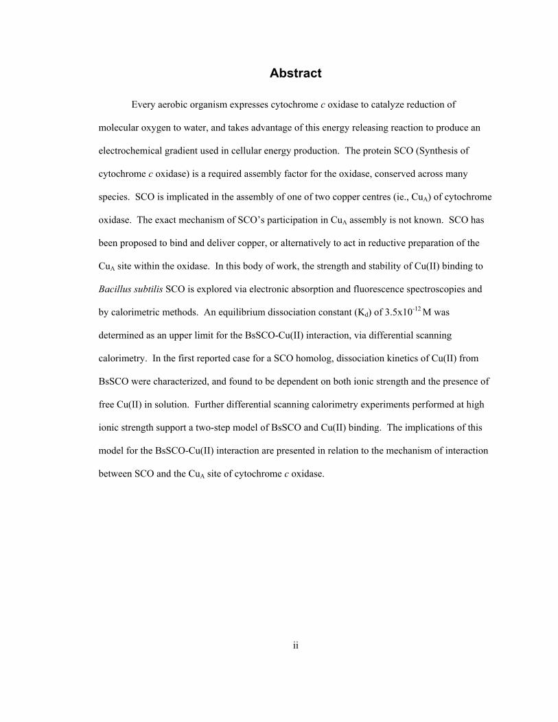
Abstract
Every aerobic organism expresses cytochrome c oxidase to catalyze reduction of
molecular oxygen to water, and takes advantage of this energy releasing reaction to produce an
electrochemical gradient used in cellular energy production. The protein SCO (Synthesis of
cytochrome c oxidase) is a required assembly factor for the oxidase, conserved across many
species. SCO is implicated in the assembly of one of two copper centres (ie., CuA) of cytochrome
oxidase. The exact mechanism of SCO’s participation in CuA assembly is not known. SCO has
been proposed to bind and deliver copper, or alternatively to act in reductive preparation of the
CuA site within the oxidase. In this body of work, the strength and stability of Cu(II) binding to
Bacillus subtilis SCO is explored via electronic absorption and fluorescence spectroscopies and
by calorimetric methods. An equilibrium dissociation constant (Kd) of 3.5x10⁻¹² M was
determined as an upper limit for the BsSCO-Cu(II) interaction, via differential scanning
calorimetry. In the first reported case for a SCO homolog, dissociation kinetics of Cu(II) from
BsSCO were characterized, and found to be dependent on both ionic strength and the presence of
free Cu(II) in solution. Further differential scanning calorimetry experiments performed at high
ionic strength support a two-step model of BsSCO and Cu(II) binding. The implications of this
model for the BsSCO-Cu(II) interaction are presented in relation to the mechanism of interaction
between SCO and the CuA site of cytochrome c oxidase.

iii
Acknowledgements
The author would like to thank Kim Munro, of the Queen’s University Protein Function
Discovery facility, as well as technician Diann Andrews and past students Bradley Poulsen and
Thomas Cawthorn. The author expresses sincere appreciation to supervisor Dr. Bruce C. Hill.

iv
Table of Contents Abstract ............................................................................................................................................ii Acknowledgements.........................................................................................................................iii Table of Contents............................................................................................................................ iv List of Tables ..................................................................................................................................vi List of Abbreviations ....................................................................................................................viii List of Figures ................................................................................................................................vii Chapter 1 Introduction ..................................................................................................................... 1 Chapter 2 Literature Review............................................................................................................ 9 Chapter 3 Materials and Methods .................................................................................................. 19
3.1 Bacterial Expression and Purification.................................................................................. 19 3.2 Preparation of Reduced and Cu(II) Bound BsSCO.............................................................. 20 3.3 Electronic Absorption Spectra (copper binding, copper dissociation)................................. 20 3.4 Determination of Free Thiol Groups.................................................................................... 21 3.5 Intrinsic Tryptophan Fluorescence....................................................................................... 21 3.6 Circular Dichroism Absorbance (unfolding) ....................................................................... 21 3.7 Stopped-flow Unfolding Fluorescence ................................................................................ 22 3.8 Copper(II) Dissociation from BsSCO in High Ionic Strength ............................................. 22 3.9 Differential Scanning Calorimetry (DSC) for Copper Binding ........................................... 23 3.10 Measurement of BsSCO Redox Potential .......................................................................... 23
Chapter 4 Results ........................................................................................................................... 25 4.1 Protein Purification and Identification................................................................................. 25 4.2 Copper Binding to BsSCO................................................................................................... 29 4.3 Equilibrium Unfolding Experiments.................................................................................... 31 4.4 Stopped-flow Unfolding of BsSCO ..................................................................................... 33 4.5 Copper Dissociation from BsSCO in High Ionic Strength Solutions .................................. 40 4.6 The Effect of Free Cu(II) in Solution with the Copper(II)-BsSCO Complex...................... 43 4.7 DSC Measurement of BsSCO Stability Changes Due to Copper(II) Binding..................... 45 4.8 DSC Measurement of BsSCO Stability Changes Due to Ionic Strength Changes .............. 48 4.9 Measurement of BsSCO Redox Potential ............................................................................ 51
Chapter 5 Discussion ..................................................................................................................... 54 5.1 Measuring Cu(II) binding to BsSCO ................................................................................... 54 5.2 Measurement of BsSCO Stability via Differential Scanning Calorimetry .......................... 56

v
5.3 Copper Dissociation from the Copper(II)-BsSCO Complex ............................................... 59 5.4 BsSCO redox potential determination ................................................................................. 67 5.5 DSC and Stability Dictated by Ionic Strength ..................................................................... 69
Chapter 6 Summary and Conclusions............................................................................................ 78

vi
List of Tables Table 1: Integrated density of slower and faster mobility species on SDS-PAGE ....................... 53
vii
List of Figures Figure 1: Representation of Cytochrome c oxidase subunits I and II .............................................. 2 Figure 2: Structures of E. coli thioredoxin and B. subtilis SCO ..................................................... 6 Figure 3: General scheme of copper transport in eukaryotic cells................................................ 12 Figure 4: SDS-PAGE of BsSCO purification stages .................................................................... 25 Figure 5: ESI MS/MS whole mass spectrum of purified BsSCO protein..................................... 26 Figure 6: Determination of free thiol groups present on BsSCO protein ..................................... 27 Figure 7: Absorption spectra of reduced and Cu(II)-bound BsSCO............................................. 29 Figure 8: Fluorescence emission spectra of forms of BsSCO....................................................... 30 Figure 9: Fluorescence emission peak wavelength of BsSCO versus guanidine concentration ... 32 Figure 10: Unfolding kinetics of reduced BsSCO observed by intrinsic fluorescence................. 34 Figure 11: Unfolding kinetics of copper(II)-bound BsSCO observed by intrinsic fluorescence.. 35 Figure 12: Component spectra generated when fitting a 2-state model........................................ 37 Figure 13: Component spectra generated when fitting a 3-state sequential model....................... 38 Figure 14: Observed rate of BsSCO unfolding plotted versus guanidine concentration .............. 39 Figure 15: Absorbance spectra of Copper(II)-bound BsSCO taken as a function of time............ 41 Figure 16: Absorbance of Cu(II)-BsSCO versus time in varying ionic strengths ........................ 42 Figure 17: The effect of free copper on Cu(II)-BsSCO versus time at high ionic strength .......... 44 Figure 18: DSC thermogram of BsSCO in buffer......................................................................... 46 Figure 19: DSC thermogram of BsSCO in 1M NaCl .................................................................... 49 Figure 20: DSC thermogram of BsSCO in 3M NaCl .................................................................... 50 Figure 21: SDS-PAGE of thiol-labelled BsSCO incubated in various redox buffers................... 52 Figure 22: Model for interaction of BsSCO with both Cu(II) and NaCl ...................................... 75

viii
List of Abbreviations AMdiS: 4-acetamido-4’-maleidylstilbene-2,2’-disulphonic acid
DPDS: 4, 4'-dithiodipyridine (dipyridyldisulfide)
DSC: Differential scanning calorimetry
DTT: Dithiothreitol
EDTA: Ethylenediamine tetraacetic acid
Gdn-HCl: Guanidine Hydrochloride
ITC: Isothermal titration calorimetry
PBS: Phosphate-Buffered Saline solution
PMSF: Phenylmethylsulphonyl fluoride
TCA: Trichloroacetic acid
qTOF MS/MS: Electrospray-ionization tandem mass spectrometry
SCO Nomenclature:
SCO – SCO family of proteins as a whole, encompassing;
ySCO1 – SCO1 protein from yeast
ySCO2 – SCO2 protein from yeast
hSCO1 – SCO1 protein from humans
hSCO2 – SCO2 protein from humans
BsSCO – SCO protein from Bacillus subtilis

1
Chapter 1 Introduction
Cytochrome c oxidase is the terminal enzyme of the electron transport chain, and is
conserved across all aerobic organisms. In mammals, cytochrome c oxidase is a thirteen subunit
complex; in yeast, a twelve subunit complex, and in bacteria, the complex contains four subunits
(1-3). Cytochrome c oxidase is a heme-copper oxidase, wherein two heme groups and a copper
(CuB) are liganded by histidine residues within subunit I (4). In Paracoccus denitrificans, the
minimal functional cytochrome c oxidase is composed of two subunits, subunits I and II (5). In
eukaryotes the core subunits (numbering I-III) are encoded on the mitochondrial DNA, whereas
the ten ancillary subunits are encoded by the nucleus of the cell (6). All redox centres within the
enzyme exist within subunits I and II, but it is unclear why eukaryotic cytochrome c oxidase
contains such a large number of subunits, when the catalytic core of the enzyme requires only
two. It has been suggested that the ancillary subunits may be involved in the regulation of the
oxidase complex (7). Cytochrome c oxidase functions in the cell in the reduction of molecular
oxygen to water, via the concomitant oxidation of cytochrome c. This reaction process begins
with the interaction of cytochrome c and its specific binding site on subunit II of cytochrome c
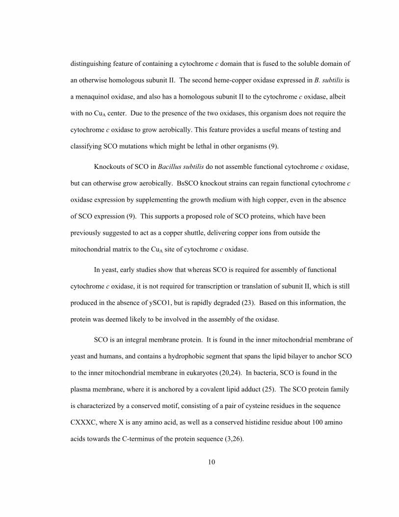
oxidase. Illustrated in Figure 1, electrons are transferred from cytochrome c to the dinuclear CuA
center of subunit II, resulting in the oxidation of cytochrome c (4).

2
Figure 1: Representation of Cytochrome c oxidase subunits I and II. Electron flow is indicated by grey arrows. Above: Structure of Paracoccus subunit II soluble domain, with side chains of CuA ligating residues displayed in blue (histidine residues) and yellow (cysteine residues).
3
These electrons progress down an electrochemical gradient, moving from the CuA center of
subunit II, through the heme center of cytochrome a in subunit I, to the O2 reaction center
cytochrome a3-CuB, located 5 Å away within the membrane in the same subunit. Cytochrome a3-
CuB is composed of a heme group as well as a single copper ion, and is the site for oxygen
binding during catalysis (4).
Cytochrome c oxidase requires many prosthetic groups as a functional enzyme; two
copper centres totaling three copper ions and two heme groups as described above, as well as one
magnesium, one zinc, and one sodium ion (6). Assembly of cytochrome c oxidase is therefore a
complex process, and has been shown to involve a number of assembly factors which are not
directly associated with the catalytic function of the oxidase (8). The core catalytic subunits of
the oxidase are similar between the mitochondrial enzyme in eukaryotes and some bacteria, such
as Bacillus subtilis. Thus, the assembly of the oxidase in B. subtilis should require some of the
same assembly factors as the eukaryotic enzyme, and this is indeed the case (9). The yeast
protein SCO1 has been shown to be required for the correct assembly of oxidase subunits I and II,
but is encoded within the nucleus of the cell (8). B. subtilis expresses a homolog of this protein,
referred to as BsSCO, short for Bacillus subtilis Synthesis of Cytochrome c Oxidase (9).
Expression of SCO homologues in Bacillus, yeast, and humans has been shown to be a
requirement for assembly of functional cytochrome c oxidase (8-10).
The specific role of cytochrome c oxidase assembly factors is a matter of some debate.
While the requirement for such factors is clearly demonstrable, the mechanism of action of these
assembly proteins is largely unknown. While some thirty complementation groups have been
identified in yeast alone that are required for assembly of the complete complex, there are few
that have been intensively studied (11). The assembly of redox-active oxidases requires several
prosthetic groups, all of which play a role in the central redox function of the enzyme complex.

4
However, these groups by their very nature have redox capabilities, and are controlled by the
enzyme by providing a very specific environment in which redox reactions can occur. For
example, the redox activity of copper is well established, and free copper within the cell has a
great deal of potential to produce oxidative damage. Therefore, there is a requirement for a class
of proteins known as metallochaperones which can bind to and limit the reactivity of such
elements within the cell (12). This concept is highlighted by the fact that the intracellular
concentration of free copper has been estimated at less than one free copper ion available per cell
(13). However, many enzymes, such as cytochrome c oxidase, require the incorporation of metal
ions in order to function properly. Thus, metallochaperones must have the ability to release and
provide these sequestered metal ions to specific proteins. Specific transporters and
metallochaperones are known for transition metals such as iron and copper, and these chaperone
proteins are conserved across many species (12).
Reactions catalyzed by transition metals are not the only redox-oriented problems faced
within the cell. Many proteins contain cysteine residues that are capable of undergoing forms of
redox chemistry other than oxidative damage. One of the best known examples of this is the
formation of disulfide bonds. These bonds occur as a covalent interaction between the thiol side
chains of two cysteine residues. Such a redox-driven change which can occur as a result of either
oxidizing cellular conditions or by a directed folding process, such as control by protein disulfide
isomerase in eukaryotes (14), thioredoxin, or by the Dsb family of proteins in prokaryotes such as
Escherichia coli (15).
Thioredoxin is one example of a protein that can act as a catalyst of redox change within
the cell. The thioredoxin protein family is found in both prokaryotic and eukaryotic cells, and is
characterized by a well-known motif that undergoes redox chemistry as part of the specific
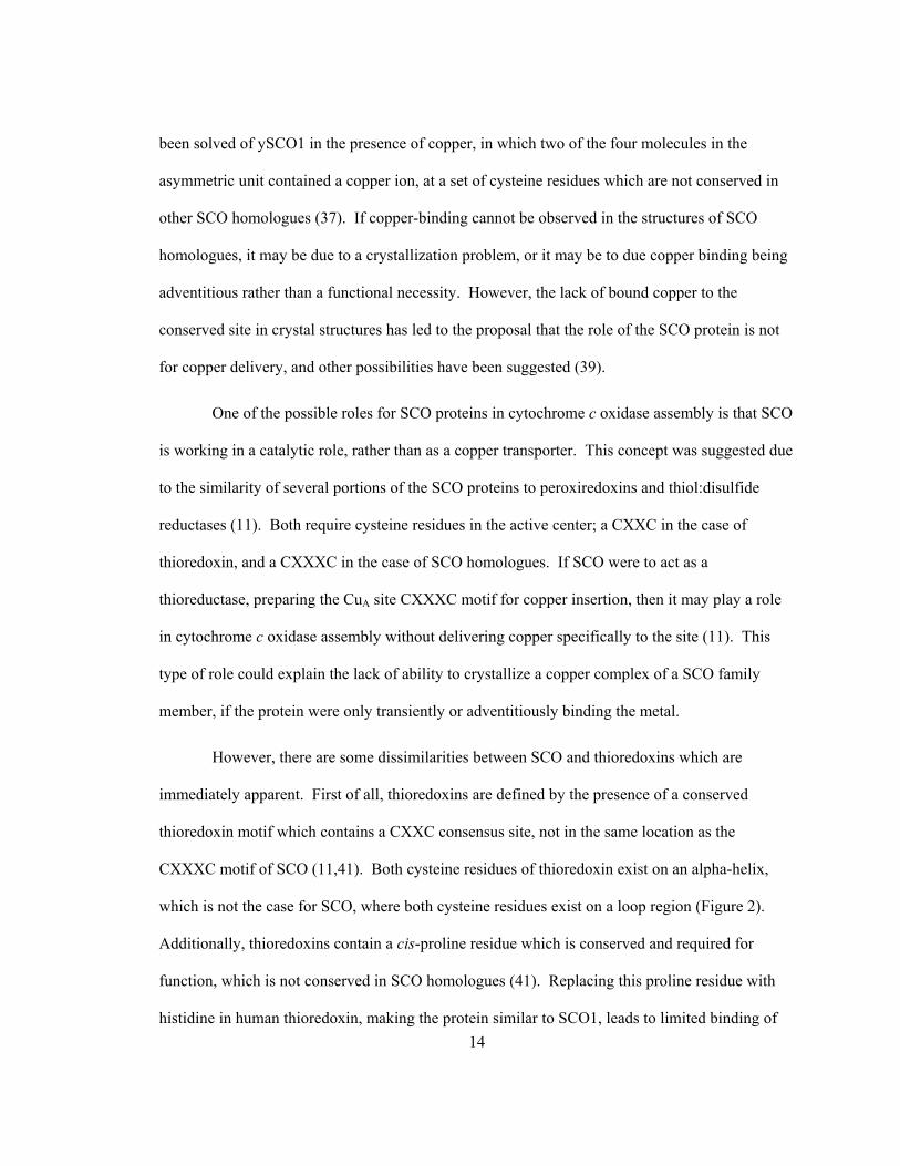
function of the protein. Proteins of this family have a characteristic thioredoxin fold, containing a

5
central beta-sheet structure surrounded by several alpha-helices, as exemplified in E. coli
thioredoxin 1, illustrated in Figure 2.

6
Figure 2: Structures of E. coli thioredoxin (top) and B. subtilis SCO (bottom) in cartoon ribbon form. Conserved cysteine motifs and conserved histidine residue have side chains displayed in blue and purple, respectively.
7
The redox-active CXXC motif lies on the N-terminus of an alpha-helix, and is generally found to
have redox activity in vivo, converting protein disulfides into reduced cysteine residues (15).
Many proteins whose role is to catalyze redox change in other proteins contain thioredoxin-like
motifs, including both protein disulfide isomerase and the Dsb family of proteins mentioned
previously. The redox-active motif in these proteins is proposed to have the same key residues as
thioredoxin, namely the CXXC motif. In fact, redox-active proteins are often characterized in
terms of their reduction potential at this site, and can be compared for redox activity (16).
Ultimately the job of redox-active proteins such as these is to maintain disulfide bonds within
cellular proteins in the correct arrangement for native protein folding, and to buffer the redox
state of the cell, which could be disrupted by influences such as catalytic amounts of transition
metal ions, or other oxidative stress.
There are multiple forms of oxidative stress which can occur within the cell. Recently,
this has been a focus of a great deal of research within fields such as cellular signaling and the
study of numerous types of cancer, as well as the cellular response to toxic environmental factors
(17). In an attempt to elucidate how these processes occur, many experiments have been
developed to test for a protein's ability to impart oxidative stress resistance. Commonly, the
application of peroxides to the cell has been used in these assays. Peroxides easily break down
into free radicals, which can quickly cause cellular damage and induce an oxidative stress
response within the cell. The class of proteins whose function is thought to be the management of
such oxidative stress is known collectively as peroxiredoxins (18). Peroxiredoxins, like
thioredoxins, are found in both prokaryotes and eukaryotes. These proteins utilize thioredoxin as
a source of reducing equivalents, in order to reduce peroxides and prevent damaging free radical
reactions within the cell. Peroxiredoxins operate via redox cycling of cysteine residues within the
protein, much like other redox proteins, but do not share a strictly conserved sequence motif.

8
Some peroxiredoxins contain two conserved cysteine residues, others contain one conserved and
one nonconserved cysteine residue, and some contain only one conserved cysteine residue and do
not require a second for function. Some of these proteins have been found to reduce other toxic
radical-forming compounds, such as peroxinitrite (which can form nitrite and sulfenic acid), in
addition to simple peroxide. Oddly, the reaction of peroxiredoxin has also been shown to
undergo oxidation as far as sulfinic acid at the catalytic cysteine residue. The sufinic acid form
can be re-reduced to the catalytic thiol state, a process which was previously not considered
reversible (18). This re-reduction is catalyzed by sulfiredoxin, and requires ATP and magnesium
in addition to a thiol for use as an electron donor (18).
Overall, the cell has a great number of ancillary proteins to manage the production of
oxidative damage within the cellular milieu. There are proteins for sequestration and transport of
catalysts for oxidative damage, such as Cox17 and SCO, proteins for the adjustment and
rearrangement of redox-driven covalent bond formation, the thioredoxins, or even proteins for the
management of radical-forming compounds, in form of peroxiredoxins. Since the heme-copper
family of oxidases is closely tied with aerobic respiration, it has been suggested that cytochrome c
oxidase may be a primary source of free radicals within the cell, and a large cause of oxidative
damage through by-products of the incomplete reduction of molecular oxygen. Additionally, the
prosthetic groups associated with the oxidase could also catalyze damaging oxidative reactions
within the cell. In this light, it becomes evident that the large number of assembly proteins which
have been discovered for cytochrome c oxidase have many important roles to fulfill in vivo, not
only for the ability to allow the cell to respire, but also to maintain chemical control of ions and to
maintain a balanced intracellular redox equilibrium.
9
Chapter 2 Literature Review
Cytochrome c oxidase is highly conserved across all aerobic organsims. It is a large
protein complex, requiring inorganic cofactors as prosthetic groups, heme molecules, and
multiple subunits for correct function; all assembled in a very specific manner. The enzyme may
at times be less than an ideal catalyst, producing peroxide and therefore helping to generate free
radicals within the cell (19), but it is also absolutely required for aerobic life. Due to its
importance, there have been many efforts directed at elucidating proteins that are required for the
assembly of the oxidase itself, and the prosthetic groups it contains. Early research was
conducted in yeast cells, and resulted in the discovery of the ySCO1 gene (8). ySCO1, for
Synthesis of Cytochrome c Oxidase, is a nuclear gene. Without ySCO1, yeast cytochrome c
oxidase subunit II cannot be assembled, resulting in respiratory deficient cells. Since its initial
discovery in yeast, research focusing on SCO1 has resulted in the discovery of homologues in
many aerobic organisms, including eukaryotes such as yeast (ySCO1 and ySCO2) and humans
(hSCO1 and hSCO2), and prokaryotes such as Bacillus subtilis (BsSCO) and Rhodobacter
sphaeroides (PrrC) (9,10,20,21).
Research has focused on the human homologues of SCO due to their relevance to
disease. Human hSCO1 has been associated with neonatal hepatic failure (22), whereas hSCO2
has been associated with fatal cardioencephalomyopathy (10). Yeast has continued to be used as
a model system, as it is a eukaryotic organism, containing mitochondria and therefore of
resemblance to the human system. Alternatively, our lab has focused on B. subtilis as a model
system, because B. subtilis expresses two members of the heme-copper oxidase family. B.
subtilis cytochrome c oxidase is similar to the mitochondrial enzyme, and also has the
10
distinguishing feature of containing a cytochrome c domain that is fused to the soluble domain of
an otherwise homologous subunit II. The second heme-copper oxidase expressed in B. subtilis is
a menaquinol oxidase, and also has a homologous subunit II to the cytochrome c oxidase, albeit
with no CuA center. Due to the presence of the two oxidases, this organism does not require the
cytochrome c oxidase to grow aerobically. This feature provides a useful means of testing and
classifying SCO mutations which might be lethal in other organisms (9).
Knockouts of SCO in Bacillus subtilis do not assemble functional cytochrome c oxidase,
but can otherwise grow aerobically. BsSCO knockout strains can regain functional cytochrome c
oxidase expression by supplementing the growth medium with high copper, even in the absence
of SCO expression (9). This supports a proposed role of SCO proteins, which have been
previously suggested to act as a copper shuttle, delivering copper ions from outside the
mitochondrial matrix to the CuA site of cytochrome c oxidase.
In yeast, early studies show that whereas SCO is required for assembly of functional
cytochrome c oxidase, it is not required for transcription or translation of subunit II, which is still
produced in the absence of ySCO1, but is rapidly degraded (23). Based on this information, the
protein was deemed likely to be involved in the assembly of the oxidase.
SCO is an integral membrane protein. It is found in the inner mitochondrial membrane of
yeast and humans, and contains a hydrophobic segment that spans the lipid bilayer to anchor SCO
to the inner mitochondrial membrane in eukaryotes (20,24). In bacteria, SCO is found in the
plasma membrane, where it is anchored by a covalent lipid adduct (25). The SCO protein family
is characterized by a conserved motif, consisting of a pair of cysteine residues in the sequence
CXXXC, where X is any amino acid, as well as a conserved histidine residue about 100 amino
acids towards the C-terminus of the protein sequence (3,26).

11
The conserved CXXXC sequence in SCO is shared with the sequence found in the CuA
site in subunit II of cytochrome c oxidase, where the metal binding site is composed of a
conserved HX34CXXXCXXHM sequence (27), and is illustrated in Figure 1. The presence of a
similar conserved motif may be important in the ligation of copper ions to both SCO and the CuA
site, and the idea has been suggested that the possible role of SCO within the cell is to deliver
copper directly to cytochrome c oxidase.
Studies of copper complexes in biological systems have demonstrated that the level of
free copper within the cell is essentially less than one free copper ion per living cell (13).
Therefore, transition metals in free solution within the cell exist at an extremely low
concentration, and instead are bound to chaperone proteins. In this way non-specific reactivity is
reduced to avoid unwanted and potentially damaging reactions within the cell. Thus, SCO has
been proposed to act as a copper shuttle, delivering copper specifically to the CuA site where it is
required as a redox centre, without allowing the passive diffusion of copper ions through the
cellular compartment (28).
SCO from yeast, human, and bacterial organisms has been shown bind to copper ions in
vitro (29-31). It does so via the cysteine motif (ie., -CXXXC-) combined with the conserved and
downstream histidine residue, evidenced by the fact that the mutation of any of these residues
results in a protein which does not exhibit copper binding activity. SCO proteins have been
shown to bind a single ion of either Cu(I) or Cu(II) oxidation states of copper (30-32), and are
proposed to work in conjunction with other proteins in order to deliver copper, imported from the
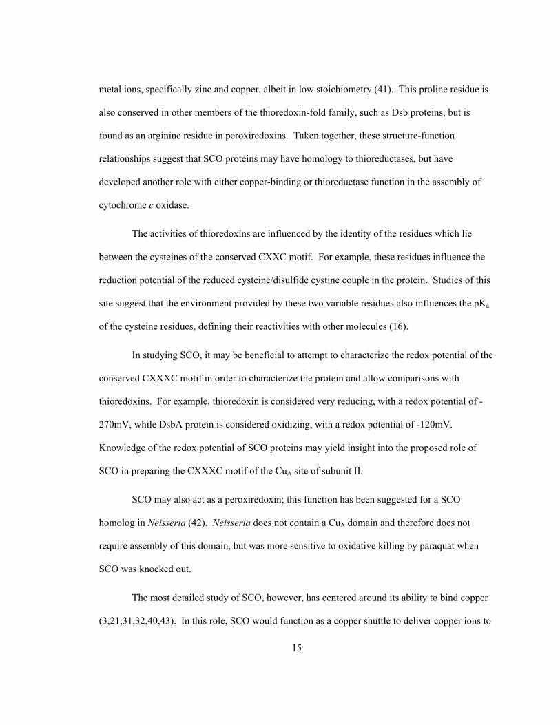
extracellular environment and delivered into the mitochondrial matrix in eukaryotes (Figure 3).

12
Figure 3: General scheme of copper transport in eukaryotic cells. Copper is acquired from the extracellular environment by Ctr1, and transported via Cox17 to SCO1, to be inserted into the CuA site of subunit II of cytochome c oxidase, within the mitochondria. Copper can also be transported via Cox17 to Cox11, to be inserted into the CuB site of cytochrome c oxidase.
The proposed series of copper-binding proteins involved in this pathway begins with a membrane
copper transporter of the Ctr family, and is one of three known pathways to distribute copper
within the cell. On the cytochrome c oxidase path, copper ions pass from Ctr to Cox17, a
cytosolic protein that exists in both yeast and humans (29,33). The yeast homolog of Cox17 has
been shown to bind copper, and is required for cytochrome c oxidase assembly. Knockouts for
Cox17 can be complemented by overexpression of the next protein in the yeast copper transporter
cascade, SCO1. However, reverse complementation does not occur: Cox17 overexpression
cannot complement ySCO1 knockouts (34). Copper ions can also pass from Cox17 to the Cox11

13
protein, implicated in the assembly of the CuB site within subunit I (35). Another protein
involved in this transport cascade is ySCO2, but the details of ySCO2’s function are not clear at
this time. It is known that yeast SCO2 can partially complement a Cox17 knockout if
supplemented with extra copper, but ySCO2 cannot complement a ySCO1 knockout. These
results suggest that ySCO1 and ySCO2 do not have identical function within the cell (34).
Human homologues exist for Cox17, ySCO1, and ySCO2 proteins. In humans, hSCO2
overexpression can correct oxidase deficiency due to hSCO2 mutations, but not hSCO1 (36).
The research described in this thesis has focused on BsSCO, the ySCO1 and hSCO1
homolog expressed in B. subtilis. In prokaryotes such as Bacillus subtilis, a homolog of SCO1 is
known, but searches for Cox17 have not yielded a protein of similar sequence (9). Since there is
only one membrane in this system, there is no requirement to transport copper ions through the
cell to a contained compartment such as the mitochondria. Since the oxidase is expressed on the
plasma membrane, metal-delivery proteins may not be absolutely required for functional
assembly of cytochrome c oxidase. Indeed, with supplementation of the growth medium by high
copper concentrations, functional oxidase can be assembled in BsSCO knockout cells (9).
The conserved CXXXC motif in SCO proteins can form an intramolecular disulfide bond
between the two cysteine residues within the motif, as well as being able to exist in a reduced
form (31,37). Only the reduced form of the protein has been reported to bind copper, while
oxidized BsSCO cannot (31).
The fold of SCO is thioredoxin-like, consisting of a central beta-sheet surrounded by
alpha-helices. The crystal structure of several SCO family members has been solved, including
BsSCO (crystal and solution structures), hSCO1 and ySCO1 (37-40). None of these structures
have been solved as a copper complex at the conserved CXXXC and H motif. One structure has
14
been solved of ySCO1 in the presence of copper, in which two of the four molecules in the
asymmetric unit contained a copper ion, at a set of cysteine residues which are not conserved in
other SCO homologues (37). If copper-binding cannot be observed in the structures of SCO
homologues, it may be due to a crystallization problem, or it may be to due copper binding being
adventitious rather than a functional necessity. However, the lack of bound copper to the
conserved site in crystal structures has led to the proposal that the role of the SCO protein is not
for copper delivery, and other possibilities have been suggested (39).
One of the possible roles for SCO proteins in cytochrome c oxidase assembly is that SCO
is working in a catalytic role, rather than as a copper transporter. This concept was suggested due
to the similarity of several portions of the SCO proteins to peroxiredoxins and thiol:disulfide
reductases (11). Both require cysteine residues in the active center; a CXXC in the case of
thioredoxin, and a CXXXC in the case of SCO homologues. If SCO were to act as a
thioreductase, preparing the CuA site CXXXC motif for copper insertion, then it may play a role
in cytochrome c oxidase assembly without delivering copper specifically to the site (11). This
type of role could explain the lack of ability to crystallize a copper complex of a SCO family
member, if the protein were only transiently or adventitiously binding the metal.
However, there are some dissimilarities between SCO and thioredoxins which are
immediately apparent. First of all, thioredoxins are defined by the presence of a conserved
thioredoxin motif which contains a CXXC consensus site, not in the same location as the
CXXXC motif of SCO (11,41). Both cysteine residues of thioredoxin exist on an alpha-helix,
which is not the case for SCO, where both cysteine residues exist on a loop region (Figure 2).
Additionally, thioredoxins contain a cis-proline residue which is conserved and required for
function, which is not conserved in SCO homologues (41). Replacing this proline residue with
histidine in human thioredoxin, making the protein similar to SCO1, leads to limited binding of
15
metal ions, specifically zinc and copper, albeit in low stoichiometry (41). This proline residue is
also conserved in other members of the thioredoxin-fold family, such as Dsb proteins, but is
found as an arginine residue in peroxiredoxins. Taken together, these structure-function
relationships suggest that SCO proteins may have homology to thioreductases, but have
developed another role with either copper-binding or thioreductase function in the assembly of
cytochrome c oxidase.
The activities of thioredoxins are influenced by the identity of the residues which lie
between the cysteines of the conserved CXXC motif. For example, these residues influence the
reduction potential of the reduced cysteine/disulfide cystine couple in the protein. Studies of this
site suggest that the environment provided by these two variable residues also influences the pKa
of the cysteine residues, defining their reactivities with other molecules (16).
In studying SCO, it may be beneficial to attempt to characterize the redox potential of the
conserved CXXXC motif in order to characterize the protein and allow comparisons with
thioredoxins. For example, thioredoxin is considered very reducing, with a redox potential of -
270mV, while DsbA protein is considered oxidizing, with a redox potential of -120mV.
Knowledge of the redox potential of SCO proteins may yield insight into the proposed role of
SCO in preparing the CXXXC motif of the CuA site of subunit II.
SCO may also act as a peroxiredoxin; this function has been suggested for a SCO
homolog in Neisseria (42). Neisseria does not contain a CuA domain and therefore does not
require assembly of this domain, but was more sensitive to oxidative killing by paraquat when
SCO was knocked out.
The most detailed study of SCO, however, has centered around its ability to bind copper
(3,21,31,32,40,43). In this role, SCO would function as a copper shuttle to deliver copper ions to

16
the CuA site where it is required. The exact nature of the copper ion, however, is a matter of some
debate.
Because the cytoplasmic compartment is a reducing environment it is assumed that
copper ions are found in the Cu(I) state. As such, copper is not believed to exist as Cu(II) within
the cell, and is thought to be bound to proteins as Cu(I) at virtually all times in the assembly
process (13). In agreement with this concept, SCO homologues have been shown in vitro to bind
to both Cu(I) and Cu(II) (30). This does not hold true of the subunit II CuA site in cytochrome c
oxidase, as this dinuclear copper centre is of mixed valence Cu1.5-Cu1.5 (27). Based on the
location of SCO within the cell, the binding of one ionic form of copper versus the other has been
called into question. If copper is always bound to chaperones within the cell and bound copper
exists in the Cu(I) form, and SCO is tethered to the inner mitochondrial membrane, then it would
seem SCO should only bind to Cu(I) in vivo regardless of its ability to bind Cu(II) in vitro.
Research focusing on delivery of copper to the cytochrome c oxidase complex by
metallochaperones has concentrated primarily on the Cu(I) state of copper (6).
In eukaryotes, if SCO homologues were only allowed access to Cu(I), as delivered from
Cox17 at the inner mitochondrial membrane, and Cu(II) binding was merely adventitious in this
case, other systems may not necessarily follow such a model. For example, in the case of
BsSCO, expressed in the plasma membrane of the bacterium, there is no limited access of the
protein to only the Cu(I) ion. Because of this, the protein could be exposed to both Cu(I) or
Cu(II) as both forms exist in the extracellular environment. If the protein were to bind Cu(II)
merely by coincidence, this would represent a problem in which normal binding and function
were being disturbed by a lack of specificity for the correct ion.

17
It has been observed that human SCO1 and SCO2 are both required for the correct
assembly of cytochrome c oxidase, and that both proteins play distinct roles in that assembly (36).
Based upon human SCO homologues, it has been proposed that Cox17 delivers copper to SCO2,
which delivers the copper directly to the CuA site of cytochrome c oxidase subunit II when
facilitated by SCO1 (36). In support of this model, Bacillus subtilis expresses both an oxidase
containing a CuA site and a SCO1 homolog, but no homologues of either Cox17 or SCO2 (9).
Furthermore, BsSCO is capable of forming functional oxidase without these other accessory
proteins. In Bacillus subtilis, in high copper conditions, functional oxidase can be formed
without even BsSCO (9). Although BsSCO is not an absolute requirement for CuA assembly its
presence allows for efficient assembly under conditions where very little copper is available.
Further study of the interaction between SCO1 or one of its homologues with copper could yield
further insight into the nature of this interaction, providing a model for the role of SCO1 in both
prokaryotic and eukaryotic systems.
Cytochrome c oxidase is an enzyme conserved across all aerobic organisms. Due to the
complexity involved in the system, many assembly factors have been discovered, and are being
characterized (35,44-46). These assembly proteins are encoded within the nucleus of the cell,
despite being required for the assembly of mitochondrially expressed oxidase subunits. These
assembly proteins, such as the SCO family of proteins, are also absolutely required for the
assembly of functional oxidase. The overarching role of SCO homologues is rather
straightforward. They are required for the assembly of the CuA site, and therefore cytochrome c
oxidase. How SCO fulfils this role is a much more complex question, with no clear answers at
this time. Many possibilities have been proposed, including the delivery of copper to the CuA
site, the redox preparation of the site without copper delivery, and even the reported ability to
sense or react with peroxides within the cell.
18
The details of copper’s interaction with BsSCO have not been fully determined. It is
known that BsSCO has an Kd for Cu(II) of less than 50nM (31). However, binding of Cu(I) to
BsSCO does not yield an observable electronic absorption spectrum, nor an observable change in
far-UV circular dichroism. Further research and critical questioning must be entertained before a
clear understanding will be developed of the SCO-copper interaction. Since the binding of Cu(I)
to BsSCO is not directly apparent, the observable signals produced by the binding of Cu(II)
provide an analog for what is considered the more physiological binding of Cu(I). Therefore, the
hypothesis in this work is that BsSCO is capable of binding Cu(II) in a high affinity interaction
that will stabilize the overall structure of BsSCO, in a reaction:
22 ,2 ( )e H Cuoxidized reducedBsSCO BsSCO BsSCO Cu II
− + +
⎯⎯⎯⎯→ ⎯⎯⎯→ −
This hypothesis will be assessed via three methods; differential scanning calorimetry to determine
a Kd for the BsSCO-copper interaction, observing the effect of copper on the rates of BsSCO
unfolding, and by examining the redox potential of the intramolecular disulfide bond within
BsSCO.
19
Chapter 3 Materials and Methods
3.1 Bacterial Expression and Purification
BsSCO was expressed as a soluble fragment of the full-length Bacillis subtilis BsSCO
protein. Primers used in the construction of soluble SCO were reported in (47). This sequence
was inserted into the pGEX-4T3 vector, giving a glutathione-S-transferase/SCO fusion construct
which was expressed in Escherichia coli DH5α cells. Cells were grown in 1.4L Fernbach flasks
in 700mL of LB media at pH 7.6 containing 100μg/mL ampicillin to an optical density of 0.6, at
which time construct expression was induced by addition of 1mM Isopropyl β-D-1-
thiogalactopyranoside. Cells were harvested 3 hours after induction by centrifugation and
resuspended in phosphate buffered saline (PBS) at pH 7.4 with 1mM EDTA and 100µM PMSF,
then frozen at -80°C.
Purification of BsSCO was accomplished by thawing cells grown from 1.4L of culture,
adding DTT to 5mM, and incubating with 1mg/mL lysozyme for 30 minutes on ice. Triton X-
100 was added to a concentration of 0.05%, and DNAse and RNAse were added to a
concentration of 37.5μg/mL and incubated for 10 minutes at 4°C, followed by 10 minutes at room
temperature. The cell extract was centrifuged at 3260 x g in a Beckman J6 centrifuge with a JS-
4.2 rotor for 20 minutes, and the pellet discarded. Supernatant was adjusted to pH 6.4 and
dialyzed twice against 10 volumes of PBS at 4°C once for 2 hours and once for 16 hours,
respectively. The supernatant was then loaded onto a 9mL Glutathione Sepharose 4 Fast Flow
column (Amersham) at 1.5mL/minute. The column was washed with 30mL of PBS at pH 7.7.
Two hundred units of thrombin in PBS pH 7.7 were added to the column and left at room
temperature for 16 hours. Cleaved BsSCO was eluted in PBS pH 7.7, and excess thrombin

20
removed by incubation with Benzamidine Sepharose 4 Fast Flow resin (Amersham) for 1 hour at
4°C, and elution in PBS (pH 7.7). Purity of the purified protein was verified by SDS-PAGE and
whole mass by qTOF MS/MS.
3.2 Preparation of Reduced and Cu(II) Bound BsSCO
All buffers used after the initial purification of BsSCO protein were passed over Chelex
resin (Sigma) to remove any trace contaminant metal ions from solution.
Samples were initially reduced by addition of 2mM DTT and incubated for 2 hours at
room temperature, followed by 5 rounds of ultrafiltration using 5mL sodium phosphate buffer at
pH 7.0 and Amicon Ultra-15 concentrators with a molecular weight cutoff of 10 000 Da
(Millipore). Alternatively samples were first denatured in 3.5M Gdn-HCl, washed with 5mL of
buffer and then reduced as described.
Cu(II)-bound BsSCO samples were generated by titrating reduced BsSCO with CuCl2
until no further absorbance change was apparent at 354 nm, representing a slight excess of CuCl2.
Samples were then passed over a Sephadex G25 desalting column (Pharmacia) to remove any
excess copper.
3.3 Electronic Absorption Spectra (copper binding, copper dissociation)
All absorbance spectra were measured on either a Hewlett-Packard HP-8452A diode
array spectrophotometer or a Cary 50 spectrophotometer. Quantification of both protein
concentration and bound copper were accomplished spectrophotometrically. BsSCO was
quantified using an extinction coefficient at 280nm of 19 200 M-1cm-1, while the quantity of
Cu(II) bound protein was determined by using extinction coefficient at 354nm of 4 780 M-1cm-1
(31).

21
3.4 Determination of Free Thiol Groups
Samples were tested for the presence of free thiol groups by their reaction with 4, 4'-
dithiodipyridine (DPDS) (48). In a split cell cuvette, 1mL of protein sample was measured
spectrophotometrically, then DPDS was added to the opposite cell and the spectrum measured.
After the mixing of protein with DPDS the resulting spectrum was recorded, and a difference
spectrum utilized to determine the number of protein thiol groups present which had
stoichiometrically interacted with the reagent, using an extinction coefficient at 324nm of
19 400 M-1cm-1 (38), and comparing this value to protein absorbance at 280nm, duplicating the
method used to measure free thiol groups on BsSCO reported in other work (31).
3.5 Intrinsic Tryptophan Fluorescence
Fluorescence data were recorded using a Horiba Jobin-Yvon Fluorolog-3 fluorescence
spectrometer, with 1nm excitation bandpass and either 2nm or 4nm emission bandpass. The
excitation wavelength was 280nm for all experiments, while emission spectra were scanned from
300 to 450nm in 1nm increments.
3.6 Circular Dichroism Absorbance (unfolding)
Circular dichroism spectra were measured using an OLIS RSM-1000 spectrometer and a
digital-subtractive circular dichroism module. For CD in the UV region the monochromator was
set up with 1.24mm slits and 2400 lines/mm gratings with a 230nm blaze angle. Spectra of 10μM
BsSCO samples in 10mM phosphate buffer (pH 7.0) were scanned from 260 to 180nm in 1nm
increments using a 0.1mm path length quartz cuvette (Hellma), in guanidine hydrochloride
solutions varying in concentration (0M to 5M) which had been mixed and allowed to equilibrate
prior to measurement.

22
3.7 Stopped-flow Unfolding Fluorescence
Stopped-flow unfolding experiments were performed in an OLIS RSM-1000
spectrometer using a fluorescence detection module and stopped-flow mixing module. Stopped-
flow mixing was accomplished ratiometrically using one syringe containing 2,3,4,5, or 6M Gdn-
HCl dissolved in buffer, mixed ten parts to one part of syringe two, containing 500μM SCO in
matched buffer. The excitation monochromator used a 1.24mm fixed slit and excitation
wavelength of 280nm, while spectra were collected from 300 to 450nm using a rapid-scanning
monochromator with a 2mm slit. Data were fit in a two step process carried out using the
SPECFIT (Spectrum Software Associates) software package. The spectral and kinetic principal
values of the matrix describing fluorescence intensity versus wavelength versus time were
estimated by singular value decomposition. These determinants were initially used to fit a kinetic
model which fits both the spectral intensity and the rates of the unfolding process for either two
or three discrete species over the timecourse of the experiment, starting with a three-state model
fit and thereafter simplified as much as possible. Final models were best generated with a two
state system in the case of reduced and oxidized BsSCO protein, whereas Cu(II)-bound BsSCO
did not conform to this model due to a slow increase in fluorescence in high ionic strength
solutions.
3.8 Copper(II) Dissociation from BsSCO in High Ionic Strength
Copper dissociation experiments were performed by preparing fully reduced BsSCO
samples via Gdn-HCl-induced unfolding, DTT reduction and washing, thiol counting, and copper
binding with removal of excess copper via size-exclusion chromatography. Copper-bound
protein fractions were pooled and concentrated to a working concentration of 1 to 2 mg/mL.
Solutions were prepared by dilution of a 6.1M (saturated) stock of NaCl with 25mM chelex-
treated sodium phosphate, pH 7.0. Absorbance spectra were taken from 240-600nm every 30s for
23
the duration of the experiment, at a constant temperature of 25°C. Two spectra of buffer were
recorded, then copper(II)-bound BsSCO protein was added to a final concentration of 10μM and
two spectra were recorded. Additional CuCl2 was mixed into the solution, and the cuvette was
sealed for the duration of the observation time.
3.9 Differential Scanning Calorimetry for Copper Binding
The redox status of samples for DSC was determined by counting available thiol groups
within an aliquot of the BsSCO sample. The remainder was then extensively washed with buffer
by ultrafiltration (at least 4 cycles of 5mL chelex-treated 25mM phosphate buffer, pH 7.0). All
buffers scanned in DSC experiments were from the same stock used to wash protein samples
prior to experiment, so as to be identically matched. 15μM BsSCO samples were scanned in a
VP-DSC Calorimeter (Microcal Inc) over a temperature range from 25°C to 95°C, at a scan rate
of 55°C per hour. All samples were thoroughly degassed and equilibrated to 18.6°C prior to
loading into the calorimeter, and initial equilibration was accomplished at 20°C for 30 minutes
prior to the start of each scan. Samples of BsSCO scanned with salt present were degassed as
described, then degassed concentrated salt solution was added to the desired final concentration
immediately prior to sample loading in the calorimeter.
3.10 Measurement of BsSCO Redox Potential
The redox potential of BsSCO was assessed by incubating a 10 μM sample of BsSCO in
25mM phosphate buffer, with various ratios of oxidized and reduced DTT at a concentration of
1mM, while shaking at a constant temperature of 37°C for a total of 48 hours to equilibrate the
solution. Protein samples were then precipitated using trichloroacetic acid (TCA) and
resuspended in SDS-PAGE sample buffer containing 8% 4-acetamido-4’-maleidylstilbene-2,2’-
disulphonic acid (AMdiS) before being run on 18% SDS-PAGE. Formation of a covalent adduct
24
of BsSCO with this reagent will induce a mobility shift upon electrophoresis in the case of free
accessible thiol groups, providing a means of differentially labeling reduced from oxidized
protein in different redox buffered conditions (49). Gels were stained with Coomassie Brilliant
Blue and imaged using the GelDoc 2000 system (Biorad), and densitometry performed using
GelDoc software (Biorad) to determine the relative intensity of the slower mobility versus higher
mobility bands.
25
Chapter 4 Results
4.1 Protein Purification and Identification
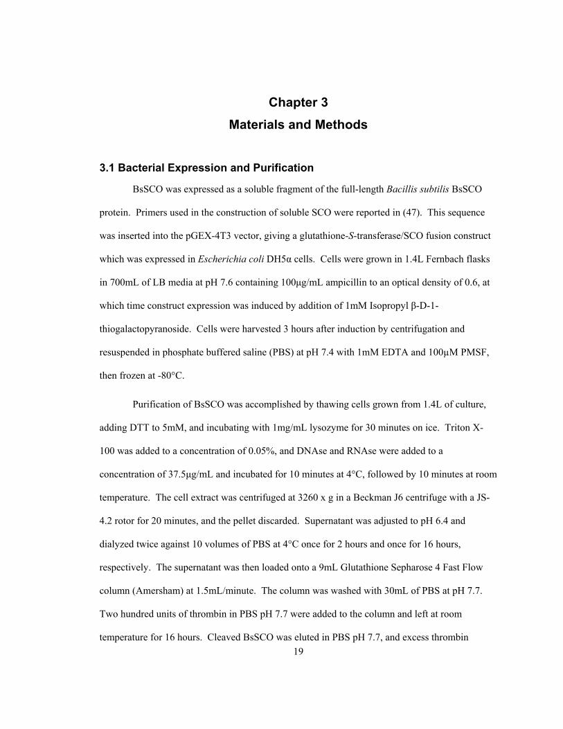
Purification of recombinant Bacillus subtilis SCO (BsSCO) protein typically yielded 5-
7mg of purified protein per liter of cell culture, verified for purity by SDS-PAGE. A Coomassie
blue stained gel of fractions from a preparation of recombinant BsSCO is illustrated in Figure 4.
Figure 4: SDS-PAGE of BsSCO purification stages. Lane 1: low range standards (Biorad) corresponding to molecular weights of 14 400, 21 500, 31 000, 45 000, 66 200, and 97 400. Lane 2, uninduced whole cells (2μg), lanes 3-5, whole cells at t=1,2,3 hours after induction with IPTG (6μg, 10μg, 10μg), lane 6, purified BsSCO protein (25µg), lane 7, cleaved GST fraction (30μg).

26
IPTG induction leads to an accumulation of a band with an apparent molecular weight of 46 000,
corresponding to the GST/BsSCO fusion protein. After cleavage with GST, the protein samples
have apparent molecular weights of 21 000 and 28 000, corresponding to BsSCO and glutathione-
s-transferase, respectively. Protein identity was also verified by use of electrospray mass
spectrometry. A mass spectrum of a purified BsSCO sample is shown in Figure 5.
Figure 5: ESI MS/MS whole mass spectrum of purified BsSCO protein. BsSCO was supplied at 1.0 mg/mL in 10mM ammonium bicarbonate buffer at pH 7.0 and diluted 1:20 with 1% formic acid prior to electrospray of sample for analysis. Scale given at left is relative to peak of greatest intensity, at 19 721.27 ± 0.15 Da.
The major peak is at 19 721.3 Da, compared to a theoretical mass of 19652.1 Da. This is
consistent with other reported preparations of the protein (47). Other minor peaks at slightly

27
higher molecular weight may correspond to sodium adducts of the protein, as they show a spacing
of 23 Da.
Purified BsSCO protein eluted from the column in varying degrees of reduction, from 0
free thiol groups per protein to 2 free thiol groups per protein. A typical spectrophotometric
determination of protein free thiol groups is illustrated in Figure 6.
Figure 6: Determination of free thiol groups present on BsSCO protein. Absorbance spectrum taken of 5µM BsSCO protein (—) before addition of 25 µM DPDS, and difference spectrum of DPDS and BsSCO in separate cells of split cuvette subtracted from BsSCO-DPDS products after mixing (- - -). The peak for the protein alone is at 280nm and the peak of thiopyridone product is at 324nm. Comparison of BsSCO absorbance peak at 280nm versus reaction product peak at 324nm allows quantification of reactive thiol groups. This sample represents 1.8 mol of available thiol per mol of BsSCO.
Absorbance at 280nm was used to determine protein concentration with an extinction coefficient
of 19.2 mM-1cm-1. Absorbance at 324nm was used to determine the free thiol concentration after

28
reaction with an excess of DPDS, using an extinction coefficient of 4.78 mM-1cm-1. The redox
state of the purified protein was not consistent between preparations, with some preparations
yielding higher numbers of reduced thiol groups per mol of protein than others. With treatment
by DTT, the number of free thiol groups per mol protein increased. Theoretically, with two
cysteine residues, reduced BsSCO protein should have yielded two mol of thiol per mol of
protein, and as such samples reduced to this extent were used as “reduced” protein in
experiments.
Oxidized samples were collected by omitting initial treatment of the cell extract with
DTT, while reduced samples were generated by treating final, purified protein with 2mM DTT. It
was observed that in many cases, use of standard Millipore-treated water to generate stock
solutions resulted in a diminution of total thiol groups available, and corresponding increase in
the absorbance at 354nm, corresponding to copper-bound BsSCO (Figure 7) (31).

29
Figure 7: Absorption spectra of reduced and Cu(II)-bound BsSCO. Dashed line (- - -) represents absorption spectrum of reduced 8.7μM BsSCO sample; solid line (—) is the same sample titrated to saturation with 10μM CuCl2. Titration with CuCl2 yields a BsSCO-copper complex with additional peaks at 354nm, 456nm, and 552nm, along with the protein peak at 280nm. Quantification of bound Cu(II) to BsSCO can be determined by measuring absorbance at 354nm.
In order to avoid this issue, all buffers used with purified protein were first passed over Chelex
resin to remove trace metal contaminants present in the solution.
4.2 Copper Binding to BsSCO
Upon titration of a BsSCO solution with CuCl2, an absorbance peak appears with a
maximum at 354nm, increasing stoichiometrically with CuCl2 addition until an equimolar BsSCO
to CuCl2 concentration is reached. Secondary and tertiary peaks are apparent in the BsSCO-
copper complex at 456nm and 552nm (Figure 7). Further addition of CuCl2 beyond 1 copper ion
per BsSCO molecule yields no further spectral changes.
30
BsSCO in solution exhibits intrinsic fluorescence arising from the aromatic residues in its
structure. A solution of BsSCO excited at 280nm yields a strong emission centred at 330nm, as
illustrated in Figure 8.
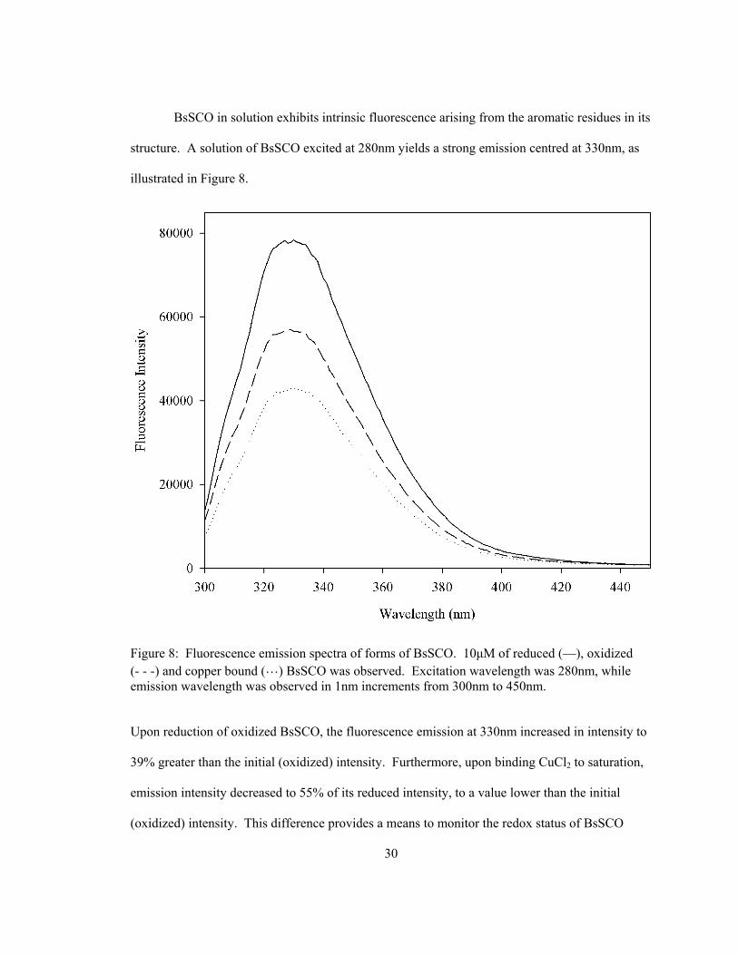
Figure 8: Fluorescence emission spectra of forms of BsSCO. 10μM of reduced (—), oxidized (- - -) and copper bound (…) BsSCO was observed. Excitation wavelength was 280nm, while emission wavelength was observed in 1nm increments from 300nm to 450nm.
Upon reduction of oxidized BsSCO, the fluorescence emission at 330nm increased in intensity to
39% greater than the initial (oxidized) intensity. Furthermore, upon binding CuCl2 to saturation,
emission intensity decreased to 55% of its reduced intensity, to a value lower than the initial
(oxidized) intensity. This difference provides a means to monitor the redox status of BsSCO

31
samples prepared from the same initial sample of protein. Individual samples could be reduced
via introduction of slight molar excess of DTT, with observation of a fluorescence increase at
330nm corresponding to protein reduction taking place over the course of approximately one
hour. Reduced protein could be titrated with CuCl2 to produce an immediate reduction in
fluorescence intensity. The fluorescence of oxidized protein did not change in the presence of
CuCl2.
4.3 Equilibrium Unfolding Experiments
Unfolding of BsSCO was initially accomplished by use of chemical denaturant
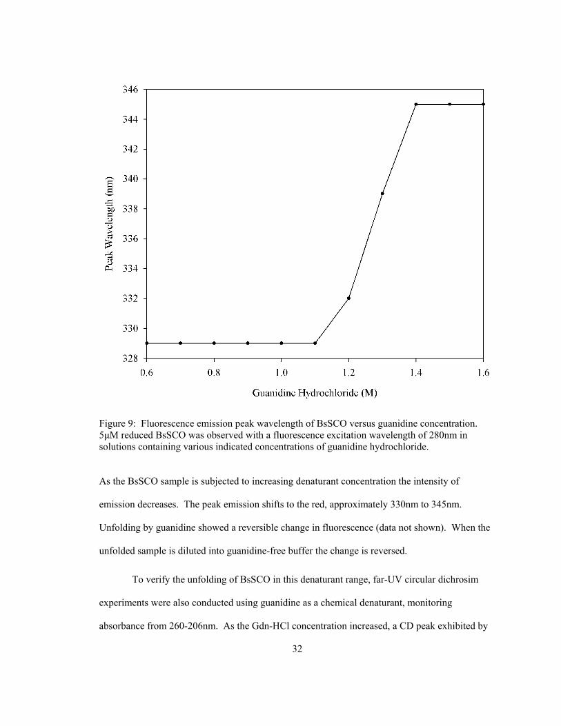
(Guanidine-HCl). The shift of the peak wavelength of intrinsic fluorescence from BsSCO is
centred at a midpoint at 1.25M Gdn-HCl (Figure 9).
32
Figure 9: Fluorescence emission peak wavelength of BsSCO versus guanidine concentration. 5μM reduced BsSCO was observed with a fluorescence excitation wavelength of 280nm in solutions containing various indicated concentrations of guanidine hydrochloride.
As the BsSCO sample is subjected to increasing denaturant concentration the intensity of
emission decreases. The peak emission shifts to the red, approximately 330nm to 345nm.
Unfolding by guanidine showed a reversible change in fluorescence (data not shown). When the
unfolded sample is diluted into guanidine-free buffer the change is reversed.
To verify the unfolding of BsSCO in this denaturant range, far-UV circular dichrosim
experiments were also conducted using guanidine as a chemical denaturant, monitoring
absorbance from 260-206nm. As the Gdn-HCl concentration increased, a CD peak exhibited by
33
the protein at 222 nm was lost. The CD-observable transition occurred with a midpoint at 1.2M
Gdn-HCl which is in agreement with the fluorescence data. The agreement of CD and
fluorescence signals support a two-state model for the unfolding equilibrium of BsSCO. These
results were consistent for reduced, oxidized, and copper(II)-bound BsSCO.
4.4 Stopped-flow Unfolding of BsSCO
Using a stopped-flow apparatus for rapid mixing of BsSCO with denaturant, intrinsic
tryptophan fluorescence was used to observe the unfolding of BsSCO with respect to time.
Fitting the data with two-state unfolding models was successful in the case of reduced (Figure 10)
and oxidized protein.

34
Figure 10: Unfolding kinetics of reduced BsSCO observed by intrinsic fluorescence. Kinetic trace of fluorescence observed at 350nm during unfolding of 50µM reduced BsSCO in 6M guanidine hydrochloride (bottom panel). Data collected at 1000 spectra per second for 4.0 seconds. Solid line (—) represents actual data collected while dotted line (…) represents modeled fit using a two-state unfolding model. Residuals of fit are displayed above the data (top panel).
Two-state unfolding model fits were unsuccessful for the copper-bound BsSCO samples, as rates
derived were not consistent and poor residuals were observed (Figure 11).
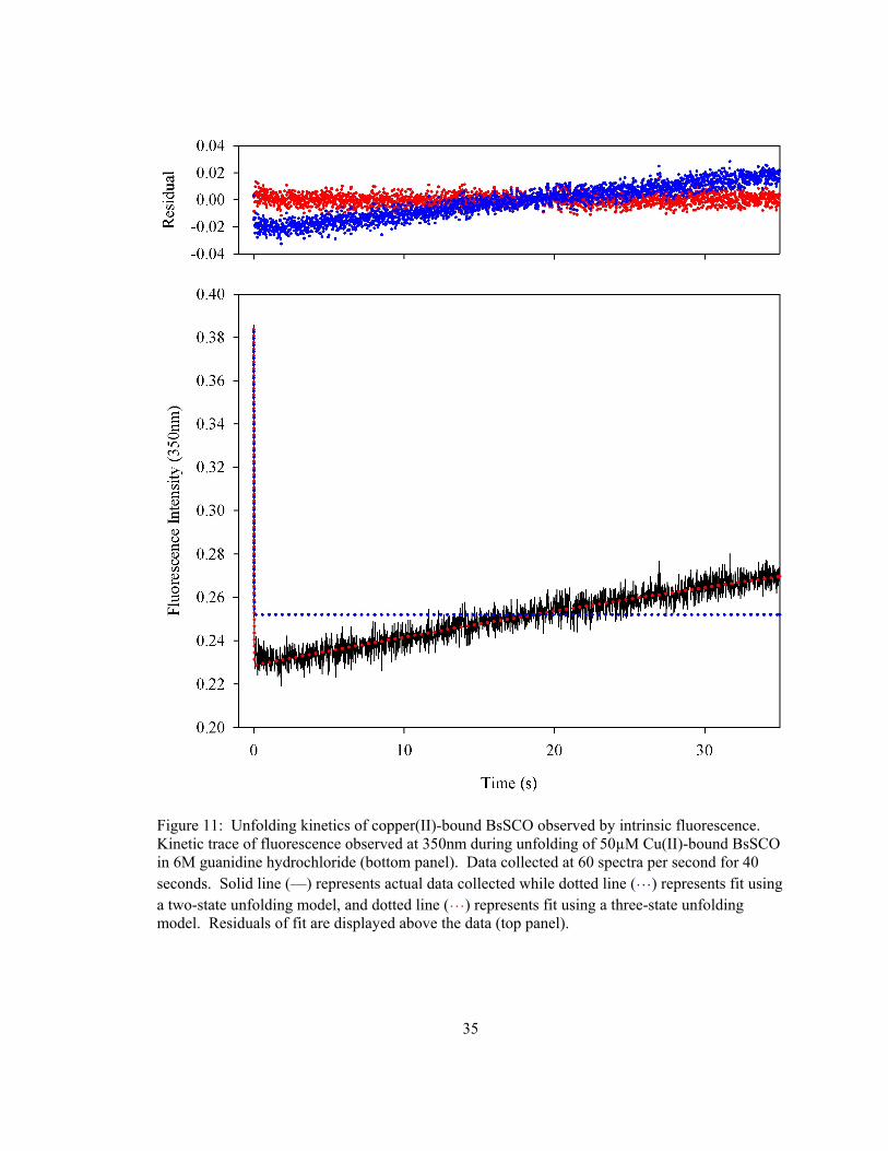
35
Figure 11: Unfolding kinetics of copper(II)-bound BsSCO observed by intrinsic fluorescence. Kinetic trace of fluorescence observed at 350nm during unfolding of 50µM Cu(II)-bound BsSCO in 6M guanidine hydrochloride (bottom panel). Data collected at 60 spectra per second for 40 seconds. Solid line (—) represents actual data collected while dotted line (…) represents fit using a two-state unfolding model, and dotted line (…) represents fit using a three-state unfolding model. Residuals of fit are displayed above the data (top panel).

36
Rate constants in this case showed some similarity if only fitting the first 0.5 seconds of data
captured, but were inconsistent if the entire timecourse was considered. Fitting a three-state
model system to the copper-bound BsSCO data was more successful, and showed a fast initial
rate with a much slower background rate, resulting in more randomly distributed residuals (Figure
11). Fitting the oxidized and reduced BsSCO data using this three-state model yielded a third
component spectrum which was essentially identical to the second.
The oxidized and reduced BsSCO sample data was fit by a two-state model. The
component spectra modeled by the singular value decomposition (SVD) algorithm of the oxidized
and reduced forms were of the two main types seen in equilibrium unfolding experiments, namely
the high-intensity peak at 330nm as an initial species, and the lower-intensity peak at 350nm as
the final species (Figure 12).

37
Figure 12: Component spectra generated when fitting a 2-state model. Data was collected during unfolding of 50µM reduced BsSCO in 6M guanidine hydrochloride (dataset identical to Figure 10). Initial species indicated by solid line (—) and final species by dotted line (…).
The three-state model does not improve the fit of data for oxidized and reduced BsSCO. In the
case of the copper-bound protein, the three component fit generated an entirely different spectral
form as the final species, particularly on the data which were collected over longer timescales,
shown in Figure 13.
38
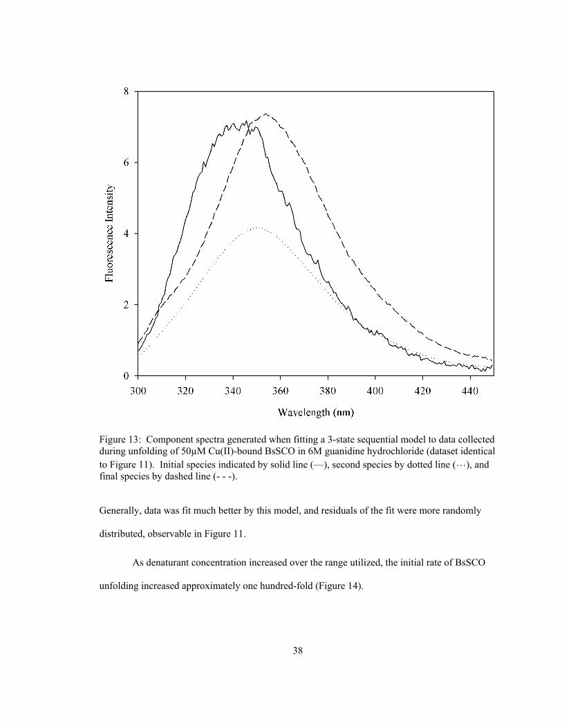
Figure 13: Component spectra generated when fitting a 3-state sequential model to data collected during unfolding of 50µM Cu(II)-bound BsSCO in 6M guanidine hydrochloride (dataset identical to Figure 11). Initial species indicated by solid line (—), second species by dotted line (…), and final species by dashed line (- - -).
Generally, data was fit much better by this model, and residuals of the fit were more randomly
distributed, observable in Figure 11.
As denaturant concentration increased over the range utilized, the initial rate of BsSCO
unfolding increased approximately one hundred-fold (Figure 14).
39
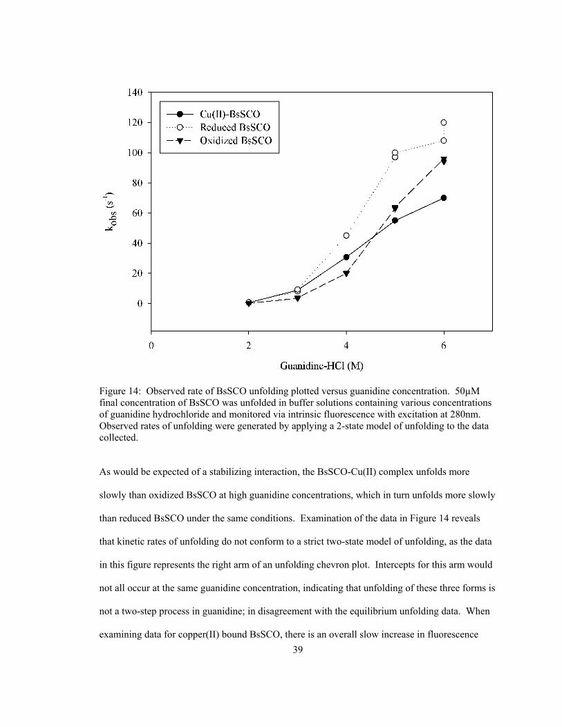
Figure 14: Observed rate of BsSCO unfolding plotted versus guanidine concentration. 50µM final concentration of BsSCO was unfolded in buffer solutions containing various concentrations of guanidine hydrochloride and monitored via intrinsic fluorescence with excitation at 280nm. Observed rates of unfolding were generated by applying a 2-state model of unfolding to the data collected.
As would be expected of a stabilizing interaction, the BsSCO-Cu(II) complex unfolds more
slowly than oxidized BsSCO at high guanidine concentrations, which in turn unfolds more slowly
than reduced BsSCO under the same conditions. Examination of the data in Figure 14 reveals
that kinetic rates of unfolding do not conform to a strict two-state model of unfolding, as the data
in this figure represents the right arm of an unfolding chevron plot. Intercepts for this arm would
not all occur at the same guanidine concentration, indicating that unfolding of these three forms is
not a two-step process in guanidine; in disagreement with the equilibrium unfolding data. When
examining data for copper(II) bound BsSCO, there is an overall slow increase in fluorescence
40
intensity at the peak wavelength which is apparent within the raw data prior to fitting models,
visible in Figure 11. When described as a three-state model, this intensity increase is fit as the
third component spectrum (Figure 13). The initial spectrum has intensity much lower than that of
the reduced form (Figure 12), as it is quenched by Cu(II) binding to the protein, while the final
spectral form of the Cu(II)-bound BsSCO is of similar magnitude to the final spectral from of the
reduced BsSCO sample. The third spectral component was investigated further, as it
distinguished the Cu(II)-bound protein sample as being different from the apo protein.
When unfolding was attempted using urea, no slow fluorescence change was observed in
the case of Cu(II)-bound BsSCO, unlike unfolding observed in guanidine.
4.5 Copper Dissociation from BsSCO in High Ionic Strength Solutions
Copper(II)-bound BsSCO samples were placed into solutions of high ionic strength,
made up by dissolving solid NaCl into buffer at pH 7.0, yielding a final protein concentration of
10µM. Absorbance spectra were taken with respect to time, and the absorbance peak at 354nm
was used in comparison to protein absorbance at 280nm to determine the quantity of copper
bound to BsSCO within the sample.
Solutions of copper(II)-bound BsSCO placed into high ionic strength conditions showed
a visible decrease in the absorbance peak centred at 354nm associated with Cu(II) binding (Figure
15).
41
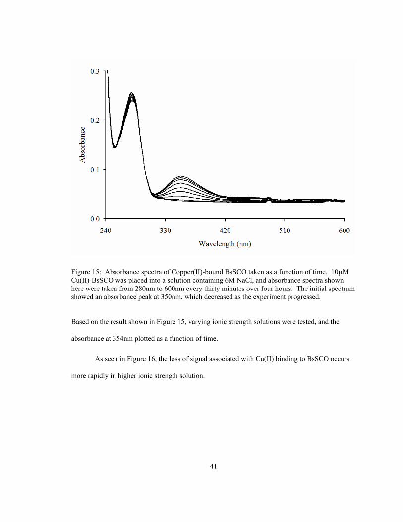
Figure 15: Absorbance spectra of Copper(II)-bound BsSCO taken as a function of time. 10µM Cu(II)-BsSCO was placed into a solution containing 6M NaCl, and absorbance spectra shown here were taken from 280nm to 600nm every thirty minutes over four hours. The initial spectrum showed an absorbance peak at 350nm, which decreased as the experiment progressed.
Based on the result shown in Figure 15, varying ionic strength solutions were tested, and the
absorbance at 354nm plotted as a function of time.
As seen in Figure 16, the loss of signal associated with Cu(II) binding to BsSCO occurs
more rapidly in higher ionic strength solution.
42
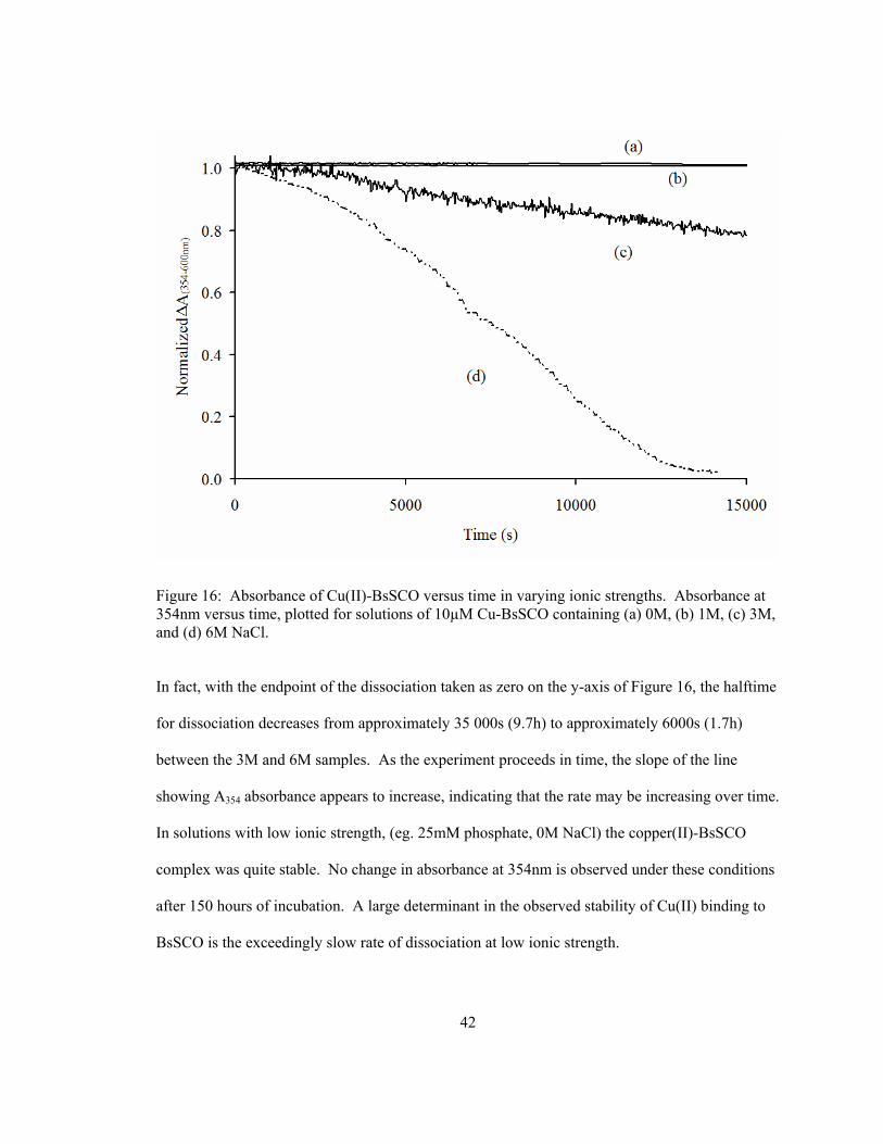
Figure 16: Absorbance of Cu(II)-BsSCO versus time in varying ionic strengths. Absorbance at 354nm versus time, plotted for solutions of 10µM Cu-BsSCO containing (a) 0M, (b) 1M, (c) 3M, and (d) 6M NaCl.
In fact, with the endpoint of the dissociation taken as zero on the y-axis of Figure 16, the halftime
for dissociation decreases from approximately 35 000s (9.7h) to approximately 6000s (1.7h)
between the 3M and 6M samples. As the experiment proceeds in time, the slope of the line
showing A354 absorbance appears to increase, indicating that the rate may be increasing over time.
In solutions with low ionic strength, (eg. 25mM phosphate, 0M NaCl) the copper(II)-BsSCO
complex was quite stable. No change in absorbance at 354nm is observed under these conditions
after 150 hours of incubation. A large determinant in the observed stability of Cu(II) binding to
BsSCO is the exceedingly slow rate of dissociation at low ionic strength.

43
In an attempt to elucidate a more specific condition required for release, poly-glutamic
acid was titrated into a BsSCO-Cu(II) solution. No release of copper was apparent by
observation of A354, nor was there in the case of poly-lysine titration (data not shown).
Furthermore, initial experiments consisting of mixing BsSCO-Cu(II) directly with the soluble
portion of Bacillus subtilis cytochrome c oxidase subunit II did not immediately generate a visible
spectrum associated with the formation of a CuA center, regardless of salt concentration or the
presence of DTT as a reductant for the site.
4.6 The Effect of Free Cu(II) in Solution with the Copper(II)-BsSCO Complex
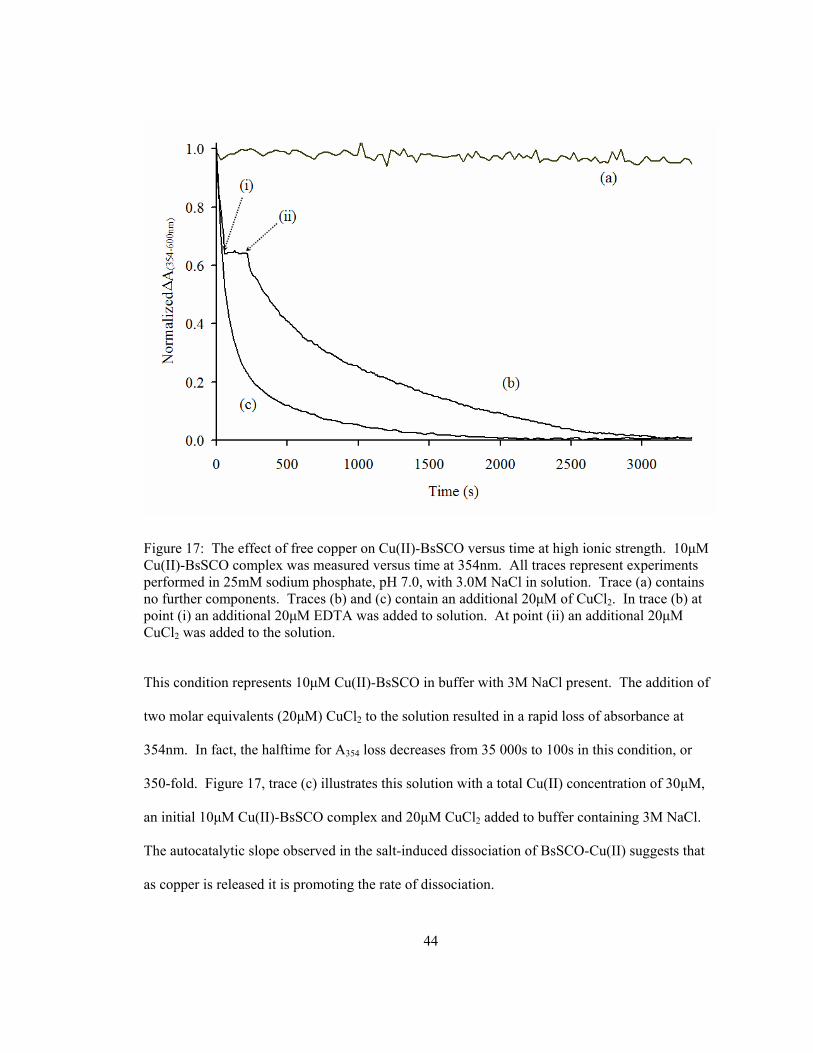
Dissociation is observed from 10μM Cu(II)-BsSCO in buffer with salt and the addition of
CuCl2, oberservable through an increase in the loss of absorbance at 354nm. Figure 17, trace (a)
is the same data plotted from Figure 16, trace (c).
44
Figure 17: The effect of free copper on Cu(II)-BsSCO versus time at high ionic strength. 10μM Cu(II)-BsSCO complex was measured versus time at 354nm. All traces represent experiments performed in 25mM sodium phosphate, pH 7.0, with 3.0M NaCl in solution. Trace (a) contains no further components. Traces (b) and (c) contain an additional 20μM of CuCl2. In trace (b) at point (i) an additional 20μM EDTA was added to solution. At point (ii) an additional 20μM CuCl2 was added to the solution.
This condition represents 10μM Cu(II)-BsSCO in buffer with 3M NaCl present. The addition of
two molar equivalents (20μM) CuCl2 to the solution resulted in a rapid loss of absorbance at
354nm. In fact, the halftime for A354 loss decreases from 35 000s to 100s in this condition, or
350-fold. Figure 17, trace (c) illustrates this solution with a total Cu(II) concentration of 30μM,
an initial 10μM Cu(II)-BsSCO complex and 20μM CuCl2 added to buffer containing 3M NaCl.
The autocatalytic slope observed in the salt-induced dissociation of BsSCO-Cu(II) suggests that
as copper is released it is promoting the rate of dissociation.

45
Figure 17 illustrates that upon addition of 20μM EDTA, complex dissociation and A354
loss stopped immediately and completely until further CuCl2 was added to the solution. Addition
of CuCl2 supplied a further source of Cu(II) ions after saturating the EDTA, and at this point
rapid dissociation continued until no remaining Cu(II)-BsSCO complex could be detected; an
interval of approximately 1 hour. This effect was tested with other metal ions, observing the
absorbance at 354nm in a 10μM Cu(II)-BsSCO in buffer with 3M NaCl present, and adding
20μM final concentration of six other metal ions (AgNO3, CaCl2, CoCl2, MgCl2, NiCl2, and
ZnCl2). None of the other metals tested were effective in promoting the dissociation of the
BsSCO-Cu(II) complex, but in every case the dissociation proceeded upon further Cu(II)
addition.
4.7 DSC Measurement of BsSCO Stability Changes Due to Copper(II) Binding
Previous ITC experiments gave an estimate for the equilibrium binding of Cu(II) by
reduced BsSCO of approximately 2x10⁷ M-1 (Kd ~ 50nM) (31). However, this determination is
the upper limit of viability for a direct ITC approach without using competitive binding
experiments. The tight binding interaction between BsSCO and Cu(II) by absorbance and
fluorescence based titration is confirmed here. In addition, we have seen that the very slow
dissociation rate contributes to the stability of binding. DSC affords another equilibrium
approach to quantitatively assess the tightness of ligand binding interactions by measuring the
increase in stability afforded to the protein upon ligand binding. The stability of the three
different forms of the BsSCO protein were assessed via the use of differential scanning
calorimetry (Figure 18).

46
Figure 18: DSC thermogram of BsSCO in buffer. 15μM BsSCO protein was scanned at 55°C per hour from 25°C to 95°C, and buffer scans subtracted from data for each of oxidized BsSCO (—), reduced BsSCO (- - -) and copper(II)-bound BsSCO (…).
The goal of the DSC experiments is to see if there is a difference in melting temperature
between the reduced and copper(II)-bound forms of SCO, in order to determine the stability
imparted to SCO by the binding of copper(II). The experiment was tested for scan rate
dependence, and the stability of the complex was determined to be under kinetic control, as with
slower scan rates than the initial rate tested (45°C per hour), the Tm observed for the copper(II)-
bound sample decreased. As such, the SCO-Cu(II) complex was likely dissociating during the
course of the experiment, and scan rate was increased to accommodate this difference and
47
maximize the difference in Tm between the reduced and copper(II)-bound forms of the protein.
The fastest scan rate to be used was determined to be 55°C per hour, as this temperature provided
the largest difference in Tm without resulting in an increase in the lower temperature protein
melting peaks.
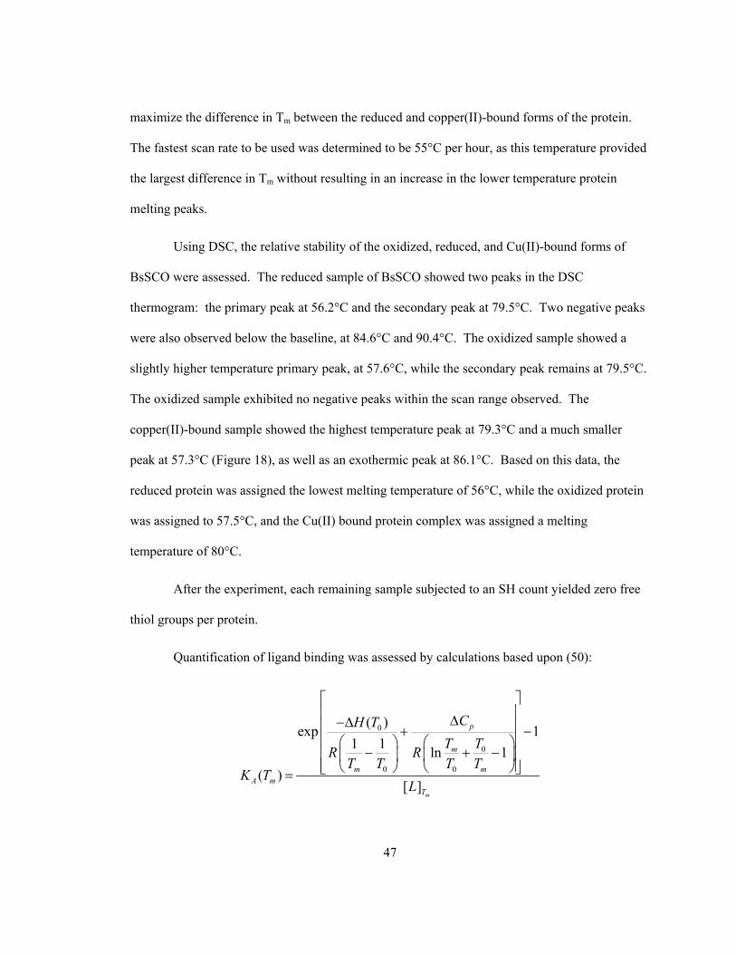
Using DSC, the relative stability of the oxidized, reduced, and Cu(II)-bound forms of
BsSCO were assessed. The reduced sample of BsSCO showed two peaks in the DSC
thermogram: the primary peak at 56.2°C and the secondary peak at 79.5°C. Two negative peaks
were also observed below the baseline, at 84.6°C and 90.4°C. The oxidized sample showed a
slightly higher temperature primary peak, at 57.6°C, while the secondary peak remains at 79.5°C.
The oxidized sample exhibited no negative peaks within the scan range observed. The
copper(II)-bound sample showed the highest temperature peak at 79.3°C and a much smaller
peak at 57.3°C (Figure 18), as well as an exothermic peak at 86.1°C. Based on this data, the
reduced protein was assigned the lowest melting temperature of 56°C, while the oxidized protein
was assigned to 57.5°C, and the Cu(II) bound protein complex was assigned a melting
temperature of 80°C.
After the experiment, each remaining sample subjected to an SH count yielded zero free
thiol groups per protein.
Quantification of ligand binding was assessed by calculations based upon (50):
0
0
0 0
( )exp 11 1 ln 1
( )[ ]
m
p
m
m mA m
T
CH TT TR R
T T T TK T
L
⎡ ⎤⎢ ⎥Δ−Δ⎢ ⎥+ −⎢ ⎥⎛ ⎞ ⎛ ⎞
− + −⎢ ⎥⎜ ⎟ ⎜ ⎟⎢ ⎥⎝ ⎠ ⎝ ⎠⎣ ⎦=

48
With T0=56.2°C, Tm=80°C, ΔH=98 440 cal/mole and ΔCp=3724 cal/mole/°C when
concentrations of both protein and ligand were 15µM, yielding a KA value of 2.86x10¹¹ M-1.
4.8 DSC Measurement of BsSCO Stability Changes Due to Ionic Strength Changes
As a difference in reduced BsSCO stability was observed via DSC with the protein bound
to Cu(II), and as the absorbance signal associated with copper(II) binding was lost in higher ionic
strength solutions, the stability of the BsSCO protein was examined under these conditions
(Figure 19).

49
Figure 19: DSC thermogram of BsSCO in 1M NaCl. 15μM BsSCO protein was scanned at 55°C per hour in 25mM phosphate buffer containing 1M NaCl, and reference scans subtracted from data for each of oxidized BsSCO (—), reduced BsSCO (- - -) and copper(II)-bound BsSCO (…).
Based upon the difference in DSC melting temperatures between apo-BsSCO protein and Cu(II)-
bound BsSCO (Figure 18), and the observation that higher ionic strength conditions result in a
loss of the absorbance signal associated with Cu(II) binding to BsSCO, it was logically expected
that higher ionic strength conditions would reduce the stability difference between apo and Cu(II)
bound protein. This was shown to be the case, as the DSC data presented in Figure 18, Figure 19,
and Figure 20 indicates a dependence of protein stability on the ionic strength of the solution.

50
Figure 20: DSC thermogram of BsSCO in 3M NaCl. 15μM BsSCO protein was scanned at 55°C per hour in 25mM phosphate buffer containing 3M NaCl, and reference scans subtracted from data for each of oxidized BsSCO (—), reduced BsSCO (- - -) and copper(II)-bound BsSCO (…).
Under 1M salt conditions, oxidized, reduced, and copper(II)-bound BsSCO no longer have
widely varying transition temperatures in the DSC thermogram. As is the case with lower ionic
strength (Figure 18), the oxidized form has a slightly higher melting temperature than the reduced
form of BsSCO, at 64.8°C versus 63.7°C, for a difference of 1.1°C. Both of these peaks are of a
higher melting temperature than the low salt condition. The copper(II)-BsSCO melting peak is
located at 65.2°C, much lower than the low salt condition, and only 0.4°C higher than the

51
oxidized form, much less than the 23.1°C shift observed between copper-bound and oxidized
forms of scans performed in lower ionic strength conditions (25mM phosphate buffer).
When these experiments were repeated 3M NaCl, the peak temperatures for oxidized,
reduced, and copper(II)-bound samples changed once more (Figure 20).
In 3M NaCl, the highest ionic strength condition which was tested, the thermogram peaks
for each protein state increased over both 0M and 1M NaCl conditions. Oxidized BsSCO showed
a melting temperature peak at 73.5°C, reduced BsSCO showed a peak at 70.8°C, and copper(II)-
bound BsSCO had a peak centred at 74.1°C. In 3M salt the peaks were much more closely
clustered than the buffer only condition, similar to the 1M salt condition.
4.9 Measurement of BsSCO Redox Potential
BsSCO samples (10µM ) were incubated in 25mM phosphate buffer (pH 7.0) containing
mixtures of oxidized and reduced DTT for 48 hours, then precipitated by addition of TCA and
labeled by resuspending the precipitated protein in buffer solution containing 8% thiol-reactive
AMdiS. Thiol-labeled BsSCO was then run on 18% SDS-PAGE and stained with Coomassie
blue (Figure 21).

52
Figure 21: SDS-PAGE of thiol-labelled BsSCO incubated in various redox buffers. SDS-PAGE of 10µM BsSCO was incubated in buffers containing mixtures of DTT and oxidized DTT, precipitated using TCA and labeled with AMdiS. Lane 1: low range standards (Biorad) corresponding to molecular weights of 14 400, 21 500, 31 000, 45 000, 66 200, and 97 400, Lane 2: 1mM/0mM DTT/ox-DTT, Lane 3: 1/1, Lane 4: 1/10, Lane 5: 1/20, Lane 6: 1/40, Lane 7: 1/80, Lane 8: 0mM/1mM DTT/ox-DTT.
Thiol-labeled BsSCO migrated with two distinct mobilities, both of which are apparent on the
stained polyacrylamide gel. The slower mobility is the predominant band in all BsSCO samples
containing any amount of DTT, while the faster mobility is more prominent in the sample

53
containing only oxidized DTT. Densitometry of these two species yielded the proportions of the
slower and faster mobility species (Table 1).
Table 1: Integrated density of slower and faster mobility species on SDS-PAGE. Data corresponds to density assessed for gel illustrated in Figure 21 of precipitated, covalently labeled BsSCO.
1mM
DTT 1:1 1:10 1:20 1:40 1:80
1mM
Oxidized
DTT
Slow (%) 88.3 88.4 87.4 85.8 91.6 86.9 9.6
Fast (%) 11.7 11.6 12.6 14.2 8.4 13.1 90.4
Densitometry performed on the faster and slower migrating bands yielded their proportions
relative to the total density between the two. In all cases where any amount of DTT was present,
greater than 85% of the density was composed of the slower moving species. Only in the absence
of DTT was the proportion of density higher in the more highly mobile species. Repetition of this
experiment using reduced and oxidized glutathione as a redox pair in place of DTT, as well as
using cysteine/cystine, did not yield any bands corresponding to the slower mobility species (data
not shown).

54
Chapter 5 Discussion
5.1 Measuring Cu(II) binding to BsSCO
In order for SCO proteins to fulfill a role in copper delivery, such as that illustrated in
Figure 3, they must achieve several important functions. For copper delivery to occur as assisted
by a SCO protein, the protein must first bind to copper. In the case of eukaryotic SCO proteins,
where copper must be delivered to internal cellular compartments, it is proposed that Cox17
protein acts as a shuttle to deliver copper ions to SCO1 (29,33). However, in prokaryotes, such as
Bacillus subtilis, there is only one cellular membrane, within which all integral membrane
proteins must reside. In either case, the SCO protein must first bind copper ions, then recognize a
target molecule for copper delivery. The final step of copper transfer would be the requirement
for release of bound copper from the SCO molecule, allowing its uptake into the target site.
Without the ability to bind copper, recognize the target site, and release copper into that site, SCO
molecules cannot function as copper metallochaperones.
Copper binding to BsSCO has been reported by multiple groups (31,32). Binding of
copper has also been reported for homologues of Bacillus SCO, including human SCO1 (30) and
yeast SCO1 (3), and has been suggested to be the primary function of these proteins in vivo.
Previous study of Bacillus SCO has shown a 1:1 stoichiometry of BsSCO-Cu(II) binding with a
Kd for the interaction of 65.0 nM given as an "upper limit" (31). This investigation suggested that
further characterization of this binding constant would likely require competitive binding
experiments.

55
The primary difficulty associated with this determination is the tight binding between
BsSCO and Cu(II). Tight binding is generally associated with conformational change, and this
may impart a difference in stability upon the protein, resulting in a measurable change in the ΔG
which can be compared under different conditions. One method to determine this value is
through an equilibrium experiment designed to measure the difference in stability between the
apo and Cu(II) bound forms of BsSCO following treatment with a chemical denaturant. If the
BsSCO-Cu(II) complex were more resistant to chemical denaturation by guanidine
hydrochloride, this difference could be translated into ΔΔG and thereby provide some measure of
stability imparted by copper binding. There is no apparent difference in equilibrium stability
versus guanidine concentration between apo and copper(II) bound BsSCO, as both samples
unfolded at the same concentrations of denaturant when measured by tryptophan fluorescence or
by circular dichroism. Data from equilibrium experiments allowed establishment of unfolding
conditions. In order to look more closely at the difference in stability the kinetics of BsSCO
unfolding in guanidine were measured both for apo-BsSCO and the BsSCO-Cu(II) complex.
Due to its high sensitivity and the rapid timescale of measurement, fluorescence was
chosen as the experimental method to measure unfolding. Circular dichroism data collected via
equilibrium experiments generally agreed with the unfolding conditions defined by fluorescence.
In equilibrium unfolding experiments, fluorescence intensity was always observed to
decrease, while the peak fluorescence wavelength shifted from 330nm to 347nm. Kinetically,
this same pattern was observed in the two-state modeled data for oxidized and reduced forms of
BsSCO. In the copper bound sample the component spectra of the 3-state model show the same
pattern of fluorescence decrease and red shift, but were then followed by a subsequent increase in
fluorescence intensity which was not observed in the unfolding of apo-BsSCO samples.
56
After unfolding, fluorescence intensity increased in the Cu(II)-bound BsSCO sample,
where in all equilibrium experiments only a decrease in intensity had been observed. Combined
with the prior knowledge that Cu(II) binding quenches the fluorescence of folded BsSCO protein,
it was inferred that the phenomenon occurring was likely a dissociation of bound Cu(II) from
unfolded BsSCO, leading to the work conducted on ionic strength dependence of Cu(II) binding
to BsSCO. A different approach was used to obtain a binding constant (Kd) for the BsSCO-
Cu(II) interaction by observing stability differences in the protein using only heat as a denaturant.
5.2 Measurement of BsSCO Stability via Differential Scanning Calorimetry
Using DSC, the relative stability of the oxidized, reduced, and Cu(II)-bound forms of
BsSCO were assessed. It was expected that the oxidized and reduced forms of the protein would
show a difference in stability due to the fact that an intramolecular disulfide bond would constrain
the protein somewhat in the oxidized form, disallowing some protein conformations that the
reduced form would be free to explore. Surprisingly, the disulfide bond formed between the
ligand-binding cysteine residues did not confer a large difference in stability between the
oxidized and reduced forms; the primary peak only differed by 1.4 C° between these two samples
(Figure 18). Based on previously reported ITC data for the interaction between BsSCO and
Cu(II), the binding of these molecules can be characterized as tight (Kd ~65nM) (31). As such, a
large shift in Tm between the apo form and Cu(II) bound form of the protein can be expected,
assuming that the tight binding of Cu(II) to BsSCO would stabilize the protein conformation.
Since the observed shift in Tm between the reduced and Cu(II)-bound forms of the protein is
23.1 C°, this seems a logical explanation. Working from this assumption, it is possible to model
the binding interaction using information derived from DSC data (50). By using the Tm values
for both the reduced and copper-bound forms of BsSCO, and assuming a stoichiometry of one

57
copper ion per BsSCO molecule (31) with ΔH and ΔCp derived experimentally, the interaction
between BsSCO and Cu(II) was modeled to have a Kd of 3.5 x 10⁻¹² M (50). This represents very
tight binding, which is expected of a protein which does not observably dissociate from its ligand
over 150 hours. Binding on a picomolar scale also indicates that fairly extreme conditions would
be expected in order to induce dissociation from the ligand; this also appears to be the case.
When the protein is completely unfolded in urea at low ionic strength (confirmed via a change in
fluorescence), it still exhibits the absorbance peak at 354nm associated with Cu(II) binding.
However, if the same experiment is repeated where Cu(II) is added to unfolded protein, no
binding event occurs and the scattering component of the absorbance spectrum becomes the
dominant feature (data not shown).
All DSC scans contain elements of both the lower and higher temperature peaks.
Presumably this is because the sample is not completely homogeneous; the reduced and oxidized
scans contain a major peak at ~56°C and ~57.5°C, with a minor peak at ~79°C (Figure 18). It
seems likely that the protein may contain trace amounts of bound Cu(II) which are not
immediately visible via absorbance at 354nm due to a relatively low extinction coefficient
compared to the extinction coefficient at 280nm. The reduced protein is highly susceptible to
contamination due to the presence of trace amounts of copper found in syringes used to handle
the material or the calorimeter cell itself. Any free copper encountered would almost certainly
result in contamination, as the binding has a dissociation constant of 3.5 pM. Finally, while the
major peak found for the copper(II)-bound protein was at ~79°C, there was also a small peak
found at ~57°C, indicative of the presence of some component fraction of oxidized BsSCO
(Figure 18). This likely represents some fraction of protein which was reduced but which
spontaneously reoxidized to the disulfide form prior to being bound to copper. A change in

58
experimental procedure wherein all steps are performed anaerobically could potentially remove
this contamination.
Of note is the presence of negative peaks above ~80°C in the reduced and Cu(II)-bound
samples, and their absence in the oxidized sample (Figure 18). These peaks were exothermic,
which indicates the presence of a spontaneous reaction. Since these peaks occur at high Tm,
rather than proceeding spontaneously at a lower temperature, this also seems to suggest that some
specific conformational change must occur before this process can occur; presumably the
unfolding change undergone by the protein which yields the endothermic peak observed at
~79°C. Following the assumption that endothermic peak is an observation of the unfolding of the
Cu(II)-bound complex, it is possible that the exothermic process being represented by the
negative peaks may be the oxidation of the free thiol residues into a disulfide bond. The Cu(II)
bound sample would be expected to have available cysteine residues to contribute to such a
thermodynamic change, as both residues must first be present in a reduced form in order to
initially bind to the Cu(II) ion, and upon its release should have the ability to allow the formation
of a disulfide bond. The oxidized protein sample, which does not contain free thiol groups, does
not exhibit the exothermic phenomenon common to the reduced or Cu(II)-bound samples. This
follows previous suggestions that human SCO ligation to Cu(II) involves bonding to these
residues, based on the observation of charge-transfer interactions between copper and sulfur
atoms (30), as well as the observation that all DSC samples were devoid of free thiol groups after
experimentation.
A Kd of 3.5pM for the interaction of BsSCO with Cu(II) has been determined via DSC.
However, it is not necessarily an absolute value, as some scan rate dependence of the interaction
was observed during experimental setup stages. This form of kinetic control resulted in the
apparent effect of the higher stability peak shifting downwards to lower temperatures, in the case

59
of the Cu(II)-bound BsSCO samples. This apparent shift was minimized by the use of higher
scan rates, but cannot be completely ignored, and time-dependent effects will still be occurring in
the background during the experiment regardless of scan rate. The implication of this observed
kinetic dependance is that high scan rates minimize but do not completely eliminate the down-
shifting of the high Tm peak, and thus the actual peak in the theoretical absence of this
phenomenon would be of a higher temperature. Therefore, a greater difference in stability would
be assessed, and a tighter binding constant would be calculated for the BsSCO-Cu(II) interaction.
By this argument, the Kd determined as 3.5 pM should be taken as an upper limit for the
interaction, despite the fact that this is a very tight binding constant. Tight binding of this
magnitude is reasonable, as other metal binding proteins also have affinities reported in this range
(51), and human SCO1 has a reported Kd for Cu(I) approaching 10-17M (52).
Despite determining a Kd, this does not necessarily provide a clear picture of the BsSCO-
Cu(II) interaction. While the melting temperature gives an indication of complex stability, and
thereby a Kd value for the components of the complex, the stability of the complex could also be
altered by other factors. This is evident in the BsSCO-Cu(II) interaction where dissociation was
observed, wherein the stability of the BsSCO-copper complex is obviously and dramatically
affected by the ionic strength of the solution.
5.3 Copper Dissociation from the Copper(II)-BsSCO Complex
This work is the first to focus on and demonstrate the release of copper from SCO, in this
case Cu(II) from BsSCO.
The initial dissociation occurs relatively slowly, but increases in rate as the time course
proceeds (Figure 16). This is visible by the increasing negative slope of the line representing the
BsSCO-Cu(II) complex with respect to time. Since the ionic strength of the solution is not

60
appreciably changing during the experimental timeframe, all other experimental components
were examined to investigate the changing rate. The initial assumption was that dissociation
would be a pseudo-first order process, because the high concentration of NaCl relative to protein
should be a constant ratio over the course of the experiment. The buffer concentration was also
high relative to the protein (a 2 500-fold excess) and therefore also not appreciably changing.
The final solution component was the copper-bound protein itself, presumably dissociating as
( ) ( )BsSCO Cu II BsSCO Cu II− ↔ +
and therefore increasing the concentrations of apo-BsSCO and free Cu(II), respectively. Based
on this analysis, the dissociation reaction should show a dependence on either free BsSCO in
solution or free Cu(II) in solution.
The easiest component to alter was copper, so the hypothesis that free Cu(II) in solution
was affecting the rate of BsSCO-Cu(II) dissociation was tested. The observation that CuCl2
increases dissociation rate while EDTA stops the dissociation suggests that there is some
requirement for free copper ions in solution in order to cause the dissociation of an otherwise
stable complex.
When the experiment was repeated with AgNO3, CaCl2, CoCl2, MgCl2, NiCl2, and ZnCl2,
no interaction was observed within a five minute timeframe, but in every case the dissociation
proceeded upon Cu(II) addition. This suggests that the rapid dissociation effect is not in fact
mediated purely by metal ions, but much more specifically by Cu(II).
The fact that high ionic strength is required for dissociation belies some other unknown
condition which must be fulfilled prior to copper ion release. BsSCO contains a large number of
charged residues, and it may be possible that the high ionic strength required to cause complex
dissociation is mimicking some other physiological process. Dissociation of the BsSCO-Cu(II)

61
complex must occur for copper delivery to be viable, but it is unlikely that BsSCO will encounter
ionic strength as high as was required for the observed complex dissociation in vitro. It is
possible that the experimental conditions determined in vitro for copper ion release could be a
mimic for the interaction between BsSCO and the CuA site of cytochrome c oxidase.
Unfortunately, experiments mixing BsSCO-Cu(II) with poly-acidic or poly-basic peptides did not
yield a dissociation of the complex, nor did the addition of purified Bacillus subtilis cytochrome c
oxidase subunit II. This data suggests that either other conditions or cofactors are required in vivo
for the formation of CuA site assembly.
However, the fact that free copper ions are required in order to induce dissociation does
lend evidence to the model for BsSCO as a copper chaperone protein. The cytochrome c oxidase
CuA site is composed of a dinuclear copper center. It has been known for some time that the
BsSCO does in fact bind to Cu(II) with high specificity, as well as binding other metals such as
Ag(I) and Cu(I) (31,32). BsSCO has also been reported to bind Ni(II) and Co(II), as well as
being reported not to bind these same two metals in a different body of work (31,32). However,
in the present study none of these metals with the exception of Cu(II) had any effect on the
observed dissociation. Such a marked impact on the interaction being observed lends itself to the
hypothesis that there may be a specific binding site for a second Cu(II) ion with the BsSCO-
Cu(II) complex. Such a site has not been reported in the case of Bacillus SCO, but a secondary
copper binding site has been suggested for yeast SCO (37). However, the proposed second Cu
site on yeast SCO1 involves two cysteine residues which are not conserved in other homologues
of the protein, and may represent a crystallographic artifact due to their appearance on only two
of the four molecules making up the asymmetric unit. In the case of Bacillus SCO, it has been
proposed that the Cu(II) ion known to bind the protein is coordinated by two sulfur atoms as well
as a nitrogen atom, as this modeling is consistent with data generated by EPR and EXAFS
62
experiments (32). The site exhibits apparent 4-coordinate square planar geometry, and as the
conserved residue ligands only compose three of these four binding partners, another ligand is
suggested. This fourth ligand has been proposed to be an endogenous oxygen ligand, combined
with the presence of a water molecule in an axial conformation (32). If this is the case, the ionic
strength dependence of dissociation may be a result of a disruption in the geometry of the site. If
the geometry of the site is such that there is a transient contact with the water in the surrounding
solution, it is possible that charged ions may enter the site, disrupting the charge-based
interactions with the fourth endogenous oxygen ligand, and allowing copper release from the site.
However, this model does not account for the strict requirement for an extraneous copper ion to
be present in order to observe dissociation.
The observation that two copper ions are required to induce dissociation may be
rationalized in terms of redox chemistry. The reduced BsSCO protein contains two free thiol
groups, which are utilized in binding Cu(II) (32). If copper were released, it could be released as
either one of two redox states: Cu(I) or Cu(II). Based on the observations that: (1) copper
binding requires reduced BsSCO; (2) after copper release no free thiol groups are present in
BsSCO; and (3) the protein is re-reduceable by DTT; it seems likely that the oxidized form of
BsSCO exists after copper release. Based on this assumption, the reducing equivalents required
to generate reduced BsSCO from oxidized BsSCO must have been taken up by another
component of the solution which is capable of undergoing redox chemistry. The first possibility
is an interaction with molecular oxygen, present as a dissolved gas due to the aerobic nature of
the experiment. The second possibility is the Cu(II) present in the solution interacting, which is
reduceable to Cu(I). Since the reduced protein has a reported half-life of approximately 32h at
room temperature (31), it seems unlikely that oxygen is the principal oxidizing component on a
timescale of one hour. In fact, in 3M NaCl the dissociation and oxidization occurs with a

63
halftime of close to ten hours, but that halftime decreases to less than five minutes with the
addition of extraneous Cu(II). This strongly indicates that copper is the oxidizing agent within
this experimental framework. A reaction centred around copper as the oxidizing agent in this
case could appear as:
2 12 2 2 2RSH Cu RSSR Cu H+ + ++ → + +
If this model holds true, any excess free copper within the solution could catalyze
dissociation of the protein-copper complex, provided the ionic interaction condition was first
satisfied at the copper binding site. By this mechanism it is possible to generate a redox cycle
based on oxidizing Cu(I) to Cu(II) via the dissolved oxygen within the solution. Such a redox
cycle would provide a source of renewable oxidizing copper(II) within the solution, such that
only a catalytic amount would be required to generate the conditions necessary to oxidize the
protein. The rate of this reaction would be dictated by the quantity of dissolved oxygen in
solution, the ionic strength of the solution, the presence of catalytic quantities of free copper, and
the continued accessibility of molecular oxygen as an electron sink.
This conceptual model could help to explain other problems encountered during the study
of BsSCO in other works. Efforts have been directed at determining the structure of BsSCO with
copper bound to the conserved CXXXC motif, but these have been largely unsuccessful (38). In
a structure of yeast SCO1, copper-bound protein crystals were obtained by soaking apo-SCO1
crystals with a solution containing Cu(I) (37). Copper bound to SCO1 was found within a site
not conserved within other SCO homologues, and only in two of four protein molecules within
the crystallographic asymmetric unit. Finally, within these structures, CuCl soaking results in the
formation of a disulfide bond between the conserved cysteine residues in the CXXXC motif of
the protein, despite an initial reduced state. All of these data with yeast SCO1 support the model

64
found for Bacillus subtilis SCO, in which copper binding and release results in the formation of a
disulfide bond and copper dissociation from the SCO-Cu(II) complex. Perhaps then, the lack of
success at obtaining a crystal or NMR structure of copper-bound BsSCO could be explained by
the model proposed in this work. In situations where observation of ligand binding is desirable,
an excess of the ligand is usually provided. For example, in the case where yeast SCO1-Cu
crystals were obtained, soaks were performed in the presence of 1mM CuCl (37), whereas other
attempts with BsSCO have involved copper concentrations from 0.5mM to 25mM (38). In all of
these conditions, an excess of copper was readily available, which is one of the two components
found to be necessary to dissociate the BsSCO-Cu(II) complex (the other necessary solution
component for dissociation being high ionic strength). In the case of the yeast SCO1
crystallographic conditions, the ionic strength was 1.5M of Li2SO4 with 0.1M sodium citrate,
whereas the BsSCO crystallization conditions contained 0.2M CaCl2 (37,38), probably satisfying
the requirements for “high ionic strength” of solution. In the latter case, ionic strength was lower
but the quantities of free copper were much higher. Overall, it is likely that over the two to three
days which may be required to form large protein crystals, the complex may have dissociated. It
is also possible that the crystallization conditions chosen only allow formation of crystals
composed of dissociated apo complex, as these were the conditions originally found for
crystallization. The ideal method to attempt crystallization may be a re-screening of
crystallization conditions for the BsSCO-Cu(II) complex itself while ensuring the absence of free
copper ions in solution and maintaining low ionic strength conditions.
Alternatively, it may be true that the free copper required for dissociation does not
interact with the primary binding site. This concept is supported by the observation that in the
case of the yeast SCO1 crystal structure reported, copper was found to be bound to a pair of
cysteine residues not commonly associated with copper binding in other SCO homologues (37).

65
Although this site is not identical in other homologues of SCO, this fact does not necessarily
preclude the existence of another copper binding site within these proteins. It may be the case
that a secondary copper binding site exists in other homologues, such as Bacillus SCO, and only
becomes accessible under specific solution conditions, such as the high ionic strength condition
reported here. An attractive feature of this theory is the fact that a secondary site in this case
would need to exhibit negative cooperativity in order to explain the data. Here, the primary site
must be completely bound to copper and the high ionic strength condition met prior to binding of
copper ions at the secondary binding site. Once copper is bound at the secondary site it may then
exhibit a negatively cooperative effect, inducing ligand dissociation from all sites on the SCO
protein. In this model both high ionic strength and extra copper would be required to observe
dissociation, and the possibility of a secondary copper binding site has been previously suggested
(37). Another characteristic of this model is the specificity for Cu(II) ions, which may be
explained as site specificity. In the case of the conserved CXXXC motif, binding can be
accomplished by different metals; Cu(I), Cu(II), and Ag(I) (31,32). However, the proposed
secondary binding site may not necessarily have the same affinity for metal ions as the primary
site; explaining why only Cu(II) induced the dissociation of the BsSCO-Cu(II) complex. While
this model has several attractive features, without a defined and well characterized secondary
binding site it is not an ideal means to explain the data obtained in this study.
Regardless of which model is applied to the experimental results, there is one interesting
implication. Though high ionic strength to induce dissociation may be an artifact, or fulfilling the
role of another more specific interaction in vivo, the strict requirement for multiple copper ions
seems more than coincidental. In cytochrome c oxidase, the CuA site which requires SCO for
assembly consists of a dinuclear copper center (27). This site is of mixed valence, with neither
copper atom strictly exhibiting a valence of Cu1+ or Cu2+, but rather both are chemically

66
indistinguishable and exhibit a valence of Cu1.5+ (27,53). These atoms are bridged by an unusual
CXXXC motif as two key ligands, while this motif is also found to be conserved and implicated
with copper binding in SCO. This is an unusual arrangement for a metal such as copper, which
commonly shows a valence state of Cu(I) or Cu(II). The requirement for two copper atoms at the
CuA center, as well as the apparent oxidation of the BsSCO molecule which occurs upon copper
dissociation, provides an indication that BsSCO plays a relatively direct role in the assembly of
the CuA site. Unfortunately this argument is not backed by experimental evidence at this time.
Experiments which involved mixing Cu(II)-bound BsSCO with the apo-cytochrome c oxidase
subunit II soluble domain did not generate an intact CuA center. While these results are
somewhat disappointing, the dissociation data still provide an interesting and exciting starting
point for further research in this area.
Based on crystal structure, a model has been proposed wherein human SCO1 may
function as a copper-dependent redox switch (39). In the proposed model, Cu(I) which was
bound to hSCO1 could be oxidized to Cu(II), and expelled from the protein, wherein reactive
cysteine residues from the CXXXC motif could participate in a redox reaction similar to
peroxidases, and vice versa, with Cu(II) conversion to Cu(I) resulting in Cu(I) blocking the
cysteine residues of hSCO1 and preventing further peroxidase activity (39). This model would
have the Cu(I) to Cu(II) redox couple used as a means of activating proposed peroxidase activity
of hSCO1 within the cell during times of oxidative stress. A similar model has been suggested
for BsSCO, prompted by the structural similarity between BsSCO and peroxiredoxins, with the
suggestion that oxidation of cysteine residues in BsSCO could promote copper transfer to the
oxidase subunit II (54).
While the concept of copper ion binding and expulsion are similar to the dissociation
reaction found for BsSCO, the model does not agree with the data presented in this investigation,
67
in two fundamental aspects. Firstly, the BsSCO molecule found in solution after the dissociation
of Cu(II) does not contain any reactive thiol groups, as determined by the DPDS assay. This
disagrees with the proposed model, which would require reduced cysteine residues to function,
but presumably also in this model there should be a means to re-reduce the catalytic cysteine
residues, for example by disulfide exchange with thioredoxin. The second concern with this
model is the high affinity with which BsSCO is known to bind Cu(II), wherein the upper limit of
Kd is reported as 65nM (31), and has since been shown to be as small as 3.5pM. Such a binding
constant is not consistent with copper expulsion from the protein on any rapid timescale, such as
that required for immediate cellular response to oxidative stress. Despite novel results which are
useful in formulating new models of the BsSCO-Cu(II) interaction and which may have further
implications on crystallographic study, this line of investigation has not drawn any closer to the
original goal of determining a Kd for the interaction of BsSCO with Cu(II).
This argument brings forth another outstanding question about BsSCO. If the primary
role of the protein is preparing the CuA site via the conserved CXXXC motif, then redox
chemistry must occur at this site within BsSCO, so the characteristics of the site need to be
determined.
5.4 BsSCO redox potential determination
Some works have focused on the interaction involving target site recognition, focusing on
electrostatic interactions between ySCO1, hSCO1, and BsSCO with subunit II of cytochrome c
oxidase (37,40). After recognition of the site, there must be some form of interaction. Since the
CXXXC motif is conserved in SCO proteins, an attempt was made to determine the redox
potential of BsSCO. A number of comparisons have been made between SCO proteins and
thioredoxin due to the similarities of their folds and conserved motifs (SCO with CXXXC and

68
thioredoxin with CXXC). As such, it has been suggested that the role of SCO homologues in CuA
site assembly is not in fact delivery of copper to the CuA site, but rather to prepare the site to
accept copper (11). In order to characterize BsSCO in a thioredoxin-like role, an attempt was
made to determine the redox potential of the protein, as redox potential is a defining characteristic
of thioredoxins (16). This attempt proved more difficult than originally anticipated. Labelling of
the reduced fraction of BsSCO using AMdiS was successful. Both labeled and unlabeled proteins
migrated to visibly different distances in 18% polyacrylamide gels. However, setting up a redox
pair for the experiment was not a straightforward matter. Traditional redox pairs such as
cysteine/cystine and reduced/oxidized glutathione were not able to reduce BsSCO, even over long
periods of time. It is suspected that these couples are not powerful enough reducing agents to
reduce BsSCO, but it is also possible that the conserved cysteine residues within BsSCO were not
accessible to these reagents, and therefore could not be reduced. DTT has been shown an
effective reducing agent of BsSCO in vitro, and it is shown that oxidized DTT is an effective
oxidizing agent of reduced BsSCO. Therefore, the redox pair used was reduced/oxidized DTT.
However, incubating with various ratios of reduced and oxidized DTT to poise the redox
potential of the solution did not produce a standard result, as no ratio of mixing generated degrees
of reduced and oxidized BsSCO (Figure 21). In all cases where any amount of reduced DTT was
present, even in a ratio with oxidized DTT as high as 1/80, nearly all BsSCO in solution was
reduced and labeled (Figure 21). Based on a redox potential for DTT of -323 mV (55), a mixture
of 1 part reduced to 80 parts oxidized DTT represents a redox potential midpoint of -264mV. At
this point the use of DTT/oxidized DTT as a redox couple becomes problematic due to the
extreme ratios involved, and a less strongly reducing redox pair would be favourable. The
reported redox potential midpoint for the reduced/oxidized glutathione pair is -220mV (56),
which would be more appropriate, but this redox pair did not experimentally prove able to reduce

69
BsSCO protein thiol groups, either due to inaccessibility or due to too low of a reduction
potential. Assuming the reducing potential of glutathione was inadequate, the redox potential of
BsSCO must lie between -264mV and -220mV. Other thioredoxins, have reported midpoint
potentials in a similar range; for example Escherichia coli thioredoxin at -285mV (57), and
human thioredoxin at -230mV (58). Unfortunately, the redox midpoint potential of BsSCO
having similarity to other thioredoxin proteins does not rule out the possibility that it may be
playing a role in preparatively reducing the CuA site within cytochrome c oxidase for copper
insertion.
5.5 DSC and Stability Dictated by Ionic Strength
Based upon the observation that higher ionic strength conditions result in a loss of the
absorbance signal associated with Cu(II) binding to BsSCO, it was expected that higher ionic
strength conditions would reduce the stability difference between apo and Cu(II) bound protein.
However, the trends observed were very dissimilar to those that were expected. Initially, it was
expected that in higher ionic strength conditions the increased stability observed due to copper
binding would be ablated, and the melting temperature peak associated with Cu(II) binding would
decrease to lie closer to the melting peaks exhibited by the apo-BsSCO samples. While the
melting temperature of the Cu(II)-BsSCO sample did decrease at higher ionic strength, the
melting temperatures of the apo-BsSCO protein dramatically and unexpectedly increased in the
presence of higher salt (Figure 19 and Figure 20). This increase is counterintuitive to what was
expected for this protein. These results indicate that under higher ionic strength conditions, the
protein is more stable than in buffer alone, with the exception of Cu(II)-bound protein. This is
unusual, as in most situations salt bridges form in such a manner as to generate a global protein
fold with higher stability (59). In the BsSCO construct used for these experiments, there are a

70
total of 174 residues, 28 of which are negatively charged, and 19 of which are positively charged.
This represents 47 charged residues in total, or over 27% of the protein. Assuming that some
these residues are capable of forming salt bridges, and that those salt bridges are stabilizing, it is
most unexpected that a high exogenous salt condition would cause an increase in protein stability.
High ionic strength solutions would be expected to disrupt salt bridges formed between charged
residues, thus decreasing the overall stability of the protein. It may be possible that the large
number of charges present within the protein cause an intrinsic instability generated through a
large quantity of surface charge. Addition of salt in such an instance could help to mask those
charges from interacting, thus increasing overall protein stability. Another possibility is that the
large amount of charge could be preventing monomeric protein subunits from forming larger
complexes, based on charge-charge repulsion between the monomers.
However, a completely different sceneario may also exist. If the conserved CXXXC
motif of SCO is specific for charged ions, perhaps a high concentration of salt is mimicking a
binding event at this site. If the Cu binding site on BsSCO is specific for only copper, then it is
likely that the environment around this binding site would carry charge in such a manner as to
adequately balance the charge of the bound ion. In a tight binding scenario, where a Cu(II) ion is
bound to the CXXXC motif of BsSCO and there is observed charge transfer between the valence
electrons of the metal ion and the sulfur atoms composing the site, the effective local
concentration of copper ion in the site may be very high. However, when other ions are able to
enter the site, such as sodium in the case of the experiments performed here, they may not be
ligated as effectively due to a different ionic radius. In the case of Cu(II), the ionic radii are much
smaller than those estimated for Na(I) (60).
Because the sodium ions are of a different size, and presumably are not able to
accommodate the geometry imposed by the residues composing the binding site, their average
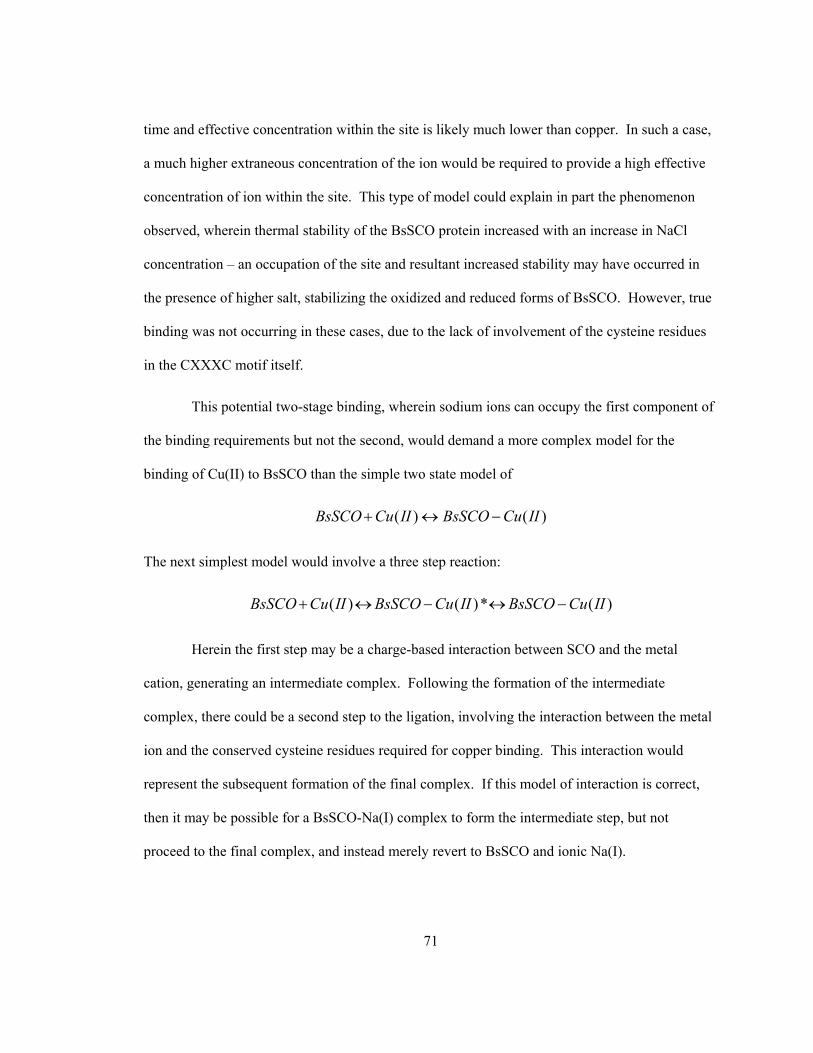
71
time and effective concentration within the site is likely much lower than copper. In such a case,
a much higher extraneous concentration of the ion would be required to provide a high effective
concentration of ion within the site. This type of model could explain in part the phenomenon
observed, wherein thermal stability of the BsSCO protein increased with an increase in NaCl
concentration – an occupation of the site and resultant increased stability may have occurred in
the presence of higher salt, stabilizing the oxidized and reduced forms of BsSCO. However, true
binding was not occurring in these cases, due to the lack of involvement of the cysteine residues
in the CXXXC motif itself.
This potential two-stage binding, wherein sodium ions can occupy the first component of
the binding requirements but not the second, would demand a more complex model for the
binding of Cu(II) to BsSCO than the simple two state model of
( ) ( )BsSCO Cu II BsSCO Cu II+ ↔ −
The next simplest model would involve a three step reaction:
( ) ( )* ( )BsSCO Cu II BsSCO Cu II BsSCO Cu II+ ↔ − ↔ −
Herein the first step may be a charge-based interaction between SCO and the metal
cation, generating an intermediate complex. Following the formation of the intermediate
complex, there could be a second step to the ligation, involving the interaction between the metal
ion and the conserved cysteine residues required for copper binding. This interaction would
represent the subsequent formation of the final complex. If this model of interaction is correct,
then it may be possible for a BsSCO-Na(I) complex to form the intermediate step, but not
proceed to the final complex, and instead merely revert to BsSCO and ionic Na(I).

72
This may also be an attractive model in determining why the ionic strength of a solution
could alter the Kd of the protein for Cu(II). It is possible that the initial interaction of the protein
with the Cu(II) ion is mediated solely by charge, and this could represent binding of copper ions
to a more "open" BsSCO complex. If this were the case, some conformational change would
likely occur after copper binding, and a more "closed" complex may form. In such a “closed”
complex, the copper ion and the cysteine residues within the CXXXC motif of BsSCO could be
forced to interact by means of proximity. Here, the presence of interfering charge via high ionic
strength could increase the stability of the apo-BsSCO protein by allowing the occupation of a
charged ion within the binding site of the protein. However, the wrong ion would not allow the
formation of the final stable BsSCO complex, due to the fact that this ion could not bond to the
sulfur atoms within the cysteine residues that compose the site. Additionally, the stability of the
complex decreased in high ionic strength, and this data could be explained by this model.
Charge-mediated interactions may have led to competitive occupation of other ions at the initial
site, rather than the site protecting the copper ion already bound to the CXXXC motif. Such
interference would leave only covalently-bound copper ion, resulting in the formation of a “semi-
open” complex in which the cysteine residues were still involved in ligation, but the charge-
stabilized "closed" form of the protein could not be correctly achieved. In such a circumstance,
the stabilization provided by the covalent Cu-S bonds would keep the Cu(II) ion in place and
bound to BsSCO, but without the stable "closed" conformation the copper atom would be
available and reactive to other components of the solution – such as free Cu(II). It is possible that
interaction between unshielded but covalently BsSCO-bound Cu(II) and free Cu(II) in solution
could occur, where free Cu(II) in solution may strip an electron from BsSCO-bound copper to
form a stable octet of valence (forming a Cu(I) ion). In a concomitant step, the copper bound to
BsSCO could also oxidize the thiol residues it was bonded to, forming another Cu(I) ion and

73
leaving behind a disulfide bonded BsSCO molecule free of covalently bound copper. In an
aerobic experimental situation this would eventually result in Cu(I) being oxidized to Cu(II) by
dissolved oxygen in solution, generating more free Cu(II) in solution. This would allow
formation of a continuous redox cycle.
Oxidization of BsSCO in these conditions would occur much more rapidly than in simple
buffer conditions. In fact, extrapolated to infinite ionic strength, where competing ion would
completely block any charge-based interaction between the BsSCO molecule and the bound
Cu(II) ion, the measured off-rate for copper would likely be identical to the off-rate that could be
measured for unfolded, copper-bound protein. So long as no free copper were present in solution,
this rate would also correspond to the off rate for the second step of the revised, two-step reaction
mechanism:
1 2
1 2( ) ( )* ( )k k
k kBsSCO Cu II BsSCO Cu II BsSCO Cu II− −
+ ←⎯→ − ←⎯→ −
In this case, where the overall Ka being measured would be a product of K1 and K2, some
estimations can be made in order to model the system. Where Kd is 3.5x10⁻¹² M, and assuming
that k1 is a fast event at at an arbitrary 1x10⁵ M-1s-1, while k-1 and k2 are slower at 1.0s-1, the
halftime for complex dissociation at 675 hours in buffer alone would leave k-2 at a value of
2.8x10⁻⁷ s-1. Since halftime decreases by addition of salt, Ka itself must change, possibly by a
process in which either k-1 or k-2 increases in rate.
This model reconciles both the DSC measurements and the copper-dissociation
experiments for BsSCO. In both DSC and spectrophotometric observation, the BsSCO-Cu(II)
complex was less stable in high ionic strength conditions. Visibly, the copper dissociated from
the complex much more quickly with higher salt and extra copper present (Figure 15), and this
was evident in DSC as well (Figure 18-Figure 20). The apo-protein stability cannot be assessed

74
visibly, but can be determined via DSC. Surprisingly, in higher ionic strength conditions the
stability of the apo-protein increased, rather than decreased (Figure 18-Figure 20). In a model
where one binding event dictates the stability of the complex, these results are counterintuitive,
because a decrease in stability in one set of measurements should not result in an increase in
stability when measuring a using a different method. However, if binding is a two-step transition,
these results can be more easily explained. By invoking a charge-charge interaction in the first
stage of binding, the higher ionic strength solutions could induce the protein to hold a more stable
conformation, visible as a higher stability in DSC measurements. Subsequently, in a second stage
of binding a covalent bond could be formed between Cu(II) ion and the sulfur atoms within the
reduced thiol residues of BsSCO, which could not be accomplished by the other salts in solution.
Furthermore, high ionic strength solutions could competitively interfere with the conformational
change dictated by charge, thereby exposing the bound Cu(II) ion within BsSCO to outside
environmental factors and eventually leading to its release. In this case, the kinetic stability of
Cu(II)-BsSCO would be supplied by the second step of binding (K2). A model for the interaction
of apo-BsSCO and Cu(II)-BsSCO in both with and without salt in solution is presented in Figure
22.

75
Figure 22: Model for interaction of BsSCO with both Cu(II) and NaCl. Panel (a): Model for BsSCO bound to Cu(II) via ligation to nitrogen from conserved histidine residue and two sulfur atoms from CXXXC motif. Panel (b): Model for BsSCO bound to Cu(II) via ligation to two sulfur atoms from CXXXC motif, while conserved histidine residue interacts with sodium counterion. Panel (c): Model for BsSCO in salt, wherein conserved histidine residue interacts with sodium counterion but sulfur atoms from CXXXC motif do not interact.
76
Using DSC, the measurement of binding affinity is determined via a difference in
stability between two conditions. However, in higher salt, the apo-protein exhibits greater
stability than it does in buffer alone. Therefore, the stability difference imparted to BsSCO by
copper(II) binding is less than would be the case in buffer alone, and dissociation occurs more
easily. Before this model can be further refined, a method must be determined to measure the
kinetic rates composing the K1 or K2 equilibrium processes.
Unfortunately, the two step model is difficult to prove directly in vitro, as spectroscopic
changes associated with copper binding are associated with the interaction of copper ion with the
conserved cysteine residues of the CXXXC motif; copper-sulfur charge transfer in the case of
electronic absorption, and copper quenching of intrinsic fluorescence. Neither of these
observable signals would be useful to indicate a conformational change occurring prior to
covalent copper binding. Circular dichroism may provide a means of observing such a transition,
and could be measured for apo-BsSCO protein, Cu(II)-bound BsSCO protein, and BsSCO protein
in the presence of high salt, such as NaF. However, a lack of a signal in CD measurements is not
necessarily indicative that no conformational change is occurring, as it is possible for a
conformational change to occur without a change in proportions of protein secondary structure.
This is particularly applicable in the case of the poorly-defined, flexible binding loop containing
the conserved histidine residue associated with copper binding.
The two-step binding model could also fit well with a view wherein BsSCO delivers
copper directly into the CuA site in vivo. The interaction between BsSCO and the CuA site could
be what exposes a bound copper within BsSCO, where it can then be transferred to the CuA site,
either by interaction with another Cu(II) ion and oxidizing the BsSCO-Cu(II) complex to free
Cu(I) and oxidized BsSCO, or by another form of transfer which has yet to be described. The
independent requirement for SCO1 and SCO2 in eukaryotes could play a role in such a model, if
77
a Cu(II) ion is required to get release of bound Cu(II) from SCO. In Bacillus subtilis, there are no
mitochondria within the cell, as well as the coincidental lack of SCO2. Perhaps the simpler
system in Bacillus can provide insight into more complex systems, as its exposure to
environmental elements such as Cu(II) is readily possible, but within the cell of eukaryotes such
elements are not freely available, and metallochaperones must transport them. SCO2 may play
such a role in eukaryotic cytochrome c oxidase assembly, and be completely unnecessary when
Cu(II) is readily available at the site of oxidase assembly in simpler prokaryotes.
While the system of CuA site assembly cannot be described completely by the work
detailed within this study, important advances have been made. A strong binding constant was
determined for the BsSCO-Cu(II) interaction, and the first description of conditions required for
copper dissociation from such a complex has been determined. While these conditions may not
be physiologically realistic, they may provide greater insight into interactions within the living
cell, as well as providing a means to probe the assembly process of the CuA site of cytochrome c
oxidase by describing what must occur to dissociate Cu(II) ion. Whether this dissociation is
assisted by other cofactors, such as SCO2 in eukaryotes, or simply by presence of metal ion in
prokaryotes, will be an interesting avenue for further research, and will hopefully yield a greater
understanding of an enzyme ubiquitous within aerobic life.

78
Chapter 6 Summary and Conclusions
Cytochrome c oxidase is a complicated enzyme, with complex assembly requirements,
and numerous assembly factors. The SCO family of proteins are conserved across aerobic
organisms and implicated in the assembly of the subunit II CuA site within the oxidase. Without
SCO, functional oxidase cannot be assembled, but the mechanism by which SCO functions has
not been elucidated. The process may even vary between organisms, as yeast and humans
express two SCO homologues, SCO1 and SCO2, while simpler bacteria only express SCO1. The
protein has been found to bind copper, both in Cu(I) and Cu(II) forms, and is proposed to directly
deliver copper to the site of requirement within the oxidase. Due to structural homology with
thioredoxins, SCO has also been suggested to prepare the CuA site to accept copper, rather than
being directly involved in copper insertion to the site.
This study has focused on the ability of Bacillus subtilis SCO (BsSCO) to bind Cu(II),
and subsequently to release it. Through study using differential scanning calorimetry, the Kd for
interaction between BsSCO and Cu(II) in phosphate buffer was determined to have an upper limit
of at most 3.5 x 10⁻¹² M. Study of the protein in high ionic strength yielded novel information;
Cu(II) will dissociate from the BsSCO-Cu(II) complex under high ionic strength conditions, if
free Cu(II) is available in solution. While these conditions may not be directly indicative of
physiological situations experienced within the cell, they do provide the first evidence of
conditions required to separate the complex, and may provide an indication of how SCO interacts
with cytochrome c oxidase in vivo.
79
References
1. Li, Y., Park, J. S., Deng, J. H., Bai, Y., Li, Y., Park, J.-S., Deng, J.-H., and Bai, Y. (2006)
J Bioenerg Biomembr 38(5-6), 283-291
2. Fontanesi, F., Soto, I. C., Horn, D., Barrientos, A., Fontanesi, F., Soto, I. C., Horn, D.,
and Barrientos, A. (2006) Am J Physiol Cell Physiol 291(6), C1129-1147
3. Nittis, T., George, G. N., and Winge, D. R. (2001) J Biol Chem 276(45), 42520-42526
4. Michel, H., Behr, J., Harrenga, A., and Kannt, A. (1998) CYTOCHROME C OXIDASE:
Structure and Spectroscopy. In.
5. Hendler, R. W., Pardhasaradhi, K., Reynafarje, B., and Ludwig, B. (1991) Biophys J
60(2), 415-423
6. Carr, H. S., and Winge, D. R. (2003) Assembly of Cytochrome c Oxidase within the
Mitochondrion. In.
7. Kadenbach, B., Huttemann, M., Arnold, S., Lee, I., and Bender, E. (2000) Free Radic
Biol Med 29(3-4), 211-221
8. Schulze, M., and Rodel, G. (1988) Mol Gen Genet 211(3), 492-498
9. Mattatall, N. R., Jazairi, J., and Hill, B. C. (2000) J Biol Chem 275(37), 28802-28809
80
10. Papadopoulou, L. C., Sue, C. M., Davidson, M. M., Tanji, K., Nishino, I., Sadlock, J. E.,
Krishna, S., Walker, W., Selby, J., Glerum, D. M., Coster, R. V., Lyon, G., Scalais, E.,
Lebel, R., Kaplan, P., Shanske, S., De Vivo, D. C., Bonilla, E., Hirano, M., DiMauro, S.,
and Schon, E. A. (1999) Nat Genet 23(3), 333-337
11. Chinenov, Y. V. (2000) J Mol Med 78(5), 239-242
12. Askwith, C., and Kaplan, J. (1998) Trends in Biochemical Sciences 23(4), 135-138
13. Rae, T. D., Schmidt, P. J., Pufahl, R. A., Culotta, V. C., O'Halloran, T. V., Rae, T. D.,
Schmidt, P. J., Pufahl, R. A., Culotta, V. C., and O'Halloran, T. V. (1999) Science
284(5415), 805-808
14. Gruber, C. W., Cemazar, M., Heras, B., Martin, J. L., and Craik, D. J. (2006) Trends
Biochem Sci 31(8), 455-464
15. Kadokura, H., Katzen, F., and Beckwith, J. (2003) PROTEIN DISULFIDE BOND
FORMATION IN PROKARYOTES. In.
16. Fomenko, D. E., and Gladyshev, V. N. (2003) Biochemistry 42(38), 11214-11225
17. Waris, G., and Ahsan, H. (2006) J Carcinog 5, 14
18. Rhee, S. G., Chae, H. Z., and Kim, K. (2005) Free Radic Biol Med 38(12), 1543-1552
81
19. Boveris, A., and Chance, B. (1973) Biochem J 134(3), 707-716
20. Paret, C., Ostermann, K., Krause-Buchholz, U., Rentzsch, A., and Rodel, G. (1999) FEBS
Lett 447(1), 65-70
21. McEwan, A. G., Lewin, A., Davy, S. L., Boetzel, R., Leech, A., Walker, D., Wood, T.,
and Moore, G. R. (2002) FEBS Lett 518(1-3), 10-16
22. Valnot, I., Osmond, S., Gigarel, N., Mehaye, B., Amiel, J., Cormier-Daire, V., Munnich,
A., Bonnefont, J. P., Rustin, P., and Rotig, A. (2000) Am J Hum Genet 67(5), 1104-1109
23. Schulze, M., and Rodel, G. (1989) Mol Gen Genet 216(1), 37-43
24. Buchwald, P., Krummeck, G., and Rodel, G. (1991) Mol Gen Genet 229(3), 413-420
25. Andrews, D., Rattenbury, J., Anand, V., Mattatall, N. R., Hill, B. C., Andrews, D.,
Rattenbury, J., Anand, V., Mattatall, N. R., and Hill, B. C. (2004) Protein Expr Purif
33(1), 57-65
26. Rentzsch, A., Krummeck-Weiss, G., Hofer, A., Bartuschka, A., Ostermann, K., and
Rodel, G. (1999) Curr Genet 35(2), 103-108
27. Farrar, J. A., Neese, F., Lappalainen, P., Kroneck, P. M. H., Saraste, M., Zumft, W. G.,
and Thomson, A. J. (1996) The Electronic Structure of CuA: A Novel Mixed-Valence
Dinuclear Copper Electron-Transfer Center. In.
82
28. Hamza, I., Gitlin, J. D., Hamza, I., and Gitlin, J. D. (2002) J Bioenerg Biomembr 34(5),
381-388
29. Beers, J., Glerum, D. M., and Tzagoloff, A. (1997) J Biol Chem 272(52), 33191-33196
30. Horng, Y.-C., Leary, S. C., Cobine, P. A., Young, F. B. J., George, G. N., Shoubridge, E.
A., and Winge, D. R. (2005) J Biol Chem 280(40), 34113-34122
31. Imriskova-Sosova, I., Andrews, D., Yam, K., Davidson, D., Yachnin, B., and Hill, B. C.
(2005) Biochemistry 44(51), 16949-16956
32. Andruzzi, L., Nakano, M., Nilges, M. J., and Blackburn, N. J. (2005) J Am Chem Soc
127(47), 16548-16558
33. Horvath, R., Lochmuller, H., Stucka, R., Yao, J., Shoubridge, E. A., Kim, S. H., Gerbitz,
K. D., and Jaksch, M. (2000) Biochem Biophys Res Commun 276(2), 530-533
34. Glerum, D. M., Shtanko, A., and Tzagoloff, A. (1996) J Biol Chem 271(34), 20531-
20535
35. Hiser, L., Di Valentin, M., Hamer, A. G., and Hosler, J. P. (2000) J Biol Chem 275(1),
619-623
36. Leary, S. C., Kaufman, B. A., Pellecchia, G., Guercin, G.-H., Mattman, A., Jaksch, M.,
and Shoubridge, E. A. (2004) Hum Mol Genet 13(17), 1839-1848

83
37. Abajian, C., and Rosenzweig, A. C. (2006) J Biol Inorg Chem 11(4), 459-466
38. Ye, Q., Imriskova-Sosova, I., Hill, B. C., and Jia, Z. (2005) Biochemistry 44(8), 2934-
2942
39. Williams, J. C., Sue, C., Banting, G. S., Yang, H., Glerum, D. M., Hendrickson, W. A.,
and Schon, E. A. (2005) J Biol Chem 280(15), 15202-15211
40. Balatri, E., Banci, L., Bertini, I., Cantini, F., and Ciofi-Baffoni, S. (2003) Structure
(Camb) 11(11), 1431-1443
41. Su, D., Berndt, C., Fomenko, D. E., Holmgren, A., and Gladyshev, V. N. (2007)
Biochemistry 46(23), 6903-6910
42. Seib, K. L., Jennings, M. P., McEwan, A. G., Seib, K. L., Jennings, M. P., and McEwan,
A. G. (2003) FEBS Lett 546(2-3), 411-415
43. Jaksch, M., Paret, C., Stucka, R., Horn, N., Muller-Hocker, J., Horvath, R., Trepesch, N.,
Stecker, G., Freisinger, P., Thirion, C., Muller, J., Lunkwitz, R., Rodel, G., Shoubridge,
E. A., and Lochmuller, H. (2001) Hum Mol Genet 10(26), 3025-3035
44. Abajian, C., Yatsunyk, L. A., Ramirez, B. E., and Rosenzweig, A. C. (2004) J Biol Chem
279(51), 53584-53592
45. Krummeck, G., and Rodel, G. (1990) Curr Genet 18(1), 13-15

84
46. Williams, S. L., Valnot, I., Rustin, P., and Taanman, J.-W. (2004) J Biol Chem 279(9),
7462-7469
47. Imriskova-Sosova, I., Ye, Q., Hill, B. C., and Jia, Z. (2003) Acta Crystallogr D Biol
Crystallogr 59(Pt 7), 1299-1301
48. Riener, C. K., Kada, G., and Gruber, H. J. (2002) Anal Bioanal Chem 373(4-5), 266-276
49. Nagamori, S., Nishiyama, K., and Tokuda, H. (2000) J Biochem (Tokyo) 128(1), 129-137
50. Brandts, J. F., and Lin, L. N. (1990) Biochemistry 29(29), 6927-6940
51. Wang, S. C., Dias, A. V., Bloom, S. L., and Zamble, D. B. (2004) Selectivity of Metal
Binding and Metal-Induced Stability of Escherichia coli NikR. In.
52. Banci, L., Bertini, I., Ciofi-Baffoni, S., Leontari, I., Martinelli, M., Palumaa, P., Sillard,
R., and Wang, S. (2007) Proc Natl Acad Sci U S A 104(1), 15-20
53. Malmstrom, B. G., and Aasa, R. (1993) FEBS Lett 325(1-2), 49-52
54. Winge, D. R. (2003) Structure (Camb) 11(11), 1313-1314
55. Szajewski, R. P., and Whitesides, G. M. (1980) Rate constants and equilibrium constants
for thiol-disulfide interchange reactions involving oxidized glutathione. In.

85
56. Schafer, F. Q., and Buettner, G. R. (2001) Free Radical Biology and Medicine 30(11),
1191-1212
57. Setterdahl, A. T., Chivers, P. T., Hirasawa, M., Lemaire, S. D., Keryer, E., Miginiac-
Maslow, M., Kim, S. K., Mason, J., Jacquot, J. P., Longbine, C. C., de Lamotte-Guery,
F., Knaff, D. B., Setterdahl, A. T., Chivers, P. T., Hirasawa, M., Lemaire, S. D., Keryer,
E., Miginiac-Maslow, M., Kim, S.-K., Mason, J., Jacquot, J.-P., Longbine, C. C., de
Lamotte-Guery, F., and Knaff, D. B. (2003) Biochemistry 42(50), 14877-14884
58. Watson, W. H., Pohl, J., Montfort, W. R., Stuchlik, O., Reed, M. S., Powis, G., Jones, D.
P., Watson, W. H., Pohl, J., Montfort, W. R., Stuchlik, O., Reed, M. S., Powis, G., and
Jones, D. P. (2003) J Biol Chem 278(35), 33408-33415
59. Kumar, S., and Nussinov, R. (1999) J Mol Biol 293(5), 1241-1255
60. Shannon, R. (1976) Revised effective ionic radii and systematic studies of interatomic
distances in halides and chalcogenides. In. Acta Crystallographica Section A


![Electrostatic Contribution to the Binding Stability of ...web2.physics.fsu.edu/~zhou/reprints/pr89.pdfthe binding stability [Fig. 1(b)].7,16 Ionic strength is found to exert a strong](https://img.pdfslide.us/doc/110x75/5ec4ce93c620f72ddb6d3880/electrostatic-contribution-to-the-binding-stability-of-web2-zhoureprintspr89pdf.jpg)

![Index [link.springer.com]978-1-59259-288-3/1.pdf · Index tissue distribution, 359 bId mutants, see Streptomyces Blue copper proteins, cupredoxins, C copper binding sites, 54 electron](https://img.pdfslide.us/doc/110x75/5ca8e00888c993e47d8c3d94/index-link-978-1-59259-288-31pdf-index-tissue-distribution-359-bid-mutants.jpg)