Embed Size (px)
Citation preview

a PATTERNS & PHENOTYPES
The Identification of Transcription FactorsExpressed in the Notochord of Cionaintestinalis Adds New Potential Players to theBrachyury Gene Regulatory NetworkDiana S. Jose-Edwards,1 Pierre Kerner,1 Jamie E. Kugler,1 Wei Deng,2 Di Jiang,2 and Anna Di Gregorio1*
The notochord is the distinctive characteristic of chordates; however, the knowledge of the complementof transcription factors governing the development of this structure is still incomplete. Here we presentthe expression patterns of seven transcription factor genes detected in the notochord of the ascidianCiona intestinalis at various stages of embryonic development. Four of these transcription factors, Fos-a,NFAT5, AFF and Klf15, have not been directly associated with the notochord in previous studies, whilethe others, including Spalt-like-a, Lmx-like, and STAT5/6-b, display evolutionarily conserved expression inthis structure as well as in other domains. We examined the hierarchical relationships between thesegenes and the transcription factor Brachyury, which is necessary for notochord development in all chor-dates. We found that Ciona Brachyury regulates the expression of most, although not all, of these genes.These results shed light on the genetic regulatory program underlying notochord formation in Ciona andpossibly other chordates. Developmental Dynamics 240:1793–1805, 2011. VC 2011 Wiley-Liss, Inc.
Key words: Ascidian; Brachyury; Ciona intestinalis; gene network; notochord; transcription factor; tunicate
Accepted 20 April 2011
INTRODUCTION
The notochord is a midline mesodermalstructure whose presence is one of thedefining features of the chordate bodyplan. This organ is critical for the em-bryonic development of all chordates,where it serves as the main axial sup-port for the growing embryos (Stemple,2005; Jiang and Smith, 2007). In verte-brates, the notochord is necessary forpatterning the neural tube, specifica-
tion of the cardiac field, and formationof the endoderm (Cleaver and Krieg,
2001; Wilson and Maden, 2005). In ver-
tebrate embryos, as ossification of the
vertebral column proceeds, the noto-
chord gradually disappears and its
remnants become incorporated into the
nucleus pulposus, the central portion of
the intervertebral discs located
between the vertebrae of the spinal col-
umn; these notochord residues can
form malignant chordomas (Risbud
et al., 2010).The ascidian embryo provides an
ideal model for studies of notochord
development and differentiation. The
notochord of these translucent
embryos consists of just 40 cells and
forms similar to that of vertebrates.
For example, notochord cells interca-
late during convergent extension
(Munro and Odell, 2002) relying upon
Additional Supporting Information may be found in the online version of this article.1Department of Cell and Developmental Biology, Weill Medical College of Cornell University, New York, New York2Sars International Centre for Marine Molecular Biology, Bergen, NorwayGrant sponsor: NIH/NICHD; Grant number: R01HD050704; Grant sponsor: March of Dimes Birth Defects Foundation; Grant number:1-FY08-430; Grant sponsor: Charles A. Frueauff Foundation; Norwegian Research Council; Grant numbers: 133335/V40, 183302/S10;Grant sponsor: NIH; Grant number: T32 GM008539.*Correspondence to: Anna Di Gregorio, Department of Cell and Developmental Biology, Weill Medical College of CornellUniversity, 1300 York Avenue, Box 60, New York, NY 10065. E-mail: [email protected]
DOI 10.1002/dvdy.22656Published online 18 May 2011 in Wiley Online Library (wileyonlinelibrary.com).
ABBREVIATIONS cDNA complementary DNA CNS central nervous system ENU N-ethyl-N-nitrosourea EST Expressed SequenceTag kb kilobase(s), or 1,000 base pairs MOPS 3-(N-morpholino) propanesulfonic acid PCR Polymerase Chain Reaction WMISH whole-mount in situ hybridization
DEVELOPMENTAL DYNAMICS 240:1793–1805, 2011
VC 2011 Wiley-Liss, Inc.
Dev
elop
men
tal D
ynam
ics

the planar cell polarity (PCP) path-way, and subsequently stretch toallow tail extension (Jiang et al.,2005). Additionally, in both Ciona andvertebrates, the notochord is sur-rounded by a basement membraneconsisting of extracellular matrix pro-teins, including laminins (Scott andStemple, 2005; Veeman et al., 2008),and a collagen-rich notochordalsheath (Miyamoto and Crowther,1985; Stemple, 2005). As developmentof the notochord proceeds, intercellu-lar pockets of extracellular matrix(lumens) form in ascidians (Jiang andSmith, 2007), and intracellularvacuoles form in vertebrates (Stem-ple, 2005); the mechanical pressureexerted by lumens and vacuolesagainst the stiff notochordal sheathprovides the rigidity necessary for theembryo to elongate (Stemple, 2005).
Despite the importance of notochordformation in shaping the chordatebody plan, the network of transcrip-tional regulators controlling its devel-opment remains incompletely charac-terized in any model system.Nevertheless, it is generally acknowl-edged that one transcriptional regula-tor necessary for notochord formationin all chordates is the T-box transcrip-tion factor Brachyury. The crucial rolefor Brachyury in notochord develop-ment is underscored by recent datathat have shown this gene to be a bio-marker of chordomas (Vujovic et al.,2006) and to be duplicated in familialchordoma (Yang et al., 2009). In Ciona,Brachyury (Ci-Bra) is expressed exclu-sively in the notochord and its precur-sors beginning at the 64-cell stage, con-current with notochord fate restriction(Corbo et al., 1997). The upstream reg-ulatory cascade leading to Ci-Braexpression is well characterized (Yagiet al., 2004; Imai et al., 2006; Matsu-moto et al., 2007), and at least 50Ciona genes have been found to be con-trolled by Ci-Bra (Di Gregorio and Lev-ine, 1999; Takahashi et al., 1999; Hottaet al., 2000, 2008; Oda-Ishii and DiGregorio, 2007; Kugler et al., 2008). Asurprisingly low number of transcrip-tion factors was included within thisfirst set of transcriptional targets; how-ever, this number has been sharplyincreased by recent whole-genomestudies of the in vivo occupancy of chro-matin by Ci-Bra in early Cionaembryos (Kubo et al., 2010).
Nevertheless, a fraction of these pre-sumptive Ci-Bra-downstream regula-tory genes have not been shown to beexpressed in notochord cells by previousstudies. Furthermore, while it has beensuggested that Ci-Bra might be control-ling expression of its late target genesvia transcriptional intermediaries (e.g.,Hotta et al., 1999), the knowledge of theprecise temporal windows of notochordexpression and the presumptive func-tions of these Ci-Bra-downstream fac-tors are still fragmentary. This repre-sents a point of interest in studies ofnotochord formation, since these inter-mediaries of Ci-Bra, alongside Ci-Bra-independent notochord transcriptionfactors, are likely to collectively directthe morphogenetic processes that beginat later developmental stages. To beginfilling this gap, we undertook an alter-native approach, as we sought to firstidentify additional transcription factorsexpressed in the Ciona notochord andsecondly to study their hierarchicalrelationship with Ci-Bra.
Here we report the previouslyuncharacterized notochord expressionof seven transcription factors in Cionaintestinalis. While for some of thesegenes this analysis provides the firstevidence of expression in this domain inany chordate, for others it underscoresthe evolutionary conservation of noto-chord expression across the chordatephylum. In either case, understandingthe hierarchical relationships of thesefactors with other components of thenotochord gene regulatory networkshould enhance our knowledge of molec-ular mechanisms fundamental to noto-chord development and to the evolutionof the chordate body plan. Toward thisaim, we began examining the relation-ship between these genes and Ci-Bra.We found that loss of Ci-Bra functionaffects the expression of some, but notall, of these genes. Together, our resultssuggest that the notochord gene regula-tory network in the simple chordateCiona is complex and multifaceted.
RESULTS AND DISCUSSION
Identification of Novel
Notochord Transcription
Factors in the Ciona Embryo
A comprehensive and detailed list ofthe transcription factor genes foundin the Ciona intestinalis genome and
their expression patterns has beenpublished by Imai et al. (2004) and ispublicly available in a searchable for-mat (http://hoya.zool.kyoto-u.ac.jp/TF_KH.html).We performed a microarray screen
on neurula and mid-tailbud FACS-sorted notochord cells aimed at identi-fying genes enriched in the notochordlineage, and we found that most ofthe genes with the highest scoreswere previously characterized noto-chord markers, including Ci-Bra(Corbo et al., 1997), Ci-leprecan(Dunn and Di Gregorio, 2009), severalCi-Noto genes (Hotta et al., 2000),and Ci-tune (Passamaneck et al.,2009), among others (unpublisheddata). We noticed that in addition toCi-Bra and its target genes, the provi-sional notochord transcriptome alsoincluded a number of transcriptionfactor genes whose expression in noto-chord cells had not been reported pre-viously. Prompted by these observa-tions, we prioritized the analysis ofthese candidate notochord transcrip-tional regulators, as they would likelybroaden the current knowledge of theCiona notochord gene regulatory net-work by shedding light on some of itsyet undiscovered branches.We began the study of these candi-
date notochord factors by performingwhole-mount in situ hybridization onembryos ranging from the 64-cellthrough the mid-tailbud II stages(Hotta et al., 2007). This analysisallowed us to validate that sevengenes, representing various familiesof transcription factors, are expressedin the Ciona notochord (Table 1; Figs.1–3 and see Supp. Fig. S1, which isavailable online). As expected, the ma-jority of these genes are expressed afternotochord fate determination is com-plete; this observation raises the possi-bility that they may govern some of themorphogenetic processes required forsubsequent stages of notochord develop-ment and differentiation.
Ci-Sall-a
Spalt-like (Sall) proteins are zinc-fin-ger transcription factors related tothe product of Drosophila spalt major(salm) and spalt-related (salr) genes(de Celis and Barrio, 2009). Whilemammalian genomes contain fourSall family members, Ciona
Dev
elop
men
tal D
ynam
ics
1794 JOSE-EDWARDS ET AL.

intestinalis has two. Through phylo-genetic reconstructions, we foundthat the two Ciona Sall genes likelyarose from a lineage-specific duplica-tion event (Supp. Fig. S2A) and areboth equally related to their verte-brate counterparts. Based on this ob-servation, here we refer to theseparalogs as Ci-Sall-a and Ci-Sall-b.
Ci-Sall-a is expressed in a numberof spatial domains during Ciona em-bryonic development (Fig. 1A). Weakexpression in notochord cells isdetected starting at the 64-cell stage;however, Ci-Sall-a transcripts becomemore evident in this territory by the110-cell stage (Fig. 1A, red arrow-heads). At these stages, faint expres-sion is also detected in some CNS pre-cursors (Fig. 1A, blue arrowheads).Notochord expression continuesthrough gastrulation, but begins to bedown-regulated during the neurulastage. Ci-Sall-a is also expressed inthe trunk ventral cells (TVCs), theprecursors of the Ciona heart (Fig.1A, orange arrowheads), beginning atthe 110-cell stage and additionally inendoderm beginning at the neurulastage (Fig. 1A, yellow arrowheads).Expression is retained in the trunkendoderm and endodermal strandthrough the mid-tailbud II stage(Supp. Fig. S1A). It has been shown
that spalt genes, such as the chickcsall3 gene, undergo alternative splic-ing (Sweetman and Munsterberg,2006). Interestingly, Ci-Sall-a mRNAsalso appear to be alternatively spliced(Satou and Satoh, 2005). Neverthe-less, in our analysis, we found thatprobes synthesized from both Ci-Sall-a cDNAs exhibited identical expres-sion patterns (Fig. 1A and data notshown).
Some members of the Sall1 groupin various vertebrates are expressedin the notochord, as is the case formouse Sall1 (Ott et al., 2001), zebra-fish sall1a (Camp et al., 2003) andchick csal1 (Sweetman et al., 2005),while the other Spalt family membersare absent from this tissue. Notably,each of the Sall1 genes mentionedabove is also present in the develop-ing heart and the neural tube (Ottet al., 2001; Camp et al., 2003; Sweet-man et al., 2005). Interestingly, thecomposite expression pattern, whichencompasses notochord, neural tube,and heart, is nearly recapitulated bythe combined expression patterns ofthe two Ciona spalt-like paralogs; Ci-Sall-a is detected in the notochordand heart precursors, and transientlyin CNS precursors (Fig. 1A) while ourprevious work has shown that Ci-Sall-b is present in the posterior neu-
ral tube in tailbud embryos althoughit is absent from the notochord(Kugler et al., 2008). The roles ofSpalt proteins in notochord develop-ment have not been examined; there-fore, Ciona represents a simplifiedmodel system in which to assess Spaltfunction in the notochord.
Ci-Lmx-like
The LIM-homeodomain transcriptionfactor gene Ciona LIM-homeobox like(Ci-Lmx-like) begins to be expressedat the 110-cell stage, where it is pres-ent in cells of the neural lineage (Fig.1B, blue arrowheads). Beginning atgastrulation, transcripts are alsodetected in notochord cells, whichgradually become the predominantexpression domain (Fig. 1B, redarrowheads); Ci-Lmx-like expressionpersists in these two areas throughthe mid-tailbud stage (Fig. 1B) andbegins to fade from the notochordaround the mid-tailbud II stage(Supp. Fig. S1B).Two members of the Lmx gene fam-
ily are found in mammalian genomes,Lmx1a and Lmx1b (Hunter and Rho-des, 2005), and two Lmx genes arealso found in Ciona (Wada et al.,2003; Imai et al., 2004). Since the mo-lecular evolutionary history of these
TABLE 1. Gene Models and ESTs for the Genes Examined in the Present Studya
Gene Name
Alternative
Name(s)
KH Gene
Modelb
JGI v1.0
Gene
Model(s)
JGI v1.0
Location
JGI v2.0
Location
cDNA
Clone
Used
Source(s) of
Previously
Published Patterns
Ci-Sall-a Ci-Spalt-like1ZF (C2H2)-18
KH.L4.17 ci0100141112 scaffold_177 scaffold_67 cieg50j03 Imai et al. (2004)
Ci-Lmx-like N/A KH.C9.485 ci0100149991 scaffold_89 chr_09q cinc025n05 Imai et al. (2004)ci0100145187
Ci-Lmx Ci-lmx1.2 KH.C9.616 ci0100150069 scaffold_89 chr_09q citb30f24 Imai et al. (2004, 2009)Ishibashi et al. (2005)Tassy et al. (2010)
Ci-Fos-a Ci-Fos KH.C11.314 ci0100130316 scaffold_63 scaffold_1690 cima839226 Imai et al. (2004)scaffold_2122
Ci-NFAT5 N/A KH.C3.133 ci0100140442 scaffold_128 chr_03q cieg069a11 Imai et al. (2004)ci0100141121 scaffold_2370
Ci-AFF N/A KH.C2.327 ci0100131909 scaffold_875 chr_02q cien95799 N/Ascaffold_117
Ci-STAT5/6-b Ci-STAT-b KH.C1.275 ci0100154492 scaffold_276 chr_01q cien84927 Imai et al. (2004)Hotta et al. (2008)
Ci-Klf15 Ci-ZF148 KH.C5.430 N/A scaffold_96 chr_05q cien222151 Miwata et al. (2006)
aZF, zinc-finger; v1.0, version 1.0; v2.0, version 2.0; N/A, not applicable.bSatou et al. (2008).
Dev
elop
men
tal D
ynam
ics
NOVEL NOTOCHORD TRANSCRIPTION FACTORS IN CIONA 1795

Fig. 1.
Fig. 2.
Dev
elop
men
tal D
ynam
ics
1796 JOSE-EDWARDS ET AL.
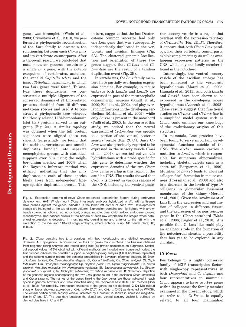
genes was incomplete (Wada et al.,2003; Srivastava et al., 2010), we per-formed a phylogenetic reconstructionof the Lmx family to ascertain therelationship between each Ciona Lmxand its vertebrate counterparts. Aftera thorough search, we concluded thatmost metazoan genomes contain onlya single Lmx gene, with the notableexceptions of vertebrates, ascidians,the annelid Capitella teleta and theinsect Tribolium castaneum, in whichtwo Lmx genes were found. To ana-lyze these duplications, we con-structed a multiple alignment of theconserved domains of 21 Lmx-relatedproteins identified from 15 differentmetazoan species and used it to con-struct a phylogenetic tree wherebythe closely related LIM-homeodomainIslet (Isl) proteins served as an out-group (Fig. 2A). A similar topologywas obtained when the full proteinsequences were aligned (data notshown). Surprisingly, we found thatthe ascidian, vertebrate, and annelidduplicates bundled into separatemonophyletic groups with statisticalsupports over 80% using the neigh-bor-joining method and 100% whenthe Bayesian inference method wasutilized, indicating that the Lmxduplicates in each of these specieslikely arose from independent line-age-specific duplication events. This,
in turn, suggests that the last Deuter-ostome common ancestor had onlyone Lmx gene that was subsequentlyindependently duplicated in the ver-tebrate and ascidian lineages (Fig.2A). The clustered genomic localiza-tion and orientation of these twogenes suggest that Ci-Lmx and Ci-Lmx-like are the result of a tandemduplication event (Fig. 2B).
In vertebrates, the Lmx family mem-bers have partially overlapping expres-sion domains. For example, in mouseembryos both Lmx1a and Lmx1b areexpressed in part of the mesencephalicdopaminergic neurons (Smidt et al.,2000; Failli et al., 2002), and play over-lapping functions in the developing cer-ebellum (Mishima et al., 2009), whileonly Lmx1a is present in the notochord(Failli et al., 2002). In the course of thisstudy, we noticed that the neuralexpression of Ci-Lmx-like was specificto a portion of the ventral posteriorsensory vesicle (Fig. 2C,C0). Since Ci-Lmx was also previously reported to beexpressed in the sensory vesicle (Imaiet al., 2004), we carried out in situhybridizations with a probe specific forthis gene to determine whether theexpression domains of the two CionaLmx genes overlap in this region of theascidian CNS. The results showed thatCi-Lmx is present in various regions ofthe CNS, including the ventral poste-
rior sensory vesicle in a region thatoverlaps with the expression territoryof Ci-Lmx-like (Fig. 2D,D0). Therefore,it appears that both Ciona Lmx paral-ogs, like their vertebrate counterparts,exhibit complementary as well as over-lapping expression patterns in theCNS, while only one family member isfound in the notochord.Interestingly, the ventral sensory
vesicle of the ascidian embryo hasbeen compared to the vertebratehypothalamus (Moret et al., 2005;Hamada et al., 2011), and both Lmx1aand Lmx1b have been shown to beexpressed in the developing mousehypothalamus (Asbreuk et al., 2002).These results suggest that functionalstudies on Ci-Lmx and Ci-Lmx-like ina simplified model system such asCiona could enhance our knowledgeof the evolutionary origins of thisstructure.In mammals, Lmx proteins have
been found to serve important devel-opmental functions outside of theCNS. The dreher mouse carries amutation in Lmx1a, which is respon-sible for numerous abnormalities,including skeletal defects such as ashort tail (Bergstrom et al., 1999).Mutation of Lmx1b leads to aberrantcollagen fibril formation in mouse cor-neas (Pressman et al., 2000) and alsoto a decrease in the levels of type IVcollagens in glomerular basementmembranes of the kidney (Morelloet al., 2001). Given the involvement ofLmx1b in the expression and matura-tion of collagen in different tissuesand the expression of various collagengenes in the Ciona notochord (Wadaet al., 2006; Kugler et al., 2010), it ispossible that Ci-Lmx-like could playan analogous role in the formation ofthe notochordal sheath, a possibilitythat has yet to be explored in anychordate.
Ci-Fos-a
Fos belongs to a highly conservedfamily of bZIP transcription factorswith single-copy representatives inboth Drosophila and C. elegans andfour representatives in mammals.Ciona appears to have two Fos geneswithin its genome; the family memberexamined in the present study, whichwe refer to as Ci-Fos-a, is equallyrelated to all four mammalian
Fig. 1. Expression patterns of novel Ciona notochord transcription factors during embryonicdevelopment. A–E: Whole-mount Ciona intestinalis embryos hybridized in situ with antisenseRNA probes against the genes indicated in the lower left corner of each row. Developmentalstages are indicated at the top of each column. Expression domains are highlighted with arrow-heads colored as follows: red: notochord; orange: muscle; blue: CNS; yellow: endoderm; purple:mesenchyme. Red dashed arrows at the bottom of each row emphasize the stages when noto-chord expression is detected. In most panels, dorsal is up and anterior to the left with theexception of the 64- and 110-cell stage embryos, where anterior is up. NP, neural plate; Tb,tailbud.
Fig. 2. Ciona contains two Lmx paralogs with both overlapping and distinct expressiondomains. A: Phylogenetic reconstruction for the Lmx genes found in Ciona. The tree was obtainedfrom neighbor-joining analyses and rooted using Islet (Isl) protein sequences as outgroups. Statisti-cal support values �75% obtained with different methods are included over conserved nodes; thefirst number indicates the bootstrap support in neighbor-joining analysis (1,000 bootstrap replicates)and the second number reports the posterior probabilities in Bayesian inference analysis. Bf, Bran-chiostoma floridae; Ce, Caenorhabditis elegans; Ci, Ciona intestinalis; Cs, Ciona savignyi; Ct, Capi-tella teleta; Dm, Drosophila melanogaster; Dp, Daphnia pulex; Hm, Hydra magnipapillata; Hs, Homosapiens; Mm, Mus musculus; Nv, Nematostella vectensis; Sk, Saccoglossus kowalevskii; Sp, Strong-ylocentrotus purpuratus; Ta, Trichoplax adhaerens; Tc: Tribolium castaneum. B: Schematic depictionof the genomic regions encompassing the two Lmx genes found in the ascidians Ciona intestinalisand Ciona savignyi. The names of the genes flanking the Lmx genes are those indicated in eachspecies’ genome browsers and were identified using the reciprocal best BLAST hit method (Borket al., 1998). For simplicity, intron/exon structures of the genes are not depicted. C–D0: Mid-tailbudstage embryos showing expression of Ci-Lmx-like (C,C0) and Ci-Lmx (D,D0) as detected by WMISH.The ventral portion of the sensory vesicle, indicated by a bracket, is shown in increased magnifica-tion in C0 and D0. The boundary between the dorsal and ventral sensory vesicle is outlined bydashed blue lines in C0 and D0.
Dev
elop
men
tal D
ynam
ics
NOVEL NOTOCHORD TRANSCRIPTION FACTORS IN CIONA 1797

paralogs, while the other, Ci-Fos-b, isa highly divergent member of the Fosfamily (Amoutzias et al., 2007).
Expression of Ci-Fos-a begins atthe 110-cell stage in precursors of themesenchyme and persists in this tis-sue through the mid-tailbud stages(Fig. 1C; see also Supp. Fig. S1C, pur-ple arrowheads). Transcripts appearin the notochord between the neurulaand early tailbud stages (Fig. 1C, redarrowheads). However, as tailbud de-velopment progresses and intercala-tion is completed, expression in thenotochord is down-regulated and onlya residual patchy expression in themesenchyme is observed (Fig. 1C;Supp. Fig. S1C). Ci-Fos-b, also knownas orphan bZIP-4, is also expressed inthe mesenchyme; however, it has notbeen reported to be expressed in thenotochord (Imai et al., 2004).
In Drosophila, Fos is crucial for dor-sal closure (Zeitlinger et al., 1997),while in C. elegans this gene is neces-sary for cell invasion through the base-ment membrane during vulvar develop-ment (Sherwood et al., 2005); therefore,Fos genes appear to be critical for cellmotility. Given this role for Fos in otheranimals and the time of its expressionduring Ciona embryogenesis, it seemspossible that Ci-Fos-a targets could beinvolved in the cell movements requiredfor notochord intercalation (Munro andOdell, 2002; Jiang et al., 2005; Shiet al., 2009). Furthermore, c-Fos hasbeen found to be expressed in both nu-cleus pulposus cells (Lee et al., 2007)and chordomas (Schwab et al., 2009),which suggests that vertebrate Fosfamily members might play as yetunexplored roles in notochord forma-tion that could be elucidated in Ciona.
In vertebrates, Fos is known to het-erodimerize with another bZIP tran-scription factor, Jun, to form the AP-1complex (e.g., Woodgett, 1990). TheAP-1 complex is involved in a varietyof processes, ranging from the tran-scriptional regulation of skeletogene-sis and bone remodeling (Karsenty,2008) to various steps of tumorigene-sis (Matthews et al., 2007). Interest-ingly, a Ciona Jun ortholog has beenpreviously reported to be expressedexclusively in part of the B-line mes-enchyme (Imai et al., 2004); this ob-servation excludes the possibility thatan AP-1-related complex might beformed in the Ciona notochord. On
the other hand, Fos has been shownto heterodimerize with additionalbZIP proteins, or even with membersof other transcription factor families,including bHLH proteins (Chinenovand Kerppola, 2001). Therefore, it ispossible that Ci-Fos-a might interactwith other notochord transcriptionfactors, such as the bZIP factor XBPa(Kugler et al., 2008) or the OrphanbHLH-1 factor (Imai et al., 2004).
Ci-NFAT5
Nuclear Factor of Activated T-cells(NFAT) proteins are part of the Relfamily of transcription factors thatinclude NF-kB (Aramburu et al., 2006).While vertebrate genomes encode fiveNFAT genes (Hogan et al., 2003), Cionahas a single NFAT that appears to bethe ortholog of NFAT5 (Yagi et al.,2003). We first detected Ci-NFAT5transcripts at the time of neurulation(Fig. 1D). Ci-NFAT5 expressionappears to be primarily confined to thenotochord (Fig. 1D, red arrowheads),although a weak signal is also presentin cells of the nerve cord (Fig. 1D, bluearrowheads). In contrast to what isseen in the case of the other genes thatwe analyzed, Ci-NFAT5 transcripts arelocalized to a narrow perinuclear area;this is particularly evident in the mid-tailbud stages (Fig. 1D; Supp. Fig.S1D).
In mammals, NFAT5 has beenshown to be crucial for the regulationof osmotic stress, allowing the adapta-tion of cells to hypertonic environments(Aramburu et al., 2006). Because ofthis, perturbation of NFAT5 affects thefunction and development of tissuessensitive to osmotic fluctuations,including the kidney (Lopez-Rodriguezet al., 2004) and eye lens (Wang et al.,2005). Of note, intervertebral discs aresurrounded by an extracellular matrixof high osmolarity (Kraemer et al.,1985) that is critical for counteractingpressure from the vertebrae (Risbudet al., 2010). Interestingly, NFAT5 isexpressed in the nucleus pulposus ofthe intervertebral discs, where it hasbeen shown to be important for theadaptation and survival of these cellsto hyperosmotic stress (Tsai et al.,2006).
Supporting the hypothesis that theancestral role of NFAT5 may be os-motic regulation, fly embryos defi-
cient in the single-copy DrosophilaNFAT are sensitive to high salt con-centrations (Keyser et al., 2007).Because of this, we can speculate thatCi-NFAT5 may contribute to the regu-lation of later stages of notochord de-velopment, such as lumen formation,where maintenance of osmolarity iscritical; this hypothesis represents anintriguing avenue to be explored inthe future. It is also possible that Ci-NFAT5 plays multiple roles duringnotochord development. For example,it has been reported that overexpres-sion of NFAT5 in breast and coloncancer cell cultures increased theirmobility, thus implicating this proteinin cellular migration (Jauliac et al.,2002). Since its expression in Cionabegins at the neurula stage, Ci-NFAT5 could contribute to generatingthe mobility required for notochordintercalation. Additionally, knock-down of NFAT5 in chondrocytesresulted in decreased expression ofchondrocytic markers, including typeII collagen (van der Windt et al.,2010). The notochord is thought torepresent a primitive form of carti-lage, as both structures share similarstructural and morphological fea-tures, such as the expression of fibril-lar collagens in vertebrates as well asin ascidians (Wada, 2010). This sug-gests that Ci-NFAT5 could also beregulating the expression of collagengenes in the notochord. As knowledgeof the functions of NFAT5 during em-bryonic development is still fragmen-tary, the examination of the role(s) ofCi-NFAT5 in notochord formation canoffer new insights into its uncharac-terized functions.
Ci-AFF
AFF (AF4/FMR2) proteins belong to afamily of transcriptional regulators,which in mammalian genomes con-sists of four genes. Recently, AF4(acute lymphoblastic leukemia 1-fused gene from chromosome 4) hasbeen shown to promote transcrip-tional elongation (Bitoun et al., 2007);however, members of the AFF familyhave been studied more extensively inrelation to human disease. Most nota-bly, mutations in FMR2 have beenindicated as causative agents of men-tal retardation and all three otherfamily members have been linked,
Dev
elop
men
tal D
ynam
ics
1798 JOSE-EDWARDS ET AL.

although to different extents, to acutelymphoblastic leukemia (Gu and Nel-son, 2003; Marschalek, 2010). Ciona,on the other hand, appears to have asingle gene belonging to this family(Supp. Fig. S2B). Figure 1E shows theexpression pattern of Ci-AFF. Hybrid-ization signal is seen in the notochordprecursors beginning at the neurulastage and persists through the mid-tailbud stage (Fig. 1E, red arrow-heads). In addition to staining in thenotochord, we often detected expres-sion of Ci-AFF in cells of the mesen-chyme and the sensory vesicle, pre-dominantly in the dorsal anteriorsensory vesicle (Fig. 1E, purple andblue arrowheads, respectively). Thispattern was also observed in mid-tail-bud II embryos (Supp. Fig. S1E).
Interestingly, in mice all AFF familymembers are expressed in the brain(Bitoun and Davies, 2005), which issuggestive that AFF genes may havean evolutionarily conserved function inthe development of the CNS. Giventhat mammalian AFF genes arethought to have overlapping roles dur-ing brain development (Bitoun and Da-vies, 2005), studying the function ofthe single Ciona AFF in the CNS couldhelp overcome the functional redun-dancy seen in higher chordates.
While the roles of AFF family mem-bers have been more readily studiedin the CNS, their additional expres-sion territories suggest that thesegenes might have a wider range ofunexplored functions. For example,AFF3 is expressed in the cartilage ofmice (Britanova et al., 2002) and thisgene was also found to be enriched inintervertebral discs of E13.5 embryos(Sohn et al., 2010). Unfortunately, noknock-out mouse model exists forAFF3 that might help in assessingthe importance of this expression do-main, and the complete early embry-onic expression patterns of the otherAFFs remain largely unknown. Giventhe evolutionary relationship betweennotochord and cartilage (Stemple,2005), elucidation of the function ofCi-AFF in notochord formation couldinform future studies on both of thesestructures.
Ci-STAT5/6-b
Signal transducers and activators oftranscription (STATs) are transcrip-
tion factors activated in response tocytokine and growth factor receptorsignaling, most notably through theaction of Janus kinases (JAKs) (Hen-nighausen and Robinson, 2008).Mammalian genomes contain sevenSTAT family genes while Drosophilahas a single STAT ortholog, dSTAT,which most closely resembles themammalian STAT5a/b genes (Yanet al., 1996). Two STAT genes havebeen found in Ciona, Ci-STAT-a andCi-STAT-b (Imai et al., 2004), bothrelated to vertebrate STAT5a/b andSTAT6 (Hino et al., 2003); Ci-STAT-bhas also been reported as Ci-STAT5/6-b (Hotta et al., 2003, 2008).
We found that Ci-STAT5/6-b isubiquitously expressed at the 110-celland gastrula stages (Fig. 3A,B), con-sistent with previous work (Imaiet al., 2004; Hotta et al., 2008); how-ever, we observed that the expressionof this gene became progressively re-stricted at later stages (Fig. 3C–F).Staining in the mesenchyme andnotochord was seen in neurula andinitial tailbud embryos (Fig. 3C,D,purple and red arrowheads, respec-tively), but only mesenchyme expres-sion persisted at the early and mid-tailbud stages (Fig. 3E,F, purplearrowheads). Embryos at the earlyand mid-tailbud stage also exhibitedweak expression of Ci-STAT5/6-b inthe endodermal strand (Fig. 3E,F, yel-low arrowheads). In mid-tailbud IIembryos, expression becomes fully re-stricted to the mesenchyme (Supp.Fig. S1F, purple arrowhead). Thisstaining pattern appears to be spe-cific, as no signal was observed whenembryos were hybridized with a Ci-STAT5/6-b sense probe (Supp. Fig.S3A).
The presence of STAT proteins inthe notochord is not unique to Ciona.STAT5 was found to be weaklyexpressed in the notochord of Xenopusembryos (Pascal et al., 2001). Addi-tionally, zebrafish STAT3, which isthought to have arisen from a dupli-cation of STAT5 (Lewis and Ward,2004), has also been reported to beexpressed in the notochord (Oateset al., 1999). The roles of these genesin notochord development remainunexplored. Of note, in fruit flies,dSTAT is required for several proc-esses, including germ cell migration,establishment of planar polarity in
the eye, and convergent extensionduring hindgut elongation (Hou et al.,2002). Interestingly, zebrafish STAT3morphants exhibit defects in conver-gent extension due to an impairmentof the PCP pathway (Miyagi et al.,2004). Ci-STAT5/6-b is down-regu-lated in the notochord after intercala-tion is completed, suggesting that oneof the ancestral roles of STAT signal-ing could be the establishment of PCPduring a variety of cellular processes.
Ci-Klf15
Kruppel-like factors (Klf) are mem-bers of a subclass of zinc-finger tran-scription factors with varied roles indevelopment, which in mammalsincludes 17 members (Pearson et al.,2008). In the Ciona genome, we foundsix Klfs, including one gene of theKlf15 group (Supp. Fig. S4).We determined that between the
110-cell and gastrula stages, Ci-Klf15is expressed weakly and ubiquitously(Fig. 3G,H). Beginning at neurula,transcripts are refined to the noto-chord and the mesenchyme (Fig. 3I–L). Notochord expression is mostprevalent at the initial-tailbud stage(Fig. 3J) and is down-regulated by themid-tailbud II stage, when thehybridization signal becomes concen-trated in the mesenchyme (Supp. Fig.S1G). No signal was observed when aCi-Klf15 sense probe was used (Supp.Fig. S3B).During mouse development, Klf15
is expressed in numerous embryonicstructures, including the CNS (vander Zwaag et al., 2005) and the heart(Fisch et al., 2007), and Klf15 nullmice are viable but show abnormalheart morphology (Fisch et al., 2007).In addition to acting as a negativeregulator of cardiomyocyte hypertro-phy, Klf15 has been shown to controladipogenesis in human cell lines(Yamamoto et al., 2010).This study provides the first evi-
dence for notochord expression ofKlf15 thus far. Of note, another mem-ber of the Ciona Klf gene family, Ci-Klf6, is also found in the notochord(Imai et al., 2004). It is not unprece-dented for members of this transcrip-tion factor family to work synergisti-cally or antagonistically, and some Klffactors have been shown to regulateone another in other model organisms
Dev
elop
men
tal D
ynam
ics
NOVEL NOTOCHORD TRANSCRIPTION FACTORS IN CIONA 1799

(Suzuki et al., 2005; Pearson et al.,2008); this suggests that Ci-Klf15 andCi-Klf6 might influence each otherduring notochord formation.
Hierarchical Relationships
Between Newly Identified
Notochord Transcription
Factors and Ci-Bra
In mouse embryos, Brachyury andFoxa2 have been shown to actupstream of another notochord tran-scription factor, Not, which is neces-sary for the development of the caudalnotochord (Abdelkhalek et al., 2004);similarly, in zebrafish, one of the twoBrachyury orthologs, No tail, hasbeen shown to control expression offloating head, a homeodomain tran-scription factor related to Not (Morleyet al., 2009). In Xenopus, screens forXbra transcriptional targets led to theidentification of the paired-likehomeobox genes Bix1 and Bix2-4(Tada et al., 1998; Casey et al., 1999).Previously published work in Cionahas identified Ci-STAT5/6-b as a Ci-Bra target (Hotta et al., 2008), andwith the present analysis we haveuncovered the notochord expressionof this gene.
In Ciona, we have the opportunityto study the relationship between Ci-Bra and the newly identified noto-chord transcription factors by analyz-ing their expression in embryosobtained from an ENU-induced ascid-ian line carrying a recessive mutationin the Ci-Bra locus (Chiba et al.,2009). This mutation inserts an early
stop codon into the Ci-Bra codingsequence, which results in a predictedprotein that lacks most of the DNA-binding domain and the C-terminalamino acid residues; for this reason,the mutation is considered a null(Chiba et al., 2009).
At the early tailbud stage, Ci-Bra�/�
embryos are characterized by a shortertail than wild-type or Ci-Braþ/� ani-mals; this phenotype is due to visiblealterations in the morphology of thenotochord cells, which fail to interca-late and fail to form lumens later on.
Fig. 3. Ci-STAT5/6-b and Ci-Klf15 are both ubiquitously expressed during early embryogenesis and become localized to specific expressiondomains at later stages. WMISH of Ciona embryos performed using antisense probes for Ci-STAT5/6-b (A–F) and Ci-Klf15 (G–L). Developmentalstages are indicated at the top of each column. The notochord territory is denoted by red arrowheads. Purple arrowheads designate mesenchymestaining, while yellow arrowheads correspond to expression in the endoderm.
Fig. 4. Expression of Ci-Lmx-like, Ci-NFAT5, and Ci-AFF in Ci-Bra mutant embryos. Expressionof Ci-Lmx-like (A–C), Ci-NFAT5 (D–F), and Ci-AFF (G–I) assessed by WMISH on wild-type con-trol embryos (left) and on the offspring of animals heterozygous mutant for Ci-Bra (right). Num-bers in the lower left corners report the number of embryos scored; the percentage of embryosexhibiting each phenotype is reported in the lower right corners. Expression in cells of the noto-chord lineage is indicated by a red arrowhead, while notochord cells lacking staining aredenoted by a white arrowhead. Insets in the upper right-hand corners of E and F show a closerview of the notochord cells boxed by the dark blue rectangles. Expression in other domains isdenoted as follows: blue arrowheads, CNS expression; purple arrowheads, mesenchymeexpression.
Dev
elop
men
tal D
ynam
ics
1800 JOSE-EDWARDS ET AL.

These characteristics render the Ci-Bra�/� embryos morphologically distin-guishable from their wild-type and het-erozygous siblings starting from approx-imately the early tailbud stage (Chibaet al., 2009). Hence, for our analysis, wechose to look at the expression of Ci-Lmx-like, Ci-NFAT5, and Ci-AFF in Ci-Bra�/� embryos, given their robustnotochord expression at the early tomid-tailbud stages (Fig. 4). Embryosfrom a heterozygous mating of Ci-Bramutants were hybridized in situ in par-allel with wild-type control embryos. Inall cases, approximately a quarter of theembryos deriving from the heterozygouscross showed a shorter tail and lack of awell-developed notochord, consistentwith what could be expected in the caseof Mendelian inheritance of the muta-tion (Fig. 4C,F,I). On the other hand,the remaining embryos (�75% of thetotal; a mixture of wild-type and hetero-zygotes) (Fig. 4B,E,H) were morphologi-cally indistinguishable from the wild-type control embryos (Fig. 4A,D,G).
In the case of Ci-Lmx-like, Ci-Bra�/�
embryos (Fig. 4C) showed a stainingpattern similar to that of the remain-ing embryos and wild-type controls(Fig. 4A,B), with staining in cells of thenotochord lineage as well as in the sen-sory vesicle. This result suggests thatCi-Lmx-like transcription in the noto-chord might be influenced by Ci-Braonly marginally, and that a notochordactivator that belongs to a Ci-Bra-inde-pendent branch of the notochord generegulatory network plays a main rolein the regulation of the expression ofthis gene.
Conversely, the expression of Ci-NFAT5 in cells of the notochord lineagedoes appear to be affected by the loss ofCi-Bra (Fig. 4D–F). In fact, expressionof this gene in wild-type control embryosand in the majority of the embryosresulting from the heterozygous matingis seen in the notochord and CNS (Fig.4D,E), while in Ci-Bra�/� embryos,expression is specifically lost from thecells of the notochord lineage but isretained in the CNS (Fig. 4F). Theseresults indicate that the loss of Ci-Brafunction specifically affects Ci-NFAT5expression in the notochord.
Similar to the case of Ci-NFAT5,expression of Ci-AFF (Fig. 4G–I) is alsolost from the notochord in Ci-Bra�/�
embryos and is detected only in cells ofthe sensory vesicle and mesenchyme
(Fig. 4I). Based upon these data, there-fore, we can begin to tentatively placeCi-NFAT5 and Ci-AFF downstream ofCi-Bra in the notochord gene regulatorynetwork and suggest that they mayfunction as transcriptional intermedia-ries for this transcription factor. Giventhe presence ofBrachyury in the nucleuspulposus (Shapiro and Risbud, 2010), itis conceivable that control ofNFAT5 andAFF genes by Brachyury might be anevolutionarily conserved feature.
Since morphological differences arenot readily observed between Ci-Bra�/�
and wild-type embryos before the mid-tailbud stage, we employed an alterna-tive approach to unravel the hierarchi-cal relationships between Ci-Bra andthe transcription factors that areexpressed prior to this developmentalperiod. To this end, we examined theexpression patterns of Ci-Fos-a and Ci-Klf15 in initial tailbud embryos electro-porated at the one-cell stage with theCi-Bra>Ci-Bra::enRD construct (abbre-viated as Bra>Bra::enRD), whichdirects expression of a repressor form ofCi-Bra in the notochord and phenocop-ies the loss of function of Ci-Bra(Kugler et al., 2008); therefore, akin tothe situation with Ci-Bra�/� animals,targets of Ci-Bra are expected to bedown-regulated in the notochord ofthose embryos expressing the Bra>Bra::enRD transgene. This approachalso offers the possibility of selectingtransgenic embryos for further analy-ses through the co-electroporation ofthe Bra>Bra::enRD construct with anappropriate marker construct, such asCi-Bra>eGFP (Corbo et al., 1997) (datanot shown). Compared to stage-matched controls (Fig. 5A,C), the Bra>Bra::enRD transgenic embryos display ashorter tail and a malformed notochord(Fig. 5B,D), with fewer distinguishablenotochord cells than the control (com-pare Fig. 5A0,C0 with 5B0,D0).
Figure 5B displays a Bra>Bra::enRD
transgenic embryo probed for Ci-Fos-a expression. While notochord cellsare generally organized as two rowsalong the midline at the initial tail-bud stage, this embryo appears tohave one normal and one aberrantrow of cells (Fig. 5B), likely due tomosaic incorporation of the trans-gene. Interestingly, the unaffectedcells are positive for Ci-Fos-a (Fig. 5B,red arrowhead) while the alterednotochord cells are not (Fig. 5B, white
arrowhead), indicating that this geneis down-regulated in cells expressingthe repressor form of Ci-Bra. Thispoint is supported by the observationthat notochord staining is virtuallyabsent in Bra>Bra::enRD transgenicembryos that exhibit a more severenotochord defect as a consequence ofa higher incorporation of the trans-gene (Fig. 5B, inset).Ci-Klf15 expression in the notochord
also appears to be influenced by thelevels of Ci-Bra, as expression of thisgene is considerably down-regulatedin Bra>Bra::enRD transgenic embryos(Fig. 5D) compared to wild-type con-trols (Fig. 5C), while mesenchymeexpression remains unperturbed.The expression of Ci-Sall-a was too
transient to be assessed in either mu-tant background or by PCR methods(data not shown); however, this genehas been previously indicated as a pu-tative Ci-Bra early target, based uponthe occupancy of its genomic locus byCi-Bra in early embryos (Kubo et al.,2010). The early occupancy of the Ci-Sall-a locus by Ci-Bra is consistentwith our detection of Ci-Sall-a innotochord precursors starting fromthe 64-cell stage.
Conclusions
Our findings are summarized by themodel shown in Figure 6. Ci-Bramight employ some of the notochordtranscription factors identified in thisstudy to control the various morpho-genetic steps required for notochorddevelopment and differentiation. Thefunction of each transcriptional inter-mediary of Ci-Bra has been tenta-tively inferred, whenever possible,from the functions previouslyassessed in other model organisms.The model suggests that the early-onset Ci-STAT5/6-b might be involvedin PCP establishment; later-onsettranscription factors, such as Ci-Fos-a, might regulate intercalation, whileCi-AFF and the Ci-Bra-independentfactor Ci-Lmx-like may contribute tothe formation of the notochordalsheath. Lastly, the multifunctionaltranscription factor Ci-NFAT5 couldcontrol one or more of the previoussteps, and/or the formation of extrac-ellular lumens.Remarkably, the notochord expres-
sion of Ci-Fos-a, Ci-STAT5/6-b, and
Dev
elop
men
tal D
ynam
ics
NOVEL NOTOCHORD TRANSCRIPTION FACTORS IN CIONA 1801

Ci-Klf15 is down-regulated in noto-chord cells at the tailbud stages,when the Ci-Bra protein is still pres-ent in the nuclei of notochord cells(our unpublished results). This sug-
gests that the activation of thesegenes by Ci-Bra might be counterbal-anced by repressive events.
In conclusion, this study has con-tributed to increasing the knowledge
of the notochord gene complement inCiona with information that is likelyapplicable to other chordates. Theseresults add new depth to the Ci-Bra-downstream gene regulatory networkthrough the efficient read-out of noto-chord-specific gene expression pro-vided by embryos of the Ci-Bra mu-tant line and by transgenic embryosthat phenocopy them. Furthermore,the results presented here highlightthe existence of transcription factorswhose expression in the notochord isless sensitive to changes in the levelof Ci-Bra.
EXPERIMENTAL
PROCEDURES
Embryo Culture, Fixation,
and Electroporation
Adult Ciona intestinalis were pur-chased from Marine Research andEducational Products (M-REP; Carls-bad, CA) and kept in an aquarium inrecirculating artificial sea water at17–18�C. Wild-type embryos for insitu experiments were obtained by invitro fertilization and fixed at thedesired stages in 4% paraformalde-hyde, 0.1M MOPS (pH 7.5), and 0.5MNaCl at 4�C overnight. Ci-Bra mutant
Fig. 6. Newly identified notochord transcription factors, their relationship with Ci-Bra and their putative functions in notochord development. Solidblack arrows lead from Brachyury to the transcription factors that it regulates. Dashed black arrows point to the possible roles of each factor innotochord formation, as described in the text. Notochord cells are depicted in red while the notochordal sheath is shown in purple; white circlesrepresent intercellular lumens.
Fig. 5. Ci-Fos-a and Ci-Klf15 are down-regulated in embryos expressing a repressor form ofCi-Bra. Wild-type (A,C) or Bra>Bra::enRD-carrying (B,D) initial tailbud stage embryos analyzedby WMISH for expression of Ci-Fos-a (A–B0) or Ci-Klf15 (C–D0). Purple arrowheads indicate mes-enchyme expression; red arrowheads indicate expression in notochord cells, while white arrow-heads denote a lack of notochord staining. A0–D0: The notochord cells are shown in greaterdetail, and dashed red lines outline the notochord territory in B0 and D0 for clarity. The inset in Bshows a Bra>Bra::enRD transgenic embryo with a more severe notochord phenotype probed forCi-Fos-a. A–C are dorsal views, while D is a dorsal-lateral view.
Dev
elop
men
tal D
ynam
ics
1802 JOSE-EDWARDS ET AL.

embryos were kindly provided by Drs.S. Chiba and W. Smith (University ofCalifornia at Santa Barbara, SantaBarbara, CA). Electroporations wereperformed as previously described(Oda-Ishii and Di Gregorio, 2007).
Probe Preparation
Digoxigenin-labeled RNA probes foreach gene were generated from thefollowing EST cDNA clones from theCiona intestinalis Gene Collectionrelease 1 (Satou et al., 2002):GC23n09 (Sall-a), GC45i22 (Lmx-like), GC31h17 (Lmx), GC40i13(NFAT5), and from the followingclones found in the Ciona intestinalisGateway-compatible Unigene collec-tion (Beckman Coulter Genomics,Grenoble, France): 63M13 (Fos-a),85P09 (AFF), 83I17 (STAT5/6-b), and104P16 (Klf15) (see also Table 1).Plasmid DNA for all clones was puri-fied using the NucleoSpin Plasmidisolation kit (Macherey-Nagel, Beth-lehem, PA). Clones GC23n09,GC45i22, GC31h17, and GC40i13were linearized by enzymatic diges-tion with NotI for the synthesis of theantisense probes (New England Biol-abs, Ipswich, MA). For 63M13, 85P09,83I17, and 104P16, fragments wereamplified by PCR using M13F andM13R reverse primers and Hi-Fi TaqDNA polymerase (Invitrogen, Carls-bad, CA). DNA templates were puri-fied by standard phenol-chloroformextraction and ethanol precipitation,and RNA probes were generated andpurified as previously described(Kugler et al., 2008).
Whole-Mount In Situ
Hybridization (WMISH)
Whole-mount in situ hybridizationswere carried out essentially as previ-ously published (Oda-Ishii and DiGregorio, 2007), using hybridizationtemperatures of either 42�C (Lmx-like, Lmx and Sall-a) or 50�C (NFAT5,Fos-a, AFF, STAT5/6-b, and Klf15).
Phylogenetic Analyses
Multiple alignments of metazoan pro-tein sequences were obtained usingthe MUSCLE 3.6 software (Edgar,2004) and modified manually when-ever necessary. Sequence alignments
are available upon request. Neighbor-joining analyses and Bayesian infer-ences were performed as previouslydescribed (Kugler et al., 2011), exceptfor the Bayesian inference analysis ofKlf, which required 500,000 genera-tions of the four Markov Chain MonteCarlo (MCMC) chains run, hencerequiring 12,500 sampled trees to bediscarded as ‘‘burn-in.’’
ACKNOWLEDGMENTSWe are indebted to Drs. S. Chiba andW. Smith (University of California atSanta Barbara) for kindly providingthe Ci-Bramutant embryos. We thankDrs. Nori Satoh and Yutaka Satou(KyotoUniversity) for the GeneCollec-tion release 1 and Drs. U. Rothbacherand P. Lemaire (IBDML) and M.Gilchrist (Wellcome Trust/CancerResearch UK Gurdon Institute) formaking the Unigene cDNA collectionavailable. This work was supported bygrant NIH/NICHD R01HD050704,grant 1-FY08-430 from the March ofDimes Birth Defects Foundation, anda grant from the Charles A. FrueauffFoundation to A.D.G., and by grants133335/V40 and 183302/S10 from theNorwegian Research Council to D.J.D.S.J.-E. was supported in part byNIH training grant T32GM008539.
REFERENCES
Abdelkhalek HB, Beckers A, Schuster-Gossler K, Pavlova MN, Burkhardt H,Lickert H, Rossant J, Reinhardt R,Schalkwyk LC, Muller I, HerrmannBG, Ceolin M, Rivera-Pomar R, GosslerA. 2004. The mouse homeobox gene Notis required for caudal notochord devel-opment and affected by the truncatemutation. Genes Dev 18:1725–1736.
Amoutzias GD, Veron AS, Weiner J 3rd,Robinson-Rechavi M, Bornberg-BauerE, Oliver SG, Robertson DL. 2007. Onebillion years of bZIP transcription fac-tor evolution: conservation and changein dimerization and DNA-binding sitespecificity. Mol Biol Evol 24:827–835.
Aramburu J, Drews-Elger K, Estrada-Gelonch A, Minguillon J, Morancho B,Santiago V, Lopez-Rodriguez C. 2006.Regulation of the hypertonic stressresponse and other cellular functions bythe Rel-like transcription factor NFAT5.Biochem Pharmacol 72:1597–1604.
Asbreuk CH, Vogelaar CF, Hellemons A,Smidt MP, Burbach JP. 2002. CNSexpression pattern of Lmx1b and coex-pression with ptx genes suggest func-tional cooperativity in the developmentof forebrain motor control systems. MolCell Neurosci 21:410–420.
Bergstrom DE, Gagnon LH, Eicher EM.1999. Genetic and physical mapping ofthe dreher locus on mouse chromosome1. Genomics 59:291–299.
Bitoun E, Davies KE. 2005. The roboticmouse: unravelling the function of AF4in the cerebellum. Cerebellum 4:250–260.
Bitoun E, Oliver PL, Davies KE. 2007.The mixed-lineage leukemia fusionpartner AF4 stimulates RNA polymer-ase II transcriptional elongation andmediates coordinated chromatin remod-eling. Hum Mol Genet 16:92–106.
Bork P, Dandekar T, Diaz-Lazcoz Y, Eisen-haber F, Huynen M, Yuan Y. 1998. Pre-dicting function: from genes to genomesand back. J Mol Biol 283:707–725.
Britanova O, Lukyanov S, Gruss P, Tara-bykin V. 2002. The mouse Laf4 gene:exon/intron organization, cDNAsequence, alternative splicing, andexpression during central nervous sys-tem development. Genomics 80:31–37.
Camp E, Hope R, Kortschak RD, Cox TC,Lardelli M. 2003. Expression of threespalt (sal) gene homologues in zebrafishembryos. Dev Genes Evol 213:35–43.
Casey ES, Tada M, Fairclough L, WylieCC, Heasman J, Smith JC. 1999. Bix4 isactivated directly by VegT and mediatesendoderm formation in Xenopus develop-ment. Development 126:4193–4200.
Chiba S, Jiang D, Satoh N, Smith WC.2009. Brachyury null mutant-induceddefects in juvenile ascidian endodermalorgans. Development 136:35–39.
Chinenov Y, Kerppola TK. 2001. Closeencounters of many kinds: Fos-Jun inter-actions that mediate transcription regula-tory specificity. Oncogene 20:2438–2452.
Cleaver O, Krieg PA. 2001. Notochordpatterning of the endoderm. Dev Biol234:1–12.
Corbo JC, Levine M, Zeller RW. 1997.Characterization of a notochord-specificenhancer from the Brachyury promoterregion of the ascidian, Ciona intestina-lis. Development 124:589–602.
de Celis JF, Barrio R. 2009. Regulationand function of Spalt proteins duringanimal development. Int J Dev Biol 53:1385–1398.
Di Gregorio A, Levine M. 1999. Regula-tion of Ci-tropomyosin-like, a Brachyurytarget gene in the ascidian, Ciona intes-tinalis. Development 126:5599–5609.
Dunn MP, Di Gregorio A. 2009. The evo-lutionarily conserved leprecan gene: itsregulation by Brachyury and its role inthe developing Ciona notochord. DevBiol 328:561–574.
Dynan WS, Tjian R. 1983. The promoter-specific transcription factor Sp1 bindsto upstream sequences in the SV40early promoter. Cell 35:79–87.
Edgar RC. 2004. MUSCLE: multiplesequence alignment with high accuracyand high throughput. Nucleic Acids Res32:1792–1797.
Failli V, Bachy I, Retaux S. 2002. Expres-sion of the LIM-homeodomain geneLmx1a (dreher) during development ofthe mouse nervous system. Mech Dev118:225–228.
Dev
elop
men
tal D
ynam
ics
NOVEL NOTOCHORD TRANSCRIPTION FACTORS IN CIONA 1803

Fisch S, Gray S, Heymans S, Haldar SM,Wang B, Pfister O, Cui L, Kumar A,Lin Z, Sen-Banerjee S, Das H, PetersenCA, Mende U, Burleigh BA, Zhu Y,Pinto YM, Liao R, Jain MK. 2007.Kruppel-like factor 15 is a regulator ofcardiomyocyte hypertrophy. Proc NatlAcad Sci USA 104:7074–7079.
Gu Y, Nelson DL. 2003. FMR2 function:insight from a mouse knockout model.Cytogenet Genome Res 100:129–139.
Hamada M, Shimozono N, Ohta N, SatouY, Horie T, Kawada T, Satake H, Sasa-kura Y, Satoh N. 2011. Expression ofneuropeptide- and hormone-encodinggenes in the Ciona intestinalis larvalbrain. Dev Biol 352:202–214.
Hennighausen L, Robinson GW. 2008. Inter-pretation of cytokine signaling throughthe transcription factors STAT5A andSTAT5B. Genes Dev 22:711–721.
Hino K, Satou Y, Yagi K, Satoh N. 2003. Agenomewide survey of developmentallyrelevant genes in Ciona intestinalis. VI.Genes for Wnt, TGFbeta, Hedgehog andJAK/STAT signaling pathways. DevGenes Evol 213:264–272.
Hogan PG, Chen L, Nardone J, Rao A.2003. Transcriptional regulation by cal-cium, calcineurin, and NFAT. GenesDev 17:2205–2232.
Hotta K, Takahashi H, Erives A, LevineM, Satoh N. 1999. Temporal expressionpatterns of 39 Brachyury-downstreamgenes associated with notochord forma-tion in the Ciona intestinalis embryo.Dev Growth Differ 41:657–664.
Hotta K, Takahashi H, Asakura T, SaitohB, Takatori N, Satou Y, Satoh N. 2000.Characterization of Brachyury-down-stream notochord genes in the Cionaintestinalis embryo. Dev Biol 224:69–80.
Hotta K, Takahashi H, Ueno N, Gojobori T.2003. A genome-wide survey of the genesfor planar polarity signaling or conver-gent extension-related genes in Cionaintestinalis and phylogenetic comparisonsof evolutionary conserved signaling com-ponents. Gene 317:165–185.
Hotta K, Mitsuhara K, Takahashi H,Inaba K, Oka K, Gojobori T, Ikeo K.2007. A web-based interactive develop-mental table for the ascidian Cionaintestinalis, including 3D real-imageembryo reconstructions: I. From fertil-ized egg to hatching larva. Dev Dyn236:1790–1805.
Hotta K, Takahashi H, Satoh N, GojoboriT. 2008. Brachyury-downstream genesets in a chordate, Ciona intestinalis:integrating notochord specification,morphogenesis and chordate evolution.Evol Dev 10:37–51.
Hou SX, Zheng Z, Chen X, Perrimon N.2002. The Jak/STAT pathway in modelorganisms: emerging roles in cell move-ment. Dev Cell 3:765–778.
Hunter CS, Rhodes SJ. 2005. LIM-homeo-domain genes in mammalian develop-ment and human disease. Mol Biol Rep32:67–77.
Imai KS, Hino K, Yagi K, Satoh N, SatouY. 2004. Gene expression profiles oftranscription factors and signaling mol-
ecules in the ascidian embryo: towardsa comprehensive understanding of genenetworks. Development 131:4047–4058.
Imai KS, Levine M, Satoh N, Satou Y.2006. Regulatory blueprint for a chor-date embryo. Science 312:1183–1187.
Imai KS, Stolfi A, Levine M, Satou Y.2009. Gene regulatory networks under-lying the compartmentalization of theCiona central nervous system. Develop-ment 136:285–293.
Ishibashi T, Usami T, Fujie M, Azumi K,Satoh N, Fujiwara S. 2005. Oligonucleo-tide-based microarray analysis of reti-noic acid target genes in theprotochordate, Ciona intestinalis. DevDyn 233:1571–1578.
Jauliac S, Lopez-Rodriguez C, Shaw LM,Brown LF, Rao A, Toker A. 2002. Therole of NFAT transcription factors inintegrin-mediated carcinoma invasion.Nat Cell Biol 4:540–544.
Jiang D, Smith WC. 2007. Ascidian noto-chord morphogenesis. Dev Dyn 236:1748–1757.
Jiang D, Munro EM, Smith WC. 2005. As-cidian prickle regulates both mediolat-eral and anterior-posterior cell polarityof notochord cells. Curr Biol 15:79–85.
Karsenty G. 2008. Transcriptional controlof skeletogenesis. Annu Rev GenomicsHum Genet 9:183–196.
Keyser P, Borge-Renberg K, Hultmark D.2007. The Drosophila NFAT homolog isinvolved in salt stress tolerance. InsectBiochem Mol Biol 37:356–362.
Kraemer J, Kolditz D, Gowin R. 1985.Water and electrolyte content of humanintervertebral discs under variableload. Spine (Phila Pa 1976) 10:69–71.
Kubo A, Suzuki N, Yuan X, Nakai K,Satoh N, Imai KS, Satou Y. 2010.Genomic cis-regulatory networks in theearly Ciona intestinalis embryo. Devel-opment 137:1613–1623.
Kugler JE, Passamaneck YJ, FeldmanTG, Beh J, Regnier TW, Di Gregorio A.2008. Evolutionary conservation of ver-tebrate notochord genes in the ascidianCiona intestinalis. Genesis 46:697–710.
Kugler JE, Gazdoiu S, Oda-Ishii I, Passa-maneck YJ, Erives AJ, Di Gregorio A.2010. Temporal regulation of the musclegene cascade by Macho1 and Tbx6 tran-scription factors in Ciona intestinalis. JCell Sci 123:2453–2463.
Kugler JE, Kerner P, Bouquet JM, JiangD, Di Gregorio A. 2011. Evolutionarychanges in the notochord genetic toolkit:a comparative analysis of notochordgenes in the ascidian Ciona and the lar-vacean Oikopleura. BMC Evol Biol 11:21.
Lee CR, Sakai D, Nakai T, Toyama K,Mochida J, Alini M, Grad S. 2007. Aphenotypic comparison of intervertebraldisc and articular cartilage cells in therat. Eur Spine J 16:2174–2185.
Lewis RS, Ward AC. 2004. Conservation,duplication and divergence of the zebra-fish stat5 genes. Gene 338:65–74.
Lopez-Rodriguez C, Antos CL, SheltonJM, Richardson JA, Lin F, Novobrant-seva TI, Bronson RT, Igarashi P, Rao A,Olson EN. 2004. Loss of NFAT5 results
in renal atrophy and lack of tonicity-re-sponsive gene expression. Proc NatlAcad Sci USA 101:2392–2397.
Marschalek R. 2010. Mixed lineage leuke-mia: roles in human malignancies andpotential therapy. FEBS J 277:1822–1831.
Matsumoto J, Kumano G, Nishida H.2007. Direct activation by Ets and Zicis required for initial expression of theBrachyury gene in the ascidian noto-chord. Dev Biol 306:870–882.
Matthews CP, Colburn NH, Young MR.2007. AP-1 a target for cancer preven-tion. Curr Cancer Drug Targets 7:317–324.
Mishima Y, Lindgren AG, Chizhikov VV,Johnson RL, Millen KJ. 2009. Overlap-ping function of Lmx1a and Lmx1b inanterior hindbrain roof plate formationand cerebellar growth. J Neurosci 29:11377–11384.
Miyagi C, Yamashita S, Ohba Y, YoshizakiH, Matsuda M, Hirano T. 2004. STAT3noncell-autonomously controls planarcell polarity during zebrafish conver-gence and extension. J Cell Biol 166:975–981.
Miyamoto DM, Crowther RJ. 1985. For-mation of the notochord in living ascid-ian embryos. J Embryol Exp Morphol86:1–17.
Morello R, Zhou G, Dreyer SD, HarveySJ, Ninomiya Y, Thorner PS, Miner JH,Cole W, Winterpacht A, Zabel B, ObergKC, Lee B. 2001. Regulation of glomer-ular basement membrane collagenexpression by LMX1B contributes to re-nal disease in nail patella syndrome.Nat Genet 27:205–208.
Moret F, Christiaen L, Deyts C, Blin M,Vernier P, Joly JS. 2005. Regulatorygene expressions in the ascidian ventralsensory vesicle: evolutionary relation-ships with the vertebrate hypothala-mus. Dev Biol 277:567–579.
Morley RH, Lachani K, Keefe D, GilchristMJ, Flicek P, Smith JC, Wardle FC.2009. A gene regulatory networkdirected by zebrafish No tail accountsfor its roles in mesoderm formation.Proc Natl Acad Sci USA 106:3829–3834.
Munro EM, Odell G. 2002. Morphogeneticpattern formation during ascidian noto-chord formation is regulative andhighly robust. Development 129:1–12.
Oates AC, Wollberg P, Pratt SJ, Paw BH,Johnson SL, Ho RK, Postlethwait JH,Zon LI, Wilks AF. 1999. Zebrafish stat3is expressed in restricted tissues duringembryogenesis and stat1 rescues cyto-kine signaling in a STAT1-deficienthuman cell line. Dev Dyn 215:352–370.
Oda-Ishii I, Di Gregorio A. 2007. Lineage-independent mosaic expression and reg-ulation of the Ciona multidom gene inthe ancestral notochord. Dev Dyn 236:1806–1819.
Ott T, Parrish M, Bond K, Schwaeger-Nickolenko A, Monaghan AP. 2001. Anew member of the spalt like zinc fin-ger protein family, Msal-3, is expressedin the CNS and sites of epithelial/
Dev
elop
men
tal D
ynam
ics
1804 JOSE-EDWARDS ET AL.

mesenchymal interaction. Mech Dev101:203–207.
Pascal A, Riou JF, Carron C, Boucaut JC,Umbhauer M. 2001. Cloning and devel-opmental expression of STAT5 in Xeno-pus laevis. Mech Dev 106:171–174.
Passamaneck YJ, Katikala L, Perrone L,Dunn MP, Oda-Ishii I, Di Gregorio A.2009. Direct activation of a notochordcis-regulatory module by Brachyuryand FoxA in the ascidian Ciona intesti-nalis. Development 136:3679–3689.
Pearson R, Fleetwood J, Eaton S, Cross-ley M, Bao S. 2008. Kruppel-like tran-scription factors: a functional family.Int J Biochem Cell Biol 40:1996–2001.
Pressman CL, Chen H, Johnson RL.2000. LMX1B, a LIM homeodomainclass transcription factor, is necessaryfor normal development of multiple tis-sues in the anterior segment of the mu-rine eye. Genesis 26:15–25.
Risbud MV, Schaer TP, Shapiro IM. 2010.Toward an understanding of the role ofnotochordal cells in the adult interver-tebral disc: from discord to accord. DevDyn 239:2141–2148.
Satou Y, Satoh N. 2005. Cataloging tran-scription factor and major signalingmolecule genes for functional genomicstudies in Ciona intestinalis. Dev GenesEvol 215:580–596.
Satou Y, Yamada L, Mochizuki Y, Taka-tori N, Kawashima T, Sasaki A, Hama-guchi M, Awazu S, Yagi K, Sasakura Y,Nakayama A, Ishikawa H, Inaba K,Satoh N. 2002. A cDNA resource fromthe basal chordate Ciona intestinalis.Genesis 33:153–154.
Schwab JH, Boland PJ, Agaram NP, SocciND, Guo T, O’Toole GC, Wang X,Ostroumov E, Hunter CJ, Block JA,Doty S, Ferrone S, Healey JH, Anto-nescu CR. 2009. Chordoma and chon-drosarcoma gene profile: implicationsfor immunotherapy. Cancer ImmunolImmunother 58:339–349.
Scott A, Stemple DL. 2005. Zebrafishnotochordal basement membrane: sig-naling and structure. Curr Top DevBiol 65:229–253.
Shapiro IM, Risbud MV. 2010. Transcrip-tional profiling of the nucleus pulposus:say yes to notochord. Arthritis Res Ther12:117.
Sherwood DR, Butler JA, Kramer JM,Sternberg PW. 2005. FOS-1 promotesbasement-membrane removal duringanchor-cell invasion in C. elegans. Cell121:951–962.
Shi W, Peyrot SM, Munro E, Levine M.2009. FGF3 in the floor plate directsnotochord convergent extension in theCiona tadpole. Development 136:23–28.
Smidt MP, Asbreuk CH, Cox JJ, Chen H,Johnson RL, Burbach JP. 2000. A sec-ond independent pathway for develop-ment of mesencephalic dopaminergicneurons requires Lmx1b. Nat Neurosci3:337–341.
Sohn P, Cox M, Chen D, Serra R. 2010.Molecular profiling of the developing
mouse axial skeleton: a role for Tgfbr2in the development of the intervertebraldisc. BMC Dev Biol 10:29.
Srivastava M, Larroux C, Lu DR,Mohanty K, Chapman J, Degnan BM,Rokhsar DS. 2010. Early evolution ofthe LIM homeobox gene family. BMCBiol 8:4.
Stemple DL. 2005. Structure and functionof the notochord: an essential organ forchordate development. Development132:2503–2512.
Suzuki T, Aizawa K, Matsumura T, NagaiR. 2005. Vascular implications of theKruppel-like family of transcription fac-tors. Arterioscler Thromb Vasc Biol 25:1135–1141.
Sweetman D, Munsterberg A. 2006. Thevertebrate spalt genes in developmentand disease. Dev Biol 293:285–293.
Sweetman D, Smith TG, Farrell ER,Munsterberg A. 2005. Expression ofcsal1 in pre limb-bud chick embryos.Int J Dev Biol 49:427–430.
Tada M, Casey ES, Fairclough L, SmithJC. 1998. Bix1, a direct target of Xeno-pus T-box genes, causes formation ofventral mesoderm and endoderm. De-velopment 125:3997–4006.
Takahashi H, Hotta K, Erives A, Di Gre-gorio A, Zeller RW, Levine M, Satoh N.1999. Brachyury downstream notochorddifferentiation in the ascidian embryo.Genes Dev 13:1519–1523.
Tassy O, Dauga D, Daian F, Sobral D, Ro-bin F, Khoueiry P, Salgado D, Fox V,Caillol D, Schiappa R, Laporte B, Rios A,Luxardi G, Kusakabe T, Joly JS, DarrasS, Christiaen L, Contensin M, Auger H,Lamy C, Hudson C, Rothbacher U, Gil-christ MJ, Makabe KW, Hotta K, Fuji-wara S, Satoh N, Satou Y, Lemaire P.2010. The ANISEED database: digitalrepresentation, formalization, and eluci-dation of a chordate developmental pro-gram. Genome Res 20:1459–1468.
Tsai TT, Danielson KG, Guttapalli A,Oguz E, Albert TJ, Shapiro IM, RisbudMV. 2006. TonEBP/OREBP is a regula-tor of nucleus pulposus cell functionand survival in the intervertebral disc.J Biol Chem 281:25416–25424.
van der Windt AE, Haak E, Das RH, KopsN, Welting TJ, Caron MM, van Til NP,Verhaar JA, Weinans H, Jahr H. 2010.Physiological tonicity improves humanchondrogenic marker expression throughnuclear factor of activated T-cells 5 invitro. Arthritis Res Ther 12:R100.
van der Zwaag B, Burbach JP, Scharfe C,Oefner PJ, Brunner HG, Padberg GW,van Bokhoven H. 2005. Identifying newcandidate genes for hereditary facialparesis on chromosome 3q21-q22 byRNA in situ hybridization in mouse.Genomics 86:55–67.
Veeman MT, Nakatani Y, Hendrickson C,Ericson V, Lin C, Smith WC. 2008. Chong-mague reveals an essential role for lami-nin-mediated boundary formation inchordate convergence and extensionmovements. Development 135:33–41.
Vujovic S, Henderson S, Presneau N,Odell E, Jacques TS, Tirabosco R, Bosh-off C, Flanagan AM. 2006. Brachyury, acrucial regulator of notochordal devel-opment, is a novel biomarker for chor-domas. J Pathol 209:157–165.
Wada H. 2010. Origin and genetic evolu-tion of the vertebrate skeleton. Zool Sci27:119–123.
Wada H, Okuyama M, Satoh N, Zhang S.2006. Molecular evolution of fibrillarcollagen in chordates, with implicationsfor the evolution of vertebrate skeletonsand chordate phylogeny. Evol Dev 8:370–377.
Wada S, Tokuoka M, Shoguchi E, Kobaya-shi K, Di Gregorio A, Spagnuolo A,Branno M, Kohara Y, Rokhsar D, Lev-ine M, Saiga H, Satoh N, Satou Y.2003. A genomewide survey of develop-mentally relevant genes in Ciona intes-tinalis. II. Genes for homeoboxtranscription factors. Dev Genes Evol213:222–234.
Wang Y, Ko BC, Yang JY, Lam TT, JiangZ, Zhang J, Chung SK, Chung SS.2005. Transgenic mice expressing domi-nant-negative osmotic-response ele-ment-binding protein (OREBP) in lensexhibit fiber cell elongation defect asso-ciated with increased DNA breaks. JBiol Chem 280:19986–19991.
Wilson L, Maden M. 2005. The mechanismsof dorsoventral patterning in the verte-brate neural tube. Dev Biol 282:1–13.
Woodgett JR. 1990. Fos and jun: two intoone will go. Semin Cancer Biol 1:389–397.
Yagi K, Satou Y, Mazet F, Shimeld SM,Degnan B, Rokhsar D, Levine M, KoharaY, Satoh N. 2003. A genomewide surveyof developmentally relevant genes inCiona intestinalis. III. Genes for Fox,ETS, nuclear receptors and NFkappaB.Dev Genes Evol 213:235–244.
Yagi K, Satou Y, Satoh N. 2004. A zincfinger transcription factor, ZicL, is adirect activator of Brachyury in thenotochord specification of Ciona intesti-nalis. Development 131:1279–1288.
Yamamoto K, Sakaguchi M, Medina RJ,Niida A, Sakaguchi Y, Miyazaki M,Kataoka K, Huh NH. 2010. Transcrip-tional regulation of a brown adipocyte-specific gene, UCP1, by KLF11 andKLF15. Biochem Biophys Res Commun400:175–180.
Yan R, Small S, Desplan C, Dearolf CR,Darnell JE Jr. 1996. Identification of aStat gene that functions in Drosophiladevelopment. Cell 84:421–430.
Yang XR, Ng D, Alcorta DA, Liebsch NJ,Sheridan E, Li S, Goldstein AM, ParryDM, Kelley MJ. 2009. T (Brachyury)gene duplication confers major suscepti-bility to familial chordoma. Nat Genet41:1176–1178.
Zeitlinger J, Kockel L, Peverali FA, Jack-son DB, Mlodzik M, Bohmann D. 1997.Defective dorsal closure and loss of epi-dermal decapentaplegic expression inDrosophila fos mutants. EMBO J 16:7393–7401.
Dev
elop
men
tal D
ynam
ics
NOVEL NOTOCHORD TRANSCRIPTION FACTORS IN CIONA 1805