Embed Size (px)
Citation preview
1
2
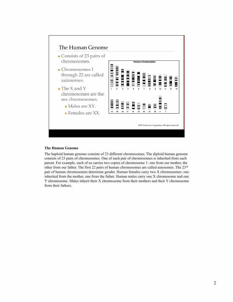
The Human Genome The haploid human genome consists of 23 different chromosomes. The diploid human genome consists of 23 pairs of chromosomes. One of each pair of chromosomes is inherited from each parent. For example, each of us carries two copies of chromosome 1: one from our mother, the other from our father. The first 22 pairs of human chromosomes are called autosomes. The 23rd pair of human chromosomes determine gender. Human females carry two X chromosomes: one inherited from the mother, one from the father. Human males carry one X chromosome and one Y chromosome. Males inherit their X chromosome from their mothers and their Y chromosome from their fathers.
3
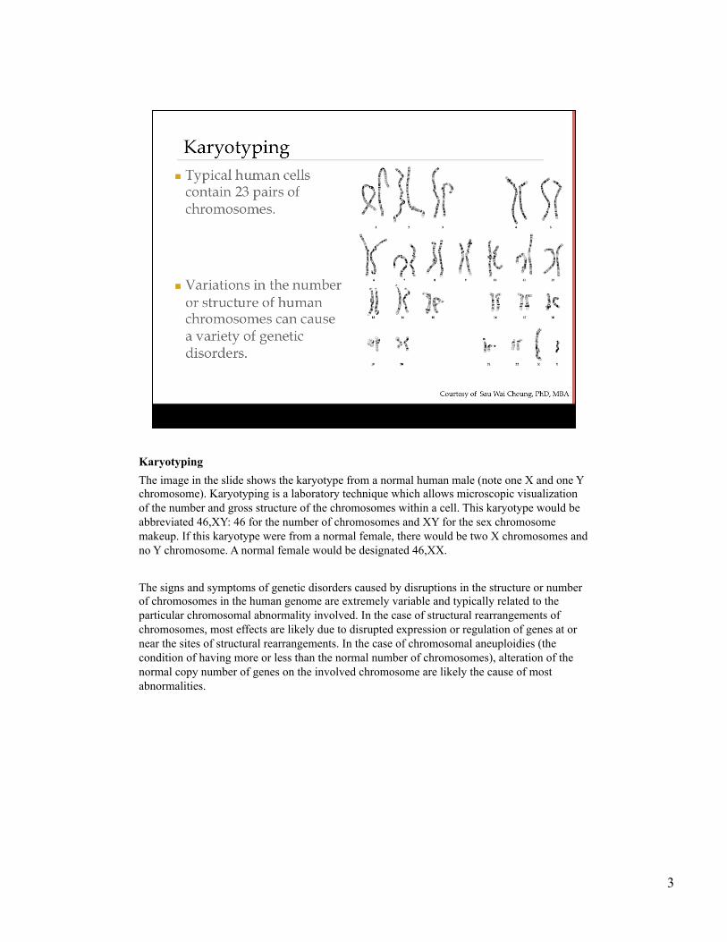
Karyotyping The image in the slide shows the karyotype from a normal human male (note one X and one Y chromosome). Karyotyping is a laboratory technique which allows microscopic visualization of the number and gross structure of the chromosomes within a cell. This karyotype would be abbreviated 46,XY: 46 for the number of chromosomes and XY for the sex chromosome makeup. If this karyotype were from a normal female, there would be two X chromosomes and no Y chromosome. A normal female would be designated 46,XX. The signs and symptoms of genetic disorders caused by disruptions in the structure or number of chromosomes in the human genome are extremely variable and typically related to the particular chromosomal abnormality involved. In the case of structural rearrangements of chromosomes, most effects are likely due to disrupted expression or regulation of genes at or near the sites of structural rearrangements. In the case of chromosomal aneuploidies (the condition of having more or less than the normal number of chromosomes), alteration of the normal copy number of genes on the involved chromosome are likely the cause of most abnormalities.
4
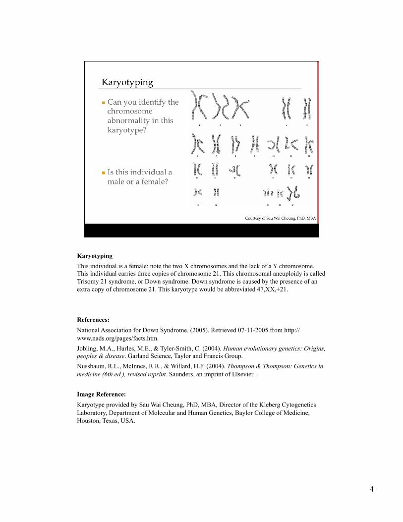
Karyotyping This individual is a female: note the two X chromosomes and the lack of a Y chromosome. This individual carries three copies of chromosome 21. This chromosomal aneuploidy is called Trisomy 21 syndrome, or Down syndrome. Down syndrome is caused by the presence of an extra copy of chromosome 21. This karyotype would be abbreviated 47,XX,+21. References: National Association for Down Syndrome. (2005). Retrieved 07-11-2005 from http://www.nads.org/pages/facts.htm. Jobling, M.A., Hurles, M.E., & Tyler-Smith, C. (2004). Human evolutionary genetics: Origins, peoples & disease. Garland Science, Taylor and Francis Group. Nussbaum, R.L., McInnes, R.R., & Willard, H.F. (2004). Thompson & Thompson: Genetics in medicine (6th ed.), revised reprint. Saunders, an imprint of Elsevier. Image Reference: Karyotype provided by Sau Wai Cheung, PhD, MBA, Director of the Kleberg Cytogenetics Laboratory, Department of Molecular and Human Genetics, Baylor College of Medicine, Houston, Texas, USA.
5
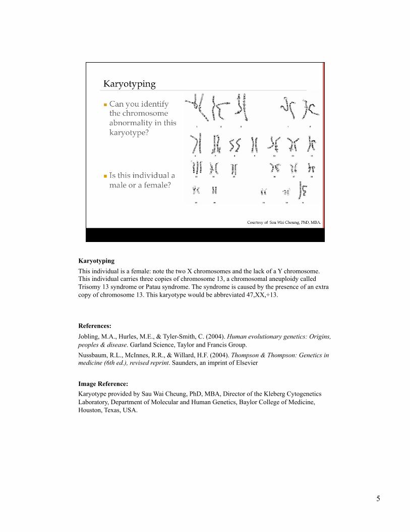
Karyotyping This individual is a female: note the two X chromosomes and the lack of a Y chromosome. This individual carries three copies of chromosome 13, a chromosomal aneuploidy called Trisomy 13 syndrome or Patau syndrome. The syndrome is caused by the presence of an extra copy of chromosome 13. This karyotype would be abbreviated 47,XX,+13. References: Jobling, M.A., Hurles, M.E., & Tyler-Smith, C. (2004). Human evolutionary genetics: Origins, peoples & disease. Garland Science, Taylor and Francis Group. Nussbaum, R.L., McInnes, R.R., & Willard, H.F. (2004). Thompson & Thompson: Genetics in medicine (6th ed.), revised reprint. Saunders, an imprint of Elsevier Image Reference: Karyotype provided by Sau Wai Cheung, PhD, MBA, Director of the Kleberg Cytogenetics Laboratory, Department of Molecular and Human Genetics, Baylor College of Medicine, Houston, Texas, USA.
6
Pedigrees
7
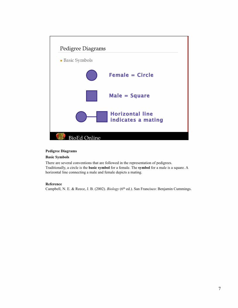
Pedigree Diagrams Basic Symbols There are several conventions that are followed in the representation of pedigrees. Traditionally, a circle is the basic symbol for a female. The symbol for a male is a square. A horizontal line connecting a male and female depicts a mating. Reference Campbell, N. E. & Reece, J. B. (2002). Biology (6th ed.). San Francisco: Benjamin Cummings.
8
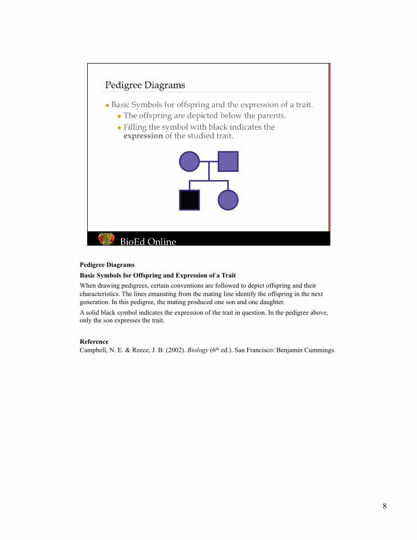
Pedigree Diagrams Basic Symbols for Offspring and Expression of a Trait When drawing pedigrees, certain conventions are followed to depict offspring and their characteristics. The lines emanating from the mating line identify the offspring in the next generation. In this pedigree, the mating produced one son and one daughter. A solid black symbol indicates the expression of the trait in question. In the pedigree above, only the son expresses the trait. Reference Campbell, N. E. & Reece, J. B. (2002). Biology (6th ed.). San Francisco: Benjamin Cummings.
9
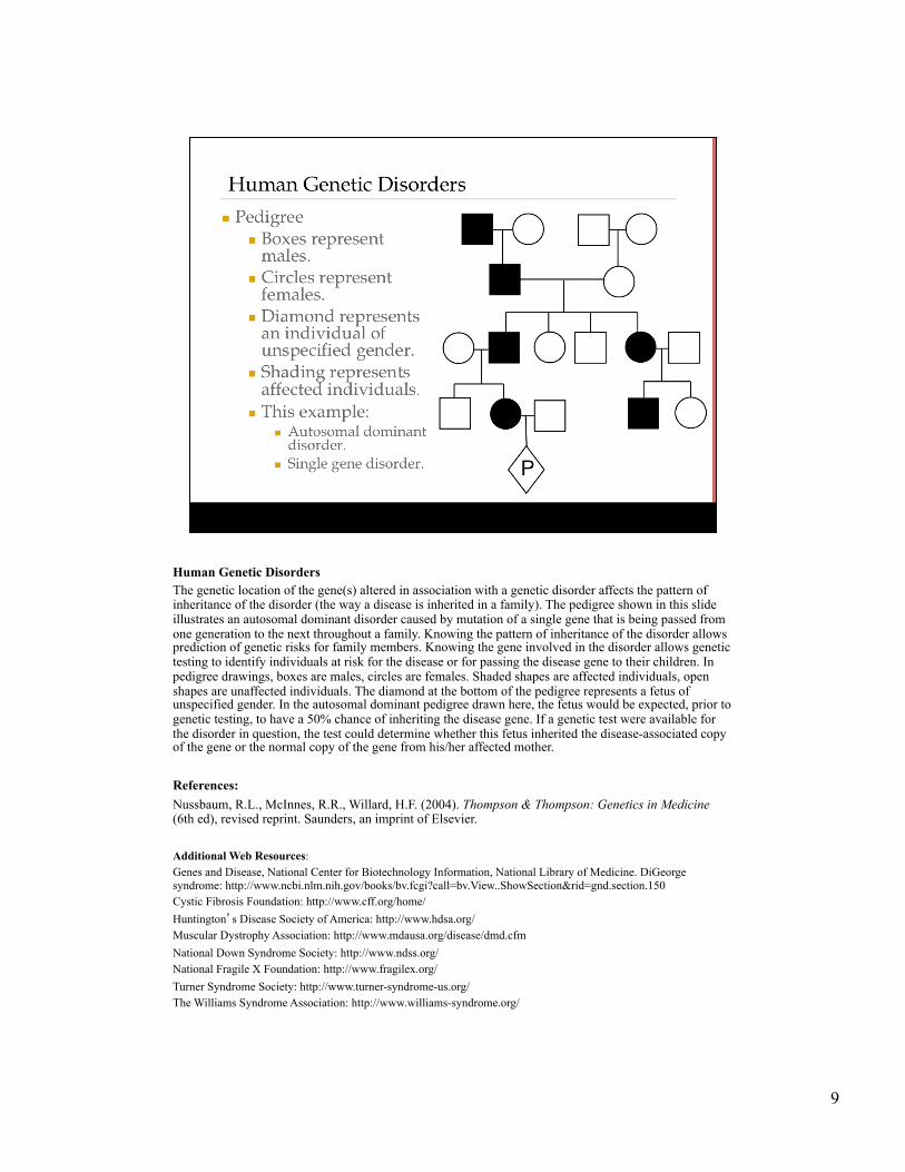
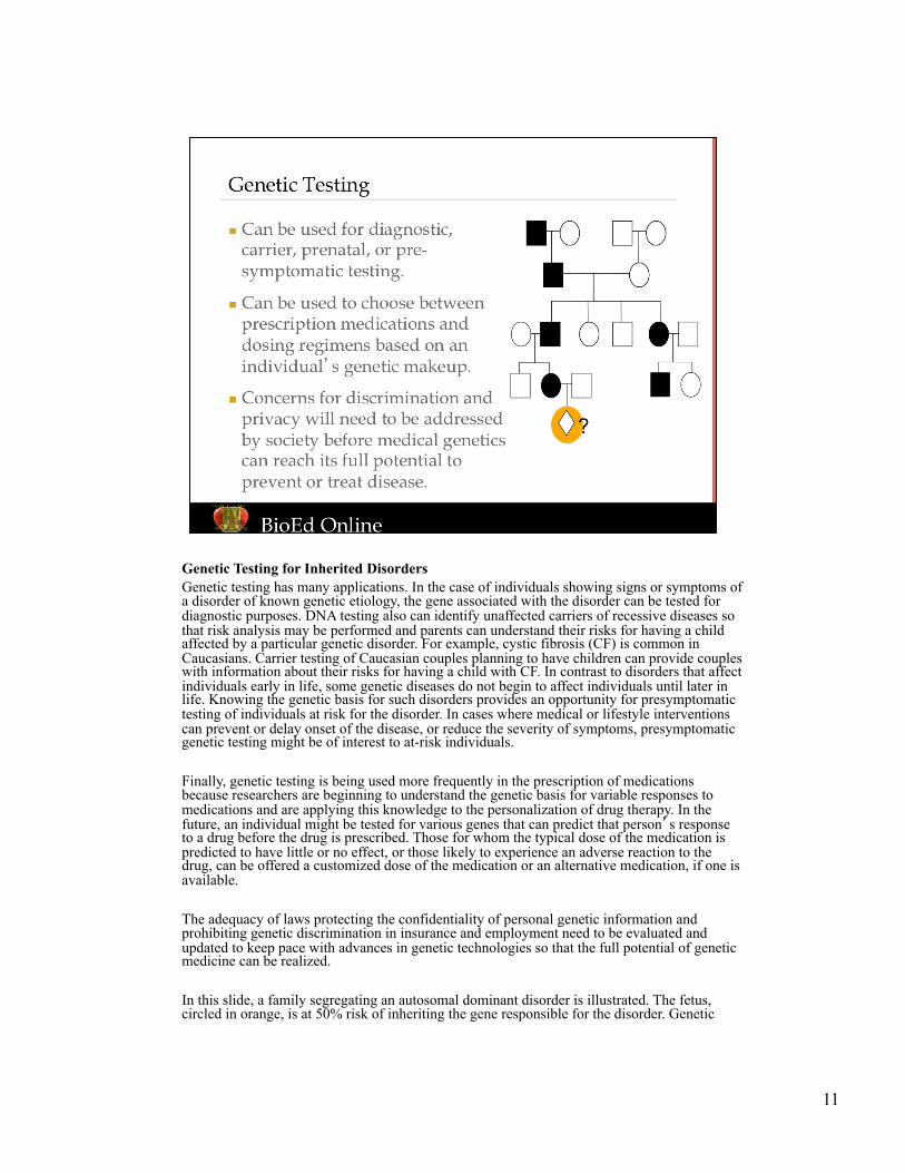
Human Genetic Disorders The genetic location of the gene(s) altered in association with a genetic disorder affects the pattern of inheritance of the disorder (the way a disease is inherited in a family). The pedigree shown in this slide illustrates an autosomal dominant disorder caused by mutation of a single gene that is being passed from one generation to the next throughout a family. Knowing the pattern of inheritance of the disorder allows prediction of genetic risks for family members. Knowing the gene involved in the disorder allows genetic testing to identify individuals at risk for the disease or for passing the disease gene to their children. In pedigree drawings, boxes are males, circles are females. Shaded shapes are affected individuals, open shapes are unaffected individuals. The diamond at the bottom of the pedigree represents a fetus of unspecified gender. In the autosomal dominant pedigree drawn here, the fetus would be expected, prior to genetic testing, to have a 50% chance of inheriting the disease gene. If a genetic test were available for the disorder in question, the test could determine whether this fetus inherited the disease-associated copy of the gene or the normal copy of the gene from his/her affected mother. References: Nussbaum, R.L., McInnes, R.R., Willard, H.F. (2004). Thompson & Thompson: Genetics in Medicine (6th ed), revised reprint. Saunders, an imprint of Elsevier. Additional Web Resources: Genes and Disease, National Center for Biotechnology Information, National Library of Medicine. DiGeorge syndrome: http://www.ncbi.nlm.nih.gov/books/bv.fcgi?call=bv.View..ShowSection&rid=gnd.section.150 Cystic Fibrosis Foundation: http://www.cff.org/home/ Huntington’s Disease Society of America: http://www.hdsa.org/ Muscular Dystrophy Association: http://www.mdausa.org/disease/dmd.cfm National Down Syndrome Society: http://www.ndss.org/ National Fragile X Foundation: http://www.fragilex.org/ Turner Syndrome Society: http://www.turner-syndrome-us.org/ The Williams Syndrome Association: http://www.williams-syndrome.org/
10
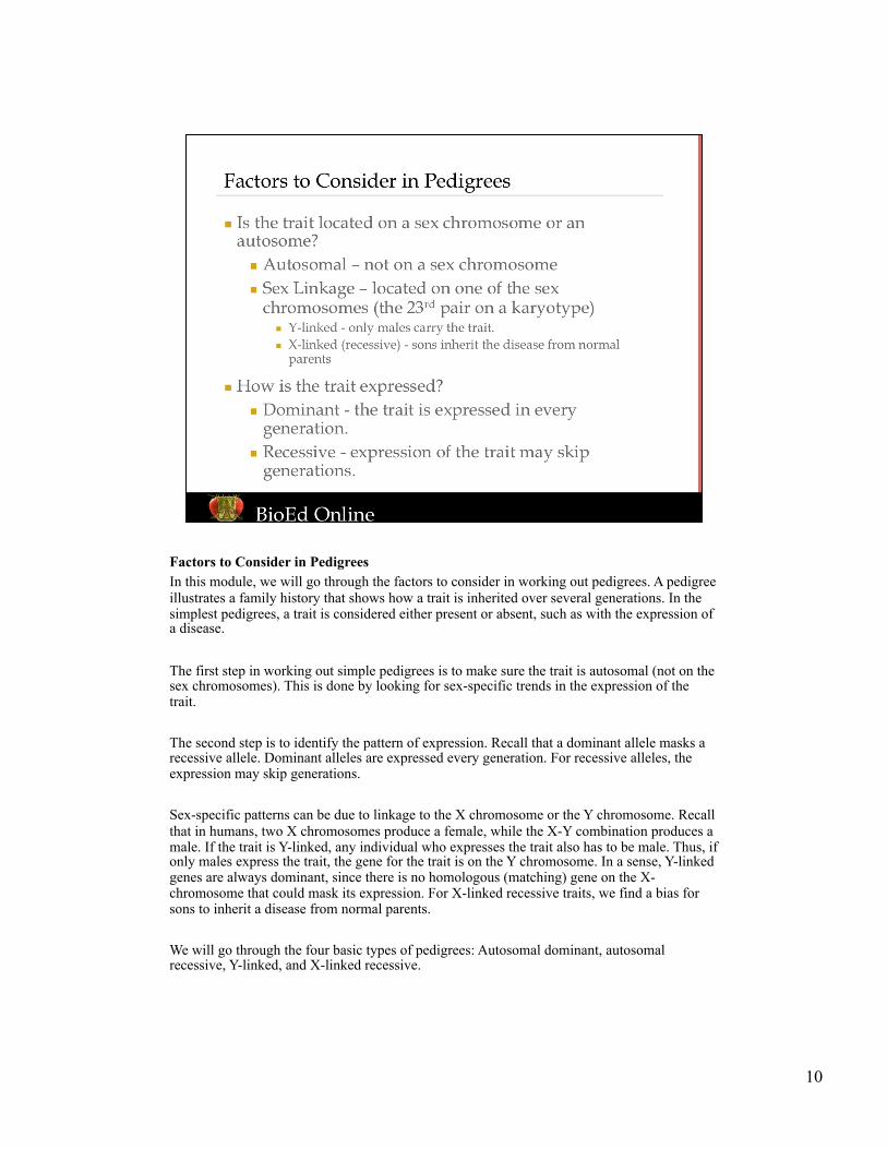
Factors to Consider in Pedigrees In this module, we will go through the factors to consider in working out pedigrees. A pedigree illustrates a family history that shows how a trait is inherited over several generations. In the simplest pedigrees, a trait is considered either present or absent, such as with the expression of a disease. The first step in working out simple pedigrees is to make sure the trait is autosomal (not on the sex chromosomes). This is done by looking for sex-specific trends in the expression of the trait. The second step is to identify the pattern of expression. Recall that a dominant allele masks a recessive allele. Dominant alleles are expressed every generation. For recessive alleles, the expression may skip generations. Sex-specific patterns can be due to linkage to the X chromosome or the Y chromosome. Recall that in humans, two X chromosomes produce a female, while the X-Y combination produces a male. If the trait is Y-linked, any individual who expresses the trait also has to be male. Thus, if only males express the trait, the gene for the trait is on the Y chromosome. In a sense, Y-linked genes are always dominant, since there is no homologous (matching) gene on the X-chromosome that could mask its expression. For X-linked recessive traits, we find a bias for sons to inherit a disease from normal parents. We will go through the four basic types of pedigrees: Autosomal dominant, autosomal recessive, Y-linked, and X-linked recessive.
11
Genetic Testing for Inherited Disorders Genetic testing has many applications. In the case of individuals showing signs or symptoms of a disorder of known genetic etiology, the gene associated with the disorder can be tested for diagnostic purposes. DNA testing also can identify unaffected carriers of recessive diseases so that risk analysis may be performed and parents can understand their risks for having a child affected by a particular genetic disorder. For example, cystic fibrosis (CF) is common in Caucasians. Carrier testing of Caucasian couples planning to have children can provide couples with information about their risks for having a child with CF. In contrast to disorders that affect individuals early in life, some genetic diseases do not begin to affect individuals until later in life. Knowing the genetic basis for such disorders provides an opportunity for presymptomatic testing of individuals at risk for the disorder. In cases where medical or lifestyle interventions can prevent or delay onset of the disease, or reduce the severity of symptoms, presymptomatic genetic testing might be of interest to at-risk individuals. Finally, genetic testing is being used more frequently in the prescription of medications because researchers are beginning to understand the genetic basis for variable responses to medications and are applying this knowledge to the personalization of drug therapy. In the future, an individual might be tested for various genes that can predict that person’s response to a drug before the drug is prescribed. Those for whom the typical dose of the medication is predicted to have little or no effect, or those likely to experience an adverse reaction to the drug, can be offered a customized dose of the medication or an alternative medication, if one is available. The adequacy of laws protecting the confidentiality of personal genetic information and prohibiting genetic discrimination in insurance and employment need to be evaluated and updated to keep pace with advances in genetic technologies so that the full potential of genetic medicine can be realized. In this slide, a family segregating an autosomal dominant disorder is illustrated. The fetus, circled in orange, is at 50% risk of inheriting the gene responsible for the disorder. Genetic
12
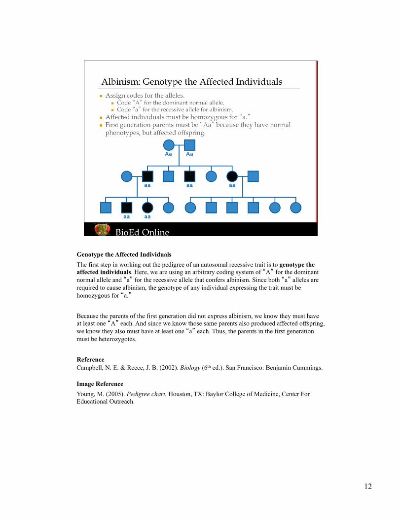
Genotype the Affected Individuals The first step in working out the pedigree of an autosomal recessive trait is to genotype the affected individuals. Here, we are using an arbitrary coding system of “A” for the dominant normal allele and “a” for the recessive allele that confers albinism. Since both “a” alleles are required to cause albinism, the genotype of any individual expressing the trait must be homozygous for “a.” Because the parents of the first generation did not express albinism, we know they must have at least one “A” each. And since we know those same parents also produced affected offspring, we know they also must have at least one “a” each. Thus, the parents in the first generation must be heterozygotes. Reference Campbell, N. E. & Reece, J. B. (2002). Biology (6th ed.). San Francisco: Benjamin Cummings. Image Reference Young, M. (2005). Pedigree chart. Houston, TX: Baylor College of Medicine, Center For Educational Outreach.
13
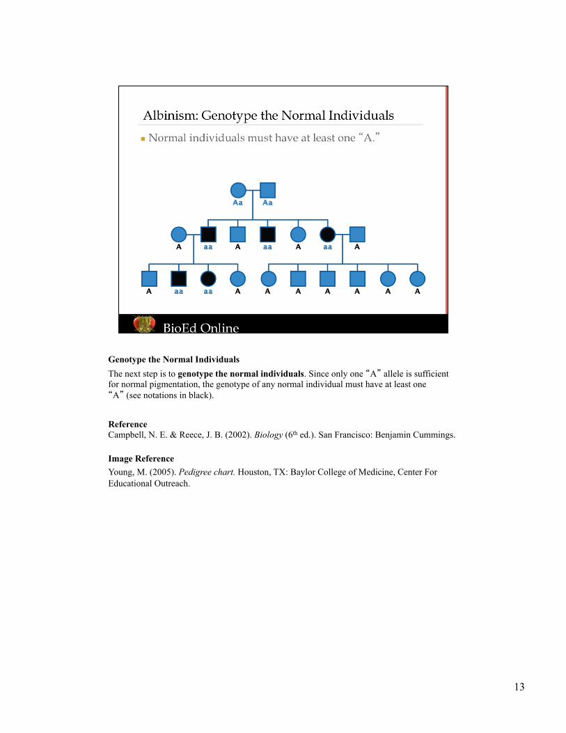
Genotype the Normal Individuals The next step is to genotype the normal individuals. Since only one “A” allele is sufficient for normal pigmentation, the genotype of any normal individual must have at least one “A” (see notations in black). Reference Campbell, N. E. & Reece, J. B. (2002). Biology (6th ed.). San Francisco: Benjamin Cummings. Image Reference Young, M. (2005). Pedigree chart. Houston, TX: Baylor College of Medicine, Center For Educational Outreach.
14
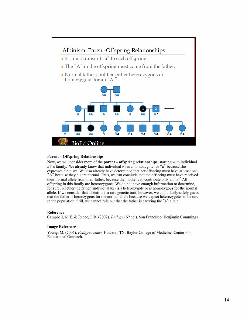
Parent – Offspring Relationships Now, we will consider more of the parent – offspring relationships, starting with individual #1’s family. We already know that individual #1 is a homozygote for “a” because she expresses albinism. We also already have determined that her offspring must have at least one “A” because they all are normal. Thus, we can conclude that the offspring must have received their normal allele from their father, because the mother can contribute only an “a.” All offspring in this family are heterozygotes. We do not have enough information to determine, for sure, whether the father (individual #2) is a heterozygote or is homozygous for the normal allele. If we consider that albinism is a rare genetic trait, however, we could fairly safely guess that the father is homozygous for the normal allele because we expect heterozygotes to be rare in the population. Still, we cannot rule out that the father is carrying the “a” allele. Reference Campbell, N. E. & Reece, J. B. (2002). Biology (6th ed.). San Francisco: Benjamin Cummings. Image Reference Young, M. (2005). Pedigree chart. Houston, TX: Baylor College of Medicine, Center For Educational Outreach.
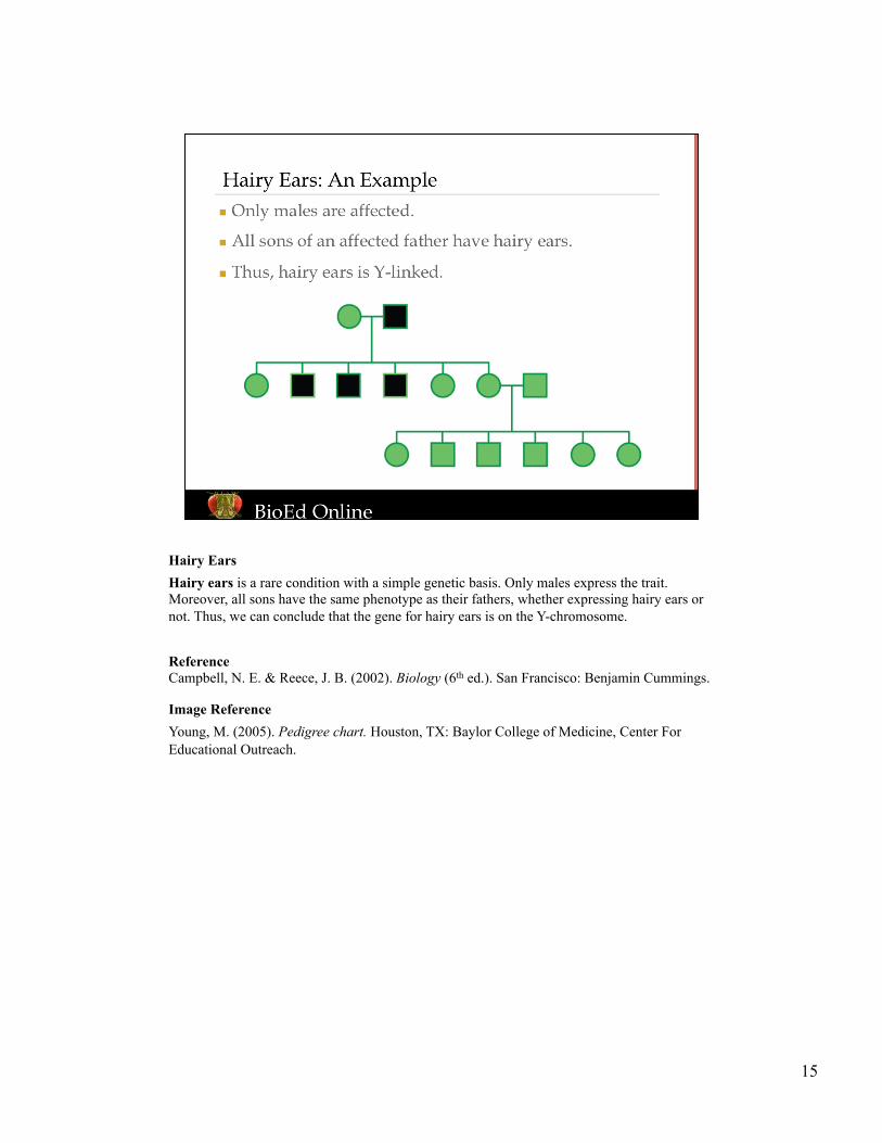
15
Hairy Ears Hairy ears is a rare condition with a simple genetic basis. Only males express the trait. Moreover, all sons have the same phenotype as their fathers, whether expressing hairy ears or not. Thus, we can conclude that the gene for hairy ears is on the Y-chromosome. Reference Campbell, N. E. & Reece, J. B. (2002). Biology (6th ed.). San Francisco: Benjamin Cummings. Image Reference Young, M. (2005). Pedigree chart. Houston, TX: Baylor College of Medicine, Center For Educational Outreach.
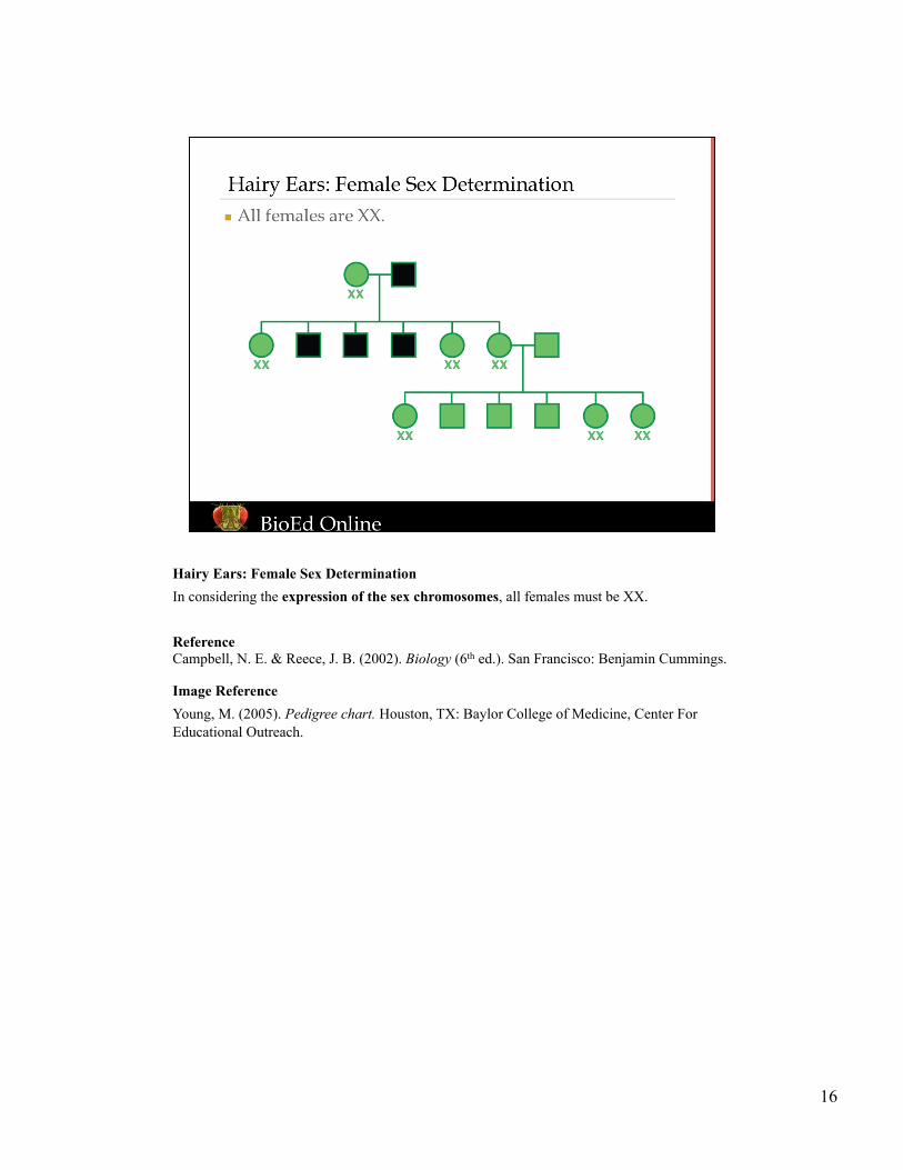
16
Hairy Ears: Female Sex Determination In considering the expression of the sex chromosomes, all females must be XX. Reference Campbell, N. E. & Reece, J. B. (2002). Biology (6th ed.). San Francisco: Benjamin Cummings. Image Reference Young, M. (2005). Pedigree chart. Houston, TX: Baylor College of Medicine, Center For Educational Outreach.

17
Hairy Ears: Male Sex Determination Under the human sex determination system, all males are XY. Reference Campbell, N. E. & Reece, J. B. (2002). Biology (6th ed.). San Francisco: Benjamin Cummings. Image Reference Young, M. (2005). Pedigree chart. Houston, TX: Baylor College of Medicine, Center For Educational Outreach.
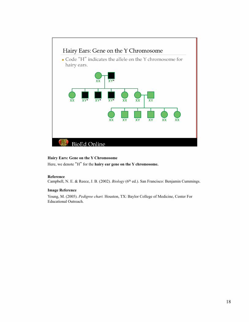
18
Hairy Ears: Gene on the Y Chromosome Here, we denote “H” for the hairy ear gene on the Y chromosome. Reference Campbell, N. E. & Reece, J. B. (2002). Biology (6th ed.). San Francisco: Benjamin Cummings. Image Reference Young, M. (2005). Pedigree chart. Houston, TX: Baylor College of Medicine, Center For Educational Outreach.
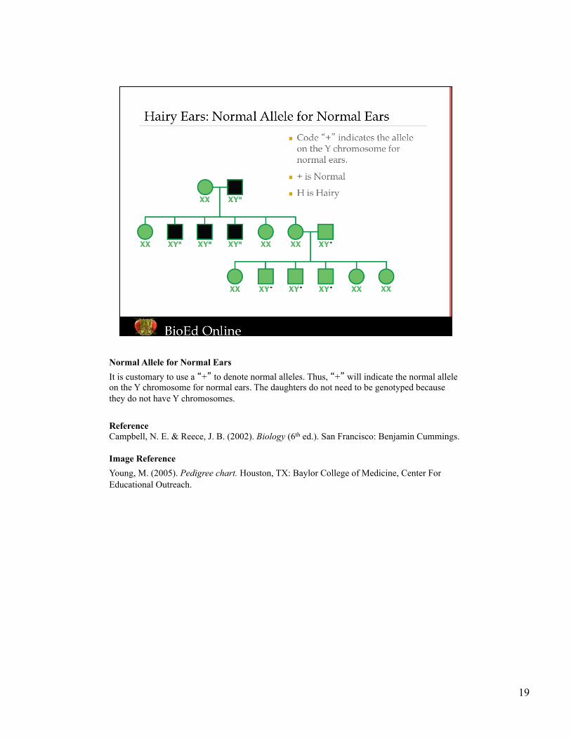
19
Normal Allele for Normal Ears It is customary to use a “+” to denote normal alleles. Thus, “+” will indicate the normal allele on the Y chromosome for normal ears. The daughters do not need to be genotyped because they do not have Y chromosomes. Reference Campbell, N. E. & Reece, J. B. (2002). Biology (6th ed.). San Francisco: Benjamin Cummings. Image Reference Young, M. (2005). Pedigree chart. Houston, TX: Baylor College of Medicine, Center For Educational Outreach.
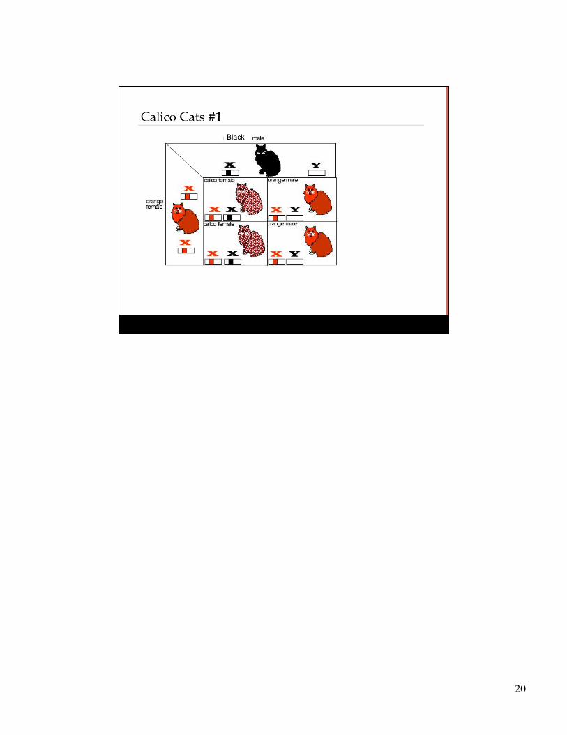
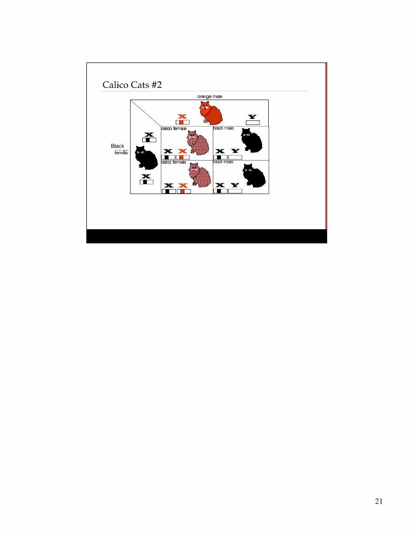
20
21