Embed Size (px)
Citation preview
Fax +41 61 306 12 34E-Mail [email protected]
Research Article
J Innate Immun 2011;3:614–622 DOI: 10.1159/000327839
The Honeybee Antimicrobial Peptide Apidaecin Differentially Immunomodulates Human Macrophages, Monocytes and Dendritic Cells
Regina Tavano a, b Daniela Segat a, b Marina Gobbo c Emanuele Papini a, b
a Centro di Ricerca Interdipartimentale per le Biotecnologie Innovative, b Dipartimento di Scienze Biomediche Sperimentali, and c Dipartimento di Scienze Chimiche, Università di Padova, Padova , Italy
low concentrations it partially antagonized LPS-stimulatory effects on both macrophages and monocytes while it stimu-lated pro-inflammatory and pro-immune functions of mac-rophages at higher concentrations.
Copyright © 2011 S. Karger AG, Basel
Introduction
A variety of innate antimicrobial peptides (AMPs) are distributed in living beings [1] . Membrane-active AMPs, such as defensins, perturb the bacterium plasma mem-brane, while non-lytic ones, like insect apidaecins, trans-locate into the microorganism cytosol [2] . Several AMPs may also regulate innate and adaptive immune cell func-tions [3–5] , such as chemotaxis, apoptosis, cytokine/che-mokine production [6, 7] , antigen presentation and Th1/Th2 balance [6, 8] .
Insect hemolymph contains AMPs ensuring protec-tion against bacterial infections. Among these, the pro-rich 18–20 residues of apidaecins are potentially exploit-able in medical and biotechnology applications as novel antibiotic drugs [9] , being poorly structured, devoid of S-S bridges, easily synthesizable and not toxic to human cells [10] . Apidaecin, like similar pro-rich peptides (dro-socin, pyrrhocoricin and Bac7), acts via a three-step pro-
Key Words Antimicrobial peptides � Apidaecin � Chemokines � Cytokines � Honeybee � Macrophages � Monocytes
Abstract We show that apidaecin binds to human macrophages, monocytes and dendritic cells, displaying different intracel-lular distributions and inducing diversified effects. An api-daecin-cell association was detectable at concentrations as low as 5 � M and increased without saturation until 60 � M , was receptor independent and required a physiological tem-perature (37 ° C). For apidaecin, cytosolic localization was prevalent in macrophages and endosomal localization in monocytes, and associations with the plasma membrane were predominant in dendritic cells. Apidaecin upregulated T-lymphocyte co-stimulatory molecule CD80 and cytokine/chemokine production in macrophages, but not in mono-cytes and dendritic cells. Suboptimal stimulatory doses (5–10 � M ) of apidaecin partially inhibited the lipopolysaccha-ride (LPS)-induced increase in major histocompatibility com-plex class II (MHCII) and CD86 in macrophages, and the release of selected cytokines/chemokines by both macro-phages [interleukin (IL)-6 and tumor necrosis factor (TNF)- � ] and monocytes [IL-6, TNF- � , basic fibroblast growth factor (FGF) and eotaxin]. Apidaecin had a double-edged effect: at
Received: October 27, 2010 Accepted after revision: March 29, 2011 Published online: June 16, 2011
Journal of InnateImmunity
Dr. Regina Tavano Dipartimento di Scienze Biomediche Sperimentali, Università di Padova Via U. Bassi 58/B IT–35131 Padova (Italy) Tel. +39 049 827 6478, E-Mail regina.tavano @ unipd.it
© 2011 S. Karger AG, Basel1662–811X/11/0036–0614$38.00/0
Accessible online at:www.karger.com/jin

Apidaecin Modulates the Human Immune System
J Innate Immun 2011;3:614–622 615
cess involving: (i) bacterial surface binding, (ii) transloca-tion into the cytoplasm and (iii) binding to DnaK, the Escherichia coli homologue of human HSP70 [2, 11–12] . While the bacteriostatic action of apidaecin has been ex-tensively studied [13–15] , its immunostimulatory activity has not been elucidated yet. Based on the structural sim-ilarity between mammalian and insect pro-rich AMPs, we hypothesized that apidaecin, although produced by phylogenetically distant organisms, can cross talk with the human immune system. This possibility is supported by the observation that pro-rich mammalian peptides Bac7 and PR39 can interact with immune cells [16–18] . We show here for the first time that apidaecin associates without inducing cytotoxic effects to three important hu-man immune cells: macrophages, monocytes and den-dritic cells. Interestingly, we found that apidaecin effec-tively translocates into the cell cytosol of macrophages, but remains mostly endosomal or plasma membrane as-sociated in monocytes and dendritic cells, respectively. Consistently, we show that apidaecin differentially regu-lates their basal and lipopolysaccharide (LPS)-induced cytokine/chemokine release and the expression of pro-teins necessary for antigen presentation and T-lympho-cyte activation, showing a higher effect on macrophages. Our data suggest that apidaecin is partially anti-inflam-matory in the presence of microbial stimuli and has a pro-immune/pro-inflammatory action when acting alone.
Materials and Methods
Synthesis of Apidaecin and Its Derivatives Full-length apidaecin Ib (GNNRPVYIPQPRPPHPRL), the
peptide sequence corresponding to amino acids 13–18 of apidae-cin and their fluoresceinated analogues were synthesized by sol-id-phase synthesis as previously reported [10] . Following the same protocol, an apidaecin derivative extended at the N-terminus with a cysteine residue (Cys-apidaecin) was prepared and used after HPLC purification in affinity chromatography. The product was characterized by electrospray ionization mass spectrometry (MARINER API-TOF workstation), and its purity ( 1 95%) was confirmed by analytical HPLC, performed on a Dionex Sum-mit Dual Gradient apparatus equipped with a Vydac C18 column (250 ! 4.6 mm, 5 � m).
Preparation of Human Monocytes, Macrophages and Dendritic Cells Monocyte-enriched preparations were isolated from buffy
coats of blood by centrifugation over a Ficoll-Hypaque (Amers-ham Biosciences) step gradient and a subsequent Percoll (Amers-ham Biosciences) gradient as described [19] . Unless otherwise specified, cells were kept at 37 ° C in a humidified atmosphere con-taining 5% (v/v) CO 2 in RPMI-1640 supplemented with 10% FCS. Human macrophages were derived from monocytes cultured for
7 days with 100 ng/ml macrophage colony-stimulating factor (M-CSF; PeproTech) in RPMI-1640 supplemented with 20% FCS. On day 4, M-CSF was added again. Human dendritic cells were ob-tained as described [20] : following this procedure 1 90% cells be-longed to the immature dendritic cell phenotype.
Cytofluorimetric Analysis The day before the experiment, human monocytes/macro-
phages or dendritic cells were incubated for different times with apidaecin (5–50 � M ) with or without LPS 0.05–0.2 � g/ml (Sigma). The cells were then incubated at 37 or 0 ° C, depending on the experiment, in RPMI medium supplemented with 10% FCS. Su-pernatants were collected and cells were then washed with PBS, resuspended in FACS buffer (PBS, 1% FBS) and 10,000 events/sample were analyzed using a FACSCanto � analyzer (Becton Dickinson). Dead cells were excluded from FACS analysis by add-ing propidium iodide. Data were processed using the FACSDiva software. For the analysis of surface marker expression, after the incubation, cells were washed, resuspended in FACS buffer, di-vided in different samples and incubated with anti-major histo-compatibility complex class II (MHCII), CD80, CD86 and ICAM-1-phycoerythrin labelled antibodies (Biolegend) for 30 min at 4 ° C. Cells were then washed, resuspended in FACS buffer and analyzed by cytofluorimetry.
Confocal Analysis Macrophages, monocytes or dendritic cells (2 ! 10 6 cells/
well) were seeded on cover glasses and incubated for 18 h at 37 ° C with fluoresceinated apidaecin. Unfixed cells were briefly rinsed with PBS and directly analyzed under a confocal microscope (SP2 Leica). As a control, cells without apidaecin have been used. Al-ternatively, adherent macrophages were incubated for 18 h with 30 � M apidaecin at 37 ° C; after three washes with PBS, cells were fixed in cold methanol for 10 min and permeabilized with cold acetone for 10 min. Before addition of primary antibodies, im-munoglobulin binding of monocytes was saturated by treatment with pooled human IgG (Sigma Chemical Co., 10 mg/ml in PBS). Cells were incubated overnight at 4 ° C with rabbit polyclonal anti-apidaecin antibody, washed three times and further incubated for 40 min with secondary FITC-labelled anti-rabbit antibody (Cal-biochem). After three washes, coverslips were mounted in mount-ing medium (KPL) and were analyzed with a confocal microscope (SP2 Leica). Images have been acquired with the different fluores-cence filters; representative pictures were collected as Tiff files and processed with standard imaging programs.
Bio-Plex Multiplex Cytokine Assays A Bio-Plex assay was performed as previously described [19] .
The fluorescence intensity of the beads was measured with a Lu-minex system (Bio-Rad) and data were analyzed with Bio-Plex Manager software.
Peptide-Carrier Conjugation Cys-apidaecin (2 mg) was conjugated with 2 mg of mcKLH
(Pierce) in conjugation buffer, following the manufacturer’s in-structions. The conjugate was subsequently purified by gel filtra-tion using Sephadex G-25 resin (Sigma). Fractions containing proteins were mixed, divided in aliquots and frozen in liquid ni-trogen.

Tavano /Segat /Gobbo /Papini J Innate Immun 2011;3:614–622 616
37°C 0°C
Pept
ide-
cell
asso
ciat
ion
(MFI
)
Apidaecin (μM)
02,0004,0006,0008,000
10,00012,00014,00016,00018,000
Macrophages
0 10 20 30 40 50 60
Time (h)0 1 2 3 4 5 6
Time (h)0 1 2 3 4 5 6
Time (h)0 1 2 3 4 5 6
0
1,000
2,000
3,000
4,000
5,000
6,000
7,000
8,000 Monocytes
0
1,000
2,000
3,000
4,000
5,000
6,000 Dendritic cells
a
b
c d
Apidaecin (μM)0 10 20 30 40 50 60
Apidaecin (μM)0 10 20 30 40 50 60
0
1,000
2,000
3,000
4,000
5,000 Macrophages
0
500
1,000
1,500
2,000 Dendritic cells
Pept
ide-
cell
asso
ciat
ion
(MFI
)
0
1,000
2,000
3,000
4,000
5,000 Monocytes
FApi FApi (13–18)
FApi
(13–
18) a
ssoc
iatio
n(%
with
resp
ect t
o FA
pi a
t 37°
C)
FApi
ass
ocia
tion
(% w
ithre
spec
t to
FApi
ass
ocia
tion
at 3
7°C
)
0
50
100
150
200 No apidaecin Apidaecin 200 μM
0
20
40
60
80
100
120 MacrophagesMonocytes
37°C0
20
40
60
80
100
120MacrophagesMonocytes
0°C37°C 0°C
* *
Fig. 1. Dose ( a )- and time-dependent uptake of apidaecin ( b ) in human monocytes, macrophages and dendritic cells. Cells were incubated at 37 or 0 ° C for 1 h with 10, 20 or 60 � M ( a ) or for 1, 3 or 6 h with 10 � M ( b ) of fluoresceinated apidaecin (FApi). Cells were then washed with PBS, trypsinized and resuspended in FACS buffer. Mean fluorescence intensity (MFI) of at least 10,000 cells/sample due to FITC-conjugated peptide uptake was quanti-fied. Data are from one of two representative experiments, run in triplicate, and bars represent ranges. c Competition assay. Human monocytes were incubated with 5 � M FApi at 37 or 0 ° C for 3 h with or without 200 � M non-labelled apidaecin. Cells were then washed, trypsinized and analyzed by FACS. MFI values are the
mean of three experiments run in duplicate, and are expressed as percent of the signal produced by labelled peptide alone at 37 ° C. Means 8 SE. d Different uptake of the whole peptide and the con-served region of apidaecin (residues 13–18). Human monocytes or macrophages were incubated with 10 � M of FApi or FApi (13–18) for 3 h at 37 or 0 ° C. Cells were then washed with PBS, trypsinized and resuspended in FACS buffer. MFI due to FITC-conjugated peptides was quantified. Data are from one of two representative experiments run in triplicate, and percentage values were calcu-lated considering 100 for FApi association at 37 ° C in monocytes and macrophages, respectively. Means 8 SE. * p ̂ 0.05.

Apidaecin Modulates the Human Immune System
J Innate Immun 2011;3:614–622 617
Immunization and Production of Polyclonal Antibodies Male adult New Zealand white rabbits (Harlan, Italy) were im-
munized by subcutaneous injection with an aliquot (750 � l) of carrier-peptide conjugate mixed 1: 1 v/v with complete Freund’s adjuvant (Sigma). Three subsequent injections were administered at 14-day intervals with incomplete Freund’s adjuvant (Sigma). Anti-sera were taken on day 68 and antibodies were purified by affinity chromatography on apidaecin-modified columns. Brief-ly, columns were prepared with 2.5 ml of 50% SulfoLink coupling gel (Pierce) and 2 mg of Cys-apidaecin dissolved in coupling buf-fer (50 m M Tris-HCl, 5 m M EDTA, pH 8.5). Columns were then washed with coupling buffer and the non-specific binding sites were blocked with a solution of 50 m M cysteine (Sigma) for 30 min at room temperature. Subsequently, columns were washed again extensively, and antisera (diluted 1: 1 v/v with PBS) were added to the columns; after extensive washing, antibodies were eluted with 0.2 M glycine, pH 2.8.
Measurement of the LPS-Binding Activities of Apidaecin The assay was performed as described [21] . Briefly, the day be-
fore the experiment, NUNC plates were incubated with 100 � l of PBS with or without 1 � g of LPS (Sigma)/well, at room tempera-ture. The day of the experiment, after some washing, wells were saturated for 1 h at room temperature with 1% BSA in PBS. Then the plate was incubated for 1 h 30 min at 37 ° C with different pep-tide concentrations. After some washing, 100 ng of anti-apidaecin were added to each well and incubated for 90 min at room tem-perature. Then 100 � l of 1/200 anti-rabbit HRP (Calbiochem) were added and incubated for 1 h at room temperature. Finally, the peroxidase substrate solution (ABTS) was added to each well. Optical density was assessed at 405 nm on an automatic micro-plate reader (Amersham).
Statistical Analysis Results were expressed as means 8 SEM. Significance (p ̂
0.05) of differences with respect to control values were calculated by two-population t tests.
Results
Flow-cytofluorimetric experiments with a fluores-cent-labelled analogue showed that the interaction be-tween apidaecin and macrophages, monocytes and den-dritic cells is comparable, although efficacy is slightly de-creasing ( fig. 1 a, b). At 37 ° C, apidaecin cell uptake reached an equilibrium after � 1–3 h and was hardly saturable at concentrations of up to 60 � M (150 � g/ml). At 0 ° C, api-daecin-cell association was markedly reduced. Competi-tion assays ( fig. 1 c) proved that this process does not re-quire high-affinity receptors, similarly to what was ob-served in epithelial cells [10] . Cell association with a fluorescent, shorter peptide corresponding to the C-ter-minal sequence 13–18, conserved in different apidaecin isoforms, was significantly decreased at 37 ° C, but com-parable to that of full-length apidaecin at 0 ° C, suggesting
Mac
roph
ages
Mon
ocyt
esD
endr
itic
cells
+ Apidaecin – Apidaecin
20 μm
20 μm
10 μm
10 μm
a
b
c
d
Fig. 2. Confocal analysis of human monocytes, macrophages or dendritic cells treated with apidaecin. Cells grown on glass cov-erslips were incubated with 30 � M apidaecin, fixed with metha-nol, and permeabilized with acetone; cells were then subjected to indirect immunostaining with specific anti-apidaecin rabbit an-tibodies and with FITC-labelled anti-rabbit antibodies and ana-lyzed by confocal microscopy ( a ). Alternatively, cells adherent to glass coverslips were incubated for 18 h with culture medium con-taining 30 � M fluoresceinated apidaecin at 37 ° C, briefly rinsed in PBS and then directly analyzed using a confocal microscope ( b–d ). Control cells were treated with no apidaecin. Arrowhead indicates the plasma membrane, arrows intracellular vesicles, and asterisks indicate the cytosol.

Tavano /Segat /Gobbo /Papini J Innate Immun 2011;3:614–622 618
that the lacking N-terminal sequence is necessary for ef-ficient cell capture of apidaecin at physiological tempera-ture ( fig. 1 d). Cell binding was not enhanced by the ab-sence of serum in the extracellular medium, suggesting that serum (10% v/v) does not interfere with peptide cell association (data not shown).
The intracellular distribution of apidaecin in the three cell types was analyzed by fluorescence confocal micros-
copy ( fig. 2 ). Pictures showed significant differences: in monocytes, the bee peptide distributes in a punctuated way, which is compatible with endocytic vesicles, but in a more diffuse way in macrophages, which is compatible with cytosolic translocation. In contrast, in dendritic cells, apidaecin is apparently mainly restricted to the plasma membrane and is found in reduced quantity in intracellular compartments.
010203040506070
MFI
Monocytes Macrophages Dendritic cells
0
1
2
3
4
5
MFI
0
10
20
30
40
50
60
MFI
020406080
100120140
MFI
MHCII
CD80
CD86
ICAM-1
020406080
100120140
MFI
02468
101214
MFI
050
100150200250300350
MFI
0
100
200
300
400
500
600
MFI
*
*
0
200
400
600
800
1,000
1,200
MFI
+ Apidaecin– Apidaecin
0
200
400
600
800
1,000
MFI
0
1,000
2,000
3,000
4,000
5,000
MFI
0
1,000
2,000
3,000
4,000M
FI
LPSControlLPSControlLPSControl
LPSControlLPSControlLPSControl
LPSControlLPSControlLPSControl
LPSControlLPSControlLPSControl
*
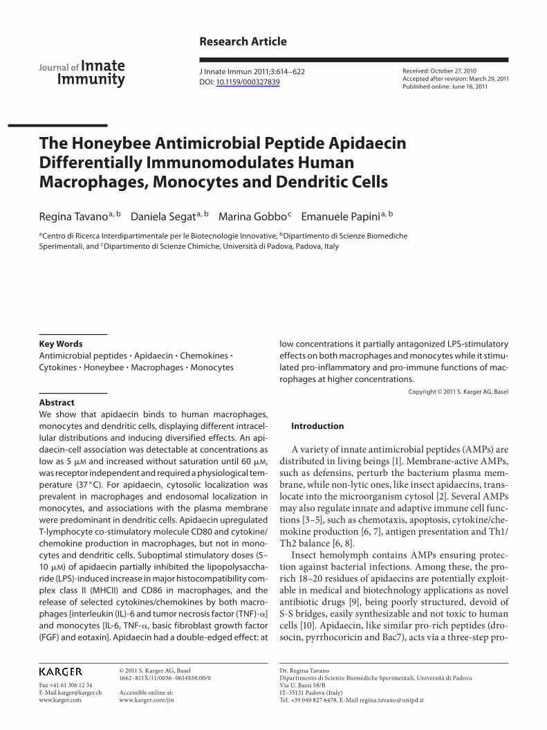
Fig. 3. Effect of apidaecin on CD80, CD86, MHCII and ICAM-1 expression in monocytes, macrophages and dendritic cells. Ad-herent human monocytes, differentiated macrophages or den-dritic cells were cultured in RPMI plus 10% FCS with 10 � M api-daecin and 0.2 � g/ml E. coli LPS, as indicated. After 24 h, cells
were collected and the indicated markers were stained with phy-coerythrin-labelled anti-CD-specific antibodies and analyzed by FACS. Data are expressed as MFI and are the mean 8 SE of four experiments run in triplicate. * p ̂ 0.05 vs. control (no stimulus or LPS stimulated only).
Apidaecin Modulates the Human Immune System
J Innate Immun 2011;3:614–622 619
ControlApidaecin
Monocytes Macrophages Monocytes Macrophages Monocytes Macrophages Monocytes Macrophages
0
10
20
30
40
50 IL-1�
0200400600800
1,0001,2001,4001,600 IL-6
0200400600800
1,0001,2001,4001,6001,800 IL-10
0
10
20
30
40
50 IL-12p70
0200400600800
1,0001,2001,400 IL-17
0100200300400500600700 TNF-�
0
200
400
600
800
1,000 G-CSF
0100200300400500600700800 Basic FGF
0100200300400500600700800900 VEGF
0
500,000
1,000,000
1,500,000
2,000,000IL-8
02,0004,0006,0008,000
10,00012,00014,000 MIP-1�
0
5,000
10,000
15,000
20,000
25,000 MCP-1
a
Monocytes Macrophages Monocytes Macrophages Monocytes Macrophages Monocytes Macrophagesb
(pg/
ml)
(pg/
ml)
(pg/
ml)
* * * *
**
* * **
LPSLPS +apidaecin(p
g/m
l)(p
g/m
l)(p
g/m
l)
01,0002,0003,0004,0005,0006,0007,000 IL-1�
01,0002,0003,000
100,000200,000300,000400,000 IL-6
0
1,000
2,000
3,000
4,000 IL-10
01020304050607080 IL-12p70
0
500
1,000
1,500
2,000
2,500 IL-17
05,000
10,000
30,00040,00050,00060,00070,000 TNF-�
01,0002,0003,0004,0005,0006,0007,0008,000 G-CSF
0100200300400500600 Basic FGF
0
100
200
300
400 VEGF
0
500,000
1,000,000
1,500,000
2,000,000 IL-8
0
10,000
20,000
30,000
40,000
50,000 MIP-1�
0
5,000
10,000
15,000 MCP-1
*
*
*
* *
*
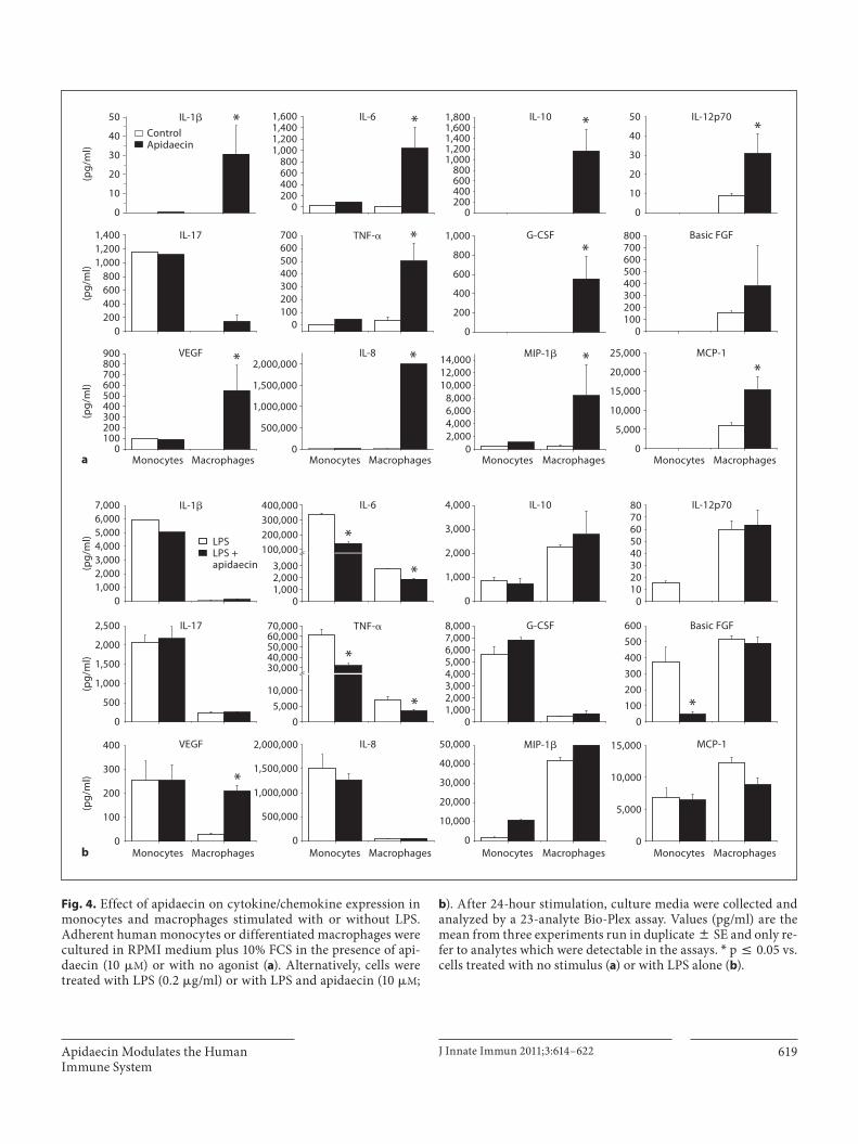
Fig. 4. Effect of apidaecin on cytokine/chemokine expression in monocytes and macrophages stimulated with or without LPS. Adherent human monocytes or differentiated macrophages were cultured in RPMI medium plus 10% FCS in the presence of api-daecin (10 � M ) or with no agonist ( a ). Alternatively, cells were treated with LPS (0.2 � g/ml) or with LPS and apidaecin (10 � M ;
b ). After 24-hour stimulation, culture media were collected and analyzed by a 23-analyte Bio-Plex assay. Values (pg/ml) are the mean from three experiments run in duplicate 8 SE and only re-fer to analytes which were detectable in the assays. * p ̂ 0.05 vs. cells treated with no stimulus ( a ) or with LPS alone ( b ).

Tavano /Segat /Gobbo /Papini J Innate Immun 2011;3:614–622 620
The effects of apidaecin on the basal or LPS-induced expression of MHCII, CD80, CD86 and ICAM-1 were an-alyzed by flow-cytofluorimetry. Figure 3 shows that mac-rophage CD80 expression was upregulated by the pep-tide, reaching a value similar to that induced by LPS, while monocytes and dendritic cells were not influenced. On the contrary, endotoxin-induced upregulation of CD86 and MHCII was significantly reduced in macro-phages ( � 70% inhibition). Again LPS-induced markers were not affected in monocytes and dendritic cells. Plas-ma membrane damage, tested by propidium iodide ex-clusion evaluation run in parallel, was never increased by apidaecin (not shown).
Suspension array assays (Bio-Plex) proved that apidae-cin stimulated a significantly higher release of interleu-kin (IL)-1 � , IL-6, IL-10, IL-12p70, tumor necrosis factor (TNF)- � , granulocyte colony-stimulating factor (G-CSF), basic fibroblast growth factor (FGF), vascular en-dothelial growth factor (VEGF), IL-8, macrophage in-flammatory protein (MIP) 1 � and macrophage chemo-tactic peptide (MCP)-1 compared to control cells ( fig. 4 ) in macrophages only. Apidaecin (10 � M ) determined a partial but significant reduction in LPS-induced secre-tion of IL-6 and TNF- � in both monocytes (–60 and –50%, respectively) and macrophages (–40 and –60%, re-spectively), with a more marked decrease in basic FGF
and eotaxin secretion in monocytes only (–85 and –90%, respectively). The release of other cytokines/chemokines remained unaffected. Apidaecin induced neither cyto-kine/chemokine production in dendritic cells nor LPS-induced effects (not shown).
Dose-response analyses of apidaecin-dependent in-duction of cytokines and inhibition of LPS-dependent ef-fects are shown in figure 5 . Apidaecin stimulated macro-phages to secrete IL-6 and TNF- � more efficiently than monocytes at all concentrations tested. The peptide effect was not significant at concentrations ! 5 � M but increased steadily at higher concentrations. Interestingly, the anti-LPS action of apidaecin is maximal at around 5–10 � M but reverts at higher concentrations, presumably for the concomitant induction of peptide-dependent cytokine stimulation.
To gain more insight into the mechanism of the mod-ulation of apidaecin-LPS effects, we determined whether it could directly bind to LPS, thus inhibiting its interac-tion with CD14, the receptor expressed on the monocyte/macrophage surface. Literature data are conflicting on this point: in fact Otvos et al. [11] demonstrated that api-daecin could bind to LPS, while Dutta et al. [14] showed that apidaecin-LPS binding was weak or absent. To ad-dress this issue we performed an ELISA: results demon-strated that apidaecin-LPS binding was not strong and
a
** *
**
0 10 20 30 40 50
0
2,000
4,000
6,000
8,000
10,000
0
500
1,000
1,500
2,000
0
2,000
4,000
6,000
8,000
10,000
12,000
0
500
1,000
1,500
2,000
2,500
3,000
IL-6
(pg/
ml)
Monocytes Macrophages
TNF�
(pg/
ml)
Macrophages
Apidaecin (μM)0 10 20 30 40 50
Apidaecin (μM)
b0 10 20 30 40 50
IL-6
(pg/
ml)
TNF�
(pg/
ml)
Apidaecin (μM) plus 50 ng/ml LPS0 10 20 30 40 50Apidaecin (μM) plus 50 ng/ml LPS
Macrophages
Fig. 5. Dose-response curves of the effect of apidaecin on IL-6 and TNF- � produc-tion in human monocytes and macro-phages. Values are the mean from three experiments run in duplicate 8 SE. a Cells were incubated for 18 h with different con-centrations of apidaecin (5–50 � M ). IL-6 and TNF- � concentrations were deter-mined in supernatants by ELISA. b Hu-man macrophages were stimulated with different concentrations of apidaecin (5–50 � M ) in the presence of 0.05 � g/ml of LPS. IL-6 and TNF- � concentrations in the supernatants were calculated by ELI-SA. * p ̂ 0.05 vs. cells treated with LPS alone.
Apidaecin Modulates the Human Immune System
J Innate Immun 2011;3:614–622 621
specific, suggesting that the anti-endotoxic activity of the bee peptide is not due to the interaction between the pep-tide and LPS (not shown).
Discussion
In this study, we document for the first time that api-daecin associates with human macrophages, monocytes and dendritic cells with no need of specific membrane receptors, extending our previous observations that this bee AMP can cross the plasma membrane of epithelial eukaryotic cells, without determining cytotoxic effects [10] . A physiological temperature (37 ° C) is required for optimal cell binding, suggesting that either membrane fluidity or cooperation of protein carriers with low affin-ity and high capacity are necessary for optimal cell inter-action. This process appears, at least in part, specific, since the removal of the 1–12 N-terminal sequence deter-mined a reduction in apidaecin cell association. Confocal microscopy suggests that part of apidaecin can translo-cate into the cell cytosol after plasma membrane binding. However, cytosolic translocation is efficient in macro-phages but less evident in monocytes and dendritic cells. In fact, in monocytes the peptide prevailed in structures resembling endolysosomal compartments, while in den-dritic cells the peptide remains more associated withcell plasma. This cell-specific cellular localization also strongly suggests that apidaecin cell translocation into the cytosol is a specific process, and further studies are necessary to elucidate its molecular mechanism. From the functional view point, we show for the first time that apidaecin has an immunomodulating effect in human cells. Macrophages, monocytes and dendritic cells, al-though similarly associating with apidaecin, were differ-entially sensitive to the peptide. Macrophages were most-ly influenced: CD80, the Th-lymphocyte co-stimulatory protein, was upregulated by apidaecin. On the contrary, we did not observe a similar action on monocytes and dendritic cells. Analysis of the modulation of LPS-in-duced effects on our cell panel confirmed the macro-phage-selective effect of apidaecin on the expression of activation markers. In this case apidaecin partially op-posed the increase in MHCII and CD86 expression in-duced by LPS. Again, such an anti-LPS action was absent in dendritic cells and monocytes, confirming the propen-sity of apidaecin to be macrophage selective. The differ-ential action of apidaecin on the LPS stimulus suggests that the bee peptide does not merely complex with LPS thus diminishing its action, because in this case dendrit-
ic cells and monocytes would have been subjected to the same inhibition of LPS effects on CDs. Indeed, we could scarcely find a binding between apidaecin and LPS.
Consistently with CD80 overexpression, we found that apidaecin-treated macrophages increased their release of many different cytokines and chemokines, while mono-cytes and dendritic cells remained unaltered. Again, api-daecin partially inhibited IL-6 and TNF- � release in-duced by LPS, but in this case not only macrophages but also monocytes were affected. Interestingly, at low pep-tide concentrations the partial LPS-inhibitory effect is predominately restricted to the ability to stimulate cyto-kines, while, on the contrary, at higher concentrations its intrinsic ability to induce cytokine stimulation annihi-lates the anti-LPS effects.
Our data suggest that apidaecin could render human macrophages more efficient in their antimicrobial action due to an increased exposure of T-lymphocyte co-stimu-latory molecules and the release of several cytokines/che-mokines. On the contrary, the lack of apidaecin effects on circulating monocytes would prevent dangerous system-ic effects, e.g. anaphylactic or septic shock. The inability of apidaecin to stimulate the immune functions of den-dritic cells suggests that it is devoid of pro-adjuvant prop-erties.
Although our data do not demonstrate the biochemi-cal target of apidaecin in the immune cells, they show a correlation between the intensity of the biological activ-ity, displayed in macrophages, and the cytosolic translo-cation. This suggests that the interaction with one or more intracellular targets mediates the cell action of api-daecin.
In conclusion, we suggest that apidaecin at doses of 5–10 � M could improve the antimicrobial effects of mac-rophages via increased antigen presentation and cytokine release. It also appears that activation of macrophages as well as monocytes by strong pro-inflammatory patho-gen-associated molecular patterns, e.g. Gram-negative LPS, is modulated in the presence of low concentrations (5–10 � M ) of apidaecin, suggesting that apidaecin may also acts as a mild anti-inflammatory agent, consistent with inhibition of NF-�B-dependent expression of adhe-sion molecules and ischemia-reperfusion injury by mam-malian analogue of apidaecin (PR 39) in a rat model [22, 23] .
Our in vitro observations have to be confirmed in vivo in future experiments to assess if the peculiar immuno-modulatory activity of bee peptides may be useful for the development and refinement of natural immune-poten-tiating or anti-inflammatory drugs.

Tavano /Segat /Gobbo /Papini J Innate Immun 2011;3:614–622 622
Acknowledgment
We thank the Centro Trasfusionale of the Hospital of Padua (ULSS 16) for providing buffy coats.
Disclosure Statement
This research was supported by a grant from the University of Padova (Ex 60%, 2010). The authors have no other relevant affili-ations or financial involvement with any organization or entity with a financial interest in or financial conflict with the subject or material discussed.
References
1 Mookherjee N, Hancock RE: Cationic host defence peptides: innate immune regulatory peptides as a novel approach for treating in-fections. Cell Mol Life Sci 2007; 64: 922–933.
2 Castle M, Nazarian A, Yi SS, Tempst P: Le-thal effects of apidaecin on Escherichia coli involve sequential molecular interactions with diverse targets. J Biol Chem 1999; 274: 32555–32564.
3 Selsted ME, Ouellette AJ: Mammalian de-fensins in the antimicrobial immune re-sponse. Nat Immunol 2005; 6: 551–557.
4 Guani-Guerra E, Santos-Mendoza T, Lugo-Reyes SO, Teran LM: Antimicrobial pep-tides: general overview and clinical implica-tions in human health and disease. Clin Im-munol 2010; 135: 1–11.
5 Yang D, Biragyn A, Hoover DM, Lubkowski J, Oppenheim JJ: Multiple roles of antimicro-bial defensins, cathelicidins, and eosinophil-derived neurotoxin in host defense. Annu Rev Immunol 2004; 22: 181–215.
6 Brown KL, Hancock RE: Cationic host de-fense (antimicrobial) peptides. Curr Opin Immunol 2006; 18: 24–30.
7 Mookherjee N, Lippert DN, Hamill P, Falsafi R, Nijnik A, Kindrachuk J, Pistolic J, Gardy J, Miri P, Naseer M, Foster LJ, Hancock RE: Intracellular receptor for human host de-fense peptide LL-37 in monocytes. J Immu-nol 2009; 183: 2688–2696.
8 Kurosaka K, Chen Q, Yarovinsky F, Oppen-heim JJ, Yang D: Mouse cathelin-related an-timicrobial peptide chemoattracts leuko-cytes using formyl peptide receptor-like 1/mouse formyl peptide receptor-like 2 as the receptor and acts as an immune adjuvant. J Immunol 2005; 174: 6257–6265.
9 Li WF, Ma GX, Zhou XX: Apidaecin-type peptides: biodiversity, structure-function relationships and mode of action. Peptides 2006; 27: 2350–2359.
10 Gobbo M, Benincasa M, Bertoloni G, Biondi B, Dosselli R, Papini E, Reddi E, Rocchi R, Tavano R, Gennaro R: Substitution of the ar-ginine/leucine residues in apidaecin Ib with peptoid residues: effect on antimicrobial ac-tivity, cellular uptake, and proteolytic degra-dation. J Med Chem 2009; 52: 5197–5206.
11 Otvos L Jr, O I, Rogers ME, Consolvo PJ, Condie BA, Lovas S, Bulet P, Blaszczyk-Thurin M: Interaction between heat shock proteins and antimicrobial peptides. Bio-chemistry 2000; 39: 14150–14159.
12 Scocchi M, Lüthy C, Decarli P, Mignogna G, Christen P, Gennaro R: The proline-rich an-tibacterial peptide Bac7 binds to and inhibits in vitro the molecular chaperone DnaK. Int J Pept Res Ther 2009; 15: 147.
13 Zhou XX, Li WF, Pan YJ: Functional and structural characterization of apidaecin and its N-terminal and C-terminal fragments. J Pept Sci 2008; 14: 697–707.
14 Dutta RC, Nagpal S, Salunke DM: Function-al mapping of apidaecin through secondary structure correlation. Int J Biochem Cell Biol 2008; 40: 1005–1015.
15 Taguchi S, Mita K, Ichinohe K, Hashimoto S: Targeted engineering of the antibacterial peptide apidaecin, based on an in vivo mon-itoring assay system. Appl Environ Micro-biol 2009; 75: 1460–1464.
16 Sadler K, Eom KD, Yang JL, Dimitrova Y, Tam JP: Translocating proline-rich peptides from the antimicrobial peptide bactenecin 7. Biochemistry 2002; 41: 14150–14157.
17 Tomasinsig L, Skerlavaj B, Papo N, Giabbai B, Shai Y, Zanetti M: Mechanistic and func-tional studies of the interaction of a proline-rich antimicrobial peptide with mammalian cells. J Biol Chem 2006; 281: 383–391.
18 Catrina SB, Refai E, Andersson M: The cyto-toxic effects of the anti-bacterial peptides on leukocytes. J Pept Sci 2009; 15: 842–848.
19 Tavano R, Franzoso S, Cecchini P, Cartocci E, Oriente F, Arico B, Papini E: The mem-brane expression of Neisseria meningitidis adhesin A (NadA) increases the proimmune effects of MenB OMVs on human macro-phages, compared with NadA-OMVs, with-out further stimulating their proinflamma-tory activity on circulating monocytes. J Leukoc Biol 2009; 86: 143–153.
20 Sallusto F, Lanzavecchia A: Efficient presen-tation of soluble antigen by cultured human dendritic cells is maintained by granulocyte/macrophage colony-stimulating factor plus interleukin 4 and downregulated by tumor necrosis factor alpha. J Exp Med 1994; 179: 1109–1118.
21 Nagaoka I, Hirota S, Niyonsaba F, Hirata M, Adachi Y, Tamura H, Heumann D: Catheli-cidin family of antibacterial peptides CAP18 and CAP11 inhibit the expression of TNF- � by blocking the binding of LPS to CD14 + cells. J Immunol 2001; 167: 3329–3338.
22 Gao Y, Lecker S, Post MJ, Hietaranta AJ, Li J, Volk R, Li M, Sato K, Saluja AK, Steer ML, Goldberg AL, Simons M: Inhibition of ubiq-uitin-proteasome pathway-mediated I kappa B alpha degradation by a naturally occurring antibacterial peptide. J Clin Invest 2000; 106: 439–448.
23 Bao J, Sato K, Li M, Gao Y, Abid R, Aird W, Simons M, Post MJ: PR-39 and PR-11 pep-tides inhibit ischemia-reperfusion injury by blocking proteasome-mediated I kappa B al-pha degradation. Am J Physiol Heart Circ Physiol 2001; 281:H2612–H2618.