Embed Size (px)
Citation preview
The History of Parkinson’s Disease: Early ClinicalDescriptions and Neurological Therapies
Christopher G. Goetz
Department of Neurological Sciences and Department of Pharmacology, Rush UniversityMedical Center, Chicago, Illinois 60612
Correspondence: [email protected]
Although components of possible Parkinson’s disease can be found in very early docu-ments, the first clear medical description was written in 1817 by James Parkinson. In themid-1800s, Jean-Martin Charcot was particularly influential in refining and expandingthis early description and in disseminating information internationally about Parkinson’sdisease. He separated Parkinson’s disease from multiple sclerosis and other disorderscharacterized by tremor, and he recognized cases that later would likely be classifiedamong the Parkinsonism-plus syndromes. Early treatments of Parkinson’s disease werebased on empirical observation, and anticholinergic drugs were used as early as the nine-teenth century. The discovery of dopaminergic deficits in Parkinson’s disease and thesynthetic pathway of dopamine led to the first human trials of levodopa. Further historicallyimportant anatomical, biochemical, and physiological studies identified additionalpharmacological and neurosurgical targets for Parkinson’s disease and allow modern clini-cians to offer an array of therapies aimed at improving function in this still incurabledisease.
Important historical anchors for the study ofParkinson’s disease concern the early descrip-
tions of the disorder, its separation from otherneurological conditions, and the evolution oftherapy from empirical observations to rationaltreatment designs based on the growing knowl-edge of anatomy, biochemistry, and physiologyof the basal ganglia. Whereas the rest of thiscollection will focus on the contemporary andfuture directions of these issues, this articleprovides the background history of Parkinson’sdisease, highlighting persons and discoveriesprimarily from the nineteenth and early twenti-eth centuries.
EARLY CLINICAL DESCRIPTIONS
Defining Parkinson’s Disease
Parkinson’s disease was first medically describedas a neurological syndrome by James Parkinsonin 1817, though fragments of Parkinsonismcan be found in earlier descriptions (Parkinson1817). As examples, Sylvius de la Boe wroteof rest tremor, and Sauvages described festina-tion (Sylvius de la Boe 1680; Sauvages 1768;Tyler 1992). Much earlier, traditional Indiantexts from approximately 1000 BC and ancientChinese sources also provide descriptionsthat suggest Parkinson’s disease (Manyam 1990;
Editor: Serge Przedborski
Additional Perspectives on Parkinson’s Disease available at www.perspectivesinmedicine.org
Copyright # 2011 Cold Spring Harbor Laboratory Press; all rights reserved; doi: 10.1101/cshperspect.a008862
Cite this article as Cold Spring Harb Perspect Med 2011;1:a008862
1
ww
w.p
ersp
ecti
vesi
nm
edic
ine.
org
on May 19, 2018 - Published by Cold Spring Harbor Laboratory Press http://perspectivesinmedicine.cshlp.org/Downloaded from
Zhang et al. 2006). In succinct and pithyEnglish, Parkinson captured the clinical picture:
Involuntary tremulous motion, with lessenedmuscular power, in parts not in action andeven when supported; with a propensity tobend the trunk forward, and to pass from a walk-ing to a running pace: the senses and intellectsbeing uninjured.
Parkinson reported on six case sketches, three ofthe patients observed in the streets of Londonand one only seen from a distance (Fig. 1).
Jean-Martin Charcot, in his teaching atthe Salpetriere over 50 years later, was morethorough in his descriptions and distinguishedbradykinesia as a separate cardinal feature of theillness (Charcot 1872):
Long before rigidity actually develops, patientshave significant difficulty performing ordinaryactivities: this problem relates to another cause.In some of the various patients I showed you,you can easily recognize how difficult it is forthem to do things even though rigidity or tremoris not the limiting feature. Instead, even a cursoryexam demonstrates that their problem relatesmore to slowness in execution of movementrather than to real weakness. In spite of tremor,a patient is still able to do most things, but heperforms them with remarkable slowness.Between the thought and the action there is aconsiderable time lapse. One would think neuralactivity can only be effected after remarkableeffort.
Charcot and his students described the clinicalspectrum of this disease, noting two proto-types, the tremorous and the rigid/akineticform. They described in full detail the arthriticchanges, dysautonomia, and pain that canaccompany Parkinson’s disease. Charcot wasalso the first to suggest the use of the term “Par-kinson’s disease” rejecting the earlier designa-tion of paralysis agitans or shaking palsy,because he recognized that Parkinson’s diseasepatients are not markedly weak and do not nec-essarily have tremor (Charcot 1872).
William Gowers, working in London, con-tributed an important study of Parkinson’sdisease demographics in his “Manual of Dis-eases of the Nervous System,” describing hispersonal experience with 80 patients in the1880s. He correctly identified the slight male
predominance of the disorder and studied thejoint deformities typical of the disease. Knownfor his descriptive prose, Gowers offered oneof the most memorable similes regarding Par-kinsonian tremor (Gowers 1888):
The movement of the fingers at the metacarpal-phalangeal joints is similar to that by whichOrientals beat their small drums.
Further clinical descriptions and studies of thepathologic changes related to Parkinson’s dis-ease were predominantly reported by the Frenchneurologic school. Richer and Meige (1895)provided clinical and morphologic details ofthe progressive stages of Parkinsonian disability,and the former provided drawings and statuesthat remain among the most important picto-rial documents related to Parkinson’s disease.Babinski commented on the strange motorfluctuations intrinsic to the disease itself (Ba-binski 1921). Brissaud first proposed damageto the substantia nigra as the anatomical seatof Parkinson’s disease, and Tretiakoff and Foixand Nicolesco pursued further pathologic stud-ies of the midbrain in relationship to the diseaseduring the 1920s (Tretiakoff 1921; Brissaud1925; Foix and Nicolesco 1925).
The most complete pathologic analysis ofParkinson’s disease and the clear delineationof the brain stem lesions was performed in1953 by Greenfield and Bosanquet (Greenfieldand Bosanquet 1953). The morbidity and clin-ical progression of Parkinson’s disease wasstudied in the important article by Hoehn andYahr in which their internationally recognizedstaging system was first introduced. This time-honored staging system is anchored in the dis-tinction between unilateral (Stage I) diseaseand bilateral disease (Stages II–V) and thedevelopment of postural reflex impairment(Stage III) as a key turning point in the disease’sclinical significance (Hoehn and Yahr 1967).
Separating Parkinson’s Disease fromOther Disorders
Prior to Charcot, the classification system, ornosology, of neurological disease was primitive,and disorders were largely grouped by primarysymptoms, for instance, tremors or weakness.
C.G. Goetz
2 Cite this article as Cold Spring Harb Perspect Med 2011;1:a008862
ww
w.p
ersp
ecti
vesi
nm
edic
ine.
org
on May 19, 2018 - Published by Cold Spring Harbor Laboratory Press http://perspectivesinmedicine.cshlp.org/Downloaded from
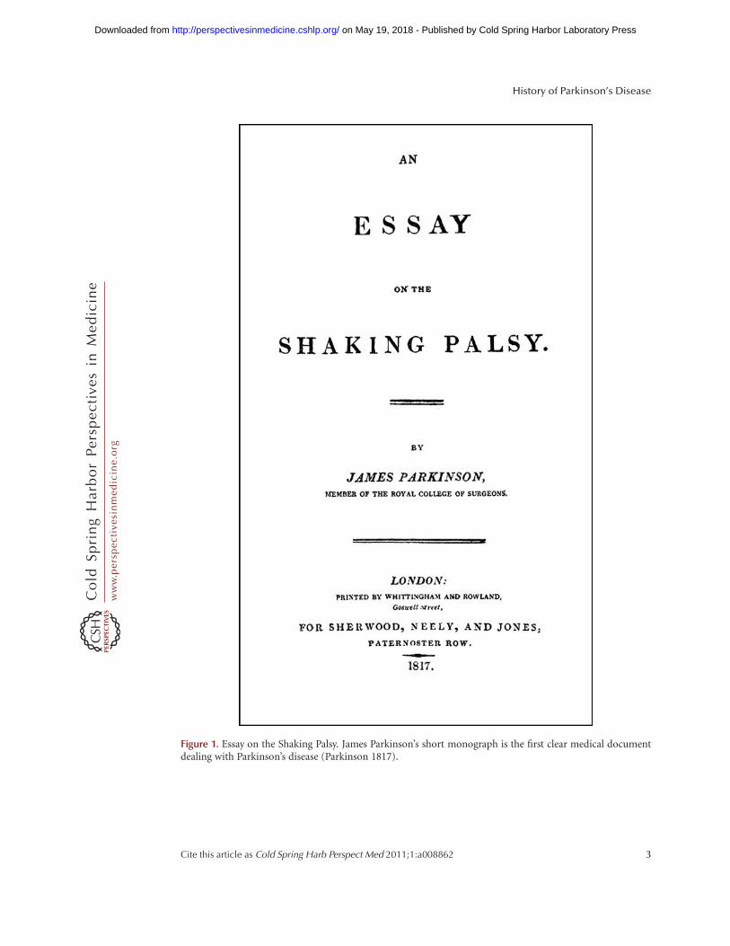
Figure 1. Essay on the Shaking Palsy. James Parkinson’s short monograph is the first clear medical documentdealing with Parkinson’s disease (Parkinson 1817).
History of Parkinson’s Disease
Cite this article as Cold Spring Harb Perspect Med 2011;1:a008862 3
ww
w.p
ersp
ecti
vesi
nm
edic
ine.
org
on May 19, 2018 - Published by Cold Spring Harbor Laboratory Press http://perspectivesinmedicine.cshlp.org/Downloaded from

Charcot’s first important contribution to thestudy of Parkinson’s disease was his differentia-tion of this disorder from other tremorous dis-orders, specifically multiple sclerosis (Charcot1872). Examining large numbers of patientswithin the vast Salpetriere Hospital in Paris,he developed a protocol to observe tremor atrest and then during action. He noted that thepatients with action tremor had accompanyingfeatures of weakness, spasticity, and visual dis-turbance. In contrast, those with rest tremor dif-fered in having rigidity, slowed movements, atypical hunched posture, and very soft speech.His early tremor studies were highly publicizedand helped to establish Parkinson’s disease as adistinct neurological entity that could be confi-dently diagnosed (Fig. 2).
Once the archetype of Parkinson’s diseasewas established, Charcot and his students iden-tified variants with features that were atypical
of classical Parkinson’s disease. These weretermed Parkinson’s disease without tremor,Parkinson’s disease with extended posture, andParkinson’s disease with hemiplegia. Thesecases are of historical interest, because they arelikely examples of disorders that would laterbe grouped under the term, Parkinsonism-plussyndromes, including progressive supranuclearpalsy, corticobasal degeneration, and multiplesystem atrophy. As one example, Charcot pre-sented a patient named Bachere on several occa-sions. On June 12, 1888, Charcot emphasizedthat Bachere did not have marked tremor, andin contrast to the usual arm flexion of typicalParkinson’s disease, he had a stiff and extendedposture.
Look how he stands. I present him in profile soyou can see the inclination of the head and trunk,well described by Parkinson. All this is typical.What is atypical, however, is that Bachere’s
Figure 2. Charcot and “myographic curves.” (Left) French neurologist Jean-Martin Charcot (1825–1893).(Right) Semi-diagrammatic “myographic curves” published by Charcot in 1887. The top tracing representsan intention tremor in multiple sclerosis. Segment AB indicates “at rest,” and BC indicates increasing oscillationsduring voluntary movement. The lower tracing represents a Parkinsonian tremor, with segment AB indicating atremor at rest, which persists in segment BC during voluntary movement. Charcot’s graphical recording methodon which these drawings were based is not described, but in other circumstances he relied on various pneumatictambour-like mechanisms (Charcot 1872; Goetz 1987).
C.G. Goetz
4 Cite this article as Cold Spring Harb Perspect Med 2011;1:a008862
ww
w.p
ersp
ecti
vesi
nm
edic
ine.
org
on May 19, 2018 - Published by Cold Spring Harbor Laboratory Press http://perspectivesinmedicine.cshlp.org/Downloaded from

forearms and legs are extended, making theextremities like rigid bars, whereas in the ordi-nary case, the same body parts are partly flexed.One can say then that in the typical case ofParkinson’s disease, flexion is the predominantfeature, whereas here, extension predominatesand accounts for this unusual presentation. The
difference is even more evident when the patientswalk (Fig. 3) (Goetz 1987; Charcot 1888a).
In addition to extended posture, this patienthad particular facial bradykinesia and con-tracted forehead muscles. Charcot commentedthat the patient had the perpetual look of
Figure 3. Atypical Parkinsonism. (A) Drawing from Charcot’s original lesson, given on June 12, 1888, in whichhe contrasted a typical Parkinson’s disease showing a flexed posture (left) with a Parkinsonian variant thatincluded the absence of tremor and extended posture (right). Charcot regularly taught his students by compar-ing and contrasting cases of patients from the Salpetriere inpatient and outpatient services. (B) Four drawings byCharcot from his lesson on atypical Parkinson’s disease, dated June 12, 1888, showing the distinctive facial fea-tures of his patient, Bachere, showing forehead muscles and superior orbicularis in simultaneous contraction,activation of the palpebral portion of the orbicularis and combined activation of the frontalis superior portionof the orbicularis and platysma, giving a frightened expression in contrast to the placid, blank stare of typicalParkinson’s disease patients. This case is a compelling case of likely progressive supranuclear palsy (Goetz,1987; Charcot 1888a).
History of Parkinson’s Disease
Cite this article as Cold Spring Harb Perspect Med 2011;1:a008862 5
ww
w.p
ersp
ecti
vesi
nm
edic
ine.
org
on May 19, 2018 - Published by Cold Spring Harbor Laboratory Press http://perspectivesinmedicine.cshlp.org/Downloaded from
surprise, because the eyes remained widelyopened and the forehead continually wrinkled(Fig. 3) (Goetz 1987; Charcot 1888a). In amodern setting, Jankovic has detailed similarfacial morphology in Parkinsonism-plus patients,specifically those with progressive supranuclearpalsy (Jankovic 1984). No specific supranucleareye movement abnormalities were described.Another Salpetriere patient with “Parkinson’s dis-ease in extension” was described by Dutil in 1889and eye movement abnormalities are mentioned,although a supranuclear lesion is not docu-mented clinically (Dutil 1889; Goetz 1996). Thiscase also had highly asymmetric rigidity of theextremities, a feature more reminiscent of cortico-basal degeneration than progressive supranuclearpalsy. In this case, the extended neck posture wasgraphically emphasized:
The face is masked, the forehead wrinkled, theeyebrows raised, the eyes immobile. . . . Thisfacies, associated with the extended posture ofthe head and trunk, gives the patient a singularlymajestic air (Dutil 1889; Goetz 1996).
With clinical features reminiscent of both pro-gressive supranuclear palsy and corticobasaldegeneration, this patient was mentioned inseveral articles from the Salpetriere school, al-though no autopsy was apparently performed.Collectively, these cases show that even theearliest diagnosticians recognized classic Parkin-son’s disease and cases that needed to be distin-guished from it. Today, these Parkinsonism-plusdiagnoses are known to have additional distinc-tive features, including poor response to dopa-minergic therapies and different pathologicallesions than seen in Parkinson’s disease.
Another important entity to be distin-guished from Parkinson’s disease was posten-cephalitic Parkinsonism, today a rare cause ofParkinsonism, but a very frequent disorder inthe period after 1916. Following the influenzaepidemic of 1916–1917, a neurologic syndromethat included Parkinsonism, along with multi-ple other signs, occurred in alarming numbers(von Economo 1919). The additional behav-ioral, ocular, and motor problems of thesepatients attested to more diffuse neurologic dis-ease than seen typically in Parkinson’s disease.
This condition has largely disappeared in thetwenty-first century, because the survivorshave died and no recurrence of an epidemic ofthis magnitude has recurred. Other importantforms of atypical Parkinsonism to be distin-guished from Parkinson’s disease include ajuvenile form of Parkinson’s disease, originallydescribed by Willige in 1911 (Willige 1911),with a more full description and its associationwith atrophy of the globus pallidus provided byRamsey Hunt and van Bogaert (Ramsey Hunt1917; van Bogaert 1930).
In the years after these pioneering papers,the concepts of neural circuits evolved withkey nuclei of importance to the clinical presen-tation of Parkinsonism being the substantianigra, the globus pallidus, and the caudatenucleus and putamen (striatum). Involvementof the striatum resulting in Parkinsonism wasdocumented in a variety of neurological disor-ders. Striatal-nigral degeneration was describedby Adams, van Bogaert, and Vander Eecken,and, though originally classified as a single dis-ease, it has since been merged into the largerdiagnosis of multiple system atrophy (Adamset al. 1964). Parkinsonian states related to stria-tal pathology were later identified in the form ofHuntington’s disease, in which a Parkinsonianpresentation is referred to as the Westphal var-iant (Westphal 1883) and in cases of striatalcalcification, either on a hereditary basis (Bruynet al. 1964) or as an acquired metabolic disorderoften related to hypoparathyroidism (Muenterand Whisnant 1968).
The historical discussion of Parkinsoniandisorders that are frequently confused withParkinson”s disease includes drug-induced andtoxin-induced cases as well. The introductionof the antipsychotic agents, originally termedneuroleptics, led to dramatic improvements inschizophrenic and other psychotic behaviors,but induced Parkinsonism largely indistin-guishable from Parkinson’s disease itself (Steck1954). Later understanding that these drugsblock dopamine receptors in the striatumexplained this clinical presentation and led tothe development of antipsychotic drugs withlower proclivity to block striatal receptors andless propensity to induce Parkinsonism. The
C.G. Goetz
6 Cite this article as Cold Spring Harb Perspect Med 2011;1:a008862
ww
w.p
ersp
ecti
vesi
nm
edic
ine.
org
on May 19, 2018 - Published by Cold Spring Harbor Laboratory Press http://perspectivesinmedicine.cshlp.org/Downloaded from

landmark observation on a cluster of youngpatients who presented with severe Parkinson-ism that appeared to be typical Parkinson’s dis-ease except for the young onset and severity ofsigns led to the discovery that the causativeagent was a self-administered narcotic deriva-tive, MPTP, that selectively damages the sub-stantia nigra (Langston et al. 1983). Thisproduct has provided a means to induce Parkin-sonism in experimental animals and remainsthe “gold standard” model to study Parkinson’sdisease in preclinical studies of new treatmentsfor Parkinson’s disease.
THE EVOLUTION OF TREATMENTS
The history of Parkinson’s disease is tightlylinked to therapeutic interventions, rangingfrom serendipitous observations to controlledclinical trials of specifically designed agents.
Parkinson devoted a chapter of his mono-graph to “considerations respecting the meansof cure” (Parkinson 1817). In humility and per-haps with a vision toward current concepts ofneuroprotection, he hoped for the identifica-tion of a treatment by which “the progress ofthe disease may be stopped” (Parkinson 1817).To this end, he advocated very early therapeuticintervention when signs were largely confinedto the arms without balance and gait impair-ments. Reflecting therapeutic approaches ofthe early nineteenth century, Parkinson rec-ommended venesection, specifically advocatingbloodletting from the neck, followed by vesica-tories to induce blistering and inflammation ofthe skin. Small pieces of cork were purposefullyinserted into the blisters to cause a “sufficientquantity” of purulent discharge (Parkinson1817). All these efforts were designed to divertblood and inflammatory pressure away fromthe brain and spinal cord, and in this way,decompress the medulla that Parkinson consid-ered the seat of neurological dysfunction.
Pharmacological Advances: Charcotand Gowers
Being the two most celebrated clinical neurol-ogists of the nineteenth century, Jean-MartinCharcot and William Gowers serve as important
icons for the study of standard and emergingtreatments for Parkinson’s disease. Charcot’sintern, Ordenstein, wrote his medical thesison the treatment of Parkinsonian tremor withbelladonna alkaloids, the first well-establishedtreatment of Parkinson’s disease (Ordenstein1972). These agents are centrally active anticho-linergic drugs that later would be understoodto affect the cholinergic/dopaminergic balancein the striatum and thereby improve Parkin-sonism. The credit of the observation of anti-cholinergic efficacy surely belongs to Charcothimself who managed his Salpetriere Schoolwith strict centralized supervision and oversawevery aspect of the neurological program. Aswith other young and aspiring students likeGilles de la Tourette and Pierre Marie, Orden-stein profited from publishing the observationwith his name as sole author, but contempora-ries would not have been deluded into thinkingof it as coming from anyone besides ProfessorCharcot. Of the many centrally active anticho-linergic agents of the era, Charcot’s preferredproduct for Parkinson’s disease was hyoscy-amine. This plant-based agent was prepared aspills, usually powder rolled into bits of whitebread, or as a syrup. As shown in a prescriptionlocated in the Philadelphia College of Physi-cians, Charcot’s anticholinergic treatment wassometimes combined with rye-based ergotproducts that in fact are the pharmacologicalbasis of some modern dopamine agonists,drugs that directly stimulate striatal dopaminereceptors and thereby simulate the activity ofdopamine itself (Fig. 4). Although Tyler hasaptly documented that Charcot was not thefirst interventionist to advocate hyoscyamine(Tyler 1992), Charcot’s name became linked tothe drug because of the widespread interna-tional publication of his lectures and classroomdemonstrations.
A unique historical opportunity to examinethe early treatment of Parkinson’s disease isprovided by a series of 18 unpublished lettersin the Charcot collection at the BibliothequeCharcot in Paris (Portfolio MAVIII: Parkinson’sdisease). These letters cover a period of at least15 months from January 1863 through March1864. Although the collection only contains
History of Parkinson’s Disease
Cite this article as Cold Spring Harb Perspect Med 2011;1:a008862 7
ww
w.p
ersp
ecti
vesi
nm
edic
ine.
org
on May 19, 2018 - Published by Cold Spring Harbor Laboratory Press http://perspectivesinmedicine.cshlp.org/Downloaded from
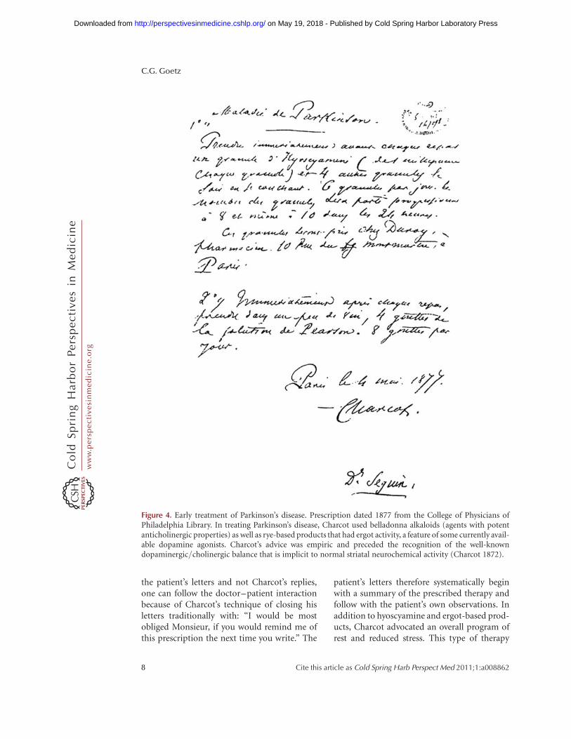
the patient’s letters and not Charcot’s replies,one can follow the doctor–patient interactionbecause of Charcot’s technique of closing hisletters traditionally with: “I would be mostobliged Monsieur, if you would remind me ofthis prescription the next time you write.” The
patient’s letters therefore systematically beginwith a summary of the prescribed therapy andfollow with the patient’s own observations. Inaddition to hyoscyamine and ergot-based prod-ucts, Charcot advocated an overall program ofrest and reduced stress. This type of therapy
Figure 4. Early treatment of Parkinson’s disease. Prescription dated 1877 from the College of Physicians ofPhiladelphia Library. In treating Parkinson’s disease, Charcot used belladonna alkaloids (agents with potentanticholinergic properties) as well as rye-based products that had ergot activity, a feature of some currently avail-able dopamine agonists. Charcot’s advice was empiric and preceded the recognition of the well-knowndopaminergic/cholinergic balance that is implicit to normal striatal neurochemical activity (Charcot 1872).
C.G. Goetz
8 Cite this article as Cold Spring Harb Perspect Med 2011;1:a008862
ww
w.p
ersp
ecti
vesi
nm
edic
ine.
org
on May 19, 2018 - Published by Cold Spring Harbor Laboratory Press http://perspectivesinmedicine.cshlp.org/Downloaded from
was generally advocated for many primary neu-rological disorders (Mitchell 1908). For thispatient, he added camphor, silver nitrate, ironcompounds, henbane pills, and zinc oxide.The rationale for using these agents was notexplained by Charcot, and their pharmacologydoes not involve the dopamine system. Theuse of iron may have been based on Romberg’searlier observation that carbonate of iron inassociation with warm baths and cold affusionsto the head and back induced “a marked dimin-ution of symptoms.” (Romberg 1846). Whetherbased on his own experience or Romberg’swarning against trying strychnine, Charcotsteered away from this therapy for Parkinson’sdisease patients. Charcot was highly specific inhis instructions, insisting that quinquina, a qui-nine derivative, must be diluted with syrupmade from orange rind and each dose of silvernitrate must be impregnated in 9 g of soft breadto form an ingestible pill. The letters communi-cate encouragement to the patient, reinforce theneed for patience in facing chronic illness, and awillingness to consider new treatment strategiesif traditional ones were unsuccessful. However,his enthusiasm to try new interventions neverclouded his objective vision of efficacy. Inreviewing pharmacologic treatments for Par-kinson’s disease in 1872, Charcot stated:
Everything, or almost everything, has been triedagainst this disease. Among the medicinal sub-stances that have been extolled and which Ihave myself administered to no avail, I needonly enumerate a few (Charcot 1872).
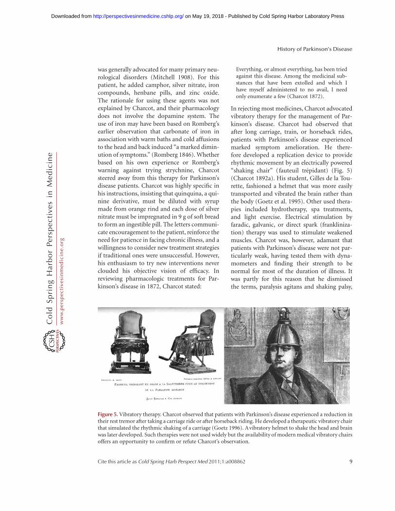
In rejecting most medicines, Charcot advocatedvibratory therapy for the management of Par-kinson’s disease. Charcot had observed thatafter long carriage, train, or horseback rides,patients with Parkinson’s disease experiencedmarked symptom amelioration. He there-fore developed a replication device to providerhythmic movement by an electrically powered“shaking chair” (fauteuil trepidant) (Fig. 5)(Charcot 1892a). His student, Gilles de la Tou-rette, fashioned a helmet that was more easilytransported and vibrated the brain rather thanthe body (Goetz et al. 1995). Other used thera-pies included hydrotherapy, spa treatments,and light exercise. Electrical stimulation byfaradic, galvanic, or direct spark (frankliniza-tion) therapy was used to stimulate weakenedmuscles. Charcot was, however, adamant thatpatients with Parkinson’s disease were not par-ticularly weak, having tested them with dyna-mometers and finding their strength to benormal for most of the duration of illness. Itwas partly for this reason that he dismissedthe terms, paralysis agitans and shaking palsy,
Figure 5. Vibratory therapy. Charcot observed that patients with Parkinson’s disease experienced a reduction intheir rest tremor after taking a carriage ride or after horseback riding. He developed a therapeutic vibratory chairthat simulated the rhythmic shaking of a carriage (Goetz 1996). Avibratory helmet to shake the head and brainwas later developed. Such therapies were not used widely but the availability of modern medical vibratory chairsoffers an opportunity to confirm or refute Charcot’s observation.
History of Parkinson’s Disease
Cite this article as Cold Spring Harb Perspect Med 2011;1:a008862 9
ww
w.p
ersp
ecti
vesi
nm
edic
ine.
org
on May 19, 2018 - Published by Cold Spring Harbor Laboratory Press http://perspectivesinmedicine.cshlp.org/Downloaded from

and advocated instead the designation, Parkin-son’s disease.
A more unusual and hazardous early treat-ment of Parkinson’s disease involved the useof a suspension apparatus to stretch the spinalcord (Goetz et al. 1995). Developed in 1883 inRussia, the apparatus gained celebrity whenCharcot examined its safety and efficacy in avariety of disorders, including Parkinson’s dis-ease. Using gravity and the patient’s weight toput excessive vertical traction on the spinalcord and nerves, the therapist hoisted the sub-ject in mid-air with a pulley and a harness thatslipped under the chin and occiput. In Parkin-son’s disease patients, rigidity and some sensorysymptoms improved, but tremor was not ame-liorated. Edmond de Goncourt described thetherapy with allusions to the macabre artworkof Goya, and the serious side effects and stresson patients led Charcot to abandon this strategyshortly after its introduction in France (de Gon-court and de Goncourt 1887–1889).
Charcot’s British contemporary, WRGowers, followed similar treatment strategies.He stressed the negative effects of mental strainand physical exhaustion, advocating that “lifeshould be quiet and regular, freed, as far asmay be, from care and work.” (Gowers 1899).For tremor, he used hyoscyamine and also notedarsenic, morphia, conium (hemlock), and“Indian hemp” (cannabis) as effective agentsfor temporary tremor abatement. Writing spe-cifically of the power of cannabis and opiumin combination, he stated: “I have several timesseen a very distinct improvement for a consid-erable time under their use.” (Gowers 1899).Today, cannabis is known to have some dopami-nergic activation properties, but opium affectsthe motor system in a generalized, sedativemanner without direct or primary dopaminer-gic involvement.
Levodopa and Dopamine-Based Therapies
Through the mid-twentieth century, the treat-ment of Parkinson’s disease remained largelythat of the nineteenth century, and though awide variety of centrally active anticholinergicdrugs were developed and used, they all were
similar in their efficacy and side effect profiles.In the Handbook of Clinical Neurology, thechapter, “Drug treatment of parkinsonism andits assessment” (published in 1968) discussesten synthetic anticholinergic compounds anda potpourri of agents under the designation“Other drugs which have been recommended,some of them without any justification.” (Onu-aguluchi 1968). The emphasis of this periodremained on supportive physical therapy andthe management of hypersalivation, seborrhea,decubiti, and infections. In the context of thisrelative stagnation, the impact of levodopa wasmagnified.
As summarized by Hornykiewicz, dopa-mine was first synthesized in 1910 by G. Bargerand J. Ewens (Hornykiewicz 2002). In the sameyear, H. Dale discovered its weak sympathomi-metic qualities. These observations were laterremembered when P. Holtz discovered theenzyme, dopa decarboxylase and documentedthat levodopa was synthesized to dopaminethrough its action. At this time, dopamine wasrelegated to a simple intermediate compoundfor the synthesis of noradrenaline and adrena-line. The consistent identification of substan-tial amounts of dopamine in various tissues,however, prompted the search for a more pri-mary role. Working in Blaschko’s CambridgeUniversity laboratory, Hornykiewicz studiedblood pressure control in experimental animalsand clearly confirmed that dopamine haddistinct effects independent of other catechol-amines. Shortly thereafter, in the late 1950s,two seminal discoveries occurred: dopaminelocalization within the brain, specifically inthe striatum; and the development of thereserpine-model, later to be used as the firstmodel of Parkinsonism that was reversed by le-vodopa treatment. In concert, these discoveriesrapidly advanced hypotheses on the role of dop-amine loss in the pathogenesis of Parkinson’sdisease itself (Carlsson et al. 1958; Sano et al.1959), and led Bertler and Rosengred to con-clude that “dopamine is concerned with thefunction of the striatum and thus with the con-trol of movement.” (Bertler and Rosengred1959). Ehringer and Hornykiewicz turned tohuman brain and after examining a series of
C.G. Goetz
10 Cite this article as Cold Spring Harb Perspect Med 2011;1:a008862
ww
w.p
ersp
ecti
vesi
nm
edic
ine.
org
on May 19, 2018 - Published by Cold Spring Harbor Laboratory Press http://perspectivesinmedicine.cshlp.org/Downloaded from
control specimens, discovered the striatal dopa-mine depletion in Parkinson’s disease and post-encephalitic parkinsonism brains (Ehringerand Hornykiewicz 1960). With the knowledgethat levodopa was the natural precursor todopamine, Hornykiewicz was now prepared tosuggest human trials in Parkinson’s diseasepatients.
Birkmayer received Hornykiewicz’s supplyof laboratory levodopa and injected it intrave-nously for the first time to Parkinsonianpatients in 1961. The antiakinetic effects werequickly published:
Bed-ridden patients who were unable to sit up,patients who could not stand up when seated,and patients who when standing could not startwalking performed all these activities with easeafter L-dopa [levodopa]. They walked aroundwith normal associated movements and theycould even run and jump. The voiceless, aphonicspeech, blurred by pallilalia and unclear articula-tion, became forceful and clear as in a normalperson (Birkmayer and Hornykiewicz 1961).
Subsequent open-label levodopa trials with oralpreparations confirmed both short and long-term benefits, and a double-blind placebo con-trolled trial followed (Barbeau 1969; Cotziaset al. 1969; Yahr et al. 1969). These reportslaunched levodopa’s establishment as the pre-mier agent to treat Parkinson’s disease symp-toms and signs. Although new formulationsand peripherally acting dopa-decarboxylaseinhibitors have added new dimensions to thetherapy, none of these events rival the firstdiscoveries.
Given that levodopa is a naturally occurringamino acid, researchers have reexamined oldertherapies to search for possible discoveriesof levodopa-containing compounds in earlymedicine. Of note, cowage or cowitch plant(Mucuna pruriens) is known under the nameof Atmagupta in Sanskrit and contains levo-dopa (Manyam 1990). One of the remediesused to treat the condition thought to be possi-ble Parkinson’s disease in traditional Indianmedicine is called Masabaldi Pacana, whichcontains beans of Mucuna pruriens. Theseobservations offer interesting, albeit indirect,evidence that patients with Parkinsonism may
have experienced the benefit of levodopa earlyin the history of medicine.
The more modern discoveries of dopamineagonists and enzyme inhibitors that enhancethe bioavailability of dopamine (monoamineoxidase inhibitors and catechol-O-methyltransferase inhibitors) date to the contempo-rary period and are of less importance to thishistorical review that emphasizes early discov-eries. These developments have been based onthe logical understanding of the dopaminesystem, metabolic pathways, and receptorpopulations. Further discoveries of modulat-ing influences by serotonin, adenosine, GABA,and glutamate systems have opened horizonsfor further pharmacological developments.The history of amantadine is of interest becauseof its serendipitous discovery as an anti-Par-kinsonian agent. Developed as an antiviralagent, it was used widely in nursing home pop-ulations, and Schwab noted its unexpectedbenefit on tremor, balance, and akinesia inboth Parkinson’s disease and postencephaliticparkinsonian patients (Schwab et al. 1969).This agent has mild dopamine effects, likelydue to inhibition of striatal synaptic dopaminereuptake so that more dopamine is left withinthe synapse to activate dopamine receptors. Ithas effects on the glutaminergic system withlikely indirect effects on dopamine functionthrough this mechanism.
SURGERY
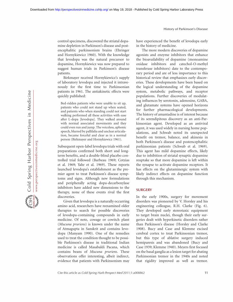
In the early 1900s, surgery for movementdisorders was pioneered by V. Horsley and hisengineering colleague, R.H. Clarke (Fig. 6).They developed early stereotaxic equipmentto target brain nuclei, though their early sur-geries dealt with hyperkinetic disorders ratherthan Parkinson’s disease (Horsley and Clarke1908). Bucy and Case and Klemme excisedcerebral cortex to treat Parkinsonian tremor,but this type of ablative surgery inducedhemiparesis and was abandoned (Bucy andCase 1939; Klemme 1940). Meyers first focusedon the basal ganglia as a lesion target for abatingParkinsonian tremor in the 1940s and notedthat rigidity improved as well as tremor.
History of Parkinson’s Disease
Cite this article as Cold Spring Harb Perspect Med 2011;1:a008862 11
ww
w.p
ersp
ecti
vesi
nm
edic
ine.
org
on May 19, 2018 - Published by Cold Spring Harbor Laboratory Press http://perspectivesinmedicine.cshlp.org/Downloaded from
Importantly, spasticity and paresis did notcompromise the improvement (Meyers 1940).In 1953, by accident, I. Cooper cut the anteriorchoroidal artery during surgery on a Parkin-sonian patient and was forced to ligate it toprevent a hematoma. The unexpected andremarkable relief of tremor and rigidity onthe contralateral side led to more widespreaduse of this procedure, though mortality wasapproximately 10% (Cooper 1953). Electricalcoagulation procedures involving the globuspallidus, thalamus, and the ansa lenticularis(ansotomy) were performed with early stereo-taxic procedures (Spiegel and Wycis 1954).Hassler and Reichert focused more directly onthe ventrolateral nucleus of the thalamus, alsoreferred to as the Ventral Oralis Anterior (Voa)nucleus (Hassler 1955; Reichert 1962). All thesereports were hampered by the lack of involve-ment by medical neurologists with resultantconcerns of incomplete reporting, lack oflong-term follow-up and potential minimaliza-tion of morbidity. Further, the role of surgerywas eclipsed by the advent of levodopa in the1960s, so that a long hiatus occurred whensurgery was not extensively used in Parkinson’sdisease. During this time, however, moreadvanced surgical techniques were developed,and these innovations would be later appliedto Parkinson’s disease patients near the end of
the twentieth century. Such treatments date tothe contemporary period and include pallidot-omy, subthalamic nucleus ablation, deep brainstimulation to thalamus, pallidum and subtha-lamic, and various transplantation procedures.Most recent are the developments of gene-basedtherapies that have entered clinical trials.
Placebo Therapy
The relationships between dopamine releaseand positive motivation, novelty seeking behav-iors, and attention have allowed researchers tounderstand the long-acknowledged placeboimpact on Parkinson’s disease. The Charcot let-ters cited above suggest that Charcot too under-stood clearly the importance of his presenceand command over the patient’s well-being.As anchored as he remained in neuroanatomi-cal concepts through the end of his career, Char-cot’s last monograph was titled “Faith Cure”and dealt with the profound improvementsthat some patients with neurological diseaseexperienced through nontraditional therapies(Charcot 1892b). Placebo-controlled trials havebecome standard in Parkinson’s disease, evenin the surgical arena, mainly because a largepercentage of patients on placebo treatmentexperience objective improvement in parkin-sonism (Goetz et al. 2008). The facilitation of
Figure 6. Early surgical interventions. (Left) Victor Horsley (1857–1916) was a celebrated British surgeonwho attempted a surgical intervention on a movement disorder patient with athetosis in 1909. He excised motorcortex with substantial improvement in involuntary movements. (Middle) Working in London with hisphysiologist colleague, Robert Henry Clarke (1850–1926), he developed early stereotaxic equipment, first foranimal experiments and then for humans. (Right) This daunting surgical apparatus taken from their reportsin Brain in 1908 guided them to deep brain centers including the basal ganglia and the cerebellum (Horsleyand Clarke 1908).
C.G. Goetz
12 Cite this article as Cold Spring Harb Perspect Med 2011;1:a008862
ww
w.p
ersp
ecti
vesi
nm
edic
ine.
org
on May 19, 2018 - Published by Cold Spring Harbor Laboratory Press http://perspectivesinmedicine.cshlp.org/Downloaded from
striatal dopaminergic activity in these settingshas been shown by neuroimaging techniques(de la Fuente-Fernandez et al. 2001). The fund-ing of federal grants for the specific study ofplacebo effects in Parkinson’s disease is, in itself,of historical significance (Goetz et al. 2008).
CONCLUDING REMARKS
Historical documents on Parkinson’s diseaseand descriptions that evoke Parkinsonismfrom eras prior to the first full medical delinea-tion of the disease provide a continuing sourceof potential neurological insights. As only oneexample, summarized in a review of traditionaland complementary therapies for Parkinson’sdisease (Manyam and Sanchez-Ramos 1999),in 1928, Lewin isolated an alkaloid from theBanisteriopsis caapi vine used in ceremonialmedicine among Amazonian tribes. He pro-vided purified banisterine to his colleague,Beringer, who tested it on patients with Parkin-son’s disease with reported marked benefit. Thedata were presented to the Berlin Medical As-sociation along with a film documenting thechanges in rigidity, bradykinesia, and gaitimpairment. Though this agent was not pur-sued further, the example underscores thepotential lessons from careful reading of tradi-tional medicine sources and the prospects fornew discoveries based on older observations.Charcot’s advocacy for vibratory therapy hasbeen tested in a modern setting (Kapur et al.2011), but Gowers’ encouraged use of Cannabishas yet to be systematically evaluated withstrong clinical trial methodology. Numerousother therapies have suggestive roles inParkinson’s disease but have not been rigorouslytested, including specific forms of physicalexercise, massage therapy, and relaxation tech-niques. The active participation of the Parkin-sonian subject in these treatments complicatesa controlled study design, but these interven-tions scientifically deserve to be tested withthe same rigor as new pharmacological or surgi-cal treatments. In the continuing search fortherapies to ameliorate current disability andto slow the natural deterioration that is implicitto Parkinson’s disease today, the guiding words
of Charcot remain modern and applicable: “Ifyou do not have a proven treatment for certainillnesses, bide your time, do what you can, butdo not harm your patients” (Charcot 1888b).
ACKNOWLEDGMENTS
Dr. Goetz acknowledges the Parkinson’s DiseaseFoundation that supports the Rush UniversityMedical Center Parkinson’s Disease and Move-ment Disorder Program with an annual grant.
REFERENCES
Adams RD, van Bogaert L, Vander Eecken H. 1964. Striato-nigral degeneration. J Neuropathol Exp Neurol 23: 584–608.
Babinski J. 1921. Kinesie parodoxale. Rev Neurol 37: 1266–1270.
Barbeau A. 1969. L-Dopa therapy in Parkinson’s disease.Can Med Assoc J 101: 59–68.
Bertler A, Rosengren E. 1959. Occurrence and distributionof dopamine in brain and other tissues. Experientia 15:10–11.
Birkmayer W, Hornykiewicz O. 1961. Der L-Dioxyphenyla-lanin-Effekt bei der Parkinson-Akinese. Wien Klin Wschr73: 787–788.
Brissaud E. 1925. Lecons sur les maladies nerveuses. Masson,Paris.
Bruyn GW, Bots GT, Staal A. 1964. Familial bilateral vascularcalcification in the central nervous system. Psychiatr Neu-rol Neurochir 67: 342–376.
Bucy PC, Case JT. 1939. Tremor: Physiologic mechanismand abolition by surgical means. Arch Neurol Psychiatr41: 721–746.
Carlsson A, Lindqvist M, Magnusson T, Waldeck B. 1958.On the presence of 3-hydroxytyramine in brain. Science127: 471.
Charcot J-M. 1872. De la paralysie agitante. In OeuvresCompletes (t 1) Lecons sur les maladies du systeme nerveux,pp. 155–188. A Delahaye, Paris. [In English: CharcotJ-M. 1877. On Parkinson’s disease. In Lectures on diseasesof the nervous system delivered at the Salpetriere (transl.Sigerson G), pp. 129–156. New Sydenham Society,London.]
Charcot J-M. 1888a. Lecons du Mardi: Policlinique de laSalpetriere, 1887–1888. Bureaux du Progres Medical,Paris. Lesson of June 12, 1888.
Charcot J-M. 1888b. Lecons du Mardi: Policlinique de la Sal-petriere, 1887–1888. Bureaux du Progres Medical, Paris.Lesson of November 15, 1887.
Charcot J-M. 1892a. La medicine vibratoire: Application desvibrations rapides et continues a traitement de quelquesmaladies du systeme nerveux. Prog Med 16: 149–151. [InEnglish: Charcot J-M. 1892. Vibratory therapeutics: Theapplication of rapid and continuous vibrations to thetreatment of certain diseases of the nervous system. JNerv Ment Dis 19: 880–886.]
History of Parkinson’s Disease
Cite this article as Cold Spring Harb Perspect Med 2011;1:a008862 13
ww
w.p
ersp
ecti
vesi
nm
edic
ine.
org
on May 19, 2018 - Published by Cold Spring Harbor Laboratory Press http://perspectivesinmedicine.cshlp.org/Downloaded from
Charcot J-M. 1892b. Faith-cure. New Rev 11: 244–262 [InFrench: Charcot J-M. 1892. La foi qui guerit. Rev Hebdo-madaire 5: 112–132.]
Cooper IS. 1953. Ligation of the anterior choroidal arteryfor involuntary movements of parkinsonism. PsychiatQuart 27: 317–319.
Cotzias GC, Papavasiliou PS, Gellene R. 1969. Modificationof parkinsonism: Chronic treatment with L-dopa. N EnglJ Med 280: 337–345.
de Goncourt E, de Goncourt D. 1887–1889. Journal: Mem-oires de la vie litteraire, Vol. 3. Flammarion, Paris.
de la Fuente-Fernandez R, Ruth TJ, Sossi V. 2001. Expecta-tion and dopamine release: Mechanism of the placeboeffect in Parkinson’s disease. Science 293: 1164–1166.
Dutil A. 1889. Sur un cas de paralysie agitante a forme hemi-plegique avec attitude anormale de la tete et du tronc(extension). Nouvelle Iconographie de la Salpetriere 2:165–169.
Ehringer H, Hornykiewicz O. 1960. Verteilung von nor-adrenalin and dopamin im gehirn des menschen undihr verhalten bei erkrankungen des extrapyramidalensystems. Klin Wschr 38: 1126–1239.
Foix MC, Nicolesco J. 1925. Les noyaux gris centraux et laregion mesencephalo-sous-optique. Masson, Paris.
Goetz CG. 1987. Charcot the clinician: The Tuesday lessons.Raven Press, New York.
Goetz CG. 1996. An early photographic case of probableprogressive supranuclear palsy. Mov Disord 11: 617–618.
Goetz CG, Bonduelle M, Gelfand T. 1995. Charcot: Con-structing neurology. Oxford University Press, New York.
Goetz CG, Wuu J, McDermott MP, Adler CH, Fahn S, FreedCR, Hauser RA, Olanow WC, Shoulson I, Tandon PK,et al. 2008. Placebo response in Parkinson’s disease: Com-parisons among 11 trials covering medical and surgicalinterventions. Mov Disord 15: 690–699.
Gowers WR. 1888. A manual of diseases of the nervous system.J and A Churchill, London.
Gowers WR. 1899. Paralysis agitans. In A system of medicine(ed. Allbutt A, Rolleston T), pp. 156–178. Macmillan,London.
Greenfield JG, Bosanquet FD. 1953. The brain-stem lesionsin Parkinsonism. J Neurol Neurosurg Psychiatry 16: 213–226.
Hassler R. 1955. The influence of stimulations and coagula-tions in the human thalamus on the tremor at rest and itsphysiopathologic mechanism. Proc Second Intl CongrNeuropath 1: 637–642.
Hoehn MM, Yahr MD. 1967. Parkinsonism: Onset, progres-sion and mortality. Neurology 17: 427–442.
Hornykiewicz O. 2002. Dopamine miracle: From brainhomogenate to dopamine replacement. Mov Disord 17:501–508.
Horsley V, Clarke RH. 1908. The structure and function ofthe cerebellum examined by a new method. Brain 31:45–124.
Hunt JR. 1917. Progressive atrophy of the globus pallidus.Brain 40: 58–148
Jankovic J. 1984. Progressive supranuclear palsy. Neurol Clin2: 473–486.
Kapur SS, Stebbins GT, Goetz CG. 2011. Vibration therapyand Parkinson’s disease. Mov Disord 26 (Suppl 2):S132.
Klemme RM. 1940. Surgical treatment of dystonia, paralysisagitans and athetosis. Arch Neurol Psychiatry 44: 926.
Langston JW, Ballard P, Tetrud JW, Irwin I. 1983. Chronicparkinsonism in humans due to a product of Meperi-dine-Analog Synthesis. Science 219: 979–980.
Manyam BV. 1990. Paralysis agitans and levodopa in “Ayur-veda”: Ancient Indian medical treatise. Mov Disord 5:47–48.
Manyam BV, Sanchez-Ramos JR. 1999. Traditional andcomplementary therapies in Parkinson’s disease. AdvNeurol 80: 565–574.
Meyers R. 1940. The modification of alternating tremors,rigidity and festination by surgery of the basal ganglia.Proc Assoc Nerv Ment Dis 21: 602–665.
Mitchell SW. 1908. Rest and psychotherapy. JAMA 50: 2034.
Muenter MD, Whisnant JP. 1968. Basal ganglia calcification,hypoparathyroidism, and extrapyramidal motor mani-festations. Neurology 18: 1075–1083.
Onuaguluchi G. 1968. Drug treatment of parkinsonismand its assessment. In Handbook of clinical neurology(ed. Vinken PJ, Bruyn GW), Vol. 6, pp. 218–226. North-Holland, Amsterdam.
Ordenstein L. 1972. Sur la paralysie agitante et la sclerose enplaque generalisee. E Martinet, Paris.
Parkinson J. 1817. An essay on the shaking palsy. Whitting-ham and Rowland for Sherwood, Needly and Jones,London.
Reichert T. 1962. Long term follow-up of results of stereo-taxic treatment in extrapyramidal disorders. ConfinNeurol 22: 336–363.
Richer P, Meige H. 1895. Etude morphologique sur la mal-adie de Parkinson. Nouvelle Iconographie de la Salpetriere8: 361–371.
Romberg M. 1846. Lehrbuch der nervenkrankheiten des men-schen. A Duncker, Berlin. [In English: Romberg M. 1853.A manual of the nervous diseases of man (trans. SievekingEH). New Sydenham Society, London.
Sano I, Gamo T, Kakimoto Y. 1959. Distribution of catecholcompounds in human brain. Biochim Biophys Acta 32:586–587.
Sauvages de la Croix FB de. 1763. Nosologia methodicaAmstelodami: Sumptibus Fratrum de Tournes.
Schwab RS, England AC, Poskanzer DC. 1969. Amantadinein the treatment of Parkinson’s disease. JAMA 208:1168–1170.
Spiegel EA, Wycis HT. 1954. Ansotomy in paralysis agitans.Arch Neurol Psychiatry 71: 598–614.
Steck H. 1954. Le syndrome extra-pyramidale et di-en-cephalique au cours des traitments au Largactil et au Ser-pasil. Ann Med-Psychchiatr 112: 737–743.
Sylvius de la Boe F. 1680. Opera Medica. Danielem Elsevi-rium et Abrahamum Wolfgang, Amsterdam.
Tretiakoff C. 1921. Contribution a l0 etude de l0anatomie dulocus niger. Rev Neurol 37: 592–608.
Tyler K. 1992. A history of Parkinson’s disease. In Handbookof Parkinson’s disease (ed. Koller WC), pp. 1–34. MarcelDekker, New York.
C.G. Goetz
14 Cite this article as Cold Spring Harb Perspect Med 2011;1:a008862
ww
w.p
ersp
ecti
vesi
nm
edic
ine.
org
on May 19, 2018 - Published by Cold Spring Harbor Laboratory Press http://perspectivesinmedicine.cshlp.org/Downloaded from
van Bogaert L. 1930. Contribution clinique et anatomique al0etude de la paralysie agitante juvenile primitive. RevNeurol 2: 315–326.
von Economo C. 1919. Grippe-encephalitis und Enceph-alitis lethargic. Wiener klinishe Wochenschrift 32: 393–396.
Westphal ACO. 1883. Uber eine dem Bilde der cerebrospina-len grauen Degeneration ahnliche Erkrankung des cen-tralen Nervensystems ohne anatomischen Befund, nebst
einigen Bemerkungen uber paradoxe Contraction. ArchPsychiatr Nervenkr 14: 87–95, 767–773.
Willige V. 1911. Ueber Paralysis Agiotans in Jugendlichenalter. Ztschr ges Neurol u Psychiatt 7: 263–265.
Yahr MD, Duvoisin RC, Schear MJ. 1969. Treatment of par-kinsonism with levodopa. Arch Neurol 21: 343–354.
Zhang Z-X, Dong Z-H, Roman GC. 2006. Early descriptionsof Parkinson’s disease in ancient China. Arch Neurol 63:782–784.
History of Parkinson’s Disease
Cite this article as Cold Spring Harb Perspect Med 2011;1:a008862 15
ww
w.p
ersp
ecti
vesi
nm
edic
ine.
org
on May 19, 2018 - Published by Cold Spring Harbor Laboratory Press http://perspectivesinmedicine.cshlp.org/Downloaded from
2011; doi: 10.1101/cshperspect.a008862Cold Spring Harb Perspect Med Christopher G. Goetz Neurological TherapiesThe History of Parkinson's Disease: Early Clinical Descriptions and
Subject Collection Parkinson's Disease
Functional Neuroanatomy of the Basal Ganglia
ObesoJosé L. Lanciego, Natasha Luquin and José A. Dysfunction in Parkinson's Disease
as a Model to Study MitochondrialDrosophila
Ming Guo
GeneticsAnimal Models of Parkinson's Disease: Vertebrate
DawsonYunjong Lee, Valina L. Dawson and Ted M. Pathways
and DJ-1 and Oxidative Stress and Mitochondrial Parkinsonism Due to Mutations in PINK1, Parkin,
Mark R. CooksonInnate Inflammation in Parkinson's Disease
V. Hugh PerryProgrammed Cell Death in Parkinson's Disease
Katerina Venderova and David S. Park
NeuropathologyParkinson's Disease and Parkinsonism:
Dennis W. DicksonDiseaseGenomics and Bioinformatics of Parkinson's
al.Sonja W. Scholz, Tim Mhyre, Habtom Ressom, et
Parkinson's DiseasePhysiological Phenotype and Vulnerability in
Sanchez, et al.D. James Surmeier, Jaime N. Guzman, Javier
DiseaseMotor Control Abnormalities in Parkinson's
CortésPietro Mazzoni, Britne Shabbott and Juan Camilo
ManagementFeatures, Diagnosis, and Principles of Clinical Approach to Parkinson's Disease:
João Massano and Kailash P. Bhatia
Parkinson's Disease: Gene Therapies
Patrick AebischerPhilippe G. Coune, Bernard L. Schneider and
The Role of Autophagy in Parkinson's DiseaseMelinda A. Lynch-Day, Kai Mao, Ke Wang, et al.
Functional Neuroimaging in Parkinson's Disease
EidelbergMartin Niethammer, Andrew Feigin and David
Parkinson's DiseaseDisruption of Protein Quality Control in
PetrucelliCasey Cook, Caroline Stetler and Leonard
Key QuestionsLeucine-Rich Repeat Kinase 2 for Beginners: Six
Lauren R. Kett and William T. Dauer
http://perspectivesinmedicine.cshlp.org/cgi/collection/ For additional articles in this collection, see
Copyright © 2011 Cold Spring Harbor Laboratory Press; all rights reserved
on May 19, 2018 - Published by Cold Spring Harbor Laboratory Press http://perspectivesinmedicine.cshlp.org/Downloaded from