Embed Size (px)
Citation preview

Rochester Institute of Technology Rochester Institute of Technology
RIT Scholar Works RIT Scholar Works
Theses
9-1-1995
The History of neurosurgery The History of neurosurgery
Savitha Viswanathan
Follow this and additional works at: https://scholarworks.rit.edu/theses
Recommended Citation Recommended Citation Viswanathan, Savitha, "The History of neurosurgery" (1995). Thesis. Rochester Institute of Technology. Accessed from
This Thesis is brought to you for free and open access by RIT Scholar Works. It has been accepted for inclusion in Theses by an authorized administrator of RIT Scholar Works. For more information, please contact [email protected].

ROCHESTER INSTITUTE OF TECHNOLOGY
AThesis Submitted to the Faculty of
The College of Imaging Arts and Sciences
In Candidacy for the Degree of
MASTER OF FINE ARTS
The History of Neurosurgery
by
SavithaViswanathan
September 1, 1995

APPROVALS
Adviser: Glen Hintz/ _
Date: lIfo/etCAssociate Adviser: Bob Wabnitz/ _
Date: iP:f/9S
Assoicate Adiviser: .Thomas Cornell/ _
Date: Z ( k..:b.o. ~S-
Departmen,tt:~~: Cleft Hintz/-:r- _, ~CnslJ~~
I, ! (SavithaViswanathari) prefer to be
contacted each time a request for production is made. I can be reached at the
following adddress:
948 Heritage Drive
Gettysburg, PA 17325
Date: _


he History of Neurosurgery

Table of Contents
Introduction i
1. The Stone Age
Prehistoric Trephining 1
2. Ancient Egypt
The Edwin Smith Surgical Papyrus 10
3. Ancient Greece
Hippocratic Medicine 20
4. Ancient Rome
Claudius Galen 31
5. The Renaissance
Ambroise Pare 38
6. The Eighteenth Century
Percivall Pott 45
7. The Nineteenth Century
Great Moments in Neurosurgery of the 19th Century 52
8. The Twentieth Century
The Father ofModern Neurosurgery: Harvey Cushing 60
Bibliography 67

The History ofNeurosurgery
The earliest evidence of the practice ofNeurosurgery are trephined skulls dating from
the Neolithic period, making this branch ofmedicine over 8,000 years old. The advances in
technique that followed were greatly influenced by the growing knowledge of internal medi
cine, and later, of neuroanatomy and neurophysiology. It is interesting to note that though
neurosurgery is one of the oldest forms of surgery practiced, it has been recognized as a spe
cialized field only since the late 19th century.
This thesis is meant to be a concise history ofneurosurgery. It includes different sec
tions which each contain an accompanying illustration.The series describes advancements
made from prehistoric trephining to those made in the early 20th century by Harvey
Cushing, the famed Father ofModern Neurosurgery.
In preparation, I researched neurosurgical history, and chose eight different historical
highhghts as the subjects for each chapter of the final thesis. Each of these subjects differed
from one another in culture and time period, allowing more freedom in style in which I was
to complete each piece. By choosing to vary artistic style between illustrations, a small
amount of research in art history was required at times. For instance, with the art for the
Edwin Smith Surgical Papyrus, I was very careful to arrange the heiroglyphs in an accurate
pattern, as well as designing the marks themselves after genuine models. .
Once the decision was made as to the content of the illustration and the style in
which is was to be handled, I then chose the media for the piece. Most of the pieces began
as either pencil drawings, or acrylic paintings which were later scanned into the computer
and manipulated using a variety of software, mostly Adobe Photoshop 3.0. I found this
method to be most useful in regards to time and craftsmanship. To complete an illustration
solely on the computer, one is at risk of allowing the illustration to become too smooth and
artificial looking. By using the computer only as a last step, I felt as though the pieces look as
though they were hand painted, though they contain interesting aspects such as transparency
and crisp type which can be easily achieved by using the computer.
The illustrations in this exhibit were prepared as if they were part of a special series

titled, "The History ofNeurosurgery"
to be published in a monthly medical journal. Each
illustration would accompany an article that featured a specific time period when a substan
tial advancement was made. Therefore, the intended audience for this body ofwork is
knowledgeable in the field of neurosurgery, although not particularly in its historical aspects.
In preparation for the exhibition of these pieces, I used a bright white window mat
for each one, which was mounted on foam board, and later placed behind glass using gallery
clips. Each piece was accompanied by a small timeline (located at the beginning of each
chapter), which I again designed to reflect the time period of its corresponding piece. I
scanned in various images as black and white files, merged them with the type using
QuarkXpress. To avoid large TIFF files, I colorized each within the layout program using
Pantone colors to save memory.
Although many different aspects of the history of neurosurgery are covered on the
following pages, it must not be overlooked that there are many different cultures and people
who are not represented here, for lack of time and space. The history ofmedicine is a very
difficult field to research thoroughly, as many people did not keep careful records of their
practices. It is also difficult to ascertain whether certain methods were developed by those
who wrote of them, or whether they had adopted them from another group ofpeople, who
are bound to be ignored by further generations of students and researchers. Medical history
is a deeply moving subject to study. It shows evidence ofhumanity and compassion coupled
with an ambitious desire to learn and explain natural mysteries. I encourage anyone who
may read this small account of surgical history to further feed their curiosity and begin to
appreciate our ancestors all over the world, from whose actions we still benefit each and
every day.

i <
H
u
5oHoo
Ph

Prehistoric Trephining-
Trephining, or creating an opening in the skull to reveal the brain and meninges
beneath, has been practiced by people since the Neolithic period. Although there are
many theories why the prehistoric surgeon chose to trephine the skulls of his patients,
There is wide agreement that these operations were generally successful. Not only did
men, women, and children survive this crude surgery, but they must also have been cured
of their symptoms, as is suggested by the fact this 10,000-year-old tradition is still prac
ticed in parts of the world today.
In the late 1800's, anthropologists began discovering skulls with curious holes
apparently made with therapeutic intentions. Though there was skepticism concerning
these proclaimed"intentions,"
this theory became widely accepted as skulls continued to
be found in many areas ofEurope including France, England, Switzerland, Denmark,
Germany, Sweden,Austria, Poland, Italy, Russia, Spain, and Portugal. Not restricted to
only one continent, skulls (some of later periods) were also found in South America
(especially Peru, Bolivia, and Ecuador), NorthAmerica, Mexico, the Orient, New Guinea,
Tahiti, and New Zealand. Some anthropologists and ethnologists believe there once
existed a natural land bridge across the Bering Strait. This would suggest that the practice
of trephining was spread throughout prehistoric civilizations over thousands of years, and
did not develop independently among separated cultures.
There are two main theories to explain trephination. The most popular explana
tion is medical treatment. It is a fact that producing an opening in the skull decreases
intracranial pressure. Increased intracranial pressure can be due to a variety of reasons,
most often from head trauma, resulting in skull fractures or intracranial bleeding.
Symptoms of these conditions can include headaches, convulsive fits, and even periods of
insanity. Trephination could have been recommended to anyone who may have possessed
these or similar symptoms. Especially common in Peruvian skulls, trephine holes can be
found lying near or on top of a scarred fracture line. In considering the brutal weapons
used by ancient people of this area, it is likely that most Peruvian trephinations were per-

formed to treat war injuries. Some experts have made the observation that many of the
trephine holes seem strategically enough placed to imply ancient surgeons may have had
some idea of regional brain function. For instance, one common site of trephine holes is
over the motor cortex, which, when irrigated(wettened), produces body movement,
which the ancient surgeon could have observed.
In some trephined skulls, however, there is no evidence of any prior injury. What
then, was the purpose of the surgery? An alternative theory supports religious motives.
Perhaps by carving out a piece of skull bone, ancient people may have been allowing
"confineddemons"
to escape the patient's body. This theory may explain the discovery
of the why bone disks have been found to be preciously placed within the burial sites of
deceased trephine patients.
After studying numerous trephined skulls from all periods, it is evident that there
were certain methods and patterns which remained fairly constant despite time period
and culture. There are four basic techniques prehistoric man used in trephination:
1 . Scraping-using a sharp stone, the thin layers of cranial bone are
successively removed in an oval pattern until the meninges is
exposed
2. Grooving-same method as above, though instead of scraping
away the surface area of the piece to be removed, an outline of the
piece is scraped away and the bone piece is removed wholly
3. Boring/cutting-long, sharp instruments are rotated until the
skull is penetrated, producing smaller, rounder holes which are
then sometimes connected as a quicker way of forming a large
opening
4. Rectangular intersecting incisions-grooved lines are cut at right
angles to each other, forming a rectangular central piece which
may then be removed

The stone instruments used were usually of flint or obsidian, and weretriangular
shaped. Sharp-edged trephines were used for scraping; knife-like trephines, for grooving;
and pointed trephines, for drilling. Some of the instruments believed to be used by
ancient Peruvians were fastened to handles, resembling present day gardening spades.
Depending on the type of incision necessary, either the sharp sided edge or the pointed
tip proved more useful. If a series of small holes were required, a longer blade was used
by positioning the pointed tip on the skull surface and rotating the handle, to produce a
drilling effect. After the surgery was complete, different surgeons dressed the open head
wound with different materials, showing concern over prolonged exposure of healthy tis
sue to open air. Some Peruvians used bundles of cotton, while others simply tied the hair
from the surrounding scalp into a knot, thereby protecting the cut edges of scalp. The
Paracas ofPeru, known for their excellent craftsmanship, used thin sheets of gold to close
the cranial separation.
Many of the trephined skulls that have been discovered show advanced stages of
healing bone. The fact that cranial bones regenerate at a much slower rate than long
bones proves the patient must have had continued to five well beyond the time of
surgery. The high success rate of these operations cannot be attributed solely to the
technique of the surgeon, since knowledge of infection and ways to prevent it did not
exist. What did allow for these practices to be considered fairly low risk was the natural
resistance to infection the patients themselves must have had. There are skulls which
have been found to have up to five or six trephine holes, all made on separate occasions.
Another reason for the procedure's success may have been the absence of attending
physicians and other extraneous people who would have risked forming the crowded
atmosphere indicative of early city hospitals, where there was a high chance of infec
tions. Also, since most civilizations left behind no indication of anesthesia, operations
were probably performed rapidly, allowing minimal exposure of the tissues, again reduc
ing the chance of infection. There is, however, one culture that did practice anesthetic
drug use. Many believe ancient Peruvian surgeons told their patients to chew on the
leaves of the erythroxylon coca plant, (today processed as cocaine), cinchona bark (source

of the modern drug quinine) and the daturas plant, all probably producing anesthetic
effects strong enough to alleviate some pain of the trephination.
Today, there are many people across the world who continue to practice the same
trephination techniques that were practiced over 10,000 years ago. Studying the princi
ples of these people help to form more accurate ideas about their ancient predecessors.
The Incas, in the spirit of the ancient Peruvians, developed trephining into a commonly
practiced skilled art. Using instruments made ofbronze, their incision patterns remain
similar to those which had been passed down for thousands of years. In nineteenth-cen
tury western Asia and northern Africa, trephination was said to have proven itself a cure
for migraines and psychosis. Formal training for this"artform"
has even become available
in Algeria. It is also interesting to note that although the basic technique has not
changed, the materials used for the instruments vary depending on their availability to the
people of an area. For instance, many of the native people ofvarious Pacific islands have
used shark's teeth as their instruments and coconut shell as part of the final dressing fol
lowing surgery. Many of these surgeons now know the risks involved in penetrating the
dura mater (the outer meningeal covering of the brain), and easily damaged areas such as
suture lines and blood sinuses are avoided during surgery. Overall, these later civilizations
have continued to perfect their methods achieving close to a 100% survival ratea suc
cess ratio unmatched today, despite numerous neurosurgical advancements.
About the Piece
This first piece is especially important in establishing the format and function of
this series of illustrations. It had to reflect a distinctive period in time, not only through
its subject matter, but also the way in which it was rendered. The overall color scheme
was to be of earth tones, though painted richly in acrylic. The textures and other effects
left by the brush were to be an important quality of the piece. Though the piece was to
be finished in Adobe Photoshop, the final copy was not to look like a computer printout,
but more like a painting.
The piece began as a stretched sheet of watercolor paper on board. After apply-

ing a thick coat of gesso, a detailed charcoal drawing of the final painting was made
directly on the paper. The background began as a map of the world.Certain areas, such
as South America and some parts ofEurope were to be kept uncovered since these were
areas where trephining was practiced most. The oceans, especially where they came in
contact with the land, were shaded dark, while the continents were left bright white.
The drawing of the trephined skull represents of a combination of a series of ref
erences. The main trephine hole does sit directly on a fracture line, correlating this exam
ple with head trauma treatment, and giving some history to this skull. The drawing does
not include the mandible, as it was not saved on this particular specimen. The location of
the skull has also changed from its original position. It was initially drawn in the upper
right hand corner of the piece, much smaller than its size in the final printout.
The profile of a South American man, is deliberately taken, not from prehistoric,
but from modern times. The contrast between the time periods of the man's face and the
trephined skull shows the length of time during which trephining has been practiced.
Although this chapter focuses on the oldest form of neurosurgery dating back to the
Neolithic Age, the illustration shows how the practices discussed are not merely some
thing of the past.
Ghosted through the man's hair is his trephined skull underneath, with a hole
made by rectangular intersecting incisions. Initially, the hair was drawn as though it was
very transparent, almost disappearing in the area of the trephine hole. The skull was then
faded into the skin around the bridge of the nose, and temple region, so the skin on the
cheek was completely opaque.
After spraying the drawing with a workable fixative, it was ready for color to be
added. The painting of the map was completed first, using very rich earth tones such as
burnt siena, yellow ochre, and a bit ofburnt umbre. Because of the value drawing under
neath, the land masses remained fairly light, while the oceans stayed darker. The colors of
the skull are taken directly from photographs ofNeolithic skull remains. Not quite as
yellow as the background, the tones used for the skull were primarily beige and brown.
The tones of the face had to be adjusted somewhat. In general, skin tones of South

American natives are of a reddish-brown, but thesecolors-
were almost exactly like those
already used for the background map. For the sake of contrast, the face was then painted
a bluer, duller brown, more like a deep African brown, and was adjusted later on the
computer.
After removing the painting from the board, it was scanned in at 150 dpi in a
series ofpieces, since it was too large to fit entirely on the scanning bed. After being
pieced back together, different selection paths were carefully traced out and saved individ
ually as"face," "skull,"
etc. Using these selections, clean, sharp, color adjusments were
made quickly. Both the map and the face were adjusted to reddish brown tones, though
the map was kept a bit yellower and darkened a great deal, leaving the two elements in a
strong enough contrast to remain visually separate.
A new grayscale file entitled, "caveart"
was then created and filled with cave art
icons taken from a reference book on the subject. They were all painted with the pencil
tool, with a thick setting.
Each cave art icon was then brought into the final piece, in the"darken"
mode,
placed, colorized, and set on various levels of opacity. The icons were to be noticeable,
but still remain subtle. This addition of the icons to the final file was probably the most
time consuming part of the piece.
The last element added to the piece was the frame. After creating the selection, it
was then lightened and shifted subtly to different colors, producing a bit more of a mod
ern effect, without losing the rough look of the painting.
After meeting with advisors and friends, it became apparent that the left lower
corner of the painting had been left empty. This emptiness at the bottom of the piece
also created a problem with the lower edge of the portrait, the face seemed to be "float
ing"
in space and became distracting. Once again, in Photoshop again, the skull was
enlarged to almost twice its original size and brought to a position much lower than
before, so the bottom edge bled off the left lower corner of the page. Moving the skull
down created a large empty space above it on the back of the profile, which I fixed using
the paintbrush and rubber stamp tools. The edge of the skull which came in contact

with the paper's edge Was then darkened, and the frame retouched. Many of the cave art
icons had to be redrawn and reimported, though placement, color, and opacity were
already preset. One last adjustment was the opacity of the hair overlying the trephine
holes. Instead of seeing the pale color ofbone directly through the hair as before, more
hair was painted on top and darkened, producing less of a distraction.
The original file measures7.5"
by 9.0". It was finally taken to a service bureau
where it was printed from a postscript file to a Fiery printer at 1200 dpi.
Sources
1 . Ballance, Sir Charles A. , The ThomasVicary Lecture: A Glimpse Into the
History of the Surgery of the Brain. MacMillan & Co. Ltd., London, England, 1922.
2. Benton, Paul and Hemlett,John H., SurgeryThrough the Ages. Hastings House
Publishers, NewYork, NY, 1944.
3. Majno, Guido, The Healing Hand: Man andWound in the AncientWorld.
Harvard University Press, Cambridge,MA, 1975.
4. Margotta, Robert, The Story ofMedicine. Golden Press, NewYork, NY, 1967.
5. Sachs. Ernest. The History and Development ofNeurological Surgery. Paul B.
Hoeber Inc. NewYork, NY, 1952.

u
luil
J'
CN
OO
Ph
JU
O
3
j/>^_
D
U
PQ
oo
u
z
<
0>-J
I I
H
w
H
U
1 ^
'dim,"
-
-
5^ ft^^'^uii'MII
# *
^S7
'
Lfc*-*
-
8I'i
C>'P
i <
* t
Soils.
It
Lm\
^
\
fH
mtz3* **
-
-
...
< *
i-:;
3l
<,
; v7:r * '*
L-JL-J:

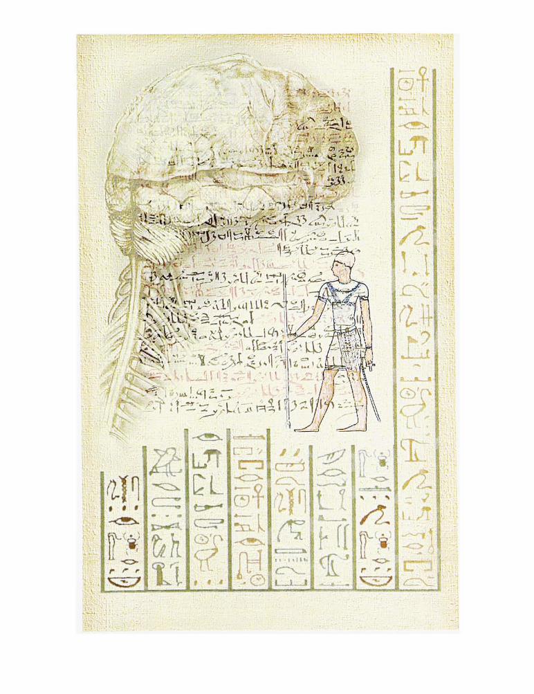
' The Edwin Smith Surgical Papyrus
The earliest written account of surgical practices is the ancient Edwin Smith
Surgical Papyrus, brought to America in the late nineteenth century from Egypt. It
shows the exemplary ways in which science was practiced at a time when much of the
world's people were still evolving into organized civilizations. The papyrus not only illu
minates the medical practices of 17th century B.C.E. Egypt, but also the societal role of
physicians and the humanistic nature of these people who existed thousands ofyears ago
Ancient Egyptian civilization developed in isolation within the Nile valley, which
was surrounded by sparsely inhabited desert. The land was like a secluded oasis, success
fully supporting its people with its own immediate resources. In reference to any foreign
country, the heiroglyph,"desert"
was used, as most foreigners were considered nonpeople.
Ancient Egyptians became so assured of their civilization's importance, they chose the
name"Egypt"
for their home, meaning"earth."
Here their civilization thrived, achieving
high success in many branches of science.
The high rate of severe injuries which occurred during the many battles Egyptian
soldiers fought, particularly called for advances in the medical sciences. Ancient Egyptians
practiced a highly organized form ofmedicine which eventually outlasted the pharaohs
and merged with that of the Greeks. Many physicians specialized within their fields. In
fact, Herodotus, the 5th century B.C.E. Greek historian, described Egyptian physicians to
be numerous and all of them specialists. From the time of the Third Dynasty (2600
B.C.E.), medical practitioners such as Hesy Re, the ChiefofDentists and Physicians to
pyramid builders, circa 1900 B.C.E., wrote and practiced procedures that were outlined in
written medical documents. The oldest of known medical texts is the Kahun Papyrus,
dating from the Middle Kingdom, 1900 B.C.E. This text ,which reads like a catalog,
consists of about 1200 paragraphs detailing roughly 900 prescriptions, diagnoses, and spells
against given diseases.
On January 20, 1862, anAmerican scholar, Edwin Smith, bought an ancient
Egyptian papyrus from a man called Mustapha Aga, in the city ofThebes. A few months
12

later,Mustapha Aga sold Smith an additional papyrus, which he later discovered was actu
ally a false document made of three separate papyri glued together. Of these three, two
proved to be pieces of the first page of the papyrus bought in January. After Dr. Smith's
death, his daughter donated the ancient manuscripts to the NewYork Historical Society
who then requested James Henry Breasted of the Oriental Institute of the University of
Chicago to translate it. In 1930, Breasted published a full translation of the document,
now known as the Edwin Smith Surgical Papyrus.
Breasted dated the document sometime between the 16th and 17th centuries
B.C.E., though the original text may be from a much earlier time. There are mainly
three important persons involved in the history of the papyrus. First, the author of the
text, who is still unknown, though some believe him to be the famed Imhotep, great
Egyptian physician of the 30th century B.C.E. The language he uses is that of the Old
Kingdom (c. 2600-2200 B. C. E.) The second person involved is the commentator, who
probably existed some centuries later. He leaves his mark in the form of69 separate
explanations or glosses included in the text, which translate obsolete phrases of the Old
Kingdom into more contemporary language. The last person involved in this surgical
text is the scribe (c. 1650 B.C.E.), whose responsibility it was to copy the text into manu
script form. He alternates between red and black ink, correcting in black the mistakes
made in red and leaving notes along the margins. Also described as careless, it was the
scribe who stopped in the middle of a sentence, actually in the middle of a word, and left
us a half finished document.
The papyrus measures 32.5-33 centimeters by 4.68 meters, though its original
length was probably over 5 meters. It is actually made of 12 separate sheets (each about
40 centimeters long) joined to form the full roll. Altogether, there are 17 columns of
writing, forming 377 lines. It is different from preexisting medical treatises for a variety
of reasons. It consists exclusively of cases, not recipes, showing, for the first time, a
reliance on knowledge in science and anatomy rather than in magic. There are 48 cases
in all, 27 ofwhich deal with head trauma and 6 with spine trauma. Modern descriptions
of some of the 33 neurologically related cases are as follows:
Case #3: A gaping head wound penetrating bone, perforating the skull
13

Case #4: A gaping head wound penetrating bone, splitting the skull
Case #5: A gaping head wound with a compound, comminuted skull fracture
Case #6: A gaping head wound with a compound, comminuted skull fracture,
with rupture ofmeningeal membranes
Case #7: A gaping head wound penetrating bone, perforating the sutures of the skull
Case #8: Compound, comminuted fracture of skull with no visible external injury
Case #9: Forehead wound with a compound, comminuted skull fracture
Case #10: A gaping head wound above the eyebrow, penetrating to the bone
Case #18: Wound in soft tissue of temple, with no bone injury
Case #19: Perforation in the temple
Case #21: Split in the temporal bone
Case #31: Dislocation of the cervical vertebrae
Case #32: Displacement of a cervical vertebra
Case #33: Crushed cervical vertebrae
The organization of these cases is systematic, beginning with the head continuing
downward, with the last unfinished case describing an injury to the spine. This type of
arrangement is still used today in describing fists of injuries and treatments. Each case is
assigned one of three verdicts by the physician: favorable ("an ailment which I will
treat"), uncertain ("an ailment which I will contend"), and unfavorable ("an ailment
which will not be treated"). This type of categorization had never been used before,
according to existing evidence. The use of the third verdict is particularly interesting,
implying scientific interest the physician may have possessed in studying the symptoms
further, since despite the absence of a described treatment details of the examination are
still recorded.
Each case follows the same format. First, an assigned title is given; then, a descrip
tion of the examination; third, the diagnosis; fourth, the treatment (except for those
described as untreatable); and last the glosses, or explanation of archaic terms. To show
the full working format of the descriptions and treatments described in the papyrus, a
14

translation ofCase #4- is given below:
Title
Instructions concerning a gaping wound in his head, penetrating to the bone,
(and) splitting his skull.
Examination
If thou examinest a man having a gaping wound in his head, penetrating to the
bone, (and) splitting his skull, thou shouldst palpate his wound. Shouldst thou find some
thing disturbing therein under thy fingers, (and) he shudders exceedingly, while the
swelling which is over it protrudes, he discharges blood from both his nostrils (and) from
both his ears, he suffers with stiffness in his neck, so that he is unable to look at his two
shoulders and his breast,
Diagnosis
Thou shouldst say regarding him: "One having a gaping wound in his head, pen
etrating to the bone, (and splitting his skull; while he discharges blood from both his nos
trils and) from both his ears, (and) he suffers with stiffness in his neck. An ailment with
which I willcontend."
Treatment
Now when thou findest that the skull of that man is split, thou shouldst not bind
him, (but) moor (him) at his mooring stakes until the period of his injury passes by. His
treatment is sitting. Make for him two supports ofbrick, until thou knowest he has
reached a decisive point. Thou shouldst apply grease to his head, (and) soften his neck
therewith and both his shoulders. Thou shouldst do likewise for every man whom thou
findest having a split skull.
GlossA
As for: "Splitting hisskull,"
it means separating shell from shell of his skull, while
15

fragments remain sticking in the flesh of his head, and do not come away.
Gloss B
As for: "The swelling which is over itprotrudes,"
it means that the swelling
which is over this split is large, rising upward.
Gloss C
As for: "(Until) thou knowest he has reached a decisivepoint,"
it means (until)
thou knowest whether he will die or he will five, for he is (a case of) "an ailment with
which I willcontend."
(Majno, Guido, The Healing Hand: Man andWound in the
AncientWorld p. 94-95)
The manner in which the Egyptian physician examined (his) patients proved to
be amazingly thorough. Aside from visual symptoms, particular smells possibly emanating
from wounds were also noted. Symptoms seemingly unrelated to the trauma wound
were taken into account as being possibly connected. For instance, it was noted that
commonly following a head injury or dislocation/fracture of the cervical spine, was the
problem ofpersistent erections, seminal emissions, and urine incontinence. Evidence of
these connections are not found again until the written accounts ofAlexandrian physi
cians of the third century B.C.E. The brain and spinal cord were described as having a
great effect, or control over many other parts of the body. This knowledge arose from the
observation that paralysis was often a result of injuries to these organs. Another major
connection noted by the Egyptians was the control that one side of the brain has over the
movement of the opposite side of the body. Again, this observation was not noted again
until centuries later. Other noteworthy descriptions given for the first time include the
structure of the meninges, brain convolutions, cranial sutures, cerebrospinal fluid, and
intracranial pulsations. It is believed metal instruments were not used at this time for
examinations. Any instrument that was used were probably made of flint. Instead, the
physician was encouraged to use mere palpitation while examining his patients, an ideal
made obvious by the words of the world's first description of the human brain as "some-
16

thing throbbing and fluttering under thyfingers."
Using one's hands also may have held
more than scientific significance, being a gesture of comfort or reassurance.
One of the greatest advances made by the Egyptian surgeon was his experimenta
tion with surgical methods of closure. Though surgical sutures were performed by
embalmers during mummification, early closure devices on the living were different.
They were a combination of a suture and a clamp as in the example described as a thorn
or needle stuck through both lips of a wound with the protruding ends tied together by a
thread placed as figure eight. Another closing technique worked like a bandage. It mere
ly called for the application of two strips of cloth over the lips of the wound in order to
allow them to close together by themselves. Tapes, which are better than sutures in the
prevention of infection, also provide a decreased tendency to produce pain or a scar.
Adhesives used in making these tapes may have been several types of resin, or gum from
the acacia tree.
It was also common to poultice a wound. Poultices which contained traces of
opium or aspirin-related substances that produced drug-induced effects were used. (Fig
and honey poultices are still used today in Egypt and Palestine). Other remedies included
the suggestion to place ox meat on a wound in order for the meat to help rot the wound.
In actuality, the meat probably acted as a clotting agent.
The Edwin Smith Surgical Papyrus is regarded as one of the most important doc
uments regarding the history of neurosurgery. Although bold and drastic measures of
entering the skull were not described (trephining was never mentioned), it was the first
account of scientific purpose behind treatment. Thorough examinations were provided
by the physician, allowing time for intelligent conclusions to be made. The role of the
Egyptian physician was not unlike the role of the modern physician, perhaps proving him
to be far ahead of his time. .
About the Piece
Of all the pieces included in this series, this one took the longest to complete,
probably because I did not plan out fully beforehand the exact composition. I had a
17

series of elements I had wanted to include, as well as an idea of overall style, but I planned
on doing the design organization on the computer. The elements to be included were
the papyrus itself, to show the way the actual script appears; the figure of Imhotep, the
ancient Egyptian surgeon who may have been the original author of the piece; and
heiroglyphs, to help place the work within a specific time frame. For the anatomical or
scientific component of the piece, I wanted to show aspects of the brain and spinal cord
such as brain convolutions, the meninges, and spinal nerves of the cervical region, which
were first observed at this time.
The anatomical drawing was originally a charcoal drawing taken directly from a
human cadaver. The drawing consisted only of the meninges and the brain, I added the
cervical nerves later. I scanned this drawing, a sketch of Imhotep taken from a photo of a
wall painting, and a full-color photograph of the papyrus, into the computer. The heiro
glyphs were hand-drawn in Photoshop using the pencil tool, to create rough, but not
fuzzy, edges, and the line tool to create the sharp vertical bars in between.
The background was created by piecing together parts of the papyrus which had
no writing on them. This background, which included in the center the image of the
full color papyrus, made it appear as though the entire piece was painted on a sheet of
papyrus. Using the layer options, I then placed each of the other elements onto this
background, assigning a layer to each so I would be able to move them around without
affecting anything surrounding them.
Each element was manipulated in a variety ofways. The heiroglyphs were scaled
down, and subtly colored in areas. I used the filter "CreateOutlines"
on the figure of
Imhotep several times, creating a heavy, almost embossed effect. I then colorized the fig
ure using similar colors found in the papyrus and the heiroglyphs, such as ochres, sienas,
browns, and blacks. I created a new file for the brain, where I merged the image (using a
variety of filters) with the plain papyrus background, so it seemed as if the brain had been
painted on the papyrus itself. Only after completing this step, did I import it into the
final piece where it was scaled down to an appropriate size.
After completing this illustration, I realized I did not benefit by completing the
18

design work on the computer. I did enjoy working with images as separate files, making
it fast and easy to change things only on a specific image, unlike the situation previously.
I do feel, however, that if I choose in the future to illustrate this way, I should have a defi
nite plan ahead of time in mind regarding size, placement, color, etc., so that the speed in
creating the images separately is not sacrificed.
The original file measures6.75"
by 10.75". It was taken to a service bureau
where it was printed from a postscript file to a Fiery printer at 1200 dpi.
Sources
1 . Ballance, Sir Charles A., The ThomasVicary Lecture: A Glimpse Into the History
of the Surgery of the Brain. MacMillan & Co. Ltd. London, England, 1922
2. Frey, Emil F, "The Earliest MedicalTexts,"
Clio Medica.. v. 20, 1985-1986, p. 79-85
3. Helgason, Cathy M., "Commentary on the Significance for Modern Neurology of
the 17th Century B.C. SurgicalPapyrus,"
The Canadian Journal ofNeurological
Sciences, v. 14, Nov. 1987, p. 560-563
4. Hughes, J., "The Edwin Smith Surgical Papyrus: An Analysis of the First Case
Reports of Spinal CordInjuries."
Paraplegia, v. 26,Apr. 1988, p. 71-82
5. Majno, Guido, The Healing Hand: Man andWound in the AncientWorld.
Harvard University Press, Cambridge, MA, 1975.
6. Pickett,A.C, "The Oath of Imhotep: In Recognition ofAfrican Contributions
toWesternMedicine."
Journal of the National MedicalAssociation, v. 84, Jul. 1992,
p. 636-637
7. Rutkow, Ira M., Surgery: An Illustrated History. MosbyYearbook Inc., St. Louis,
MO, 1993
8. Sachs, Ernest, The History and Development ofNeurological Surgery. Paul B.
Hoeber Inc. NewYork, NY 1952
19

IllCQ
J?
mm
u
"pel-SO
PO:is;'*
J
o
0
ro
ro
'c


HippocraticMedicine-
The Ancient Greeks made many contributions to the development ofneuro
surgery. One man, in particular, made exceptional strides, not only in neurosurgicalprac
tices, but also in medicine more generally. This man was Hippocrates, who lived from
460 to 370 B.C.E. Hippocrates carried on traditional practices, as well as developing new
ways of treating other ailments, such as spinal cord injury. Today's western philosophy of
medical practice is largely based on his teachings. He disassociated medicine from reli
gious mysticism, and organized existing medical knowledge into a systematic science. He
was also the first to emphasize the high moral inspiration under which the practice of
medicine stands. The Hippocratic Oath, a summary of this philosophy, is still recited
today by young doctors acquiring their licenses.
The Ancient Greeks inherited their system ofmedicine, pharmacology, and
surgery from a remote ancestry, possibly prehistoric from Minoan civilization.
Hippocrates lived during what is now known as the classical period ofAncient Greece, a
time when this civilization flourished, producing some of the greatest minds in history.
Hippocrates, himself, was thought to have been descended directly fromAesculapius, the
Greek god ofmedicine. Before this time, medicine practiced withinAesculapian temples
still relied heavily on superstition and magic. Hippocrates and his followers changed this
ideal by treating medicine as a science, based not on theory, but on observations acquired
in a systematic and logical way. Followers of the Hippocratic method have been said to
remain patient observers of fact, skeptical of the unverifiable, hesitating to theorize
beyond concrete data, yet eager to generalize from actual experience.
After observations were made, diagnoses and treatments were then assigned, just as
in modern medicine. Hippocrates, along with some of his disciples and successors, creat
ed a detailed and voluminous body ofwritings which described methods of examination
and the treatments which followed. These writings,The Corpus Hippocraticum, con
tained a series of surgical books, all believed to have been completely written by
Hippocrates himself. One of these books, entitled, "Injuries of theHead"
describes dif-
22
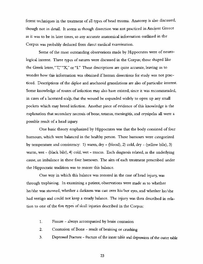
ferent techniques in the treatment of all types of head trauma. Anatomy is also discussed,
though not in detail. It seems as though dissection was not practiced inAncient Greece
as it was to be in later times, so any accurate anatomical information outlinedin the
Corpus was probably deduced from direct medical examination.
Some of the most outstanding observations made by Hippocrates were of neuro
logical interest. Three types of sutures were discussed in the Corpus; those shaped like
the Greek letter,"T," "X,"
or"I."
These descriptions are quite accurate, leaving us to
wonder how this information was obtained ifhuman dissections for study was not prac
ticed. Descriptions of the diploe and arachnoid granulations are also ofparticular interest.
Some knowledge of routes of infection may also have existed, since it was recommended,
in cases of a lacerated scalp, that the wound be expanded widely to open up any small
pockets which may breed infection. Another piece of evidence of this knowledge is the
explanation that secondary necrosis ofbone, tetanus, meningitis, and erysipelas all were a
possible result of a head injury.
One basic theory emphasized by Hippocrates 'was that the body consisted of four
humours, which were balanced in the healthy person. These humours were categorized
by temperature and consistency: 1) warm, dry- (blood), 2) cold, dry - (yellow bile), 3)
warm, wet- (black bile), 4) cold, wet - mucus. Each diagnosis related, as the underlying
cause, an imbalance in these four humours. The aim of each treatment prescribed under
the Hippocratic tradition was to restore this balance.
One way in which this balance was restored in the case ofhead injury, was
through trephining. In examining a patient, observations were made as to whether
he/she was stunned, whether a darkness was cast over his/her eyes, and whether he/she
had vertigo and could not keep a steady balance. The injury was then described in rela
tion to one of the five types of skull injuries described in the Corpus:
1 . Fissure - always accompanied by brain contusion
2. Contusion ofBone - result ofbruising or crushing
3. Depressed Fracture - fracture of the inner table and depression of the outer table
23

4. Impression (by sharp or pointed weapons)- not penetrating the
skull
5. Fractures
Trephining the skull was recommended for all the above cases except #4, and those
which involved extensively comminuted bones. Trephining was also discouraged if the
injury was directly over a suture. The reasoning behind this could be that either
Hippocrates (like some Greek and Roman physicians who followed him) thought the
sutures were sites of respiratory exchange, or, perhaps in earlier attempts to trephine in
these areas, the dura was torn (being especially adherent at these sites), and may have
released large amounts ofvenous blood from its sinuses. Damage to the dura mater was
rightly of great concern to Hippocrates. He developed a series of trephines which had an
extra flange placed at a precise distance from the sharp tip, thereby preventing it from
being drilled too deeply into the skull and injuring the underlying meninges and brain.
This principle in trephine construction was carried on for centuries.
Other methods developed by Hippocrates were for the treatment of head injuries
which damaged, but did not penetrate the skull bones. In these cases, the wound was
enlarged to expose a greater area of the skull. A heavy, black salve was then apphed
directly on the bone. After it set, the physician wiped off the excess, leaving only what
had sunken into the fracture lines. The physician then scraped the bone until all the
black lines disappeared. Those marks made by the salve which sank into the suture lines
looked quite different from the fracture lines, so there was no risk of confusion.
Aside from head injuries, treatment of spine dislocation was also described.
Connections were made between spinal injuries and various symptoms throughout the
body, demonstrating some early knowledge of the paths of the nervous system. For
example, leg paralysis was described as being associated with possible urinary inconti
nence or sensory disturbances. Cervical spine dislocations were believed to be the cause
of paralyzed deglutition, paresis of tongue, and paralysis to the four extremities, bladder,
and rectum. Extension and counter extension was recommended for these conditions.
Over 2,000 years following his death, Hippocrates is still remembered as an out
standing physician, surgeon, scholar,and humanist. His basic principles are still the basis
24

ofAmerican medical practice today: observe all; study the patient, not the disease; and/
evaluate honestly. The following is a copy of the Hippocratic Oath. It is important to
realize the theory upon which it is based may be apphed to all professions, and not just
those related to medicine.
25

THE HIPPOCRATIC OATH
I swear by Apollo Physician, by Aesculapius, by Health, by Heal-all, and by all the
gods and goddesses, making them witnesses, that I will carry out, according to my ability
and judgment, this oath and this indenture:To regard my teacher in this art as equal to
my parents; to make him partner in my livelihood, and when he is in need ofmoney to
share mine with him; to consider his offspring equal to my brothers; to teach them this
art, if they require to learn it, without fee or indenture; and to impart precept, oral
instruction, and all the other learning, to my sons, to the sons ofmy teacher, and to pupils
who have signed the indenture and sworn obedience to thephysicians'
Law, but to none
other. I will use treatment to help the sick according to my ability and judgment, but I
will never use it to injure or wrong them. I will not give poison to anyone though asked
to do so, nor will I suggest such a plan. Similarly I will not give a pessary to a woman to
cause abortion. But in purity and in holiness I will guard my life and my art. I will not
use the knife on sufferers from stone, but I will give place to such as are craftsmen there
in. Into whatsoever houses I enter, I will do so to help the sick, keeping myself free from
all intentional wrongdoing and harm, especially from fornication with woman or man,
bond or free.Whatsoever in the course ofpractice I see or hear (or even outside my prac
tice in social intercourse) that ought never to be published abroad, I will not divulge, but
consider such things to be holy secrets. Now if I keep this oath, and break it not, may I
enjoy honor, in my life and art, among all men for all time; but if I transgress and for
swear myself, may the opposite befall me.
26

About the Piece
The illustration representing Hippocratic contributions to neurosurgerywas
almost completely done by hand, with minimal computer manipulation. I began by
sketching on an ungessoed, stretched piece ofwatercolor paper. I had decided to work
out the composition fully before working, avoiding the problems I had run into on the
Egyptian piece. Once again, there were a variety of elements I had wanted to include,
some I already had the visual reference for, others were more abstract ideals or theories
for which I had to create visual symbols.
The portrait ofHippocrates came from an engraving by an unknown artist. Since
there is no direct evidence for what Hippocrates actually looked like, I had license to use
any reference I could find, or to develop a face myself, without conveying inaccurate
information. I decided to use a very classic portrait which could easily be considered
recognizable as an ancient Greek scholar.
The figure to the lower left of the piece was taken from an Ancient Greek sculp
ture of a professional boxer. It is especially interesting to see the facial abnormalities that
resulted from such a career, including the swollen brow, broken nose, and cauliflower ear.
These men were frequent patients ofGreek physicians, along with soldiers and gladiators.
Unlike the somewhat realistic face ofHippocrates, I chose to illustrate this figure as
though he were a statue, an ancient piece of physical evidence not only ofGreek medical
practice, but also of the clinical accuracy of the Greek sculptor.
Since the main treatments ofneurosurgery practiced in this time period was
trephining, I did want to include this in the illustration. I did not, however, want it to
look too similar to the first piece of the series. I decided to illustrate a cross section of
the procedure itself, showing how the special trephines (with depth gauges) developed by
Hippocrates worked. I completed this part of the illustration in a very stylized manner,
making the width of the subarachnoid space almost as wide as the thickness of the crani
um, in order to show the meningeal vessels clearly. I drew two additional trephines with
similar designs, but later removed them from the illustration digitally because since no
matter where I placed them, they still seemed to disturb the composition of the piece.
27

The surface view of the skull (in the box), was an afterthought. I knew I had wanted to
show the technique of surrounding a fracture with trephine holes in order to release pres
sure, but I did not want to show it on the skull of the boxer. I feel the color correlation
ties the two separate anatomical references in this piece together.
I wanted to be sure to illustrate the staff ofAesculapius in this piece, to distin
guish it from the caduceus, which many confuse with the Greek symbol for medicine.
Aesculapius was the ancient god ofmedicine and healing. His staff was made of a crude
wooden stick, which had a single serpent coiled around it. The caduceus is the staffof
Hermes and is represented as a polished wand, usually with some sort of ornamentation
at one end, and a pair ofwings (the symbol for the messenger of the gods) attached to
this same end. Coiled around the staff are a pair of serpents which face each other at the
top. This staffhad no medical symbolism behind it, and actually represented aspects of
society such as business and money. Perhaps since it looked so much like the staff of
Aesculapius, people today simply confused the two, and it now is used as the symbol for
many medical groups, including the American MedicalAssociation (The Royal College
of Surgeons ofEngland correctly use the staff ofAesculapius as their symbol) . I wanted
to make this element visually very subtle compared to the other images used in this
piece. By blending it into the background a bit, it appears as though it was drawn or
carved directly on that stone itself.
The type, though the last to be discussed, was the first to be drawn. The Corpus
Hippocraticum, the written medical texts which Hippocrates had left behind, was a very
important part of this chapter, and of the history of neurosurgery. Though the language
used is ancient, the alphabet used is far from obsolete. By including the actual title of
the written work in the illustration, an emphasis is placed not only on the text itself, but
also on the variety of things modern men have taken from the ancient Greeks, scientific
practices included. I used several fonts as references for this type, though I did draw it
freehand (without tracing it directly), so minor details and proportions were ofmy own
creation. Since the words were so long, I decided to overlap the letters a bit, so the piece
would not have to be stretched horizontally in order to fit the type on one line. The size
of the type was also kept large since lower case letters were not used in ancient Greece
28

and I did want to simulate (though not imitate) carved letters.
After completing the drawing, I then used acrylic paint to colorize everything but
the portrait. The marbleized effect of the background and the sculpture was created by
applying the paint with a rag, creating a"splotchy"
look, then tightened up by carefully
painting in the delicate veins so characteristic of this stone.
Once the piece was scanned, I colorized the face ofHippocrates using the Fill,
Variations, Color Balance, and Hue/Saturation commands in Adobe Photoshop 3.0. I
used these same commands to tint the sculpture slightly, trying hues from pink to green
marble, finally deciding on a blue-grey, so not to distract from the color of the anatomical
images, which are the focus of the piece. I also cleaned and sharpened the type by simply
creating a path around each letter, coloring the letters a bright white, and painting a sub
tle dark grey haze behind them. Once these details were complete, I used the
Brightness/Contrast controls and adjusted the entire piece until I felt each color was
enhanced to the correct level.
The original file measures6.5"
by 11.0". It was taken to a service bureau where
it was printed from a postscript file to a Fiery printer at 1200 dpi.
Sources
1. Ballance, Sir Charles A., The ThomasVicary Lecture: A Glimpse Into the History
of the Surgery of the Brain. MacMillan & Co. Ltd. London, England, 1922
2. Majno, Guido, The Healing Hand: Man andWound in the AncientWorld.
Harvard University Press, Cambridge,MA, 1975.
3. Margotta, Robert, The Story ofMedicine. Golden Press, NewYork, NY, 1967.
4. Rutkow, Ira M., Surgery: An Illustrated History. MosbyYearbook Inc., St. Louis,
MO, 1993.
29

5. Sachs. Ernest. The History and Development ofNeurological Surgery. Paul B.
/
Hoeber Inc. NewYork, NY, 1952.
6. Zimmerman, Leo M., andVeith, Ilza, Great Ideas in the History of Surgery.
Dover Publications Inc., NewYork, NY, 1967.
30

oo
Q
WHP-*
O


Claudius Galen
Following the time of the Alexandrians, there were few accomplishments made in
the field of neurosurgery until the work of Claudius Galen (c. 130-200 A.D.). Galen had
much respect for his predecessors, and spread the knowledge handed down to him from
the Alexandrian and Hippocratic schools. Through his education and his steady practice
ofmedicine throughout the Roman Empire, Galen accomplished much in the fields of
neuroanatomy, neurophysiology, and neurosurgery. He kept greatly detailed written
accounts of his teachings. There are over 400 separate treatises which come directly from
Galen. The subjects of these treatises include all aspects ofmedicine, making it the most
complete medical text created until that time.
Unlike Hippocrates, Galen included much autobiographical information within
his texts, which illuminates the type ofman he was and, more importantly, how he prac
ticed medicine. Galen was born in Pergamum, a city famous for its Aesculapian temple.
He was educated at fine schools, and was very proud of this fact. After travelling a great
deal, he ended up in Alexandria, where autopsies were a common practice, and he was
able to learn much. In 157 A.D., Galen returned to Pergamum, where he was appointed
chief surgeon to the gladiators. In the years following, Galen began practicing in Rome.
He had become good friends with Marcus Aurelius, who requested his move to Rome.
Here, he held pubhc anatomical demonstrations, which made him even more well
known. He later gave up all his teaching and left the city due to the outbreak of the
plague.
By closely observing and studying both human and animal cadavers, Galen was
able to speculate on many different physiological matters. He deduced that the brain
affected emotions and behavior, and that it controlled intelligence, thinking, fantasy, mem
ory, and judgement. Galen practiced dividing nerves, and he also traced their course,
including the recurrent laryngeal nerve, which follows a long path to the brainstem.
Galen was the first to name and identify different forms ofhydrocephalus. He identified
swelling between the brain and the meninges, the membranes and bone, the bone and the
33

pericranium, and the bone and skin all as forms of this disease, though today we would
only call the first by that term.
Some of his most advanced experiments included the sectioning of the spinal
cord at various sites. By studying the effects, Galen was able to deduce that areas below a
certain site on the spinal cord were controlled by nerves which were originating from
that site. He realized that the hemisection of the spinal cord resulted in loss ofmovement
to only one side of the body, where full section of the cord resulted in complete paralysis.
Galen, like so many of his predecessors, treated head trauma with trephining. He
described the various head injuries much more elaborately than Hippocrates, even
including details such as a description of each instrument that was used. The types of
fracture which he chose to treat by trephination were:
1 . depressed fracture
2. fractures with hematoma
3. comminuted fractures
4. trichiasis
Trephine holes 'were made using a variety of instruments. Those he used most often
included a standard trephine, auger, terebra (drill), terebra abaptista (drill with guards)
chisels, cutting forceps, and a blunt director (used to elevate a depressed fracture).
Explanations of these instruments and instructions on when to use them were included
in his published treatises. By treating such large numbers ofpatients at a time, Galen also
became an expert in suturing wounds, using silk, linen or catgut.
Despite all that he accomphshed in the field ofmedicine, Galen's reputation has
suffered over the years. Near the end of his life, he Galen survived two major losses in his
life. First, Marcus Aurelius died in 180 A.D. and, in addition, a great portion ofhis origi
nal manuscripts were destroyed in a fire. He did continue to study and write, and died in
Pergamum at the age of 70.
The first thing to affect his reputation was the discovery of some of bis writings
which included some observations which implied he believed in a single god. Though
34

never mentioned as a Christian god, Christians so interpreted it, and he gained a great
deal of respect. As respect turned to veneration, those in medical occupations began to
take Galen's word as law. In the following years, some errors were discovered in his
observations. Many say Galen was suddenly blamed for various inaccuracies he may or
may not have been responsible for. Only following the Renaissance has this attitude
changed to a positive and respectful appreciation for Galen's accomplishments. As the
first to emphasize the importance of anatomy and physiology in surgical considerations,
Galen is now known as one of the greatest contributors to the history ofmedicine in
general, and ofneurosurgery in particular.
About the Piece
Since I had concentrated on brain surgery for the first three illustrations, I decided
to show some aspect ofperipheral nerve surgery or anatomy for this piece showing the
neurosurgical accomplishments ofGalen. One ofGalen's major achievements was to
trace the root of the recurrent laryngeal nerve, which begins in the brainstem and heads
downward to the aortic arch, under which it loops under before continuing upwards to
innervate structures of the laryngeal region. This nerve is actually a branch of the tenth
cranial nerve, or the vagus nerve. This illustration is a schematic representation of the
path of these nerves.
After planning the design on paper, I completed the final drawing in charcoal on
stretched, ungessoed watercolor paper. The anatomical drawing is proportionately inaccu
rate. Since the vagus nerve is one of the most widely distributed nerves in the body, it
would have had to have been painted extremely small in order to show its full course in
proportion. After painting the paper with a flat, brown wash, I began to paint in only the
darkest shadows and brightest highlights within the drawing. Initially, I had planned on
using some color in this area, though I changed my mind at this point. I decided leaving
the extraneous anatomy monotone, allowing the colored nerve to attract more attention.
The cross section of the spinal cord was included in the illustration to show not only did
35

Galen know it existed; but that he knew the difference between afferent and efferent
nerves (sensory and motor neurons).
The portrait ofGalen was included in a distinct and characteristically Roman
way. Portraits of emporers were often carved on ancient coins, and though Galen was
never a political figure, he was a very important and highly regarded professional by the
citizens of ancient Rome. I used a photograph of a bronze coin as a reference in painting
the correct highhghts on the coin itself. The only portrait ofGalen I could find was a
three quarter view of his head. By studying his features, I rotated his head mentally, and
sketched a simple profile, which I later transferred onto the coin drawing.
Completing the mosaic frame was a very time-consuming procedure. I knew I
had wanted to include this element, though I neither wanted the anatomy to be drawn as
a mosaic, nor obscured by closely surrounding mosaic. For this reason, I faded the mosaic
tiles into the anatomical drawing, but continued them in a pattern around the periphery.
After the drawing was complete, I began colorizing it with acrylic paint. The dif
ference between this illustration and the three before it, is that I used colored pencils to
give the surface a bit of texture. By darkening the blank area beneath the nerve, and
between the tiles, the fighter areas appeared much brighter and provided a richer and
deeper contrast.
Digital manipulation of this piece was kept to a minimum. Aside from decreasing
the size of the spinal cord section, I used Photoshop only to enhance color in various
areas. Using the paintbrush tool, I went in between each mosaic tile and darkened the
area with a deeper shade ofbrown. I used the "Brightness/Contrast, Fill, Color Balance
andHue/Saturation"
commands with the coin, deciding between bronze, silver, or even
clay. I finally decided on a rustic bronze. I continued to use these controls in completing
the final touches.
The original file measures8.75"
by 8.0". It was taken to a service bureau where
it was printed from a postscript file to a Fiery printer at 1200 dpi.
36

Sources
1 . Ballance, Sir Charles A. , The ThomasVicary Lecture: A Glimpse Into the History
of the Surgery of the Brain. MacMUlan & Co. Ltd. London, England, 1922.
2. Majno, Guido, The Healing Hand: Man andWound in the AncientWorld.
Harvad University Press, Cambridge, MA 1975
3. Margotta, Robert, The Story ofMedicine. Golden Press, NewYork, NY, 1967.
4. Rutkow, IraM., Surgery: An Illustrated History. MosbyYearbook Inc., St. Louis,
MO, 1993.
5. Sachs, Ernest, The History andDevelopment ofNeurological Surgery. Paul B.
Hoeber Inc. NewYork, NY, 1952.
6. Zimmerman, Leo M., andVeith, Ilza, Great Ideas in the History of Surgery.
Dover Publications Inc., NewYork, NY, 1967.
37

:.:
in
t-4.. -*2
a)o
'3/>
a)
^>
o : n
p ?!(1) 0
0'"X
c
c o
0)k_ o0
tfc(1)
"UD
>>c
oCD
b
:.^.$..
~8E
<2
o ~8-fe- JO
:1-2
<Z
^ Q-
2
E<c
o
E
m>.-:
c g
oc
o
1
roc
_2
cr
.'C
j>
o
ro
0)
c
D
0)
E3
-fc
CD
..C.,,,
_2
o
<uu
2CL
"5u
'ro
ro
o
0)
;D
'r-
:**; J>
~ i
"5 8
<. 8
0 1!. ^CD o
E u
Ia;
"5 O
c
o
oc
D
<U
"if
roL_
D
_8
D
roD
O-C
St01
J3o
Q.
O
-oO
-if
...J....
"8
c0)
Ec3..
0)
_Q
OC
1T>


Ambroise Pare/
In considering the medical and surgical advances made during the age of the
Renaissance, one man stands out as an exceptional contributor. Although he did not
contribute much specifically to neurosurgery,Ambroise Pare (1510-1590) played a major
role in modernizing all surgery practiced in his day He represents the end of the lasting
links to ancient tradition and the decisive push toward modernism.
Pare was born in a village in northwest France, as the son of a humble cabinet
maker. He worked as an apprentice with several barber-surgeons, an occupation shared
by two of his siblings. At the age of 15, Pare went to Paris to receive further formal
training in anatomy and surgery. He went on to practice primarily as a military surgeon,
beginning in 1536, and continuing until just before his death.
Some of the earliest observations of certain neurological symptoms and conditions
ever made were described by Pare in his books. For instance, it was Pare who first
observed the occurrence of fiver abscess due to a pyemic infection following a head
injury, as well as brain abscess which occurred as complications of skull fractures. Such
associations were rarely made before the birth ofphysiology.
It seems as though Pare carried out various operations on the skull, brain and
spine successfully. Aside from surgically removing osteomyelitic piecesofbone, he was
also bold enough to incise the dura to remove and evacuate blood clots or pus. He
emphasized the importance of debridement, and described the removal ofmany different
foreign bodies, including clothing, bone splinters, and dead tissue from within the skull as
well as those that were impinging on the spinal cord. He recommended trephining for a
variety of reasons, the mostcommon being cases in which there was matter lodged
between the dura and the brain. For matter lodged superficial to the dura and beneath
the bone, Pare devised a new way of release. He would insert a lead cannula into the
extradural space and then ask the patient to strain with nose and mouth closed, and, with
the dura depressed from the bone surface, the matter was then expelled.
In addition to describing his surgical techniques, Pare also described in great detail
various types of instruments and which ones he recommended for which types of frac
tures. Some of these instruments included trepans, razors, shavers, and scrapers. One
40

instrument designed by Pare created a circular flap by swinging a cutting blade in an arc
about a fixed point. It consisted of a simple compass with cutting points. He devised an
apparatus used to apply traction to the spine in cases of dislocations. It consisted of a
wooden frame on which the patient was placed while traction was then exerted by assis
tants who were directed by the surgeon. For cases of spinal deformities, usually due to
vertebral caries, Pare recommended the use of a brass cuirasse, and continued to develop
other types ofbraces, frames and supportive or corrective devices for such conditions.
One man who had a great effect on Pare and his work was AndreasVesalius. Pare
felt very strongly that surgeons required extensive anatomical knowledge. Aside from
some of his own books on anatomy, including Anatomie Universelle Du Corps Humain.
1561, he also wrote an epitome onVesalius'
Fabrica. This work had a profound effect on
Renaissance surgery, making the work ofVesalius accessible to surgeons for the first time.
Pares humble background and beginnings as a barber-surgeon, unfortunately won
him minimal respect from the professional French surgeons, at first. In 1554, he was
finally, yet reluctantly, accepted as a member of the College of St. Come, a prestigious sur
geon's organization. He later went on to become the chief surgeon to four successive
French kings: Henry II, Francis II, Charles IX, and Henry III. This was an outstanding
achievement for someone with such a meager formal education.
Much may be observed about Pare's nature with his famous motto, "Je le pansay,
Dieu leguerit,"
(I treated him, God cured him). Despite his grand success as royal sur
geon, he remained quite humble and true to the noble ideals ofmedicine and the healing
arts. In 1575, he had all his works published in French, not in Latin, which created quite
an uproar in the medical community. Even his fellow members of the College of St.
Come fought to have the works legally banned. This led Pare to write his last manu
script, a lengthy denial to these requests to have his work published in Latin. Apologie Et
Voyages was published in 1585, and is considered, in many ways, an autobiographical
work. It is said to reveal the true nature of a man who changed the history of surgery
through his innovative ideas and remarkable dedication.
41

About the Piece
Of all the pieces in the series, this piece is probably the most unique. . Not only is
it black and white, it was also completed in media very different from those I had used
for the previous pieces. In researching visual references on the time period, I discovered I
could choose from a variety of styles. I chose to treat the illustration as though it were a
title page to a medical manuscript. These title pages consisted usually ofvery intricately
designed engravings, with hand set type which varied from elegant scripts to block-like
capital fonts. To avoid pure imitation, I decided to keep the intricacy and detail of the
original designs to a minimum, making the piece appear more modern upon completion.
The portrait ofAmbroise Pare was my first attempt at scratchboard. I found this
medium to be fairly simple to practice, though I did have a tendency to make some lines
too thin. The reference which I used for this portrait is an etching, also containing some
delicate fines, which I unconsciously continued to imitate. The neurosurgical scene is
taken from an actual woodcut believed to be the first artistic rendition of a neurosurgical
operation. This drawing was also completed in scratchboard, at about the same size as the
portrait.
After completing the scratchboard illustrations, I began the surgical illustrations
which were later placed behind the portrait. Using a variety of references, I sketched the
removal of a piece of osteomyelitic bone. After laying a piece of double sided mylar over
it, I began to trace the two separate drawings in ink with a sable brush and rapidographs.
These were then laid aside with the scratchboard pieces.
Though the piece is primarily about Pare, I felt as though it was important to
includeVesalius and his work somehow in the illustration as well. The two anatomical
drawings are the only elements in the entire thesis series which were not created by
myself. I scanned these directly into the computer, where I brightened and clarified
them.
I scanned all the illustrations into the computer as a 256-gray file at 150 dpi. I
created a blank page in Photoshop first, then imported each illustration as a separate layer.
Since the file was a grayscale file and required less memory, there was no problem with
the speed of the software. It was a time consuming process to both resize and place each
42
illustration on the board. I had decided I did want to"compartmentalize"
the piece after
the style of the renaissance manuscripts, as well as include some sort of title block con
taining the names of the men this piece was based on. Although I had sketched the final
composition, it was useful only as a guide, and not as a strict plan. One of the best deci
sions made was the choice of a black background inserted behind the portrait. After
doing this, the separate illustrations really seemed related to each other more and
appeared more united. The frames I built using the stroke command, varying color,
width and placement (inside, outside, or centered on the edge of the selection). The
alternating black and white bars which surround theVesalian drawing of the brain and
spinal cord imitate those used in anatomical drawings of the period. They were used as
instruments for measurement; which the artist lined up the drawing to in order to main
tain correct proportions.
The last element completed for this piece was the type. In studying the type
used in the manuscripts, I realized that most were based on standard serif faces, but had a
shght hand-rendered look to them. I thought the best way to achieve this effect was to
either hand draw some myself, or rework digitally set type on the computer. In the
interest of time and resolution quality, I chose the latter option. I started by typing out
the two names in the font, Palatino, using the software Adobe Illustrator 5.5. I then used
the command, "CreateOutlines"
to transform the letters as separate outlined drawings,
rather than as a line of text. Using the pen tool, I moved the points of the outlined let
ters randomly, creating thick and thin lines and uneven serifs. I imported this type into
the final document as an EPS file and set the other italicized type in Photoshop itself.
The original file measures9,0"
by 7.0". It was taken to a service bureau
where it was printed from a postscript file to a Fiery printer at 1200 dpi.
43

Sources
1. Rutkow, Ira M., Surgery: An Illustrated History. MosbyYearbook Inc., St. Louis,
MO, 1993.
2. Sachs, Ernest, The History and Development ofNeurological Surgery. Paul B.
Hoeber, Inc., NewYork, NY, 1952.
3. Walker. Earl A.. A History ofNeurological Surgery. TheWilliams and Wilkins
Co., Baltimore, MD, 1951.
4. Zimmerman, Leo M., andVeith, Ilza, Great Ideas in the History of Surgery.
Dover Publications Inc., NewYork, NY, 1967.
44

ml*\vp^Vy.
/{/ /

Percivall Pott
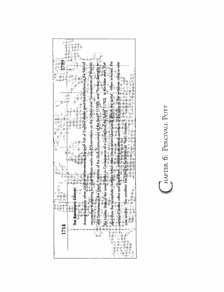
Many advancements were made during the 17th and 18th centuries in the field of
neurosurgery. A major change occurring at this time were the attempts at using neuro
surgery to treat ailments other than those associated with trauma. In England, Percivall
Pott (1714-1788) made many contributions in regards to this new approach to surgery.
He was the first to describe in detail the pathology related to certain diseases. Though
the practice of choosing a specialty still did not exist, some consider Pott, who made spe
cific contributions to the field of neurosurgery, worthy ofbeing called a very early neuro
surgeon.
Like Ambroise Pare, Pott began his career humbly. As an adolescent, he "was
apprenticed to a surgeon for a small sum. For the next seven years, he continued the
apprenticeship as the surgeon's primary assistant in preparing cadavers for dissection.
Eventually, Pott was accepted into the Company of the Barber-Surgeons, and soon
opened his own practice which he ran from a rented house where he lived with his
brother and half sister. Pott worked the majority of his career as an assistant surgeon, then
full surgeon at St. Bartholomew's Hospital. At his retirement, he is said to have stated he
had served the institution "as a boy and aman."
One of Pott's most famous descriptions is that of the "puffytumor."
This refers to
the outward appearance of a brain abscess, differentiating it from an extradural hematoma.
The tumor appears as a circumscribed swelling of the scalp indicating either an underly
ing osteitis of the skull, or an extradural abscess. This description was included in one of
his most extensive works, "Observations on the Nature and Consequences ofWounds and
Contusions of the Head, Fractures of the Skull, Concussions of theBrain."
The best known of Pott's published works is probably his two volumes on spinal
diseases, Remarks on That Kind ofPalsy of the Lower Limbs.Which Is Frequendy Found
to Accompany a Curvature of the Spine (1779), and Farther Remarks on the Useless State of
the Lower Limbs in Consequence of a Curvature of the Spine (1782). In the first volume,
tuberculosis of the spine, later referred to as "Pott'sDisease"
is described. This disease is
47

characterized by a compression of the spinal cord due to the collapse ofvertebral bodies
which are infected with tuberculosis. Although Pott did not understand the true (bacteri
al) cause of the disease, he did depict the condition of the collapsed vertebrae and spinal
curvature. Additionally, he observed the spasticity of the muscles which occurred when
the disease affected the spinal cord, and associated the spinal deformity,"gibbus"
("hump
back") with the disease. Pott's choice of treatment included making a paraspinal incision
into the swelling of the trunk to drain the pus, inserting a kidney bean into the wound
(to maintain a draining sinus tract in communication with the paravertebral abscess), ,and
then sprinkling it with cantharides ( a counterirritant) .
The manner in which Pott chose to treat head injuries was most often trepliining.
It proved to be the most efficient in draining purulent matter, and was often completed
by producing more than one trephine hole. He stated his intentions regarding this prac
tice: "The reasons for trepanning in these cases are, first, the immediate relief of present
symptoms arising from pressure of extravasated fluid; or second, the discharge ofmatter
formed between the skull and dura mater, in consequence of inflammation; or third, the
prevention of such mischief, as experience has shown may most probably be expected
from such kind ofviolence offered to the last-mentioned membrane...In the ...mere frac
ture without depression ofbone, or the appearance of such symptoms as indicate com
motion, extravasation, or inflammation, it is used as a preventative, and therefore is a mat
ter of choice, more than immediatenecessity."
There were certain principles Pott discussed in regard to the practice of surgery
that showed the dedication of his practices. He emphasized the need for better surgical
technique, not greater speed, describing the custom ofmeasuring the motion of a sur
geon's hand with a stopwatch, as"absurd."
Pott also pointed out that a surgeon needs
more than manual skill to be successful. He stressed the ability to judge whether or not
to operate as important, and said this ability comes only with experience, and not from
books. Regarding his own contributions to the field of surgery, Pott once remarked:
"Many and great are the improvements which the chirurgic art has received within these
last fifty years; and much thanks are due to those who have contributed to them; but
48

when we reflect how much still remains to be done, it should rather excite our industry
than inflame ourvanity."
About the Piece
In selecting the elements to be included in the piece on Percivall Pott, I chose to
emphasize his studies on tuberculosis of the spine. During the period in which he lived,
there were many surgeons making great advancements in their respective fields, though
visual references of these people and their accomplishments were fairly limited. The por
traits were typical Baroque influenced oil paintings, and other visual icons reflective of the
period were also of this heavy, ornamental nature. Instead of following this style, specifi
cally, I chose to treat this illustration primarily as a medical illustration, using the portrait
and the type as the only indicators as to the period of the discovery.
I initially planned on painting the illustration in acrylic, but instead I completed
the original charcoal drawing on a sheet of gessoed watercolor paper. I used a three
dimensional plastic model of the spine as a reference in completing a view of a healthy
spine. The second and third drawings show the deterioration of the bony spinal bodies, a
characteristic process of spinal tuberculosis. For these drawings, I used references not only
on this disease specifically, but also on other degenerative bone disorders.
I completed the drawing of Percivall Pott as a very rough pencil sketch, modeled
after a formal painted portrait. His features were very distinct, and I wanted to be sure to
include an image of his face in the piece.
I scanned in both black and white drawings as 256-gray files at 150 dpi, and
brought them into Photoshop. Since I had wanted this piece to stay light, as though it
were still a sketch, I decided to tint and tone parts of the images, rather than fully paint
over, or re-illustrate them. Before merging the two images, I decided on which two col
ors I would use to replace the blacks and whites. Instead ofmaking the piece a duotone,
which would mix the two colors I chose, and therefore create new colors, I first tinted
the entire screen (ofboth scanned images) the background color. Then, using the"Fill"
command in its"Lighten"
mode, I replaced all the black, or dark, areas with brown.
49

Using a limited palette, I began to tone only thosevertebraewhich were affected
by the disease. Trying again to keep the colorization subtle, I used the burn and dodge
tool to enhance shadows and highlights, preventing the chance of too many colors being
introduced into the drawing.
I set the type using Dorchester Script in Adobe Illustrator 5.5, then made addi
tional changes to it in the same program. First, I created the type to outlines, as I had
done for the last piece. Then, using the skew tool, I stretched the type diagonally, making
it appear more slanted than the original. I added the large, swirling loop to each initial
using the pen tool. After importing this type into Photoshop as an EPS, I then colorized it
in the same way as the other files.
I used the spine drawing as my background layer in Photoshop, and imported the
portrait and the type as separate layers overlying it. After flipping the portrait horizontal
ly, and resizing the type, I was satisfied with the composition.
I had decided to use the oval shape as though it were a vignette, a common ele
ment in artwork of this time period. In order to create it, I built a path, which I later
saved as a selection, that followed the contour of the three vertebral columns. Then, after
drawing the oval several times deciding on the size, I subtracted the"spine"
selection from
the oval selection, and what remained selected is what was then filled with blue at 25%
opacity. After saving this selection, I used the paintbrush tool, set on a large size, and put
an extra dark blue ridge around the edge of the shape.
Since the type and portrait were already separate from the background layer, it
was much simpler to create the brush stroke that lies behind them. I set the pencil tool
to fade to transparent after a certain number of pixels. This way, it appears darker on the
left than on the right. The shape of the stroke took some time to perfect. Because of the
all the complex negative shapes which were created by the type, it became quite impor
tant to notice exactly what areas the blue covered.
The original file measures10.0"
by 7.0", and was taken to a service
bureau where it was printed from a postscript file to a Fiery printer at 1200 dpi.
50

Sources
/
1. Flamm, Eugene S., "Percivall Pott: an 18th centuryJournal of
Neurosurgery, v. 76, Feb. 1992, p. 319-326
2. Rutkow, Ira M., Surgery: An Illustrated History. MosbyYearbook Inc., St. Louis,
MO, 1993.
3. Sachs. Ernest. The History and Development ofNeurological Surgery. Paul B.
Hoeber, Inc., NewYork, NY, 1952.
4. Walker. Earl A.. A History ofNeurological Surgery. TheWilliams and
Wilkins Co., Baltimore, MD, 1951.
51

c^
co
ooCO
p
1Ul
u"I-
fczUl
Ul
tZ :
c
_g
800
o-
</) .E
*- fc
t
"i:S:V:,i
Li
0
0)
O _Q
C
CN 0
CO fl)
2 .
~T"
Vf)
-IS
OSr-,..g
03 1-
... ...<D..
fi
-* s3 0
:""
E
1
=iS
ro ^3 0}
5 >Tj >
:> =
V5 2
0) SI15
- <uoo
!s-'
.Z.S..
C O
c O~
-.'
i a)
1 i
IS *
O AC
' f :H "5
5 6 ;*
"8N
:,c 3
o
E2
<D D
D-
D
CD: N.,,g....
ro
o
4)
-o
CD
E5
sr -2
<D
o
c
E oo *-
8c o ii
Tfi ID
"
"1 E
j ^
O tt!
e >
<W-'
CO
b .;
:>:;.HCL:
a^
3 . oo? oo
c?
a,
> j-
<DCD C
11
II
S OW:
*
:
mp':
I.CD
E
c
cp*D
80)


Great Moments in Neurosurgery of the 19th Century
The 19th century was a time in which many advances were made in all fields of
surgery, due to the improvement of the conditions under which surgeons were able to
practice. With the development of ether in 1846, and chloroform in 1847, patients could
painlessly undergo long hours of surgery, allowing surgeons increased time to concentrate
on completing successful operations. In 1867, the English surgeon, Joseph Lister, devel
oped the use of carbolic acid as an antiseptic. He studied Louis Pasteur's writings which
disproved the concept of spontaneous generation, and established bacteria as a cause for
infection. This affected surgical methods immensely, introducing, for the first time, the
practice of antiseptic surgery.
Though these discoveries led to important breakthroughs in general surgery, neu
rological surgery developed more slowly, since this branch depended a great deal more on
the allied sciences. Such technical procedures as roentgenology, ventriculography, and
angiography that are involved with neurological diagnosis, were not perfected until some
time later. Nevertheless, neurosurgeons all over the world still were able to continue to
improve the survival rate ofpatients, developing a variety ofmethods which have gone
down in history as being some of the most bold and innovative surgical techniques ever
attempted.
William MacEwen (1848-1924) of Scotland, is considered by many to be the
father ofneurological surgery. He was one a pioneer in establishing the field as a spe
cialty and developed many ways of diagnosis and treatment ofbrain tumors. One of
these diagnostic procedures include tapping the skull and listening for a "cracked-pot
sound", that would indicate hydrocephalus. MacEwen also is remembered for being the
first to make the decision to perform a craniotomy based solely on neurological signs and
symptoms, without any external manifestations of a lesion.
The patient was a 14-year old girl who had a swelling at the upper inner portion
of the left orbital cavity. While in the hospital, she suffered from recurring seizures, based
on which MacEwen made the clinical diagnosis of a brain tumor and decided to
54

trephine. On July 27,' 1879,MacEwen removed the meningioma, allowing the patient to
be dismissed shortly after. She was able to return to a normal life in a matter of a few
months. MacEwen wrote that his decision to operate was based on a variety of symp
toms, including the contraction of the left pupil, a dull, fixed pain on the left side of the
brow, and convulsions which affected the opposite side of the body. This deductive way
of thinking demonstrated the dependent relationship between neurologists and neurosur
geons, and was one ofMacEwen's greatest contributions.
Aside from conducting many neurological experiments involving cerebral func
tion and the electric currents produced in the brain,Victor Horsley (1857-1916) was one
ofEngland's earliest neurosurgeons. He had begun his career primarily as a neurologist,
and it was not until 1885, at the age of 28, that he received his first appointment as
Assistant Surgeon to the University College Hospital, and was made Surgeon to the
National Hospital for the Paralysed and Epileptic (England's primary hospital for the
treatment of nervous diseases) the following year. During his years as a surgeon, Horsley
developed specific techniques and materials which greatly improved the success rate of
neurosurgery. One of these important developments was bone wax, used to control
bleeding from the bone edges, which continues to be used in cranial surgery today. He is
also is known as one of the pioneers ofmodern spinal surgery, completing the first suc
cessful operation for the removal of an extramedullary tumor of the spinal cord in 1888.
The patient, in this case, had complained of intense pain and partial paralysis in his
lower limbs and abdomen. Upon further examination, Horsley came to the conclusion
that these symptoms were due to a tumor of the spinal cord, at about the level of the 5th
vertebra. After opening the vertebral column and exposing the cord, He boldly incised
the dura, but did not find any sign of a lesion. He continued to open the meninges more
fully by lengthening the incision upward. Finally, he found the tumor, which he then
removed with no complications. After one year, Horsley received a letter from this
patient who described himself as being in excellent health, able to work up to 16 hours a
day.
InAmerica,WilliamWilliams Keen (1837-1932) was making great strides in neu-
55

rosurgery as well. Aside from his medical practice, one of-Keen's greatest contributions
was his vast array ofpublished works, ranging in a variety of topics. He jointly edited
AmericanText-Book ofSurgery (1892), the first surgical text written only byAmerican
surgeons. From 1906 to 1921, he authored the eight volume, Surgery. Its Principles and
Practice, which became known in the first few decades of the twentieth century as the
"clinicalbible"
ofAmerican surgeons in the first few decades of the twentieth century.
Keen remained very involved with academic activities and taught in many different
schools. In 1866, he acquired ownership of the Philadelphia School ofAnatomy, the
nation's only remaining independent anatomical school. One of his more interesting aca
demic endeavors was his teaching of artistic anatomy to students at the Pennsylvania
Academy of Fine Arts in Philadelphia. From 1876 to 1881, he provided a series of lec
tures which included visual aids such as actual dissections, plaster casts, galvanic muscle
stimulation, and live models. Eventually, the success of his surgical practice required him
to end this project.
Keen's surgical ventures are numerous and varied. One particular case encouraged
Keen to develop new approaches to ventricular puncture. The patient had been diag
nosed as having a tumor which, though eventually removed, had caused so many compli
cations following surgery, the patient finally died. During the autopsy, Keen observed the
extensive enlargement of the ventricular system, especially of the lateral ventricles. He
later proposed tapping these lateral ventricles directly in order to relieve intracranial pres
sure. After considering the site of the puncture, he decided on making a small burr hole
1.25"
above and behind the external auditory meatus. He advised to aim the trocar and
cannula at a point2.5"
above the opposite external auditory meatus. This burr hole site
proved to be accurately placed, and came to be known as "Keen'spoint."
It is important to note that accomplishments such as those described here are only
a few examples of the advancements made during this century. In addition to these three
men, there were many others who continued throughout this century to perform many
life-risking surgeries, while steadily decreasing the fatality rate. It was at this time neuro
surgery began to be recognized as a specialty separate from other branches of surgery.
56
Those who chose to specialize in this field fought intensely for this recognition, which
did not fully come into being until early in the next century.
About the Piece
For the illustration on the 19th century, I spent much ofmy time narrowing
down those surgeons who should be profiled in the piece. Since it was a time ofmany
major achievements, this was a difficult decision to make. I decided upon three surgeons
who contributed three very different types ofprocedures to the development of neuro
surgery. This way, not only would the portraits of the men themselves differ, but also
would the corresponding surgical illustrations.
I sketched out the composition ahead of time, and decided on doing the portraits
on one sheet ofpaper, and simply planned the sizes of the surgical pieces carefully, in
order to draw the originals to the right proportions. I used colored pencil on colored
paper for the portraits, keeping them as tritones using one black, one siena, and one
white pencil only. Since the surgical illustrations were going to be colorized in
Photoshop, I decided the type ofpaper must be able to take charcoal and graphite. For
this reason, I continued to use the watercolor paper I had been using for the other pieces,
even though I had not planned on doing any painting.
After scanning in the portraits, I also scanned the blank, flip side of the drawing
for the future background of the piece. Since both the surgical illustrations and the por
traits would require reorganization for placement, I had decided to build this file in layers,
since it would allow more freedom to move separate elements without affecting others.
I first cleaned up the portrait piece by erasing smudge marks, and enhancing the
hghts and darks. Because I was not satisfied with the dull grey color that resulted from
the black pencil on the colored background, I used the"Fill"
command in the"Lighten"
mode and colorized these dark areas a deep, dull, red. This not only eliminated the black
areas, but also enriched the color of the background. I then imported each of these por
traits into the blank background page as one layer.
57

The first surgical illustration I colorized was the ventricular puncture, then the
/
removal of the meningioma beneath the frontal bone, and lastly, the surgical removal of
the spinal cord tumor. I tried to use similar palettes among the three of them, containing
colors which would also relate to the colors used in the portraits. The tool mainly used
for this job was the paintbrush, although many additional commands were used following
the initial painting of the pieces. The background color I picked up with the eyedropper
tool from the darkest darks of the portraits. Choosing a darker hue of the same color, I
then filled the background areas again in the"dissolve"
mode at a low opacity, to create a
speckled look. I carried this effect through the entire piece by using the "addnoise"
to
each illustration upon completion.
Each of the three files was then imported into the file which contained the back
ground and portraits. Once again, each piece was resized and repositioned until the com
position was satisfactory. The last step was to add the frames around the surgical illustra
tions. I did this by using the"stroke"
option after selecting each rectangle. To create the
double frame, I simply used the "expandselection"
option, selected a small number of
pixels, and then"stroked"
it once again.
The original file measures9.0"
by 7.0", and was taken to a service bureau where
it was printed from a postscript file to a Fiery printer at 1200 dpi.
Sources
1. Al-Rodham, NayefA.F, and Laws, Edward R., "Meningioma: A Historical Study
of the Tumor and Its SurgicalManagement,"
Neurosurgery, v. 16, May 1990,
p. 832-847
2. Bingham,William F, "WW Keen and the Dawn ofAmericanNeurosurgery,"
Journal ofNeurosurgery, v. 64, May, 1986, p.705-712
3. Bucy Paul C, "Scotland: The Birthplace of SurgicalNeuroloev."
Tournal of
Neurology. Neurosurgery, and Psychiatry, v. 48, Oct. 1985, p. 965-976
58

4. Laws, EdwardR.,Tradition in Neurosurgery: "DoingWellWhat Has Been Done
Before,"
Neurosurgery, v. 16, Jun. 1985, p. 739-753
5. Rutkow, Ira M., Surgery: An Illustrated History. MosbyYearbook Inc., St. Louis,
MO, 1993.
6. Sachs, Ernest, The History and Development ofNeurological Surgery. Paul B.
Hoeber, Inc., NewYork, NY 1952.
7. Stone, James L.,"WW. Keen: America's Pioneer NeurologicalSurgeon,"
Neurosurgery, v. 17, Dec. 1985, p.997-1007
8 . Walker, Earl A. , A History ofNeurological Surgery. TheWilliams andWilkins
Co., Baltimore, MD, 1951.
9. Wilkins, Robert H., Neurosurgical Classics. Johnson Reprint Corporation, New
York NY, 1965.
10. Zimmerman, Leo M., andVeith, Ilza, Great Ideas in the History of Surgery.
Dover Publications Inc., NewYork, NY, 1967.
59

;.tH
::0
O


The Father ofModern Neurosurgery: -Harvey Cushing
Many have contributed to the history ofneurosurgery, however, one man has
almost single-handedly brought the practice of neurosurgery into the modern era.
Harvey Cushing (1869-1939) developed many of the neurosurgical procedures still prac
ticed today. HarveyWilliams Cushing was born in Cleveland, Ohio onApril 8, 1869, as
the fourth generation ofCushings destined for a career in medicine. Raised in a relative
ly strict atmosphere, Cushing had the ability to meet the intense demands of his under
graduate education atYale, where he graduated with a Bachelor ofArts Degree in 1891.
He then went on to Harvard Medical School, where he received his degree cum laude in
1895.
Cushing's interest in neurosurgery began when he went to England and studied
underVictor Horsley. When he returned to the United States, he was given the opportu
nity to work solely on neurological cases atThe Johns Hopkins Hospital in Baltimore,
Maryland. At this time, surgery of the brain was quite difficult, largely due to ineffective
methods ofhemorrhage control. In his attempts to solve this problem, Cushing made
some of his most innovative contributions to neurosurgery.
First, in 1904, he devised a pneumatic cranial tourniquet to decrease bleeding.
This was an inflatable rubber tourniquet fastened to the scalp with a simple mechanism
to control tension. In 1908, he replaced this with a simpler tourniquet, consisting of a
heavy rubber ring adjustable by a buckle. The ring was prevented from rolling onto the
orbits of the eyes by a tape which ran perpendicularly from one side of the ring to the
other. To prevent further bleeding of the scalp, Cushing developed a method whereby
firm digital compression was maintained by his assistant on either side of the incision. The
hemostats chpped to either side of the cut scalp were reflected, and, by their own weight,
controlled the bleeding. By allowing these instruments to suspend, the scalp edges were
folded sharply against the head, occluding all vessels within the scalp. Clamping individ
ual vessels became unnecessary, making this method of creating an almost bloodless inci
sion superior to any other procedure used until this time.
62

Once the cranium was entered, there was another irisk of hemorrhage, this time
due to rupture of the meningeal vessels. In 1911, Cushing devised the silver hemostatic
clips, used to control this bleeding. First, silver wire was wrapped around a flat metal
pencil and cut, producing small, U-shaped pieces. A special type of forceps was then used
to position and clamp the clip at thevessels'
openings. The inner surfaces of the forceps
tips contained grooves which allowed the clip to be held securely. Cushing also went on
to develop other hemostatic agents, such as astringents, coagulants, and suction mecha
nisms, all having a profound effect on the ability of a neurosurgeon to operate for longer
amounts of time than ever before.
Cushing's other accomplishments include his studies on trigeminal neuralgia and
his consequent method of treatment (Gasserian ganglion resection), along with his exten
sive work on the pituitary body, and treatments of its abnormalities. He successfully
experimented with infiltration anesthesia and operative intervention in intracranial hem
orrhage of the newborn, as well as devising an "etherchart"
for the operating room, on
which pulse, respiration, blood pressure, and other vital signs could be recorded.
Cushing was also a great author and teacher. Some ofhis numerous pubhcations
on neurology, include The Pituitary Body and its Disorders. (1912), Classification of the
Tumors of the Glioma Group. (1926), Studies in Intracranial Physiology and Surgery.
(1926), and Meningiomas. (1938). He also wrote books on the history ofmedicine,
including biographies ofAndreasVesalius, the renowned Renaissance anatomist, and Luigi
Galvanas, an 18th century Italian physiologist who contributed greatly to the study of
electricity and its uses in medicine. In 1926, Cushing was awarded the Pulitzer prize for
his two volume work, The Life of SirWilliam Osier. He gave much to the institutions
which serviced his education. To his undergraduate alma mater, he bequeathed his exten
sive personal library, now known collectively as the "Harvey Cushing Collection of
Books andManuscripts"
atYale University. He also began a widely respected residency
program at Harvard, and in 1905, he established the Hunterian Laboratory of
Experimental Medicine at the Johns Hopkins University.
Cushing was artistically gifted as well, and was said to be able to draw on a black-
63

board simultaneously with both hands. He often provided -his ownillustrations for his pub-
/
lications and lecture presentations. He was frequently found sketching in the operating
room following a surgery, often without even stopping to take offhis gloves.
Cushing has repeatedly been described as austere, and cold, though warm and gra
cious to those he knew well. All leading men in surgery around the world held great
respect and admiration for him and inAmerica, he was honored by the establishment of
The Harvey Cushing Society, an organization ofAmerican neurosurgeons. Though many
of his contemporaries also contributed to the field, Cushing's combined medical, literary
and educational achievements stand alone. He raised neurosurgery to new and distin
guished heights, becoming not only one of the most outstanding men in medical history,
but in history in general.
About the Piece
Harvey Cushing contributed so much to the field ofneurosurgery, it was difficult
to decide which to illustrate. I decided early I would choose one accomplishment to
illustrate, and also include his portrait somehow. Although this piece is about a time peri
od before computers and other technological breakthroughs in surgery, it was the period
that led directly to the use of these materials. It can be said that Harvey Cushing's
achievements set the stage for our present times and marked the end of old values and
traditions. For this reason, I chose to complete this piece in a somewhat modern style.
In completing the other pieces, I tried not to allow the computer technique to overshad
ow the hand-rendered quality of the original drawing or painting. In this piece, however,
I did just the opposite. I tried to give it a very modern, computer-aided look to reflect
this notion of the modern era and changing ideas.
I began by choosing Cushing's development of the silver hemostatic clips as the
subject of the piece. For the image of the brain, I used a photograph of the brain's sur
face which, because of the camera angle and the lighting, was a bit distorted. This distor
tion did not affect the overall accuracy of the specimen, but rather exaggerated certain
qualities, like color. For the dura, I used a variety ofmedical texts as references, and the
64

portrait of Cushingis' taken from a fairly well-known photograph of the surgeon sketch
ing in the operating room. Unlike the portraits completed in the earlier pieces, I wanted
this one to have a less formal look, to show this aspect of the man's personality.
Once I collaged these images into a layout, I drew it out as a single image with
charcoal and pencil. After scanning it into the computer as a grayscale image at 150 dpi, I
used a variety of controls to colorize the piece. For the brain, I followed quite closely the
colors of the photo reference. Once again, these were not the most realistic colors, but
they did succeed in producing a distinct effect and still left the area recognizable as brain
tissue. First, I colorized all the vessels using the pencil tool and the fill command. I
accentuated highlights and shadows using the burn and dodge tool. I painted the rest of
the cerebral surface in the same way. Once this was complete, I realized I had made the
colors too intense, a situation I corrected by using the "Map/Adjust, Brightness/Contrast,
Color Balance, and Hue/Saturation"
controls. The portrait ofCushing I completed in
much the same way. I was able to choose the color of his scrubs, and spent some time
choosing the his skin tone color. Completing the dura, by far, was the most time-con
suming process of the piece. After trying to colorize my initial drawing, I realized I had
left it too sketchy, and needed more references. Instead of redrawing and rescanning the
entire image, I simply erased that part of the image and created completely in Photoshop,
along with the silver clips. The forceps were the last to be completed, again in much the
same way as the other elements.
The original file measures9.0"
by 7.5", and was taken to a service bureau where
it was printed from a postscript file to a Fiery printer at 1200 dpi.
Sources
1 . Al-Rodham, NayefA.F., and Laws, Edward R., "Meningioma: A Historical Study of
the Tumor and Its SurgicalManagement,"
Neurosurgery v. 16,May 1990, p. 832-847
2. Bucy Paul C, "Scotland: The Birthplace of SurgicalNeurology."
Tournal of
Neurology. Neurosurgery, and Psychiatry, v. 48, Oct. 1985, p. 965-976
65

3. Rutkow, Ira M., Surgery: An Illustrated History MosbyYearbook Inc., St. Louis,
MO, 1993.
4. Sachs, Ernest, The History and Development ofNeurological Surgery. Paul B.
Hoeber, Inc., NewYork, NY, 1952.
5. Walker, Earl A., A History ofNeurological Surgery. TheWilliams and Wilkins
Co., Baltimore, MD, 1951.
6. Wilkins, Robert H., Neurosurgical Classics. Johnson Reprint Corporation, New
York NY, 1965.
66

BIBLIOGRAPHY
1. Al-Rodham, NayefA.F., and Laws, Edward R., "Meningioma: A Historical Study of
theTumor and Its SurgicalManagament."
Neurosurgery, v. 16, May 1990, p. 832-847
2. Ballance, Sir Charles A., The ThomasVicary Lecture: A Glimpse Into the History
of the Surgery of the Brain. MacMillan & Co. Ltd. London, England, 1922.
3. Benton, Paul and Hemlett, John H., SurgeryThrough the Ages. Hastings House
Publishers, NewYork, NY, 1944.
4. Bettman, Otto L.. A Pictorial History ofMedicine. Charles C.Thomas
Publishers, Springfield, IL, 1956.
5. Bingham,William F, "WW. Keen and the Dawn ofAmericanNeurosurgery,"
Journal ofNeurosurgery, v. 64, May, 1986, p.705-712
6. Bucy Paul C, "Scotland: The Birthplace of SurgicalNeurology."
Journal of
Neurology. Neurosurgery, and Psychiatry, v. 48, Oct. 1985, p. 965-976
7. Corsi, Pietro, Jones, Edward G., and Shepherd, Gordon M., The Enchanted
Loom: Chapters in the History ofNeuroscience. Oxford University Press, New
York, NY, 1991.
8. Curran, R.C., Color Atlas ofHistopathology. Oxford University Press, NewYork,
NY, 1966.
9. Flamm, Eugene S., "Percivall Pott: an 18th centuryneurosurgeon"
Journal of
Neurosurgery, v. 76, Feb. 1992, p. 319-326
67

10. Frey Emil E, "The Earliest MedicalTexts,"
Clio Medica. v. 20 1985-1986 p. 79-85
1 1 . Gluhbegoric, Nedzad, andWilliams,Terence H. , The Human Brain: A
Photographic Guide. Harper and Row Publishers, Hagerstown, MD, 1980.
12. Helgason, Cathy M., "Commentary on the Significance for Modern Neurology of
the 17th Century B.C. SurgicalPapyrus,"
The Canadian Journal of Neurological
Sciences, v. 14, Nov. 1987, p. 560-563
13. Hughes, J., "The Edwin Smith Surgical Papyrus: An Analysis of the First Case
Reports of Spinal CordInjuries,"
Paraplegia, v. 26,Apr. 1988, p. 71-82
14. Laws, Edward R.,Tradition in Neurosurgery: "DoingWellWhat Has Been Done
Before,"
Neurosurgery v. 16, Jun. 1985, p. 739-753
15. Lyons,Albert S., and Petrucelli, R.Joseph, Medicine: An Illustrated History
Abradale Press, Harry N. Abrams Inc. Publishers, NewYork, NY, 1978.
16. Majno, Guido, The Healing Hand: Man andWound in the AncientWorld.
Harvard University Press, Cambridge, MA, 1975.
17. Margotta, Robert, The Story ofMedicine. Golden Press, NewYork, NY, 1967.
18. Meyer.Alfred. Historical Aspects ofCerebralAnatomy Oxford University Press,
NewYork, NY, 1971.
19. O'Malley. CD.. AndreasVesalius ofBrussels: 1514-1564. University of
California Press, Berkeley, CA, 1964.
20. Payne, E.E., An Atlas ofPathology of the Brain. Sandoz Publishing, Cardiff,
Wales, 1969.
68

21. Pickett,A.C., "The Oath of Imhotep: In Recognition ofAfrican Contributions
toWesternMedicine."
Journal of the National MedicalAssociation, v. 84,Jul. 1992,
p. 636-637
22. Quest, Donald O., "Commitment andContribution,"
Neurosurgery, v. 22,June,
1988, 981-5
23. Rawhngs, C, Rossitch, E, and Nashold B.S., "The History ofNeurosurgical
Procedures for the Relief ofPain,"
Surgical Neurology, v. 38, Dec. 1992,p. 454-463
24. Rutkow, Ira M., Surgery: An Illustrated History MosbyYearbook Inc., St. Louis,
MO, 1993.
25. Sachs. Ernest. The History and Development ofNeurological Surgery. Paul B.
Hoeber Inc. NewYork, NY, 1952.
26. Shapiro, R.,"Talmudic and Other Ancient Concepts of the Number ofVertebrae
In the HumanSpine,"
Spine, v. 15, Mar. 1990, p. 246-7
27. Singer, Charles, A Short History ofAnatomy and Physiology from the Greeks to
Harvey. Dover Publications Inc. NewYork, NY, 1957.
28. Spector, Benjamin, "One Hour ofMedicalHistory,"
Surgical Neurology, v. 33,
Jan. 1990 p. 64-73
29. Stone, James L.,"W.W Keen: America's Pioneer NeurologicalSurgeon,"
Neurosurgery, v. 17, Dec. 1985, p.997-1007
30. Walker, Earl A. , A History ofNeurological Surgery. TheWilliams andWilkins
Co., Baltimore, MD, 1951.
69

31 . Wilkins, Robert H., Neurosurgical Classics. Johnson Reprint Corp., NewYork,
/
NY, 1965.
32. Zacks. Sumner I.. Atlas ofNeuropathology. Harper and Row Publishers,NewYork
NY, 1971.
33. Zimmerman, Leo M. andVeith, Ilza, Great Ideas in the History ofSurgery.
Dover Publications, Inc., NewYork, NY, 1967.
70