Embed Size (px)
Citation preview
The Highly Conserved MraZ Protein Is a Transcriptional Regulator inEscherichia coli
Jesus M. Eraso,a Lye M. Markillie,b Hugh D. Mitchell,b Ronald C. Taylor,b Galya Orr,b William Margolina
Department of Microbiology & Molecular Genetics, University of Texas Medical School at Houston, Houston, Texas, USAa; Environmental Molecular Sciences Laboratoryand Fundamental & Computational Sciences Directorate, Pacific Northwest National Laboratory, Richland, Washington, USAb
The mraZ and mraW genes are highly conserved in bacteria, both in sequence and in their position at the head of the divisionand cell wall (dcw) gene cluster. Located directly upstream of the mraZ gene, the Pmra promoter drives the transcription of mraZand mraW, as well as many essential cell division and cell wall genes, but no regulator of Pmra has been found to date. AlthoughMraZ has structural similarity to the AbrB transition state regulator and the MazE antitoxin and MraW is known to methylatethe 16S rRNA, mraZ and mraW null mutants have no detectable phenotypes. Here we show that overproduction of Escherichiacoli MraZ inhibited cell division and was lethal in rich medium at high induction levels and in minimal medium at low inductionlevels. Co-overproduction of MraW suppressed MraZ toxicity, and loss of MraW enhanced MraZ toxicity, suggesting that MraZand MraW have antagonistic functions. MraZ-green fluorescent protein localized to the nucleoid, suggesting that it binds DNA.Consistent with this idea, purified MraZ directly bound a region of DNA containing three direct repeats between Pmra and themraZ gene. Excess MraZ reduced the expression of an mraZ-lacZ reporter, suggesting that MraZ acts as a repressor of Pmra,whereas a DNA-binding mutant form of MraZ failed to repress expression. Transcriptome sequencing (RNA-seq) analysis sug-gested that MraZ also regulates the expression of genes outside the dcw cluster. In support of this, purified MraZ could directlybind to a putative operator site upstream of mioC, one of the repressed genes identified by RNA-seq.
Escherichia coli varies the timing of cell division, DNA replica-tion, and peptidoglycan (PG) synthesis, depending on the
phase of growth, nutrient availability, and the respiratory or fer-mentative mode of growth (1–5). An �17.8-kb region located atapproximately min 2 on the E. coli chromosome, called the divi-sion and cell wall (dcw) cluster, consists of 16 genes expressed inthe same orientation that are involved in the biosynthesis of PGand assembly of the cell division apparatus, also called the divi-some (Fig. 1A). This cluster is highly conserved in prokaryotes interms of both gene content and gene order (6–8).
The mraZ (yabB) and mraW (yabC, rsmH) genes, which areusually the first two genes in the dcw cluster in many bacterialspecies, are no exception in terms of conservation (9, 10) (seeTables S2 and S3 in the supplemental material). Because of thespecific location of their genes, it has been assumed that the MraZand MraW proteins might have functions related to cell divisionand PG synthesis (7, 11, 12). Nonetheless, their presence in my-coplasmas (13), which usually lack cell walls, is suggestive of ad-ditional and/or alternative functions. Considering their conserva-tion, knowledge about their functions is relatively limited. TheN-terminal end of MraZ, encompassing approximately 45 resi-dues, is similar to the N-terminal DNA-binding domains of thetransition state regulator AbrB from Bacillus subtilis (14, 15) andthe antidote protein of the MazE/F addiction module, MazE, fromE. coli (16). AbrB and MazE are members of a family of transcrip-tional regulators that have a dimeric N-terminal region consistingof a four-stranded � sheet and a C-terminal DNA-binding do-main forged from one � helix and a looped hinge, constituting aso-called “looped-hinge helix fold.” It has been proposed for AbrBthat this looped-hinge helix motif reorients with respect to thefour-stranded �-sheet, allowing a localized induced fit betweenthe protein and DNA target sites. This, in turn, would allow AbrBto bind unrelated DNA sequences with high specificity and affin-ity. This DNA recognition fold is present in bacteria and archaea
(17). Because of the similarity at their N-terminal ends, AbrB,MazE, and MraZ had been grouped into a superfamily of proteins,and by inference, it had been suggested previously that MraZmight also bind to DNA (14).
MraZ from E. coli (MraZEc) and Mycoplasma pneumoniae(MraZMp), which share 28% sequence identity, have both beencrystallized (6, 18), and the solved structures are unusual. MraZcontains two tandem homologous copies of the UPF0040 fold, anovel protein fold with no significant similarity to any other pro-teins whose three-dimensional structures are known (19). Fur-thermore, both proteins have a tendency to multimerize; MraZEc
oligomerizes as a dodecamer, whereas MraZMp assembles as anoctamer. Both oligomers adopt a toroidal structure.
MraW, one of 21 16S rRNA methyltransferases (MTs) presentin E. coli (20), methylates 16S rRNA at position C1402 in vitro inan S-adenosyl-L-methionine (AdoMet)-dependent manner (21).Interestingly, MraW might also use proteins as substrates formethylation. Two unknown proteins of 20 and 60 kDa were meth-ylated when MraW was overproduced in vivo (22), although thismay reflect either direct or indirect methylation. Similar to otherAdoMet-dependent MTs, MraW releases S-adenosyl-L-homocys-teine as a methylation by-product. The structure of MraW is alsounusual. Conserved amino- and carboxy-terminal domains ofMraW form the MT fold, as well as an internal, approximately
Received 19 November 2013 Accepted 19 March 2014
Published ahead of print 21 March 2014
Address correspondence to William Margolin, [email protected].
Supplemental material for this article may be found at http://dx.doi.org/10.1128/JB.01370-13.
Copyright © 2014, American Society for Microbiology. All Rights Reserved.
doi:10.1128/JB.01370-13
June 2014 Volume 196 Number 11 Journal of Bacteriology p. 2053–2066 jb.asm.org 2053
on August 25, 2018 by guest
http://jb.asm.org/
Dow
nloaded from
110-amino-acid domain of unknown function that is novelamong MTs but conserved in all MraW orthologs, including thosein eukaryotes (see Table S3 in the supplemental material). Crystalstructures of MraW from both Thermotoga maritima (19) and E.coli (23) are available, where MraW has been shown to formdimers in solution.
Transcription within the dcw cluster is complex, with as manyas 12 different transcripts having been described (Ecogene data-base; www.ecogene.org), and reviewed in references 24 and 25.This transcript complexity may be necessary for proper regulationof the cell cycle in response to growth rate changes (26). The sta-tionary-phase sigma factor RpoS and the LuxR family proteinSdiA regulate the expression of the last genes in the dcw cluster,ftsQA and possibly ftsZ (27). SOS boxes precede the ftsI gene, anda gearbox promoter is located upstream of ftsQ (25). RNase Ecleaves the polycistronic ftsA-ftsZ transcripts, affecting the decayof the ftsA and ftsZ mRNAs (28). Overall, the stability of thesteady-state mRNAs from the dcw cluster varies (29).
Previous studies in other laboratories identified a �70 pro-moter, Pmra, located upstream of mraZ, the first gene in the dcwcluster. Pmra drives the transcription of a polycistronic mRNA (11,30, 31) extending through the first nine genes of the dcw cluster,including ftsW (11, 25) and possibly also through murC, but notreaching the essential ftsQAZ genes at the distal end of the cluster(32). Thus, the Pmra promoter is unlikely to be one of the upstreampromoters driving the expression of the ftsZ gene (33). A putative�70 promoter (11, 25) and a transcriptional start site located 38 bpupstream of the mraZ start site (25) have been identified previ-ously. Intriguingly, there is an unusually long stretch of DNA be-tween mraZ and its upstream gene, cra (fruR), with no apparentcoding capacity (11). This, along with the importance of the 5= dcw
genes for cell division and PG synthesis, suggests that the Pmra
promoter might be subject to regulation under some growth con-ditions. In this respect, recent microarray, promoter enrichment,and DNA-binding data implicate the pyruvate dehydrogenasecomplex regulator PdhR as a possible transcriptional regulator ofPmra, although the effects are very subtle (34).
Because of the inferred importance of MraZ and MraW inselective aspects of cell division and PG synthesis gene expression,as well as their widespread conservation in diverse bacteria, wedecided to investigate their functions. In the present study, wefocused mainly on MraZ and asked whether it is a transcriptionalregulator on the basis of its structural homology to AbrB andMazE. We indeed found new evidence of a role for MraZ as atranscriptional regulator. We report that approximately 2% of thegenes in the genome are differentially regulated when comparing astationary-phase-grown mraZ null mutant to the wild-type (WT)parent. In addition, �23% of E. coli genes are regulated by MraZupon its overproduction during the early logarithmic phase. Wealso provide evidence that MraZ autoregulates its own expressionfrom the Pmra promoter and can potentially repress the first 11genes in the dcw cluster. We identify the locations of specific MraZbinding sequences involved in Pmra repression and demonstratethe DNA-binding activity of purified MraZ in electrophoretic mo-bility shift assays (EMSAs). Finally, we characterize a second MraZbinding site upstream of mioC.
MATERIALS AND METHODSBacterial strains, plasmids, and growth conditions. The bacterial strainsand plasmids used in this study are described in Table 1. E. coli strains weregrown at the required temperatures on either LB or minimal M-63 me-dium supplemented with either 0.2% glucose or 0.4% glycerol (35). When
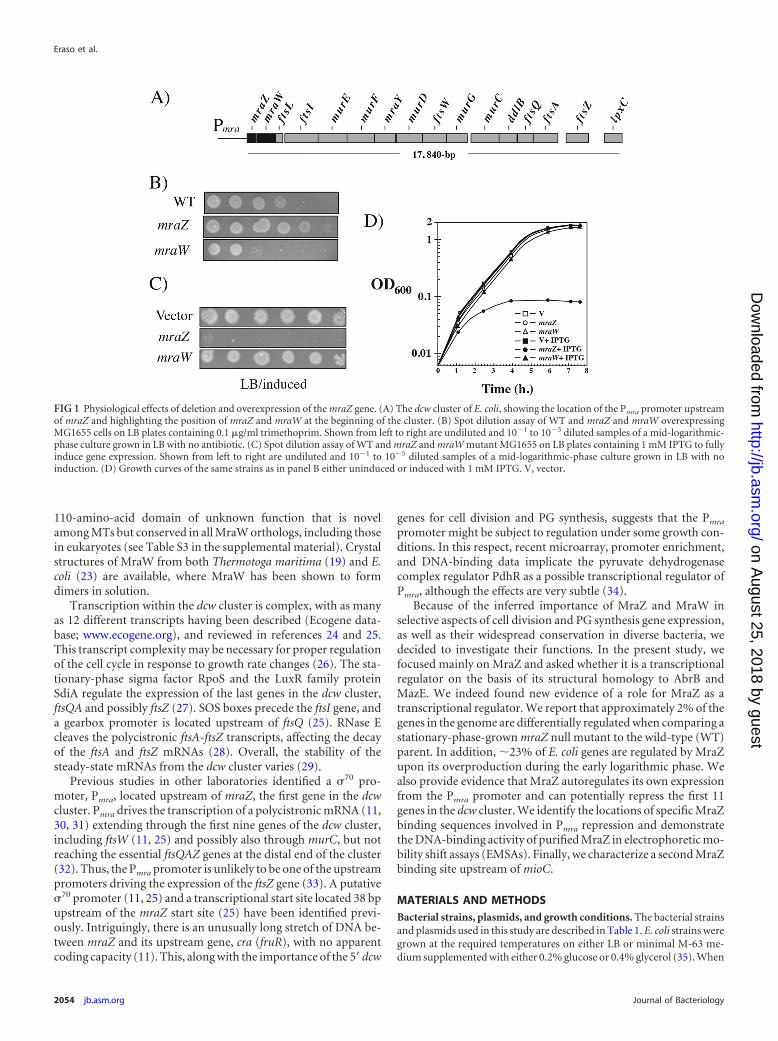
FIG 1 Physiological effects of deletion and overexpression of the mraZ gene. (A) The dcw cluster of E. coli, showing the location of the Pmra promoter upstreamof mraZ and highlighting the position of mraZ and mraW at the beginning of the cluster. (B) Spot dilution assay of WT and mraZ and mraW overexpressingMG1655 cells on LB plates containing 0.1 �g/ml trimethoprim. Shown from left to right are undiluted and 10�1 to 10�5 diluted samples of a mid-logarithmic-phase culture grown in LB with no antibiotic. (C) Spot dilution assay of WT and mraZ and mraW mutant MG1655 on LB plates containing 1 mM IPTG to fullyinduce gene expression. Shown from left to right are undiluted and 10�1 to 10�5 diluted samples of a mid-logarithmic-phase culture grown in LB with noinduction. (D) Growth curves of the same strains as in panel B either uninduced or induced with 1 mM IPTG. V, vector.
Eraso et al.
2054 jb.asm.org Journal of Bacteriology
on August 25, 2018 by guest
http://jb.asm.org/
Dow
nloaded from
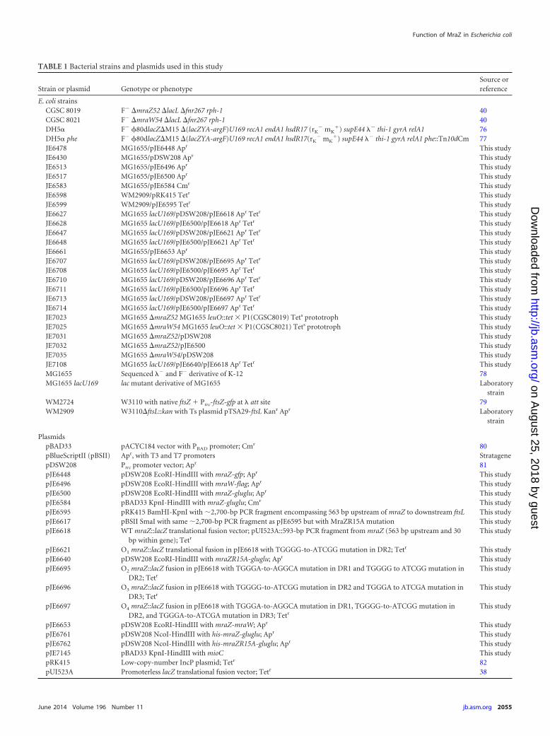
TABLE 1 Bacterial strains and plasmids used in this study
Strain or plasmid Genotype or phenotypeSource orreference
E. coli strainsCGSC 8019 F� �mraZ52 �lacL �fnr267 rph-1 40CGSC 8021 F� �mraW54 �lacL �fnr267 rph-1 40DH5� F� 80dlacZ�M15 �(lacZYA-argF)U169 recA1 endA1 hsdR17 (rK
� mK) supE44 �� thi-1 gyrA relA1 76
DH5� phe F� 80dlacZ�M15 �(lacZYA-argF)U169 recA1 endA1 hsdR17(rK� mK
) supE44 �� thi-1 gyrA relA1 phe::Tn10dCm 77JE6478 MG1655/pJE6448 Apr This studyJE6430 MG1655/pDSW208 Apr This studyJE6513 MG1655/pJE6496 Apr This studyJE6517 MG1655/pJE6500 Apr This studyJE6583 MG1655/pJE6584 Cmr This studyJE6598 WM2909/pRK415 Tetr This studyJE6599 WM2909/pJE6595 Tetr This studyJE6627 MG1655 lacU169/pDSW208/pJE6618 Apr Tetr This studyJE6628 MG1655 lacU169/pJE6500/pJE6618 Apr Tetr This studyJE6647 MG1655 lacU169/pDSW208/pJE6621 Apr Tetr This studyJE6648 MG1655 lacU169/pJE6500/pJE6621 Apr Tetr This studyJE6661 MG1655/pJE6653 Apr This studyJE6707 MG1655 lacU169/pDSW208/pJE6695 Apr Tetr This studyJE6708 MG1655 lacU169/pJE6500/pJE6695 Apr Tetr This studyJE6710 MG1655 lacU169/pDSW208/pJE6696 Apr Tetr This studyJE6711 MG1655 lacU169/pJE6500/pJE6696 Apr Tetr This studyJE6713 MG1655 lacU169/pDSW208/pJE6697 Apr Tetr This studyJE6714 MG1655 lacU169/pJE6500/pJE6697 Apr Tetr This studyJE7023 MG1655 �mraZ52 MG1655 leuO::tet � P1(CGSC8019) Tets prototroph This studyJE7025 MG1655 �mraW54 MG1655 leuO::tet � P1(CGSC8021) Tets prototroph This studyJE7031 MG1655 �mraZ52/pDSW208 This studyJE7032 MG1655 �mraZ52/pJE6500 This studyJE7035 MG1655 �mraW54/pDSW208 This studyJE7108 MG1655 lacU169/pJE6640/pJE6618 Apr Tetr This studyMG1655 Sequenced �� and F� derivative of K-12 78MG1655 lacU169 lac mutant derivative of MG1655 Laboratory
strainWM2724 W3110 with native ftsZ Ptrc-ftsZ-gfp at � att site 79WM2909 W3110�ftsL::kan with Ts plasmid pTSA29-ftsL Kanr Apr Laboratory
strain
PlasmidspBAD33 pACYC184 vector with PBAD promoter; Cmr 80pBlueScriptII (pBSII) Apr, with T3 and T7 promoters StratagenepDSW208 Ptrc promoter vector; Apr 81pJE6448 pDSW208 EcoRI-HindIII with mraZ-gfp; Apr This studypJE6496 pDSW208 EcoRI-HindIII with mraW-flag; Apr This studypJE6500 pDSW208 EcoRI-HindIII with mraZ-gluglu; Apr This studypJE6584 pBAD33 KpnI-HindIII with mraZ-gluglu; Cmr This studypJE6595 pRK415 BamHI-KpnI with �2,700-bp PCR fragment encompassing 563 bp upstream of mraZ to downstream ftsL This studypJE6617 pBSII SmaI with same �2,700-bp PCR fragment as pJE6595 but with MraZR15A mutation This studypJE6618 WT mraZ::lacZ translational fusion vector; pUI523A::593-bp PCR fragment from mraZ (563 bp upstream and 30
bp within gene); Tetr
This study
pJE6621 O1 mraZ::lacZ translational fusion in pJE6618 with TGGGG-to-ATCGG mutation in DR2; Tetr This studypJE6640 pDSW208 EcoRI-HindIII with mraZR15A-gluglu; Apr This studypJE6695 O2 mraZ::lacZ fusion in pJE6618 with TGGGA-to-AGGCA mutation in DR1 and TGGGG to ATCGG mutation in
DR2; Tetr
This study
pJE6696 O3 mraZ::lacZ fusion in pJE6618 with TGGGG-to-ATCGG mutation in DR2 and TGGGA to ATCGA mutation inDR3; Tetr
This study
pJE6697 O4 mraZ::lacZ fusion in pJE6618 with TGGGA-to-AGGCA mutation in DR1, TGGGG-to-ATCGG mutation inDR2, and TGGGA-to-ATCGA mutation in DR3; Tetr
This study
pJE6653 pDSW208 EcoRI-HindIII with mraZ-mraW; Apr This studypJE6761 pDSW208 NcoI-HindIII with his-mraZ-gluglu; Apr This studypJE6762 pDSW208 NcoI-HindIII with his-mraZR15A-gluglu; Apr This studypJE7145 pBAD33 KpnI-HindIII with mioC This studypRK415 Low-copy-number IncP plasmid; Tetr 82pUI523A Promoterless lacZ translational fusion vector; Tetr 38
Function of MraZ in Escherichia coli
June 2014 Volume 196 Number 11 jb.asm.org 2055
on August 25, 2018 by guest
http://jb.asm.org/
Dow
nloaded from
required, tetracycline (Tet) at 20 �g/ml, kanamycin (Kan) at 25 �g/ml,chloramphenicol (Cm) at 20 �g/ml, and ampicillin (Ap) at 150 �g/mlwere added to the growth medium. Cultures were grown anaerobically inanaerobic chambers with BD BBL Plus anaerobic system envelopes andindicators, and LB and minimal M-63 plates were supplemented with 20mM KNO3 as the terminal electron acceptor.
DNA manipulations and analysis. For the primers used in this study,see Table S1 in the supplemental material. Standard protocols or the man-ufacturer’s instructions were followed to isolate plasmid DNA, as well asfor restriction endonuclease, DNA ligase, PCR, and other enzymatic treat-ments of plasmids and DNA fragments. To eliminate potential impurities,DNA fragments were drop dialyzed against autoclaved water for 1 h on0.025-�m disc filters (Millipore, Billerica, MA) prior to ligation reactions.Enzymes were purchased from New England BioLabs, Inc. (Beverly, MA);Promega Corp. (Madison, WI); and Invitrogen (Carlsbad, CA). Plasmidsand DNA fragments were purified with the Wizard SV miniprep and PCRCleanup kits from Promega (Madison, WI). Phusion DNA polymerasewas used as the high-fidelity PCR enzyme (New England BioLabs). Thefinal versions of all relevant clones were verified by sequencing. Modifiedand unmodified oligonucleotides were purchased from Sigma-Aldrich.
Transcriptome sequencing (RNA-seq) experiments. Whole-genometranscriptome analyses were performed at two different stages of the cellcycle. Gene expression was assayed in an mraZ null mutant (JE7031)grown to a high cell density (optical density at 600 nm [OD600] of �1.4)and compared to that in the WT (JE6430). Ten-milliliter volumes of therespective cultures were spun, treated with RNAlater (Life Technologies,Grand Island, NY) by following the manufacturer’s instructions, frozen,and stored at �70°C prior to RNA extraction. In addition, gene expres-sion was also assayed in MraZ-overproducing cells (JE6517) and com-pared to that in WT cells containing the empty vector (JE6430); both weregrown to early log phase (OD600 of �0.1). Two-hundred-milliliter vol-umes of the respective cultures were processed in a manner similar to thatdescribed above. All cultures were grown in triplicate and processed in-dependently.
RNA extraction. RNA was extracted with Invitrogen TRIzol Reagent(catalog no. 15596018), followed by genomic DNA removal and cleaningwith the Qiagen RNase-Free DNase Set kit (catalog no. 79254) and theQiagen Mini RNeasy kit (catalog no. 74104). An Agilent 2100 Bioanalyzerwas used to assess the integrity of the RNA samples. Only RNA sampleswith integrity numbers between 8 and 10 were used.
RNA sequencing. The Applied Biosystems SOLiD Total RNA-Seq kit(catalog no. 4445374) was used to generate the cDNA template library.The SOLiD EZ Bead system was used to perform emulsion clonal beadamplification to generate bead templates for SOLiD platform sequencing.Samples were sequenced on the 5500XL SOLiD platform. The 50-baseshort-read sequences produced by the 5500XL SOLiD sequencer weremapped in color space with SOLiD LifeScope software version 2.5 byusing the default parameters against the genome of E. coli K-12 strainMG1655 (WIS_MG1655_m56) reference genome, and both the FASTAand GFF files were obtained from the E. coli Genome Project at the Univer-sity of Wisconsin—Madison (http://www.genome.wisc.edu/sequencing.htm). The output of the whole-transcriptome analysis generated a genecount file with the base counts summed to a single value across the entiregene length and with the number of reads per kilobase of transcript permillion mapped reads (RPKM) also given for each gene, a BAM file con-taining the sequence of every mapped read and its mapped location, onepair of *.wig files giving the mapped counts at each base position, and astatistical summary on alignment and filtering report.
Transcriptome and pathway enrichment analyses. RNA-seq datawere filtered to remove genes in the high- or low-OD sample sets withfewer than two replicates with nonzero counts per million for each con-dition. The bioconductor package edgeR (36) was used to identify thedegree of statistical differential expression between the WT and the mraZmutant, as well as WT and mraZ overexpression conditions, for each gene.Separate analyses of data from high- and low-OD600 conditions were con-
ducted. Genes were ranked according to the resulting P values, and theserankings were used as input to Gene Set Enrichment Analysis (GSEA)software (37). This algorithm identifies gene sets whose members are sta-tistically significantly enriched among genes at the top (or bottom) of a listof ranked genes. We used the “GSEA preranked” setting so that we couldenter our ranked list as direct input, thus bypassing the ranking step set byGSEA. As input, GSEA takes user-defined gene sets grouped according toareas of potential interest. For gene sets, we used the files “func-associa-tions.col” and “pathways.col” in the set of E. coli-specific files from theEcoCyc web resource (ecocyc.org). Gene sets were included only if thenumber of genes in the set present in the expression data was at least 10.
Microscopy. Cells expressing green fluorescent protein (GFP) ormCherry fusions were grown in LB plus antibiotics and embedded in 1%LB-agarose on a microscope slide. To image these cells, we used an Olym-pus BX60 fluorescence microscope equipped with a 100� numerical ap-erture 1.4 objective and GFP, 4=,6-diamidino-2-phenylindole (DAPI),and tetramethyl rhodamine isocyanate (for mCherry) filter sets. Grayscaleimages from each channel were captured with a Hamamatsu ORCA IIcharge-coupled device camera and HCImage software and imported intoAdobe Photoshop for pseudocoloring and image layering.
Construction of translational lacZ fusions and �-galactosidase as-says. Plasmid pJE6618 harbors the WT mraZ-lacZ translational fusion,which contains a 593-bp PCR fragment from the mraZ gene (563 bpupstream and 30 bp within the gene) fused to lacZ in pUI523A (38).Combinatorial PCR, as described previously (39), was used to mutate thethree direct repeats (DRs) in the mraZ operator to construct fusions O1,O2, O3, and O4 in plasmids pJE6621, pJE6695, pJE6696, and pJE6697,respectively (Table 1).
�-Galactosidase assays were performed as described elsewhere (38).The data provided are the averages of at least two separate experimentseach performed in duplicate. Standard deviations were always �15%.Protein concentrations of cell extracts were determined with the bicin-choninic acid (BCA) protein assay kit (Thermo Scientific, Waltham, MA)with bovine serum albumin as the standard.
P1 transductions. Standard protocols were used to mobilize geneticmarkers from existing strains by phage P1vir transductions (35). All of thestrains used are listed in Table 1. JE7023 (MG1655 mraZ) and JE7025(MG1655 mraW) were constructed by P1 transduction with CGSC8019and CGSC8021 (40), respectively, as donors and MG1655 leuO::Tn10 asthe recipient. After selecting for prototrophy and scoring for Tets, theresulting mraZ and mraW deletion mutant genes were PCR amplifiedfrom the chromosome of the transductants and verified by sequencing.
DNA labeling. The mraZ and mioC regulatory regions were PCR am-plified with biotinylated primers. A 240-bp mraZ biotinylated fragmentwas amplified with primers 1922 and 1923, whereas mioC was amplifiedwith primers 2045 and 2046 to form a 238-bp fragment. Competitor DNAfragments were also PCR amplified with nonbiotinylated primers with theexact same DNA sequences. The DNA amplification efficiencies were sim-ilar when using labeled and unlabeled primers. DNA concentrations werecalculated after reading the OD260s of several dilutions of the purifiedfragments with a UV-1601 UV-visible spectrophotometer (Shimadzu,Kyoto, Japan).
MraZ binding and competition assays. The final reactant concentra-tions in the 25-�l DNA-binding reaction mixtures were 1� binding buf-fer, 5 mM MgCl2, 0.04% Nonidet P-40 (NP-40), 2 mM spermidine trihy-drochloride, �0.35 pmol of a biotin-labeled double-stranded 240-bpmraZ or 238-bp mioC fragment, 40 ng/ml LightShift poly(dI · dC) non-specific competitor DNA (Thermo Scientific, Waltham, MA), water, andpurified MraZ in storage or dilution buffer at the appropriate concentra-tions (see recipes below). The presence of spermidine in the binding re-action mixtures increases the binding specificity for other transcriptionalregulators (41). The MraZ dilutions were done in 1� dilution buffer.After the addition of MraZ, the reaction mixtures were incubated at roomtemperature for 20 min, and immediately 4 �l (�1/6) of each reactionmixture was loaded into the prerun gels described in the next section. For
Eraso et al.
2056 jb.asm.org Journal of Bacteriology
on August 25, 2018 by guest
http://jb.asm.org/
Dow
nloaded from
each experiment, both gels in the electrophoresis tank contained identicalsamples and therefore were run in duplicate. For competition experi-ments, the specific competitor DNA at the appropriate concentration wasadded after MraZ addition, prior to incubation. The 10� MraZ bindingbuffer contained 200 mM Tris (pH 7.5), 50 mM KCl, 5 mM MgCl2, and 1mM dithiothreitol (DTT). The 1� MraZ dilution buffer contained 25mM Tris (pH 7.5), 150 mM KCl, 5 mM MgCl2, and 0.5 mM DTT. The10� gel loading buffer contained 250 mM Tris (pH 7.5), 0.2% bromophe-nol blue, and 40% glycerol.
Electrophoretic mobility shift assays (EMSAs). Electrophoresis wasperformed in Mini Protean 3 cells (Bio-Rad, Hercules, CA) with 5% gelsmade with a 40% stock of 29:1 acrylamide-bisacrylamide and 0.5� Tris-borate-EDTA (TBE). Gels were run at 4°C for approximately 20 min at 85V and for an additional 1 h at 60 V. Both the gels and the running buffercontained a final concentration of 200 �M spermidine. After electropho-resis, the DNA fragments were wet transferred in 0.5� TBE to Biodyne Bnylon membranes (Thermo Scientific, Waltham, MA) for 30 min at 385mA and UV-cross-linked with a UV Stratalinker 1800 (Agilent Technol-ogies, Santa Clara, CA) by using the auto-cross-link setting. The Chemi-luminescent Nucleic Acid Detection Module (Thermo Scientific, Wal-tham, MA) was used for detection in accordance with the manufacturer’sinstructions. The blots were exposed on Hyblot CL film (Denville Scien-tific Inc., South Plainfield, NJ).
Protein purifications. His6-MraZ-GluGlu and His6-MraZR15A-GluGlu were purified from 8 liters of MG1655 containing plasmidpJE6761 or pJE6762 (Table 1) encoding full-length WT or R15A mutantMraZ, respectively. These plasmids are derivatives of pDSW208, whichcontains an isopropyl-�-D-thiogalactopyranoside (IPTG)-inducible Ptrc
promoter. Cells were grown at 30°C for approximately 3 h to an OD600 of�0.6, induced with 1 mM IPTG for �90 min, and collected by centrifu-gation. Pellets were washed once in MraZ lysis buffer (25 mM Tris [pH7.5], 200 mM NaCl, 5 mM MgCl2) and stored at �70°C. Phenylmethyl-sulfonyl fluoride was used at a 1 mM final concentration. Cells were lysedby three passages through a French pressure cell (SLM Aminco, Roches-ter, NY). The cell lysates were clarified by centrifugation at 11,000 � g for20 min at 4°C and incubated on ice for 30 min in the presence of 2 mg/mllysozyme and DNase. TALON metal affinity resin (Clontech, MountainView, CA) was used for purifications by following manufacturer’s instruc-tions. Crude extracts were incubated with the resin for 2 h at 4°C prior tocolumn purification in the presence of 5 mM imidazole. After washes inMraZ lysis buffer containing 5, 20, and 30 mM imidazole and elution inMraZ lysis buffer with 500 mM imidazole, the proteins were dialyzedtwice at 4°C for �18 h against buffer I (25 mM Tris [pH 7.5], 150 mM KCl,5 mM MgCl2, 1 mM DTT, 20% glycerol, 100 mM NaCl) and then bufferII (same as buffer I but with no NaCl). The purified proteins were resus-pended in 100-�l aliquots and quick frozen in a dry-ice– ethanol bathprior to storage at �80°C. The BCA assay (Thermo Scientific, Rockford,IL) was used to determine protein concentration. The purification proce-dure yielded proteins that were �95% pure (data not shown).
Immunoblot analysis. Crude extracts and purified protein were re-suspended in SDS loading buffer, boiled for 10 min, and separated bySDS-PAGE in Mini Protean 3 cells (Bio-Rad, Hercules, CA) with 12 or18% gels made with a 40% stock of 29:1 acrylamide-bisacrylamide. Pro-teins were transferred to nitrocellulose membranes with a wet apparatus.In addition to the N-terminal His6 tag, a C-terminal GluGlu epitope (EEEVMPME) (42) was used to tag MraZ for Western blot analysis. Thesequence encoding the GluGlu tag was added by using combinatorial PCRas described previously (39). The DNA fragments were amplified from anmraZ-containing PCR product obtained by colony PCR. Plasmid pJE6500contains carboxy-terminally GluGlu-tagged full-length mraZ. The pri-mary antibodies used for immunoblotting were monoclonal anti-His6
(Sigma-Aldrich, St. Louis, MO) or monoclonal anti-GluGlu (CovanceResearch Products, Inc., Emeryville, CA) antibodies at 1:5,000. Anti-mouse secondary antibodies conjugated to horseradish peroxidase (HRP)
were used at 1:10,000. SuperSignal West Pico Chemiluminescent Sub-strate (Thermo Scientific) was used as the substrate for HRP detection.
Other reagents. o-Nitrophenyl-�-D-galactopyranoside, spermidinetrihydrochloride (minimum 98%), and antibiotics were purchased fromSigma Chemical Co., St. Louis, MO. NP-40 was purchased from ThermoScientific, Waltham, MA. GeneMate LE agarose was purchased from Bio-Express, Kaysville, UT. All of the other chemicals used in this work werereagent grade.
RESULTSPhenotypic analysis of cells after loss of MraZ or MraW. The dcwcluster (Fig. 1A) and its first two genes, mraZ and mraW, arehighly conserved in prokaryotes (9, 10). Using the web serverhmmer.janelia.org, we identified orthologs of MraZ in 2,184 outof 2,259 significant query matches within bacterial species (seeTable S2 in the supplemental material) and orthologs of MraW in4,184 out of 4,402 (see Table S3). Interestingly, MraW was alsofound in 161 eukaryote species (see Table S3), consistent with aprevious report of MraW homologs in eukaryotes (6). Thus, mraZand mraW are very highly conserved and MraW is more universal.
Despite their conservation and prevalence, nonpolar deletionsof mraZ and mraW have no detectable phenotypes under standardlaboratory conditions in E. coli (40, 43, 44) and B. subtilis (45). Weconfirmed that unmarked deletion mutations in both genes (40)in the MG1655 strain background are indeed nonessential for vi-ability and subsequently searched for conditional phenotypes as-sociated with the removal of these genes. The growth rates of cellslacking mraZ or mraW were indistinguishable from those of theWT parent during aerobic growth in LB at 37°C, anaerobic growthon LB with nitrate as the terminal electron acceptor, or both aer-obic and anaerobic growth on M-63 minimal medium supple-mented with glycerol as the sole carbon source (data not shown).Additionally, viability after a 12-day extended period in stationaryphase was not affected in the mraZ and mraW mutants comparedto the WT parent strain, although general viability was down by 2logs (data not shown).
We then searched for differences in antibiotic susceptibility.We found that the MICs for both mutants and the WT parentstrain were the same in the presence of Ap; the �-lactam amoxi-clav; the cephalosporin cephapirin; cefoxitin, which interfereswith cell wall synthesis; colistin, which alters both inner and outermembrane integrity; and the DNA gyrase inhibitor nalidixic acid.Notably, however, we did find that the mraZ mutant was moreresistant to the dihydrofolate reductase (DHFR) inhibitor trim-ethoprim than the WT parent (Fig. 1B). In contrast, the mraWmutant was more sensitive to trimethoprim than was the WTstrain, consistent with data from the Porteco website (Porteco.org).
MraZ overproduction is lethal and perturbs cell division.The difficulty in uncovering a growth phenotype for the mraZ ormraW null mutant prompted us to investigate the effect of mraZor mraW overexpression. Although overproduction of MraWby IPTG induction of plasmid pDSW208-Ptrc-mraW (calledpDSW208-mraW here) had no phenotype in rich (LB) growthmedium, overproduction of pDSW208-Ptrc-mraZ (called pDSW208-mraZ here) with IPTG induction was lethal (Fig. 1C). IPTGconcentrations of �50 �M prevented colony formation (data notshown). Cessation of growth after the overexpression of mraZ wasalso observed in growth curves (Fig. 1D). Notably, MraZ-over-producing cells filamented (see Fig. S1 in the supplemental mate-rial), suggesting that cell division is affected and may be a signifi-
Function of MraZ in Escherichia coli
June 2014 Volume 196 Number 11 jb.asm.org 2057
on August 25, 2018 by guest
http://jb.asm.org/
Dow
nloaded from
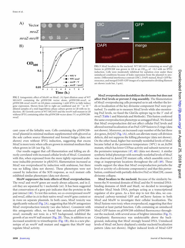
cant cause of the lethality seen. Cells containing the pDSW208-mraZ plasmid in minimal medium supplemented with glycerol asthe sole carbon source filamented and formed bulges (data notshown) even without IPTG induction, suggesting that excessMraZ is more toxic when cells are grown in minimal medium thanwhen grown in LB (see Fig. S2).
Our results suggest that cell filamentation and killing are di-rectly correlated with increased cellular levels of MraZ. Consistentwith this, when expressed from the more tightly repressed arabi-nose-inducible promoter in pBAD33, filamentation increased asMraZ was overproduced by induction with arabinose, but therewas no killing (data not shown). These phenotypes were notcaused by induction of the SOS response, as recA mutant cellsexhibited similar phenotypes (data not shown).
MraW suppresses the toxic effects of MraZ overproduction.The mraZ and mraW genes are nearly always adjacent, and in E.coli they are separated by 1 nucleotide (nt). It has been suggestedthat conservation of a gene pair indicates that the proteins in thepair interact (46). To test this notion, we co-overexpressed mraWand mraZ, either in cis in their native mraZ-mraW gene context orin trans on separate plasmids. In both cases, MraZ toxicity wassignificantly reduced (Fig. 2A), suggesting that MraW antagonizesMraZ overproduction toxicity (see also below). Consistent withthis antagonistic effect, we found that uninduced pDSW208-mraZ, normally not toxic in a WT background, inhibited thegrowth of an mraW null mutant (Fig. 2B). Thus, in addition to anincreased sensitivity to trimethoprim (Fig. 1B), this is a new phe-notype of an mraW null mutant and suggests that MraW mayregulate MraZ activity.
MraZ overproduction destabilizes the divisome but does notaffect FtsZ levels or prevent Z ring assembly. The filamentationof MraZ-overproducing cells prompted us to ask whether the lev-els or localization of the key divisome component FtsZ were per-turbed. To enable us to measure MraZ levels while also monitor-ing FtsZ levels, we fused the GluGlu epitope tag to the 3= end ofmraZ (Table 1 and Materials and Methods). This fusion conferredthe same overproduction phenotype as untagged MraZ. We foundthat MraZ overproduction did not affect cellular FtsZ levels andallowed normal localization of an FtsZ-GFP fusion to Z rings (datanot shown). Moreover, an increased copy number of the last threedcw genes, ftsQAZ (Fig. 1A), which can alleviate many cell divisiondefects, did not suppress the killing phenotype (data not shown).On the other hand, higher but normally nonlethal levels of MraZbecame lethal at the permissive temperature (30°C) in an ftsZ84mutant, which has lower GTPase activity and subunit turnover atthe permissive temperature (47, 48) (data not shown). A similarsynthetic lethal phenotype with normally nonlethal levels of MraZwas observed in �minCDE mutant cells, which assemble extra Zrings at inappropriate locations throughout the cell (49). Theseresults support the ideas that MraZ overproduction perturbs thelevel or activity of a divisome component(s) and that this pertur-bation, combined with partially defective FtsZ or MinCDE, causesdivisome collapse.
MraZ localizes to the nucleoid. Because of the similarity be-tween the N-terminal domain of MraZ and the N-terminal DNA-binding domains of AbrB and MazE, we decided to investigatewhether MraZ binds DNA, perhaps acting as a transcriptionalregulator of dcw genes. As a first step to test this idea, we con-structed carboxy-terminal GFP and mCherry fusions to bothMraZ and MraW to investigate their cellular localization. TheMraZ fusions were toxic when overproduced, suggesting that theyretained at least partial function. Cells containing an uninducedMraZ-GFP fusion on pDSW208 exhibited fluorescence through-out the nucleoid, with several areas of brighter intensities (Fig. 3).Cytoplasmic fluorescence was undetectable above the back-ground, indicating that MraZ strongly bound DNA. Uninducedlevels of MraZ-mCherry displayed a similar nucleoid localizationpattern (data not shown). Higher induced levels of MraZ-GFP
FIG 2 Antagonistic effect of MraW on MraZ. (A) Spot dilution assay of WTMG1655 containing the pDSW208 vector alone, pDSW208-mraZ, orpDSW208-mraZ-mraW on LB plates containing 1 mM IPTG to fully inducegene expression. Shown from left to right are undiluted and 10�1 to 10�5
diluted samples of a mid-logarithmic-phase culture grown in LB with no in-duction. (B) Growth curves of WT MG1655 and the mraW null mutant in LBwithout IPTG containing either the pDSW208 vector alone (V) or pDSW208-mraZ.
FIG 3 MraZ localizes to the nucleoid. WT MG1655 containing an mraZ-gfpfusion in pDSW208 was grown in LB to an OD600 of �0.2 with no IPTGinduction. Cells were moderately inhibited for septation even under theseuninduced conditions because of leaky expression from the plasmid trc pro-moter. Differential interference contrast (DIC), DAPI-stained, MraZ-GFP flu-orescence, and merged DAPI-GFP images of a representative dividing filamentare shown (scale bar, 3 �m).
Eraso et al.
2058 jb.asm.org Journal of Bacteriology
on August 25, 2018 by guest
http://jb.asm.org/
Dow
nloaded from
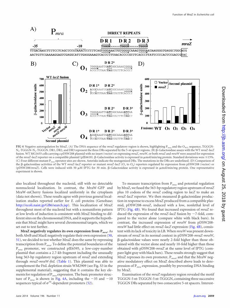
also localized throughout the nucleoid, still with no detectablenonnucleoid localization. In contrast, the MraW-GFP andMraW-mCherry fusions localized uniformly in the cytoplasm(data not shown). These results agree with previous general local-ization studies reported earlier for E. coli proteins (Genobase;http://ecoli.naist.jp/GB6/search.jsp). This localization of MraZthroughout most of the nucleoid but with a nonuniform patternat low levels of induction is consistent with MraZ binding to dif-ferent sites on the chromosomal DNA, and it supports the hypoth-esis that MraZ might have several chromosomal targets, which weset out to test further.
MraZ negatively regulates its own expression from Pmra. Asboth AbrB and MazE negatively regulate their own expression (50,51), we decided to test whether MraZ does the same by repressingtranscription from Pmra. To define the potential boundaries of thePmra promoter, we constructed pJE6595, a low-copy-numberplasmid that contains a 2.7-kb fragment including the unusuallylong 563-bp regulatory region upstream of mraZ and extendingthrough mraZ-mraW-ftsL (Table 1). This plasmid was able tocomplement the FtsL depletion strain WM2909 (see Fig. S3 in thesupplemental material), suggesting that it contains the key ele-ments for regulation of Pmra expression. The basic promoter struc-ture of Pmra is shown in Fig. 4A, including the �35 and �10sequences typical of �70-dependent promoters (52).
To measure transcription from Pmra and potential regulationby MraZ, we fused the 563-bp regulatory region upstream of mraZplus 10 codons of the mraZ coding region to lacZ to make anmraZ-lacZ reporter. We then measured �-galactosidase produc-tion in response to excess MraZ produced from a compatible plas-mid, pDSW208-mraZ, induced with a low, nonlethal level ofIPTG (Fig. 4B). We found that increased expression of mraZ re-duced the expression of the mraZ-lacZ fusion by �7-fold, com-pared to the vector alone (compare white with black bars). Incontrast, the increased expression of mraW from pDSW208-mraW had little effect on mraZ-lacZ expression (Fig. 4B), consis-tent with its lack of toxicity in LB. When mraW was present down-stream of mraZ in its normal context in pDSW208-mraZ-mraW,�-galactosidase values were nearly 2-fold higher than those ob-tained with the vector alone and nearly 10-fold higher than thoseobtained with pDSW208-mraZ at the same level of IPTG (com-pare light gray with black bars). These results strongly suggest thatMraZ represses its own promoter, Pmra, and that the MraW neg-ative modulatory effect on MraZ described above leads to dere-pression of Pmra expression, possibly by preventing DNA bindingby MraZ.
Examination of the mraZ regulatory region revealed the motifTGGGN-5 nt-TGGGN-5 nt-TGGGN, containing three successiveTGGGN DRs separated by two consecutive 5-nt spacers. Interest-
FIG 4 Negative autoregulation by MraZ. (A) The DNA sequence of the mraZ regulatory region is shown, highlighting Pmra and the OWT sequence, TGGGN-N5-TGGGN-N5-TGGGN. DR1, DR2, and DR3 represent the three DRs separated by the 5-nt spacer regions. (B) �-Galactosidase assays with the WT mraZ-lacZfusion. WT MG1655 cells carrying a pDSW208 plasmid with no insert (vector) or expressing mraZ, mraW, or both mraZ and mraW were assayed for expressionof the mraZ-lacZ reporter on a compatible plasmid (pJE6618). �-Galactosidase activity is expressed in �mol/min/mg protein. Standard deviations were �15%.(C) Four different mutant Pmra operator sites are shown. Asterisks indicate the mutagenized DRs. The mutations in the DRs are underlined. (D) Comparison ofthe �-galactosidase activities of the WT mraZ-lacZ reporter or mutant mraZ-lacZ (O1 to O4) reporters regulated by expression from pDSW208 (vector) or(pDSW208/mraZ). Cells were induced with 50 �M IPTG for 30 min. �-Galactosidase activity is expressed in �mol/min/mg protein. One representativeexperiment is shown.
Function of MraZ in Escherichia coli
June 2014 Volume 196 Number 11 jb.asm.org 2059
on August 25, 2018 by guest
http://jb.asm.org/
Dow
nloaded from
ingly, this repeat resembles the TGGNA motif involved in DNArecognition by AbrB in B. subtilis (53). As this repeat region islocated 8 nt downstream from the Pmra promoter (Fig. 4A), it isconsistent with the idea that these DRs constitute an operator sitefor MraZ binding.
To test this hypothesis, we constructed additional mraZ-lacZfusions containing mutations in the TGGGN DRs singly or incombination. We called the WT sequence operator OWT and themutant sequences O1 to O4 (Fig. 4C). The expression results areshown in Fig. 4D; in general, the mutant mraZ-lacZ fusions werederepressed, even when mraZ was overexpressed, compared to theWT fusion. Interestingly, the effect of mutation of the DRs is ad-ditive; the lacZ fusion to the O1 operator, in which the middle DR(DR2) is mutated, was still repressed, but the lacZ fusion to the O4
mutant operator, in which all of the DRs are mutated, was totallyderepressed, irrespective of whether MraZ was overproduced ornot. Thus, MraZ represses the expression of Pmra and the mraZgene and the DRs constitute DNA elements necessary for this re-pression. As we cannot rule out the possibility that promoter func-tion and/or a transcription start site(s) may have been perturbedas a consequence of the mutagenesis, we next tested DNA bindingwith purified MraZ (see below).
A point mutation in the putative DNA-binding domain ofMraZ eliminates its toxicity. If MraZ is a transcriptional regula-tor, then the killing phenotype observed upon its overproductionis most likely caused by inappropriate regulation of gene expres-sion at the OWT operator located upstream of mraZ and/or atadditional genomic targets. Thus, overproduction of a mutantMraZ protein unable to bind to DNA should not be toxic. Argi-nine 15 (R15) in MraZ is conserved in AbrB from B. subtilis (14,15) and MazE from E. coli (16). An R16A mutation at the equiv-alent location in MazE abolishes DNA binding (16, 51). R15 ofMraZ forms part of the first DXXXR DNA-binding motif of MraZpresent in the two tandem homologous copies of the UPF0040fold (Fig. 5A) (6, 18). We therefore decided to mutate this argi-nine, predicting that it would abolish MraZ binding to DNA.
We found that MraZR15A is not toxic to cells, even when over-produced by full IPTG induction, in contrast to the WT protein(Fig. 5B). The stability of the MraZR15A mutant protein is similarto that of WT MraZ under uninduced and induced conditions(Fig. 5C), suggesting that the mutation inactivates a specific func-tion of MraZ. We calculate that there was an �10-fold increase inprotein concentration in induced versus uninduced cells, with thecaveat that MraZ was lethal to cells at this concentration of IPTG.
FIG 5 Characterization of the MraZR15A DNA-binding mutant. (A) Sequence of the MraZ protein from E. coli, representing the two copies of the UPF0040 fold(19), including amino acids 1 to 76 (top row) and 77 to 152 (bottom row). The two DXXXR motifs are highlighted; they correspond to amino acids D11 to R15and D87 to R91. Arginine R15 (shaded square) in the first motif was mutated to alanine in the MraZR15A mutant. (B) Spot dilution assay of WT MG1655containing the pDSW208 vector, pDSW208-mraZ, or pDSW208-mraZR15A on LB plates containing 1 mM IPTG to fully induce gene expression. (C) Levels ofMraZR15A are similar to those of MraZ. WT MG1655 cells containing the pDSW208 vector (lane 1), pDSW208-mraZ-gluglu (lanes 2 and 3), or pDSW208-mraZR15A-gluglu (lanes 4 and 5) were grown in LB to an OD600 of �0.2. Cells expressing mraZ on the plasmid were either left uninduced (lanes 2 and 4) orinduced (lanes 3 and 5) with 1 mM IPTG and grown for an additional 2 h. Aliquots were normalized for total protein and subjected to SDS-PAGE. Blots weredetected with anti-GluGlu antibodies. The location of 20 Kd is where the 20-kDa molecular size marker runs. (D) �-Galactosidase assays comparing the effectsof pDSW208 (vector), (pDSW208/mraZ), and (pDSW208/mraZR15A) on the mraZ-lacZ reporter after induction with 50 �M IPTG for 30 min. �-Galactosidaseactivity is expressed in �mol/min/mg protein. One representative experiment is shown.
Eraso et al.
2060 jb.asm.org Journal of Bacteriology
on August 25, 2018 by guest
http://jb.asm.org/
Dow
nloaded from
In addition, the expression of an mraZ-lacZ fusion is not repressedby the overproduction of MraZR15A, in contrast to that of WTMraZ (Fig. 5D). These results indicate that the killing phenotypeobserved upon MraZ overproduction probably results from inap-propriate regulation of gene expression because of DNA bindingand, importantly, is not a nonspecific effect of protein overpro-duction.
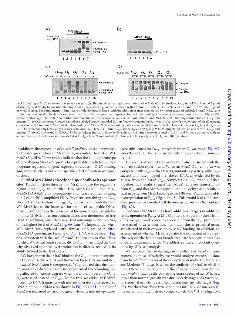
Purified MraZ binds directly and specifically to its operatorsites. To demonstrate directly that MraZ binds to the regulatoryregion near Pmra, we purified His6-MraZ-GluGlu and His6-MraZR15A-GluGlu to homogeneity and measured their bindingto a 240-bp PCR-amplified DNA fragment containing the OWT
DRs by EMSAs. As shown in Fig. 6A, increasing concentrations ofWT MraZ led to the increased formation of two stable DNA-protein complexes in the presence of the noncompetitive inhibi-tor poly(dI · dC) and a concomitant decrease in the amount of freeDNA. In addition, unlabeled OWT DNA outcompeted the bindingof the highest level of MraZ (Fig. 6A, lane 7). Importantly, whenWT MraZ was replaced with similar amounts of purifiedMraZR15A protein, no binding to OWT DNA was detected (Fig.6B), consistent with the lack of MraZR15A toxicity in vivo. Thus,purified WT MraZ binds specifically to OWT in vitro, and the tox-icity observed upon its overproduction is directly related to itsability to bind to its DNA site(s).
We have shown that MraZ binds to the OWT operator contain-ing three consecutive DRs and that when these DRs are mutated,the mraZ-lacZ fusion is derepressed. We predicted that the dere-pression was a direct consequence of impaired DNA binding, be-ing affected to various degrees when the mutant operators O1 toO4 were used instead of OWT. To test this, we added WT MraZprotein to DNA fragments with mutant operators and measuredDNA binding in EMSAs. As shown in Fig. 6C and D, binding ofMraZ was impaired to various degrees when the mutant operators
were substituted for OWT, especially when O4 was used (Fig. 6C,lanes 9 and 10). This is consistent with the mraZ-lacZ fusion ac-tivities.
The results of competition assays were also consistent with thereporter fusion experiments. When an MraZ-OWT complex wascompeted with OWT or the O1 to O4 mutants separately, only OWT
successfully outcompeted the labeled DNA, as evidenced by itsrelease from the MraZ-OWT complex (Fig. 6D, lane 2). Takentogether, our results suggest that MraZ represses transcriptionfrom Pmra and that MraZ overproduction toxicity might result, atleast in part, from MraZ binding to the DRs near Pmra and possiblyoverrepression of Pmra (Fig. 4 and 5). This would lead to the un-derexpression of essential cell division genes such as ftsL and ftsI(Fig. 1A).
Evidence that MraZ may have additional targets in additionto the operator at Pmra. As MraZ binds to the operator site in frontof its own gene and represses expression from the Pmra promoter,we wanted to determine how many dcw cluster-proximal genesare affected in their expression by MraZ binding. In addition, anassessment of whether MraZ regulates the expression of Pmra ex-clusively or whether it has a broader regulatory spectrum was alsoof paramount importance. We addressed these important ques-tions by RNA-seq analysis.
We reasoned that to distinguish the effects of MraZ on geneexpression most effectively, we would analyze expression datafrom two different stages of the cell cycle as described in Materialsand Methods. This was based on the similarity of MraZ to AbrB intheir DNA-binding region and the aforementioned observationthat mraW mutant cells containing extra copies of mraZ have alower than normal growth rate during early stages of growth be-fore normal growth is resumed during later growth stages (Fig.2B). We therefore chose two conditions for RNA-seq analysis, (i)the mraZ null mutant strain compared with the WT at a high cell
FIG 6 Binding of MraZ to the mraZ regulatory region. (A) Binding of increasing concentrations of WT MraZ to biotinylated OWT in EMSAs. About 0.3 pmolof a biotinylated 240-bp fragment containing the mraZ regulatory region was incubated with 3.2 (lane 2), 6.4 (lane 3), 10.7 (lane 4), 32 (lane 5), or 96 (lane 6) pmolof MraZ protein. The components in lane 7 were similar to those in lane 6, with the addition of an approximately 8� molar excess of unlabeled mraZ DNA. Lane1 is biotinylated mraZ DNA alone. Complexes 1 and 2 are the two specific complexes observed. (B) Binding of increasing concentrations of mutant MraZR15Ato biotinylated OWT. The protein concentrations were similar to those in panel A. Lane 1 is biotinylated mraZ DNA alone. (C) Binding of MraZ to WT (OWT) andmutant (O1 to O4) operators. About 0.3 pmol of a labeled double-stranded 240-bp fragment containing OWT was incubated with �0.09 nmol of MraZ protein,equivalent to the amount of MraZ used in lane 6 of panel A (lane 2). The mutant operators were incubated similarly (O1, lane 4; O2, lane 6; O3, lane 8; O4, lane10). The corresponding DNA-only lanes are as follows: OWT, lane 1; O1, lane 3; O2, lane 5; O3, lane 7; O4, lane 9. (D) Competition with unlabeled WT (OWT) andmutant (O1 to O4) operators. MraZ-OWT DNA complexes similar to those depicted in panel A, lane 6 (shown in lanes 1, 3, 5, 7, and 9), were competed with anapproximately 8� molar excess of unlabeled WT (OWT, lane 2) and mutant (O1, lane 4; O2, lane 6; O3, lane 8; O4, lane 10) operators.
Function of MraZ in Escherichia coli
June 2014 Volume 196 Number 11 jb.asm.org 2061
on August 25, 2018 by guest
http://jb.asm.org/
Dow
nloaded from
density and (ii) the WT strain overproducing MraZ comparedwith the WT in the early log phase. Overproduction of MraZ-GluGlu was confirmed by reverse transcription-PCR and immu-noblotting with anti-GluGlu antibodies, similar to the data shownin Fig. 5C.
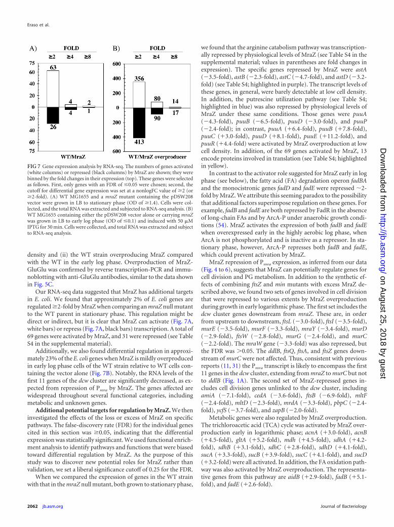
Our RNA-seq data suggested that MraZ has additional targetsin E. coli. We found that approximately 2% of E. coli genes areregulated �2-fold by MraZ when comparing an mraZ null mutantto the WT parent in stationary phase. This regulation might bedirect or indirect, but it is clear that MraZ can activate (Fig. 7A,white bars) or repress (Fig. 7A, black bars) transcription. A total of69 genes were activated by MraZ, and 31 were repressed (see TableS4 in the supplemental material).
Additionally, we also found differential regulation in approxi-mately 23% of the E. coli genes when MraZ is mildly overproducedin early log phase cells of the WT strain relative to WT cells con-taining the vector alone (Fig. 7B). Notably, the RNA levels of thefirst 11 genes of the dcw cluster are significantly decreased, as ex-pected from repression of Pmra by MraZ. The genes affected arewidespread throughout several functional categories, includingmetabolic and unknown genes.
Additional potential targets for regulation by MraZ. We theninvestigated the effects of the loss or excess of MraZ on specificpathways. The false-discovery rate (FDR) for the individual genescited in this section was �0.05, indicating that the differentialexpression was statistically significant. We used functional enrich-ment analysis to identify pathways and functions that were biasedtoward differential regulation by MraZ. As the purpose of thisstudy was to discover new potential roles for MraZ rather thanvalidation, we set a liberal significance cutoff of 0.25 for the FDR.
When we compared the expression of genes in the WT strainwith that in the mraZ null mutant, both grown to stationary phase,
we found that the arginine catabolism pathway was transcription-ally repressed by physiological levels of MraZ (see Table S4 in thesupplemental material; values in parentheses are fold changes inexpression). The specific genes repressed by MraZ were astA(�3.5-fold), astB (�2.3-fold), astC (�4.7-fold), and astD (�3.2-fold) (see Table S4; highlighted in purple). The transcript levels ofthese genes, in general, were barely detectable at low cell density.In addition, the putrescine utilization pathway (see Table S4;highlighted in blue) was also repressed by physiological levels ofMraZ under these same conditions. Those genes were puuA(�4.3-fold), puuB (�6.5-fold), puuD (�3.0-fold), and puuP(�2.4-fold); in contrast, puuA (6.4-fold), puuB (7.8-fold),puuC (3.0-fold), puuD (8.1-fold), puuE (11.2-fold), andpuuR (4.4-fold) were activated by MraZ overproduction at lowcell density. In addition, of the 69 genes activated by MraZ, 13encode proteins involved in translation (see Table S4; highlightedin yellow).
In contrast to the activator role suggested for MraZ early in logphase (see below), the fatty acid (FA) degradation operon fadBAand the monocistronic genes fadD and fadE were repressed �2-fold by MraZ. We attribute this seeming paradox to the possibilitythat additional factors superimpose regulation on these genes. Forexample, fadB and fadE are both repressed by FadR in the absenceof long-chain FAs and by ArcA-P under anaerobic growth condi-tions (54). MraZ activates the expression of both fadB and fadEwhen overexpressed early in the highly aerobic log phase, whenArcA is not phosphorylated and is inactive as a repressor. In sta-tionary phase, however, ArcA-P represses both fadB and fadE,which could prevent activation by MraZ.
MraZ repression of Pmra expression, as inferred from our data(Fig. 4 to 6), suggests that MraZ can potentially regulate genes forcell division and PG metabolism. In addition to the synthetic ef-fects of combining ftsZ and min mutants with excess MraZ de-scribed above, we found two sets of genes involved in cell divisionthat were repressed to various extents by MraZ overproductionduring growth in early logarithmic phase. The first set includes thedcw cluster genes downstream from mraZ. These are, in orderfrom upstream to downstream, ftsL (�3.0-fold), ftsI (�3.5-fold),murE (�3.5-fold), murF (�3.3-fold), mraY (�3.4-fold), murD(�2.9-fold), ftsW (�2.8-fold), murG (�2.4-fold), and murC(�2.2-fold). The mraW gene (�3.3-fold) was also repressed, butthe FDR was 0.05. The ddlB, ftsQ, ftsA, and ftsZ genes down-stream of murC were not affected. Thus, consistent with previousreports (11, 31) the Pmra transcript is likely to encompass the first11 genes in the dcw cluster, extending from mraZ to murC but notto ddlB (Fig. 1A). The second set of MraZ-repressed genes in-cludes cell division genes unlinked to the dcw cluster, includingamiA (�7.1-fold), cedA (�3.6-fold), ftsB (�6.9-fold), mltF(�2.4-fold), mltD (�2.3-fold), mrdA (�3.3-fold), pbpC (�2.4-fold), ycfS (�3.7-fold), and zapB (�2.0-fold).
Metabolic genes were also regulated by MraZ overproduction.The trichloroacetic acid (TCA) cycle was activated by MraZ over-production early in logarithmic phase; acnA (3.0-fold), acnB(4.5-fold), gltA (5.2-fold), mdh (4.5-fold), sdhA (4.2-fold), sdhB (3.1-fold), sdhC (2.8-fold), sdhD (4.1-fold),sucA (3.3-fold), sucB (3.9-fold), sucC (4.1-fold), and sucD(3.2-fold) were all activated. In addition, the FA oxidation path-way was also activated by MraZ overproduction. The representa-tive genes from this pathway are aidB (2.9-fold), fadB (5.1-fold), and fadE (2.6-fold).
FIG 7 Gene expression analysis by RNA-seq. The numbers of genes activated(white columns) or repressed (black columns) by MraZ are shown; they werebinned by the fold changes in their expression (top). These genes were selectedas follows. First, only genes with an FDR of �0.05 were chosen; second, thecutoff for differential gene expression was set at a nonlogFC value of �2 (or�2-fold). (A) WT MG1655 and a mraZ mutant containing the pDSW208vector were grown in LB to stationary phase (OD of �1.4). Cells were col-lected, and the total RNA was extracted and subjected to RNA-seq analysis. (B)WT MG1655 containing either the pDSW208 vector alone or carrying mraZwas grown in LB to early log phase (OD of �0.1) and induced with 50 �MIPTG for 30 min. Cells were collected, and total RNA was extracted and subjectto RNA-seq analysis.
Eraso et al.
2062 jb.asm.org Journal of Bacteriology
on August 25, 2018 by guest
http://jb.asm.org/
Dow
nloaded from
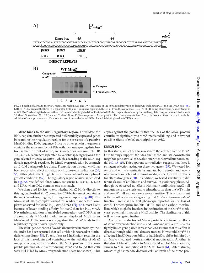
MraZ binds to the mioC regulatory region. To validate theRNA-seq data further, we inspected differentially expressed genesby scanning their regulatory regions for the presence of a putativeMraZ-binding DNA sequence. Since no other gene in the genomecontains the same number of DRs with the same spacing distribu-tion as that in front of mraZ, we searched for any multiple DRT-G-G-G-N sequences separated by variable spacing regions. Onegene selected this way was mioC, which, according to the RNA-seqdata, is negatively regulated by MraZ overproduction by as muchas 12-fold during early log phase. Transcription through mioC hasbeen reported to affect initiation of chromosome replication (55,56), although its effect might be more prevalent under suboptimalgrowth conditions (57). The regulatory region of mioC is depictedin Fig. 8A. We defined three MraZ consensus DRs as DR1, DR2and DR3, where DR2 contains one mismatch.
We then used EMSAs to test whether MraZ binds directly tothis region. Purified MraZ bound to a 238-bp fragment containingthe mioC regulatory region, forming one complex (Fig. 8B). TheMraZ-mioC DNA complex formed less readily than the two com-plexes observed for MraZ-Pmra-mraZ DNA (Fig. 6A), most likelybecause of lower binding affinity for mioC than for Pmra-mraZ.Nevertheless, addition of unlabeled competitor mioC DNA at anapproximately �10-fold molar excess displaced MraZ fromMraZ-mioC DNA complexes, suggesting that MraZ binds to theregulatory region of mioC specifically.
The mioC gene encodes a flavodoxin involved in biotin synthe-sis, and it has been reported that cell division is retarded in biotin-deficient medium (58). To test if excess MraZ might result in thelack of this flavodoxin and contribute to the lethality of MraZoverproduction, we overproduced the MioC protein from a com-patible plasmid while overproducing MraZ and found that cellswere still killed by MraZ overproduction (data not shown). This
argues against the possibility that the lack of the MioC proteincontributes significantly to MraZ-mediated killing, and in favor ofpossible effects of mioC transcription on oriC.
DISCUSSION
In this study, we set out to investigate the cellular role of MraZ.Our findings support the idea that mraZ and its downstreamneighbor gene, mraW, are evolutionarily conserved but nonessen-tial (40, 43–45). This apparent contradiction suggests that there isstringent selection acting on these two genes (59). We tested formraZ and mraW essentiality by assaying both aerobic and anaer-obic growth in rich and minimal media, as performed by othersfor alternative genes (60). In addition, we tested sensitivity to dif-ferent classes of antibiotics and survival in stationary phase. Al-though we observed no effects with many antibiotics, mraZ nullmutants were more resistant to trimethoprim than the WT strainand mraW null mutants were more sensitive. This is consistentwith our other evidence suggesting that MraW antagonizes MraZfunction, and it is the first phenotype reported for the loss ofmraZ. Trimethoprim inhibits DHFR and one-carbon metabo-lism, which might be involved in the function of the MraW meth-ylase, potentially impacting MraZ activity. The significance of thiswill be investigated further.
As co-overproduction of MraW protects cells from the effectsof MraZ overproduction in vivo and mraZ and mraW are usually atightly linked gene pair, it is reasonable to assume that this effect isdirect, although additional data are needed. How could MraW beaffecting MraZ? One possibility is that MraZ could be inhibited byan MraW-mediated posttranslational modification. Another isthat direct MraW binding to MraZ could inhibit MraZ activity,similar to MazE inhibition of the MazF toxin (61). Alternatively,MraW might somehow decrease cellular levels of the MraZ pro-
FIG 8 Binding of MraZ to the mioC regulatory region. (A) The DNA sequence of the mioC regulatory region is shown, including PmioC and the DnaA box (56).DR1 to DR3 represent the three DRs separated by 8- and 5-nt spacer regions. DR2 is 1 nt from the consensus TGGGN. (B) Binding of increasing concentrationsof WT MraZ to biotinylated mioC. About 0.3 pmol of a biotinylated double-stranded 238-bp fragment containing the mioC regulatory region was incubated with3.2 (lane 2), 6.4 (lane 3), 10.7 (lane 4), 32 (lane 5), or 96 (lane 6) pmol of MraZ protein. The components in lane 7 were the same as those in lane 6, with theaddition of an approximately 10� molar excess of unlabeled mioC DNA. Lane 1 is biotinylated mioC DNA only.
Function of MraZ in Escherichia coli
June 2014 Volume 196 Number 11 jb.asm.org 2063
on August 25, 2018 by guest
http://jb.asm.org/
Dow
nloaded from
tein. Any of these scenarios might result in more mraW gene ex-pression because of the loss of MraZ repression at Pmra, whichwould induce a positive feedback loop. Irrespective of the mech-anism, our results suggest that MraZ and MraW regulate eachother and argue that MraW is above MraZ in the putative regula-tory cascade.
What might be the molecular mechanism of MraZ transcrip-tional regulation? We have observed high-molecular-weightMraZ multimers in denaturing SDS gels with purified MraZ-Glu-Glu (data not shown), consistent with the reported dodecamericstructure of E. coli MraZ in solution (18). Dodecameric transcrip-tional regulators from bacteria have rarely been reported. Threeknown examples adopting this quaternary structure are anti-TRAP (trp RNA-binding attenuation protein) in Bacillus species(62), the YjiE hypochlorite-specific transcription factor (63), andthe Dps DNA condensation protein (64), the latter two of whichare both from E. coli. If MraZ indeed binds to DNA as a dodeca-mer, it might wrap the DNA around its toroidal structure similarto the E. coli transcriptional repressor RcnR (65) and DNA gyrase(66). In addition, as a dodecamer, six times more protein would benecessary for DNA binding than for a dimer, which could explainthe relatively high MraZ concentration needed to detect bandshifts in our EMSAs. Alternatively, MraZ may assemble into poly-mers of dimers similar to MazE (67) or ArcA-P, the latter beingable to occupy two, three, and even four consecutive DR se-quences separated by variable spacer regions, all located on oneside of the helix (68). In our experiments, the existence of twodistinct MraZ-OWT complexes would be consistent with the bind-ing of two MraZ dimers to the operator site.
The three TGGGN repeats in the mraZ OWT operator are sep-arated by 5-nt spacers, indicating that the repeats face the sameside of the DNA helix; similarly, AbrB also binds to residues lo-cated on one side of the helix (50). Thus far, we have not been ableto define a consensus DNA-binding sequence for MraZ, as thenumber and distribution of DRs are highly variable in all of thegenes examined, similarly to AbrB DNA-binding regions (15, 69).AbrB, which integrates environmental and metabolic informationto minimize inappropriate gene expression during log phase (70),shows similarity to MraZ in its “looped-hinge helix fold” DNA-binding domain. Therefore, MraZ might use a similar strategy forDNA recognition, allowing it to bind unrelated DNA sequences ina specific manner. It may even act like AbrB, with higher or loweractivity, depending on the growth phase.
The toxicity from MraZ overproduction was more pronouncedin minimal medium. When E. coli cells grow in minimal glycerolmedium, they exhibit a carbon stress response (71). Cells readjusttheir metabolism by using the transcriptional regulators Cra(FruR), which, among its other functions, downregulates the TCAcycle (72); the CRP-cyclic AMP complex; and the ArcBA two-component system. Our RNA-seq data showed that MraZ over-production represses cra transcription by 4.4-fold, either directlyor indirectly, and as would be expected, upregulates TCA cyclegenes. Thus, altered expression of TCA cycle genes, which hasbeen shown to involve other regulators (73), might be partly re-sponsible for the sensitivity to MraZ overproduction under theseparticular growth conditions.
MraZ, at physiological levels, has the potential to autoregulateits expression and that of the first 11 genes in the dcw cluster,including mraW, by binding to the OWT operator at Pmra. Suchrepression by MraZ might limit the amount of some essential cell
division and/or PG biosynthesis proteins, and this is the likelycause of cell division inhibition after the overproduction of MraZ.Despite this phenotype, an mraZ null mutant shows no significantchanges in dcw gene expression, at least in stationary phase. Thissuggests that MraZ may be more active as a repressor during stressconditions or other stages of growth, and/or MraZ function isredundant with another transcriptional regulator.
Although they do not affect dcw transcription in stationaryphase, it is important to emphasize that physiological levels ofMraZ do repress the transcription of arginine and polyamine ca-tabolism, the ast and puu genes, respectively, and activate othergenes encoding proteins involved in translation. This indicatesthat increases in polyamine concentration and some regulation oftranslation in stationary phase might be a consequence of MraZactivity. These differences in gene expression between an mraZnull mutant and the isogenic WT parent suggest that MraZ mighthave a role in cell adaptation or survival in suboptimal environ-ments or under suboptimal growth conditions, as has been pro-posed for the MazEF toxin-antitoxin system (74, 75).
In summary, here we show that MraZ, the product of thehighly conserved mraZ gene, acts as a transcriptional regulator inE. coli. By binding to a DNA sequence organized with differentcombinations of TGGGN DRs located immediately downstreamfrom the Pmra promoter, MraZ represses its own expression andthat of the 10 subsequent genes in the proximal part of the dcwcluster. In addition, overproduction of MraZ inhibits cell division,but co-overproduction with MraW suppresses MraZ toxicity,suggesting that MraZ and MraW may have antagonistic functions.Finally, several lines of evidence, including MraZ localizationthroughout the nucleoid, activation or repression of multiplegenes by RNA-seq, and in vitro binding to the mioC gene nearoriC, suggest that MraZ may also regulate the expression of genesoutside the dcw cluster and perhaps influence other cell cycle func-tions.
ACKNOWLEDGMENTS
We thank members of the Margolin laboratory, David Bates, and GeorgePhillips for helpful discussions; Christopher C. Overall for help withbioinformatic analysis; and Audrey Wanger for generously providing an-tibiotic Etest strips.
A portion of the research described here was performed with EMSL, anational scientific user facility sponsored by the Department of Energy’sOffice of Biological and Environmental Research and located at PacificNorthwest National Laboratory. The remainder of the research was sup-ported by NIH grant GM61074 to W.M.
REFERENCES1. Blasco B, Pisabarro AG, de Pedro MA. 1988. Peptidoglycan biosynthesis
in stationary-phase cells of Escherichia coli. J. Bacteriol. 170:5224 –5228.2. Clark DJ. 1968. The regulation of DNA replication and cell division in E.
coli B-r. Cold Spring Harbor Symp. Quant. Biol. 33:823– 838. http://dx.doi.org/10.1101/SQB.1968.033.01.094.
3. Lee YS, Han JS, Jeon Y, Hwang DS. 2001. The arc two-componentsignal transduction system inhibits in vitro Escherichia coli chromo-somal initiation. J. Biol. Chem. 276:9917–9923. http://dx.doi.org/10.1074/jbc.M008629200.
4. Loeb A, McGrath BE, Navre JM, Pierucci O. 1978. Cell division duringnutritional upshifts of Escherichia coli. J. Bacteriol. 136:631– 637.
5. Walker HH, Winslow CE, Mooney MG. 1934. Bacterial cell metabolismunder anaerobic conditions. J. Gen. Physiol. 17:349 –357. http://dx.doi.org/10.1085/jgp.17.3.349.
6. Chen S, Jancrick J, Yokota H, Kim R, Kim SH. 2004. Crystal structureof a protein associated with cell division from Mycoplasma pneumoniae
Eraso et al.
2064 jb.asm.org Journal of Bacteriology
on August 25, 2018 by guest
http://jb.asm.org/
Dow
nloaded from
(GI: 13508053): a novel fold with a conserved sequence motif. Proteins55:785–791. http://dx.doi.org/10.1002/prot.10593.
7. Mingorance J, Tamames J, Vicente M. 2004. Genomic channeling inbacterial cell division. J. Mol. Recognit. 17:481– 487. http://dx.doi.org/10.1002/jmr.718.
8. Tamames J, Gonzalez-Moreno M, Mingorance J, Valencia A, VicenteM. 2001. Bringing gene order into bacterial shape. Trends Genet. 17:124 –126. http://dx.doi.org/10.1016/S0168-9525(00)02212-5.
9. Szklarczyk D, Franceschini A, Kuhn M, Simonovic M, Roth A, MinguezP, Doerks T, Stark M, Muller J, Bork P, Jensen LJ, von Mering C. 2010.The STRING database in 2011: functional interaction networks of pro-teins, globally integrated and scored. Nucleic Acids Res. 39:D561–D568.http://dx.doi.org/10.1093/nar/gkq973.
10. Tatusov RL, Koonin EV, Lipman DJ. 1997. A genomic perspective onprotein families. Science 278:631– 637. http://dx.doi.org/10.1126/science.278.5338.631.
11. Hara H, Yasuda S, Horiuchi K, Park JT. 1997. A promoter for the firstnine genes of the Escherichia coli mra cluster of cell division and cell enve-lope biosynthesis genes, including ftsI and ftsW. J. Bacteriol. 179:5802–5811.
12. Wang S, Arends SJ, Weiss DS, Newman EB. 2005. A deficiency inS-adenosylmethionine synthetase interrupts assembly of the septal ring inEscherichia coli K-12. Mol. Microbiol. 58:791–799. http://dx.doi.org/10.1111/j.1365-2958.2005.04864.x.
13. Alarcon F, Ribeiro de Vasconcelos AT, Yim L, Zaha A. 2007. Genesinvolved in cell division in mycoplasmas. Genet. Mol. Biol. 30:174 –181.http://dx.doi.org/10.1590/S1415-47572007000200003.
14. Bobay BG, Andreeva A, Mueller GA, Cavanagh J, Murzin AG. 2005.Revised structure of the AbrB N-terminal domain unifies a diverse super-family of putative DNA-binding proteins. FEBS Lett. 579:5669 –5674.http://dx.doi.org/10.1016/j.febslet.2005.09.045.
15. Vaughn JL, Feher VA, Bracken C, Cavanagh J. 2001. The DNA-bindingdomain in the Bacillus subtilis transition-state regulator AbrB employssignificant motion for promiscuous DNA recognition. J. Mol. Biol. 305:429 – 439. http://dx.doi.org/10.1006/jmbi.2000.4305.
16. Loris R, Marianovsky I, Lah J, Laeremans T, Engelberg-Kulka H, GlaserG, Muyldermans S, Wyns L. 2003. Crystal structure of the intrinsicallyflexible addiction antidote MazE. J. Biol. Chem. 278:28252–28257. http://dx.doi.org/10.1074/jbc.M302336200.
17. Hsu CH, Wang AH. 2011. The DNA-recognition fold of Sso7c4 suggestsa new member of SpoVT-AbrB superfamily from archaea. Nucleic AcidsRes. 39:6764 – 6774. http://dx.doi.org/10.1093/nar/gkr283.
18. Adams MA, Udell CM, Pal GP, Jia Z. 2005. MraZ from Escherichia coli:cloning, purification, crystallization and preliminary X-ray analysis. ActaCrystallogr. Sect. F Struct. Biol. Cryst. Commun. 61:378 –380. http://dx.doi.org/10.1107/S1744309105007657.
19. Miller DJ, Ouellette N, Evdokimova E, Savchenko A, Edwards A,Anderson WF. 2003. Crystal complexes of a predicted S-adenosylmethionine-dependent methyltransferase reveal a typicalAdoMet binding domain and a substrate recognition domain. Protein Sci.12:1432–1442. http://dx.doi.org/10.1110/ps.0302403.
20. Sergiev PV, Golovina AY, Sergeeva OV, Osterman IA, Nesterchuk MV,Bogdanov AA, Dontsova OA. 2012. How much can we learn about thefunction of bacterial rRNA modification by mining large-scale experi-mental datasets? Nucleic Acids Res. 40:5694 –5705. http://dx.doi.org/10.1093/nar/gks219.
21. Kimura S, Suzuki T. 2009. Fine-tuning of the ribosomal decoding centerby conserved methyl-modifications in the Escherichia coli 16S rRNA. Nu-cleic Acids Res. 38:1341–1352. http://dx.doi.org/10.1093/nar/gkp1073.
22. Carrión M, Gomez MJ, Merchante-Schubert R, Dongarra S, Ayala JA.1999. mraW, an essential gene at the dcw cluster of Escherichia coli codesfor a cytoplasmic protein with methyltransferase activity. Biochimie 81:879 – 888. http://dx.doi.org/10.1016/S0300-9084(99)00208-4.
23. Wei Y, Zhang H, Gao ZQ, Wang WJ, Shtykova EV, Xu JH, Liu QS,Dong YH. 2012. Crystal and solution structures of methyltransferaseRsmH provide basis for methylation of C1402 in 16S rRNA. J. Struct. Biol.179:29 – 40. http://dx.doi.org/10.1016/j.jsb.2012.04.011.
24. Dewar SJ, Dorazi R. 2000. Control of division gene expression in Esche-richia coli. FEMS Microbiol. Lett. 187:1–7. http://dx.doi.org/10.1111/j.1574-6968.2000.tb09127.x.
25. Vicente M, Gomez MJ, Ayala JA. 1998. Regulation of transcription of celldivision genes in the Escherichia coli dcw cluster. Cell. Mol. Life Sci. 54:317–324. http://dx.doi.org/10.1007/s000180050158.
26. Vicente M, Errington J. 1996. Structure, function and controls in micro-bial division. Mol. Microbiol. 20:1–7. http://dx.doi.org/10.1111/j.1365-2958.1996.tb02482.x.
27. Sitnikov DM, Schineller JB, Baldwin TO. 1996. Control of cell division inEscherichia coli: regulation of transcription of ftsQA involves both rpoS andSdiA-mediated autoinduction. Proc. Natl. Acad. Sci. U. S. A. 93:336 –341.http://dx.doi.org/10.1073/pnas.93.1.336.
28. Cam K, Rome G, Krisch HM, Bouche JP. 1996. RNase E processing ofessential cell division genes mRNA in Escherichia coli. Nucleic Acids Res.24:3065–3070. http://dx.doi.org/10.1093/nar/24.15.3065.
29. Selinger DW, Saxena RM, Cheung KJ, Church GM, Rosenow C. 2003.Global RNA half-life analysis in Escherichia coli reveals positional patternsof transcript degradation. Genome Res. 13:216 –223. http://dx.doi.org/10.1101/gr.912603.
30. de la Fuente A, Palacios P, Vicente M. 2001. Transcription of theEscherichia coli dcw cluster: evidence for distal upstream transcripts beinginvolved in the expression of the downstream ftsZ gene. Biochimie 83:109 –115. http://dx.doi.org/10.1016/S0300-9084(00)01212-8.
31. Mengin-Lecreulx D, Ayala J, Bouhss A, van Heijenoort J, Parquet C,Hara H. 1998. Contribution of the Pmra promoter to expression of genesin the Escherichia coli mra cluster of cell envelope biosynthesis and celldivision genes. J. Bacteriol. 180:4406 – 4412.
32. Flärdh K, Palacios P, Vicente M. 1998. Cell division genes ftsQAZ inEscherichia coli require distant cis-acting signals upstream of ddlB for fullexpression. Mol. Microbiol. 30:305–315. http://dx.doi.org/10.1046/j.1365-2958.1998.01064.x.
33. Dai K, Lutkenhaus J. 1991. ftsZ is an essential cell division gene in Esch-erichia coli. J. Bacteriol. 173:3500 –3506.
34. Göhler A-K, Kokpinar O, Schmidt-Heck W, Geffers R, Guthke R, RinasU, Schuster S, Jahreis K, Kaleta C. 2011. More than just a metabolicregulator— elucidation and validation of new targets of PdhR in Esche-richia coli. BMC Syst. Biol. 5:197–208. http://dx.doi.org/10.1186/1752-0509-5-197.
35. Silhavy TJ, Berman ML, Enquist LW. 1984. Experiments with genefusions. Cold Spring Harbor Laboratory, Cold Spring Harbor, NY.
36. Robinson MD, McCarthy DJ, Smyth GK. 2010. edgeR: a Bioconductorpackage for differential expression analysis of digital gene expression data.Bioinformatics 26:139 –140. http://dx.doi.org/10.1093/bioinformatics/btp616.
37. Subramanian A, Tamayo P, Mootha VK, Mukherjee S, Ebert BL,Gillette MA, Paulovich A, Pomeroy SL, Golub TR, Lander ES, MesirovJP. 2005. Gene set enrichment analysis: a knowledge-based approach forinterpreting genome-wide expression profiles. Proc. Natl. Acad. Sci.U. S. A. 102:15545–15550. http://dx.doi.org/10.1073/pnas.0506580102.
38. Tai TN, Havelka WA, Kaplan S. 1988. A broad-host-range vector systemfor cloning and translational lacZ fusion analysis. Plasmid 19:175–188.http://dx.doi.org/10.1016/0147-619X(88)90037-6.
39. Eraso JM, Kaplan S. 2002. Redox flow as an instrument of gene regula-tion. Methods Enzymol. 348:216 –229. http://dx.doi.org/10.1016/S0076-6879(02)48640-5.
40. Merlin C, McAteer S, Masters M. 2002. Tools for characterization ofEscherichia coli genes of unknown function. J. Bacteriol. 184:4573– 4581.http://dx.doi.org/10.1128/JB.184.16.4573-4581.2002.
41. Eraso JM, Kaplan S. 2009. Regulation of gene expression by PrrA inRhodobacter sphaeroides 2.4.1: role of polyamines and DNA topology. J.Bacteriol. 191:4341– 4352. http://dx.doi.org/10.1128/JB.00243-09.
42. Grussenmeyer T, Scheidtmann KH, Hutchinson MA, Eckhart W, Wal-ter G. 1985. Complexes of polyoma virus medium T antigen and cellularproteins. Proc. Natl. Acad. Sci. U. S. A. 82:7952–7954. http://dx.doi.org/10.1073/pnas.82.23.7952.
43. Baba T, Ara T, Hasegawa M, Takai Y, Okumura Y, Baba M, DatsenkoKA, Tomita M, Wanner BL, Mori H. 2006. Construction of Escherichiacoli K-12 in-frame, single-gene knockout mutants: the Keio collection.Mol. Syst. Biol. 2:2006.0008. http://dx.doi.org/10.1038/msb4100050.
44. Dassain M, Leroy A, Colosetti L, Carole S, Bouche JP. 1999. A newessential gene of the ‘minimal genome’ affecting cell division. Biochimie81:889 – 895. http://dx.doi.org/10.1016/S0300-9084(99)00207-2.
45. Daniel RA, Williams AM, Errington J. 1996. A complex four-geneoperon containing essential cell division gene pbpB in Bacillus subtilis. J.Bacteriol. 178:2343–2350.
46. Dandekar T, Snel B, Huynen M, Bork P. 1998. Conservation of geneorder: a fingerprint of proteins that physically interact. Trends Biochem.Sci. 23:324 –328. http://dx.doi.org/10.1016/S0968-0004(98)01274-2.
Function of MraZ in Escherichia coli
June 2014 Volume 196 Number 11 jb.asm.org 2065
on August 25, 2018 by guest
http://jb.asm.org/
Dow
nloaded from
47. RayChaudhuri D, Park JT. 1992. Escherichia coli cell-division gene ftsZencodes a novel GTP-binding protein. Nature 359:251–254. http://dx.doi.org/10.1038/359251a0.
48. Stricker J, Maddox P, Salmon ED, Erickson HP. 2002. Rapid assemblydynamics of the Escherichia coli FtsZ-ring demonstrated by fluorescencerecovery after photobleaching. Proc. Natl. Acad.Sci. U. S. A. 99:3171–3175. http://dx.doi.org/10.1073/pnas.052595099.
49. Yu X-C, Margolin W. 1999. FtsZ ring clusters in min and partitionmutants: role of both the Min system and the nucleoid in regulating FtsZring localization. Mol. Microbiol. 32:315–326. http://dx.doi.org/10.1046/j.1365-2958.1999.01351.x.
50. Strauch MA, Perego M, Burbulys D, Hoch JA. 1989. The transition statetranscription regulator AbrB of Bacillus subtilis is autoregulated duringvegetative growth. Mol. Microbiol. 3:1203–1209. http://dx.doi.org/10.1111/j.1365-2958.1989.tb00270.x.
51. Zhang J, Zhang Y, Inouye M. 2003. Characterization of the interactionswithin the mazEF addiction module of Escherichia coli. J. Biol. Chem.278:32300 –32306. http://dx.doi.org/10.1074/jbc.M304767200.
52. McClure WR, Hawley DK, Youderian P, Susskind MM. 1983. DNAdeterminants of promoter selectivity in Escherichia coli. Cold Spring Har-bor Symp. Quant. Biol. 47(Pt 1):477– 481. http://dx.doi.org/10.1101/SQB.1983.047.01.057.
53. Chumsakul O, Takahashi H, Oshima T, Hishimoto T, Kanaya S,Ogasawara N, Ishikawa S. 2011. Genome-wide binding profiles of theBacillus subtilis transition state regulator AbrB and its homolog Abh re-veals their interactive role in transcriptional regulation. Nucleic Acids Res.39:414 – 428. http://dx.doi.org/10.1093/nar/gkq780.
54. Cho B-K, Knight EM, Palsson BO. 2006. Transcriptional regulation ofthe fad regulon genes of Escherichia coli by ArcA. Microbiology 152:2207–2219. http://dx.doi.org/10.1099/mic.0.28912-0.
55. Ogawa T, Okazaki T. 1994. Cell cycle-dependent transcription from thegid and mioC promoters of Escherichia coli. J. Bacteriol. 176:1609 –1615.
56. Stuitje AR, de Wind N, van der Spek JC, Pors TH, Meijer M. 1986.Dissection of promoter sequences involved in transcriptional activation ofthe Escherichia coli replication origin. Nucleic Acids Res. 14:2333–2344.http://dx.doi.org/10.1093/nar/14.5.2333.
57. Bates DB, Boye E, Asai T, Kogoma T. 1997. The absence of effect of gidor mioC transcription on the initiation of chromosomal replication inEscherichia coli. Proc. Natl. Acad. Sci. U. S. A. 94:12497–12502. http://dx.doi.org/10.1073/pnas.94.23.12497.
58. Summers JW, Wyss O. 1967. Biotin-deficient growth of Bacillus poly-myxa. J. Bacteriol. 94:1908 –1914.
59. Jordan IK, Rogozin IB, Wolf YI, Koonin EV. 2002. Essential genes aremore evolutionarily conserved than are nonessential genes in bacteria.Genome Res. 12:962–968. http://dx.doi.org/10.1101/gr.87702.
60. Tong X, Campbell JW, Balazsi G, Kay KA, Wanner BL, Gerdes SY,Oltvai ZN. 2004. Genome-scale identification of conditionally essentialgenes in E. coli by DNA microarrays. Biochem. Biophys. Res. Commun.322:347–354. http://dx.doi.org/10.1016/j.bbrc.2004.07.110.
61. Zhang Y, Zhang J, Hoeflich KP, Ikura M, Qing G, Inouye M. 2003.MazF cleaves cellular mRNAs specifically at ACA to block protein synthe-sis in Escherichia coli. Mol. Cell 12:913–923. http://dx.doi.org/10.1016/S1097-2765(03)00402-7.
62. Snyder D, Lary J, Chen Y, Gollnick P, Cole JL. 2004. Interaction of thetrp RNA-binding attenuation protein (TRAP) with anti-TRAP. J. Mol.Biol. 338:669 – 682. http://dx.doi.org/10.1016/j.jmb.2004.03.030.
63. Gebendorfer KM, Drazic A, Le Y, Gundlach J, Bepperling A, Kasten-muller A, Ganzinger KA, Braun N, Franzmann TM, Winter J. 2012.Identification of a hypochlorite-specific transcription factor from Esche-richia coli. J. Biol. Chem. 287:6892– 6903. http://dx.doi.org/10.1074/jbc.M111.287219.
64. Grant RA, Filman DJ, Finkel SE, Kolter R, Hogle JM. 1998. The crystalstructure of Dps, a ferritin homolog that binds and protects DNA. Nat.Struct. Biol. 5:294 –303. http://dx.doi.org/10.1038/nsb0498-294.
65. Iwig JS, Chivers PT. 2009. DNA recognition and wrapping by Escherichiacoli RcnR. J. Mol. Biol. 393:514 –526. http://dx.doi.org/10.1016/j.jmb.2009.08.038.
66. Orphanides G, Maxwell A. 1994. Evidence for a conformational changein the DNA gyrase-DNA complex from hydroxyl radical footprinting.Nucleic Acids Res. 22:1567–1575. http://dx.doi.org/10.1093/nar/22.9.1567.
67. Lah J, Marianovsky I, Glaser G, Engelberg-Kulka H, Kinne J, Wyns L,Loris R. 2003. Recognition of the intrinsically flexible addiction antidoteMazE by a dromedary single domain antibody fragment. Structure, ther-modynamics of binding, stability, and influence on interactions withDNA. J. Biol. Chem. 278:14101–14111. http://dx.doi.org/10.1074/jbc.M209855200.
68. Park DM, Akhtar MS, Ansari AZ, Landick R, Kiley PJ. 2013. Thebacterial response regulator ArcA uses a diverse binding site architectureto regulate carbon oxidation globally. PLoS Genet. 9:e1003839. http://dx.doi.org/10.1371/journal.pgen.1003839.
69. Vaughn JL, Feher V, Naylor S, Strauch MA, Cavanagh J. 2000. NovelDNA binding domain and genetic regulation model of Bacillus subtilistransition state regulator abrB. Nat. Struct. Biol. 7:1139 –1146. http://dx.doi.org/10.1038/81999.
70. Strauch MA, Hoch JA. 1993. Transition-state regulators: sentinels ofBacillus subtilis post-exponential gene expression. Mol. Microbiol. 7:337–342. http://dx.doi.org/10.1111/j.1365-2958.1993.tb01125.x.
71. Martínez-Gómez K, Flores N, Castaneda HM, Martínez-Batallar G,Hernandez-Chavez G, Ramirez OT, Gosset G, Encarnacion S, Bolivar F.2012. New insights into Escherichia coli metabolism: carbon scavenging,acetate metabolism and carbon recycling responses during growth onglycerol. Microb. Cell Fact. 11:46. http://dx.doi.org/10.1186/1475-2859-11-46.
72. Shimada T, Yamamoto K, Ishihama A. 2011. Novel members of the Craregulon involved in carbon metabolism in Escherichia coli. J. Bacteriol.193:649 – 659. http://dx.doi.org/10.1128/JB.01214-10.
73. Perrenoud A, Sauer U. 2005. Impact of global transcriptional regulationby ArcA, ArcB, Cra, Crp, Cya, Fnr, and Mlc on glucose catabolism inEscherichia coli. J. Bacteriol. 187:3171–3179. http://dx.doi.org/10.1128/JB.187.9.3171-3179.2005.
74. Hazan R, Sat B, Engelberg-Kulka H. 2004. Escherichia coli mazEF-mediated cell death is triggered by various stressful conditions. J. Bacteriol.186:3663–3669. http://dx.doi.org/10.1128/JB.186.11.3663-3669.2004.
75. Ramisetty BC, Natarajan B, Santhosh RS. 25 June 2013. mazEF-mediated programmed cell death in bacteria: “what is this?” Crit. Rev.Microbiol. (Epub ahead of print.) http://dx.doi.org/10.3109/1040841X.2013.804030.
76. Sambrook J, Fritsch EF, Maniatis T. 1989. Molecular cloning: a labora-tory manual, 2nd ed. Cold Spring Harbor Laboratory Press, Cold SpringHarbor, NY.
77. Eraso JM, Kaplan S. 1994. prrA, a putative response regulator involved inoxygen regulation of photosynthesis gene expression in Rhodobacter spha-eroides. J. Bacteriol. 176:32– 43.
78. Blattner FR, Plunkett G, III, Bloch CA, Perna NT, Burland V, Riley M,Collado-Vides J, Glasner JD, Rode CK, Mayhew GF, Gregor J, DavisNW, Kirkpatrick HA, Goeden MA, Rose DJ, Mau B, Shao Y. 1997. Thecomplete genome sequence of Escherichia coli K-12. Science 277:1453–1462. http://dx.doi.org/10.1126/science.277.5331.1453.
79. Geissler B, Shiomi D, Margolin W. 2007. The ftsA* gain-of-functionallele of Escherichia coli and its effects on the stability and dynamics of theZ ring. Microbiology 153:814 – 825. http://dx.doi.org/10.1099/mic.0.2006/001834-0.
80. Guzman L-M, Belin D, Carson MJ, Beckwith J. 1995. Tight regulation,modulation, and high-level expression by vectors containing the arabi-nose PBAD promoter. J. Bacteriol. 177:4121– 4130.
81. Weiss DS, Chen JC, Ghigo JM, Boyd D, Beckwith J. 1999. Localizationof FtsI (PBP3) to the septal ring requires its membrane anchor, the Z ring,FtsA, FtsQ, and FtsL. J. Bacteriol. 181:508 –520.
82. Keen NT, Tamaki S, Kobayashi D, Trollinger D. 1988. Improved broad-host-range plasmids for DNA cloning in Gram-negative bacteria. Gene70:191–197. http://dx.doi.org/10.1016/0378-1119(88)90117-5.
Eraso et al.
2066 jb.asm.org Journal of Bacteriology
on August 25, 2018 by guest
http://jb.asm.org/
Dow
nloaded from


![93 Million Miles [Jason Mraz]](https://img.pdfslide.us/doc/110x75/563dbaf8550346aa9aa92ba1/93-million-miles-jason-mraz-566db3b371766.jpg)