Embed Size (px)
Citation preview

The Heart A very effective pump, moves blood through the CS and also by aspiration Phylogenetically, the heart probably began as a contractile muscle, much like those found within the CS of amphioxus do not have a heart. The prototype vertebrate heart is inferred from embryological development. It is believed to be a nearly straight tube consisting of four chambers. a. sinus venosus b. atrium c. ventricle d. conus/bulbus arteriosus
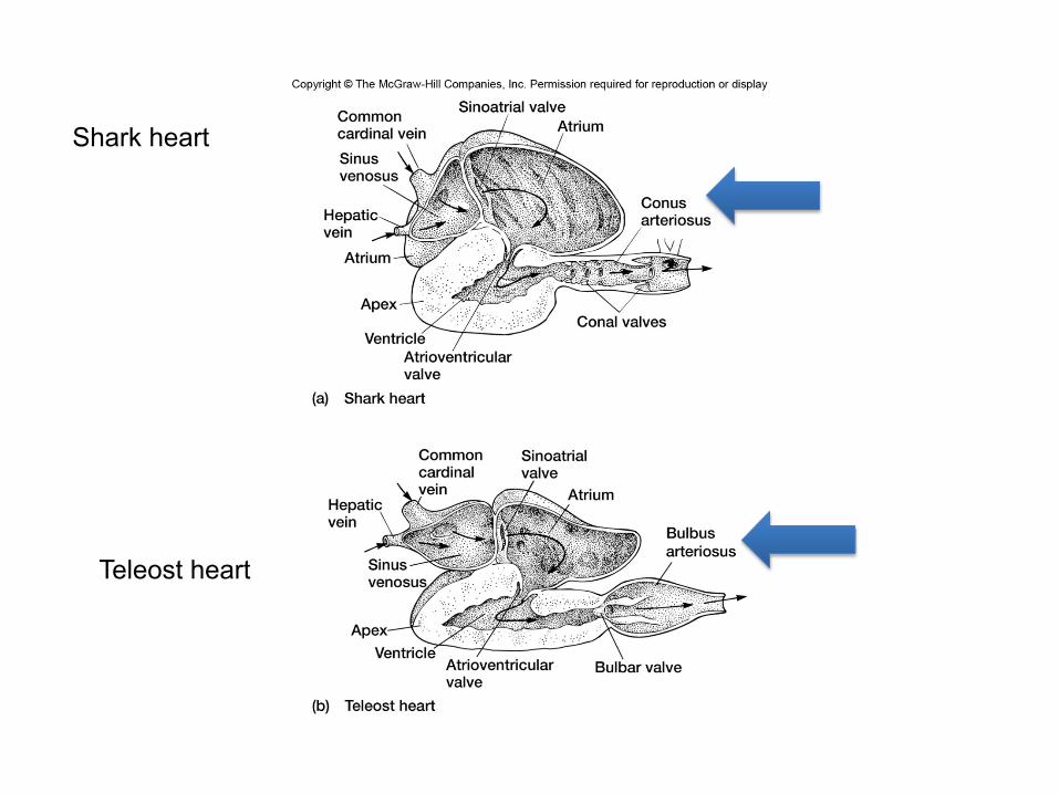
Shark heart
Teleost heart

The general circulatory pathway is as follows: venous blood enters the thin-walled sinus venosus via the common cardinal vein. Contraction of the sinus venosus forces blood across the sinatrial valve into the atrium. The atrium contracts, forcing blood across the atrioventricular valve and into the ventricle. The ventricle is the most powerful pump in the sequence since it must contract with enough force to pump the blood though the speripheral circulation and back to the heart. Blood leaving the ventricle passes a series of semilunar valves and enters the truncus arteriosus. From here the blood enters the gills for gas exchange. This type of system is referred to as a single-circuit pump.

Cyclostomes and Fish -The proposed ancestral prototype is essentially maintained in cyclostomes and fish. -The heart is undivided and is located in close approximation of the gills. No R and L halves. -The general shape of the heart is linear, although the ventricle tends to be positioned below the atrium in an “S” shape.
Lamprey heart

-The position close to the gills makes sense because the blood must pass through the gills for oxigenation before going out to the rest of the body. -Fish do not require high pressures to overcome gravitational force since they are supported in the water. -Transition from water to land presents a problem in how to deliver O2 to the tissues and remove CO2 produced by metabolism. -In Fish, blood flows through the heart in nearly a straight line to the gills, then to the body, and finally back to to the heart. It’s a single circuit system
Gills Heart

-Very efficient type of system. Water and blood flow in what is termed a counter-current system. Important because O2 concentrations in H2O is limited. -This system always maintains a concentration gradient of O2 which favors O2 delivery to the blood, CO2 is readily soluble in water, leaves via countercurrent exchange

Changes in the heart related to the transition from water to air respiration -The transitional form of the heart was probably present in the rhipidistrians, which gave rise to the amphibians. However, the fossil record of the form of gas exchange and heart structure in these organisms is incomplete. -Most of the hypothesis about how the transition occurred is inferred from the extant lung-fish or Dipnoi. -Dipnoi survive conditions of drought and water stagnation by using pulmonary ventilation. -Dipnoi use gill respiration when conditions exist with high oxygen tension in the water. They switch to pulmonary circulation when water is poorly oxygenated.

• The original amphibians probably had a heart similar to lungfish heart.
• This is not true for modern amphibians that are specialized.
• Modern amphibians have an incompletely divided heart which is believed to represent a degenerate condition.
• The ventricle is undivided. Various levels of division are found in the truncus arteriosus and the atria.
Amphibians

• Amphibians rely on cutaneous gas exchange, on gills, on
lungs, or on all three modes.
• The general cardiovascular pathway: venous blood enters the sinus venosus right atrium --> ventricle --> spiral septum of the truncus arteriosus shunts deoxygenated blood to the 6th aortic arch --> lungs --> pulmonary veins --> left atria --> ventricle where oxygenated and deoxygenated blood are mixed to some degree, streams of oxygenated and deoxygenated blood are separated by ridges in the ventricular walls --> truncus arteriosus where the spiral septum directs oxygenated blood to the aortic arches 3,4 --> systemic circulation.
• In the ventricle, pulmonary and sytemic streams of blood are separated by ridges in the ventricular wall, some mixing occurs

Reptiles
The three major groups of reptiles have three different schemes of circulation. Some features that the three groups have in common include: Division of the atria into right and left chambers. In the squamata (lizards, snakes) and the chelonia (turtles) the division is by an interatrial septum. The right and left atria are separate structures in the crocodilians. The truncus is divided into pulmonary and systemic divisions by the spiral septum and is split into vessels. Two systemic vessels and one pulmonary vessel leave the heart.

Lizard Heart. Ventral view Three interconnected compartments: Cavum Venosum, separated by a muscular ridge from the Cavum Pulmonale, and deeper Cavum Arteriosum. The solid arrow indicates the route of blood flow from the CA via the interventricular canal into the CV entering at the bases of the aortic arches

The differences in the squamata and chelodonia are primarily related to the position of the vessels leaving the ventricles. The ventricle is divided in both groups by a septum. The pulmonary trunk leaves the right ventricle in both groups. In the squamata, both systemic trunks leave the left ventricle. In the chelodonia, one systemic trunk leaves the right ventricle, the other leaves the left ventricle. Both groups have muscular flaps that extend into the left ventricle. The flaps divide the ventricle into the cavum arteriosum on the left and the cavum venosum on the right.

The crocodilian heart differs in that the ventricular septum completely separates the right and left ventricle. Foramen of Panizza links the right aorta which leaves the left ventricle and the left aorta which leaves the right ventricle (adjacent to the pulmonary artery) During diastole blood pressure in the left and right aortas equilibrates through the foramen of Panizza. This keeps the left aortic pressure high enough to keep the left aortic valve closed. During ventricular contraction, a cusp on the aortic valve of the right aorta closes the foramen of Pinazza. The pressure generated by the right ventricle (low pressure system) is still lower than the back pressure in the left aorta and most of the blood is directed to the lungs through the pulmonary artery.
During diving, the pulmonary system is not required. When the animal dives, the heart rate decreases. Crocodilians have a valve called the founs in the aperture region of the pulmonary artery which, when closed, increases resistance in the pulmonary aorta. When the right ventricular pressure exceeds the systemic pressure blood form the right ventricle will by-pass the pulmonary system and flow into the left aorta and systemic circulation. This is believed to provide additional systemic blood and oxygen capacity during the dive.


Birds
The birds, like the reptiles, probably evolved from archosaurian ancestors. However, birds have high metabolic rate, are endothermic and have lungs that are continuously ventilated. Birds have evolved a pumping system in which equal amounts of blood are pumped to the systemic and pulmonary circulation at all times by using a double circuit pump. 1. Pump 1 - is the pulmonary circuit that operates at low pressure to pump to the lungs and back to the heart. 2. Pump 2 - is the high pressure systemic circuit.

The principle factor that leads to the double circuit pump in the birds appears to be the elimination of the left systemic trunk. The right aortic trunk arises from the left ventricle. The pulmonary artery arises from the right ventricle. The sinus venosus is present in birds but is greatly reduced.

Mammals They also employ a double circuit pump Superficially, the mammalian and bird hearts appear to be very similar. However, the synapsid line of reptiles that gave rise to the mammals branched off from the stem reptiles long before the lines that led to the modern reptiles and birds did. Evidence suggests that the double circuit pumps found in mammals and birds evolved independently: The interventricular septum in birds and mammals have separate embryological origins. The systemic trunk leaving the left ventricle in mammals is the counterpart of the left systemic trunk rather than the right systemic trunk found in birds.

19
Overview • The right side receives
oxygen-‐poor blood from the body and <ssues and then pumps it to the lungs to pick up oxygen and dispel carbon dioxide
• Its le@ side receives oxygenated blood returning from the lungs and pumps this blood throughout the body to supply oxygen and nutrients to the body <ssues
The heart=a muscular double pump with 2 functions

20
simplified…
• Cone shaped muscle • Four chambers
– Two atria, two ventricles • Double pump – the ventricles • Two circula<ons
– Systemic circuit: blood vessels that transport blood to and from all the body <ssues
– Pulmonary circuit: blood vessels that carry blood to and from the lungs

21
Heart’s posi<on in thorax

22
Heart’s posi<on in thorax • In medias<num – behind sternum and poin<ng le@, lying on the diaphragm
• It weighs 250-‐350 gm (about 1 pound) Feel your heart beat at apex
(this is of a person lying down)

23
24
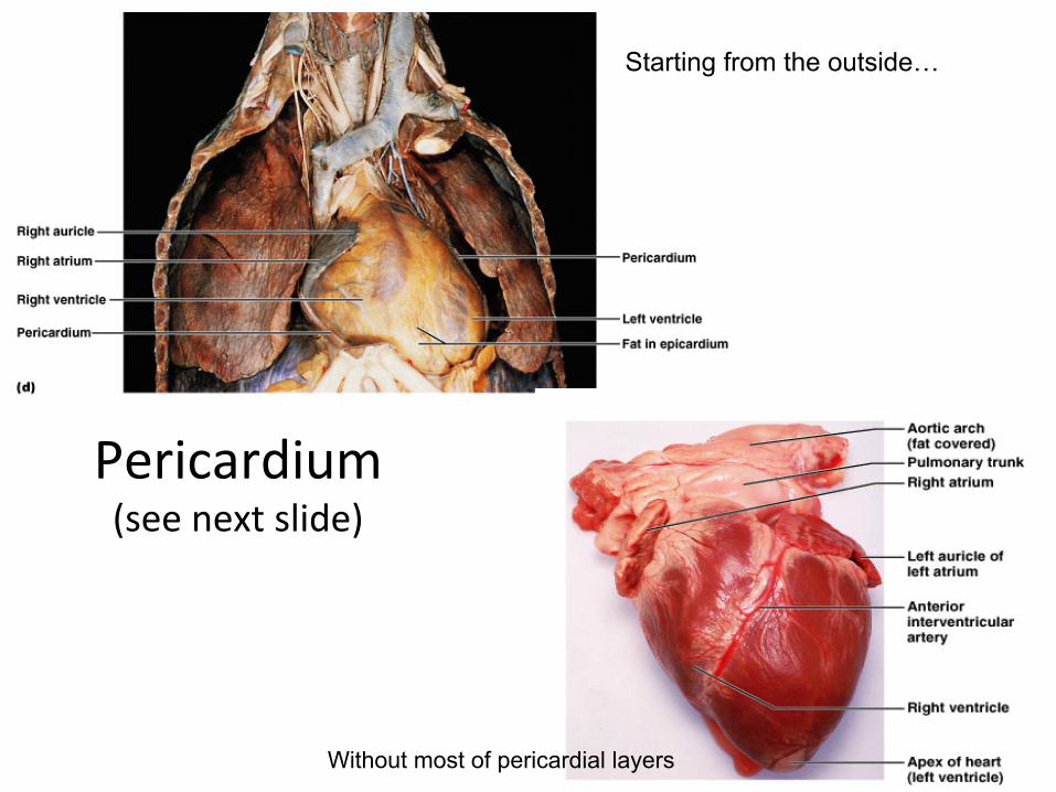
Pericardium (see next slide)
Starting from the outside…
Without most of pericardial layers
25
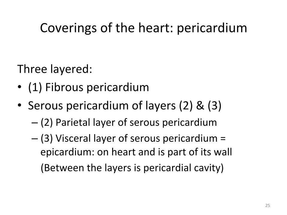
Coverings of the heart: pericardium
Three layered: • (1) Fibrous pericardium • Serous pericardium of layers (2) & (3)
– (2) Parietal layer of serous pericardium – (3) Visceral layer of serous pericardium = epicardium: on heart and is part of its wall (Between the layers is pericardial cavity)

26
Layers of the heart wall
• Muscle of the heart with inner and outer membrane coverings
• Muscle of heart = “myocardium” • The layers from out to in:
– Epicardium = visceral layer of serous pericardium – Myocardium = the muscle – Endocardium lining the chambers

27
Layers of pericardium and heart wall

28
Chambers of the heart sides are labeled in reference to the pa8ent facing you
• Two atria – Right atrium – Le@ atrium
• Two ventricles – Right ventricle – Le@ ventricle
--------------------------------------------------------------------------------

29
Chambers of the heart divided by septae:
• Two atria-‐divided by interatrial septum – Right atrium – Le@ atrium
• Two ventricles-‐divided by interventricular septum – Right ventricle – Le@ ventricle

30
4 Valves three tricuspid one bicuspid
• A heart valve normally allows blood flow in only one direc<on through the heart.
• The two atrioventricular valves (AV), which are between the atria and the ventricles, are the mitral valve (bicuspid, LA to LV) and the “tricuspid” valve (RA to RV).
• The two semilunar (SL) valves, which are in the arteries leaving the heart, are the aor2c valve (LV to aorta) and the pulmonary valve( RV to pulmonary trunk (branches R and L))
(cusp means flap)

31
Function of AV valves

32
Function of semilunar valves (Aortic and pulmonic valves)

33
Pa5ern of flow (simple to more detailed)
• Body • RA • RV • Lungs • LA • LV • Boby
Body to right heart to lungs to left heart to body
Body, then via vena cavas and coronary sinus to RA, to RV, then to lungs via pulmonary arteries, then to LA via pulmonary veins, to LV, then to body via aorta
From body to RA; then to RV through tricuspid valve; to lungs through pulmonic valve and via pulmonary arteries; to LA via pulmonary veins; to LV through mitral valve; to body via aortic valve then aorta

34
Chambers with embryologic changes added fetal in pink; postnatal in blue
(see next slide)
• Two atria-‐-‐-‐-‐-‐-‐-‐-‐-‐-‐-‐-‐divided by interatrial septum • Fossa ovalis le@ over from fetal hole in septum, the foramen ovale
– Right atrium-‐-‐-‐-‐-‐-‐-‐-‐in fetus RA received oxygenated blood from mom through umbilical cord, so blood R to L through the foramen ovale
– Le@ atrium • Two ventricles-‐-‐-‐-‐-‐divided by interventricular septum
– Right ventricle-‐-‐-‐-‐-‐in fetus pulmonary trunk high resistance & ductus arteriosus shunts blood to aorta
• Ductus arteriosus becomes ligamentum arteriosum a@er birth
– Le@ ventricle

35
In the fetus, the RA received oxygenated blood from mom through umbilical cord, so blood R to L through the foramen ovale: fossa ovalis is le@ a@er it closes
The pulmonary trunk had high resistance (because lungs not func<oning yet) & ductus arteriosus shunted blood to aorta; becomes ligamentum arteriosum a@er birth

36
• Note posi<ons of valves • Valves open and close in response to pressure differences • Trabeculae carnae • Note papillary muscles, chordae tendinae (heart strings): keep
valves from prolapsing (purpose of valve = 1 way flow)

37
Rela<ve thickness of muscular walls
LV thicker than RV because it forces blood out against more resistance; the systemic circulation is much longer than the pulmonary circulation Atria are thin because ventricular filling is done by gravity, requiring little atrial effort

38

39
more on valves

40
Heartbeat
• Systole: contrac<on • Diastole: filling • Normal rate: 60-‐100 • Slow: bradycardia • Fast: tachycardia
***Note: blood goes to RA, then RV, then lungs, then LA, then LV, then body; but the fact that a given drop of blood passes through the heart chambers sequentially does not mean that the four chambers contract in that order; the 2 atria always contract together, followed by the simultaneous contraction of the 2 ventricles
Definition: a single sequence of atrial contraction followed by ventricular contraction

41
Heart sounds
• Called S1 and S2 • S1 is the closing of atrioventricular (Mitral and Tricuspid) valves at the start of ventricular systole
• S2 is the closing of the semilunar (Aor<c and Pulmonic) valves at the end of ventricular systole
• Murmurs: the sound of flow – Can be normal – Can be abnormal

42
Cardiac muscle (microscopic)
Automaticity: inherent rhythmicity of the muscle itself

43
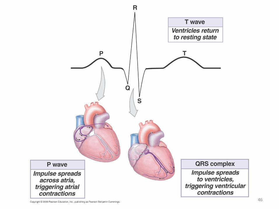
Electrical conduc2on system:
(Explanation in next slides)
specialized cardiac muscle cells that carry impulses throughout the heart musculature, signaling the chambers to contract in the proper sequence

44
Conduc<on system
• SA node (sinoatrial) – In wall of RA – Sets basic rate: 70-‐80 – Is the normal pacemaker
• Impulse from SA to atria • Impulse also to AV node via internodal pathway
• AV node – In interatrial septum

45
Conduc<on con<nued
• SA node through AV bundle (bundle of His) – Into interventricular septum – Divides
R and L bundle branches
become subendocardial branches (“Purkinje fibers”)
• Contrac<on begins at apex
46

47
Autonomic innerva<on
• Sympathe<c – Increases rate and force of contrac<ons
• Parasympathe<c (branches of Vagus n.) – Slows the heart rate

48
Anterior view
Each atrium has an “auricle,” an ear-like flap

49
Posterior view Note: the coronary sinus (largest cardiac vein) –
delivers blood from heart wall to RA, along with SVC & IVC)

50
another flow chart