Embed Size (px)
Citation preview
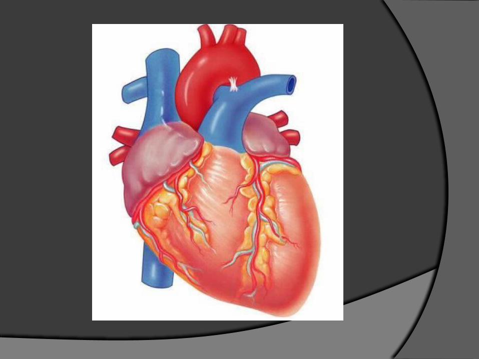
THE HEART
Position of the Heart
human heart is about the size of a fist
lies in the thoracic cavity w/in the mediastinum (area from sternum to vertebrae, between the lungs)
tilted @ angle so its inferior surface lies against the diaphragm
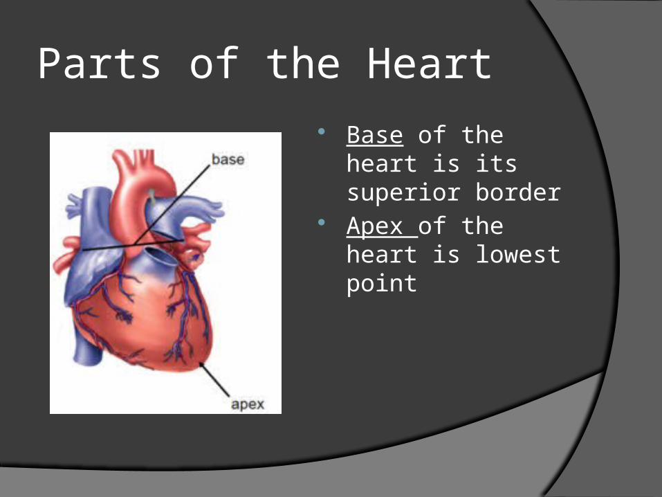
Parts of the Heart Base of the heart
is its superior border
Apex of the heart is lowest point
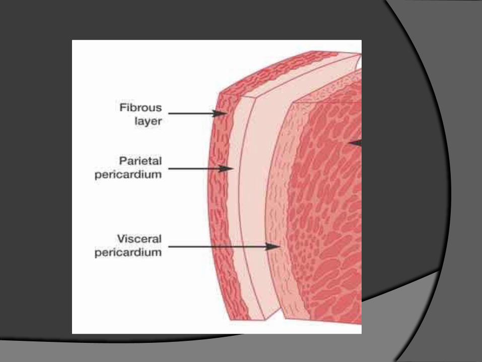
Major Heart Structures:the Pericardium Outer Layer: Fibrous Pericardium
tough, attaches to diaphragm
Inner Layer: Serous Pericardiumdbl membrane: ○ outer parietal: attaches to fibrous pericardium ○ inner visceral layer: covers cardiac muscle
between the 2: pericardial cavity filled with serous fluid
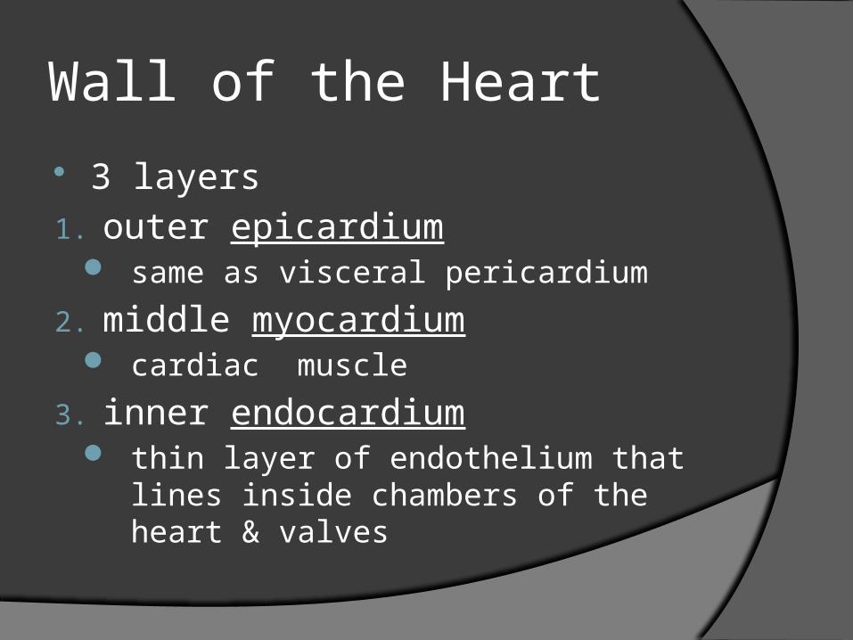
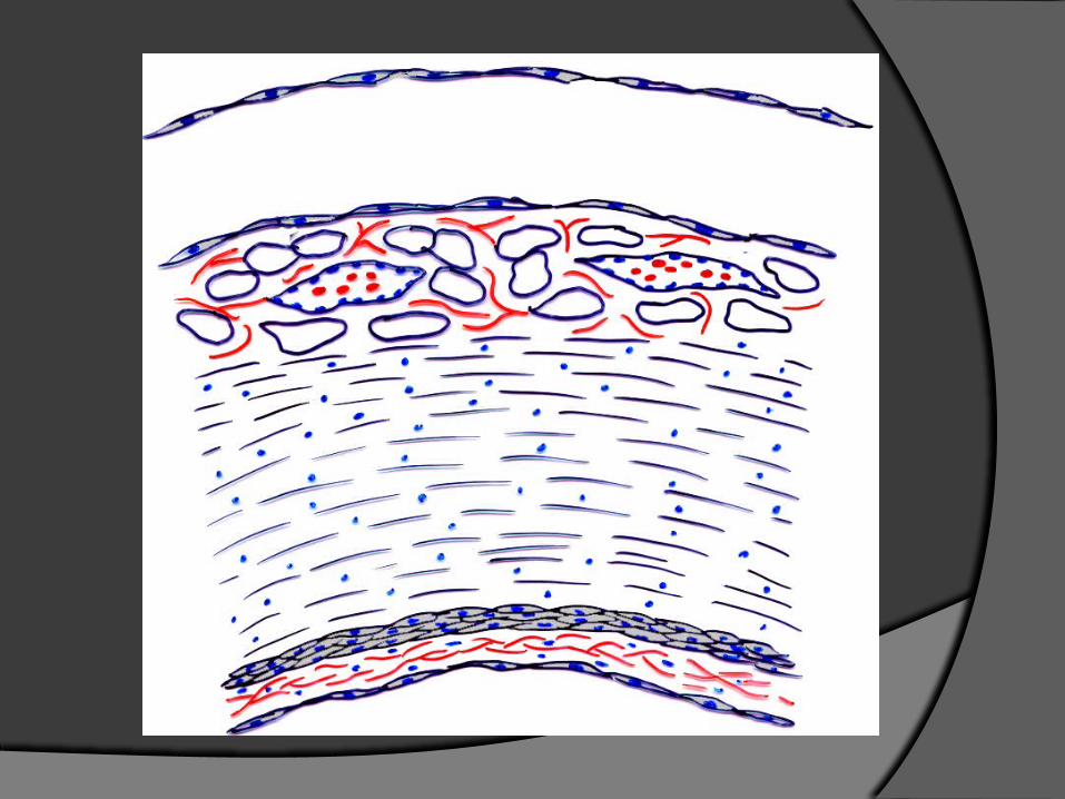
Wall of the Heart
3 layers1. outer epicardium
same as visceral pericardium
2. middle myocardium cardiac muscle
3. inner endocardium thin layer of endothelium that lines
inside chambers of the heart & valves
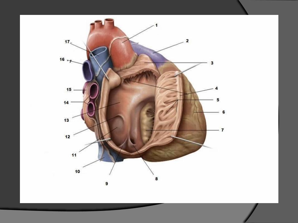
Surface Features of the Heart
4 chambers of heart:2 atria form the base ○ Auricles (ear-like) pouch-like extensions
tip of left ventricle forms the apexSulci: grooves where coronary blood vessels
& adipose tissue that externally mark the boundaries between the 4 heart chambers○ coronary sulcus: separates atria from
ventricles○ anterior & posterior interventricular sulcus:
separate 2 ventricles
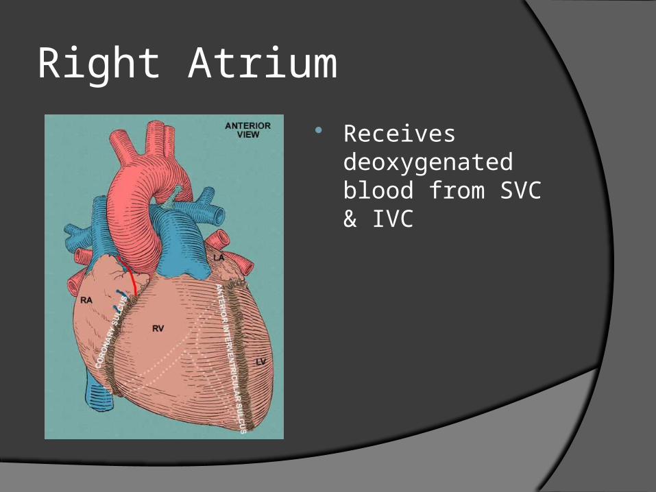
Right Atrium Receives
deoxygenated blood from SVC & IVC
Right Atrium
inside surface has honeycombed appearance & ridges called pectinate muscles
wall separating rt & lt atrium= interatrial septum: in fetus hole called foramen ovale (blood shunts from rt atrium lt atrium avoiding pulmonary circulation); when scarred over called fossa ovalis
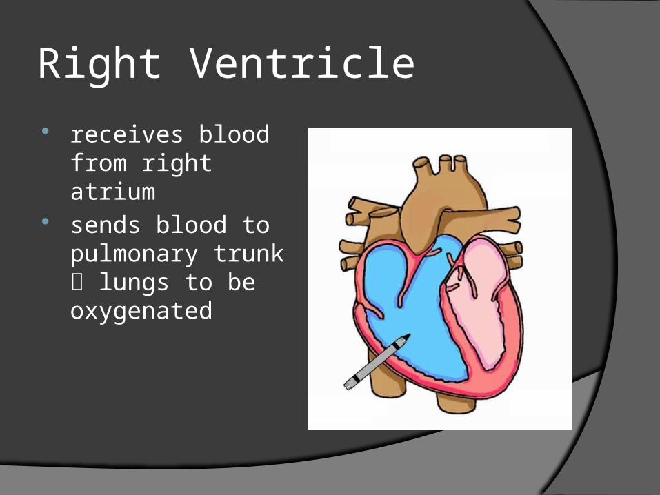
Right Ventricle receives blood
from right atrium sends blood to
pulmonary trunk lungs to be oxygenated
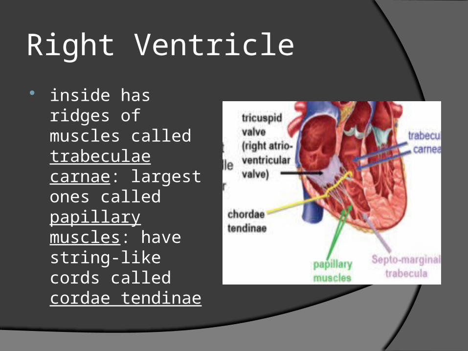
Right Ventricle inside has ridges
of muscles called trabeculae carnae: largest ones called papillary muscles: have string-like cords called cordae tendinae
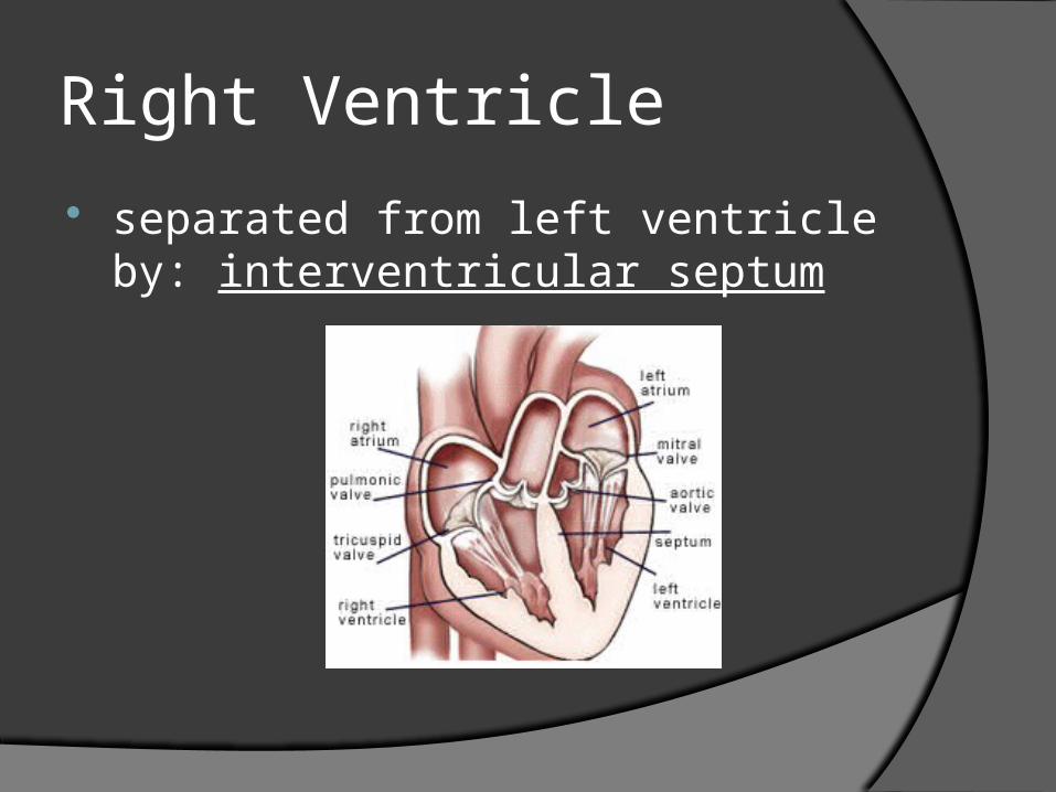
Right Ventricle
separated from left ventricle by: interventricular septum
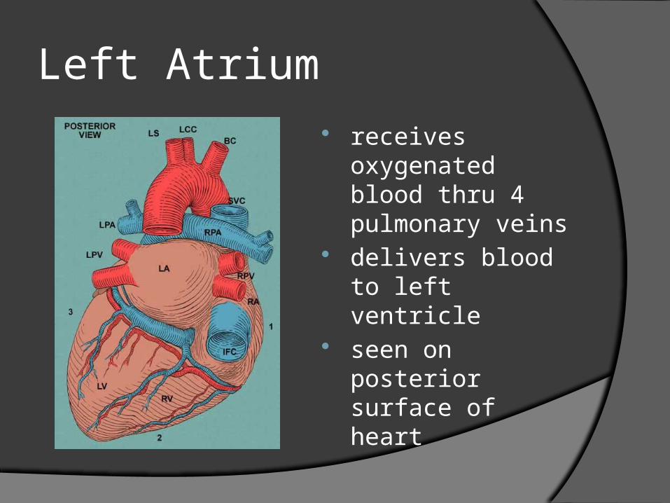
Left Atrium receives
oxygenated blood thru 4 pulmonary veins
delivers blood to left ventricle
seen on posterior surface of heart
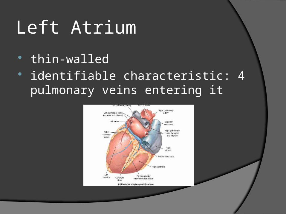
Left Atrium
thin-walled identifiable characteristic: 4
pulmonary veins entering it
Left Ventricle
receives oxygenated blood from left atrium
sends blood to systemic circulation thru Aorta
has thickest muscle (pumps blood the farthest)
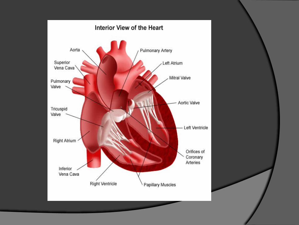
4 Heart Valves control 1-way flow of blood 2 AV valves
between atria & ventriclesTricuspid : rt AV valveMitral : lt AV valve, aka bicuspid
2 semilunar valvesblood exits rt ventricle thru Pulmonary
(semilunar) valveblood exits lt ventricle thru Aortic
(semilunar) valve
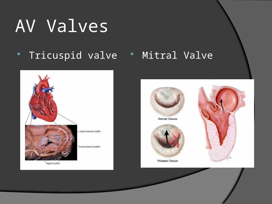
AV Valves Tricuspid valve Mitral Valve
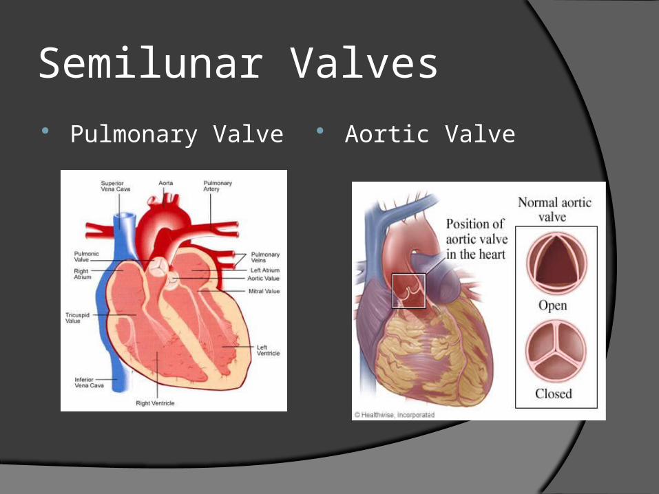
Semilunar Valves Pulmonary Valve Aortic Valve
Blood Flow thru the Heart
thinner walled atria receive blood returning to heart from veins
pressure of blood in filled atria opens the AV valves & most of the blood flows into ventricles
both atria contract simultaneously to pump remaining blood into ventricles
Blood Flow thru the Heart
when atria have stopped contracting AV valves close
Ventricles contract together forcing semilunar valves open
walls of left ventricle thicker providing more force to pump blood thru systemic circulation
Blood Flow thru the Heart
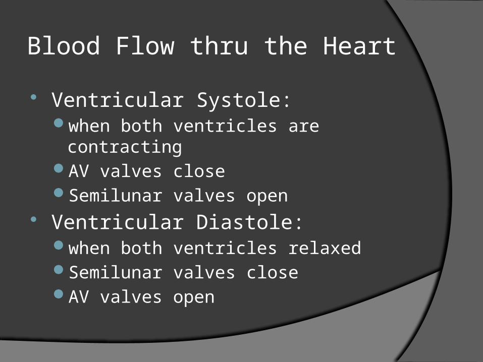
Ventricular Systole: when both ventricles are contractingAV valves closeSemilunar valves open
Ventricular Diastole: when both ventricles relaxedSemilunar valves closeAV valves open
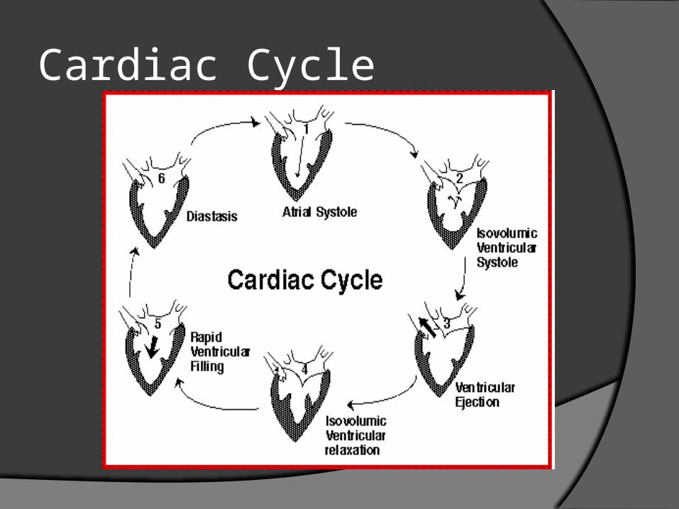
Cardiac Cycle
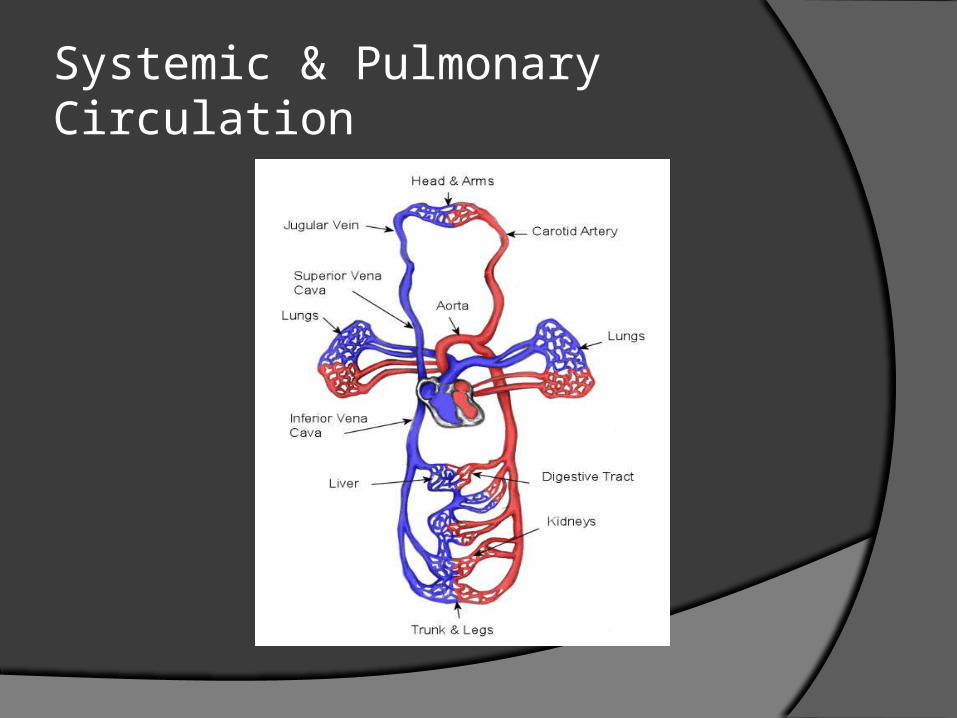
Systemic & Pulmonary Circulation
Heart Animations
http://www.nhlbi.nih.gov/health//dci/Diseases/hhw/hhw_pumping.html
http://www.hybridmedicalanimation.com/work/animation/beating-heart-with-blood-flow/
Heart Sounds
Auscultation: listening to body sounds
1 heartbeat produces 2 heart sounds: lub-dub
heart murmurs: abnl heart sounds usually due to valve abnl
http://www.blaufuss.org/tutorial/index1.html
Heart Sounds
http://www.dnatube.com/video/9217/Review-of-heart-sounds
http://familymedicine.osu.edu/products/physicalexam/exam/flash/heart/heart2.cfm
http://www.blaufuss.org/tutorial/index2.html
Pulse
when ventricles contract a blood pressure wave is produced that travels in the arteries and can be felt as your pulse
radial pulse: check over radial artery
carotid artery pulse: check over carotid artery

Calculate Pulse
Count the # of beats in 15 s and multiply x 4
If the math is too difficult count for 30 s and multiple x 2
Blood Pressure
pressure exerted by blood against blood vessel walls
highest in the aorta & large elastic arteries & decreases as arteries get smaller & further from heart
Systolic Blood Pressure
top # on a BP pressure generated by ventricular
systole normal adult: ~120
Diastolic BP
bottom # on BP pressure exerted during ventricular
diastole normal adult: 60- 80
Arterial Blood Pressure
normal adult ~ 120/80
normal venous BP: ~16 mm Hg
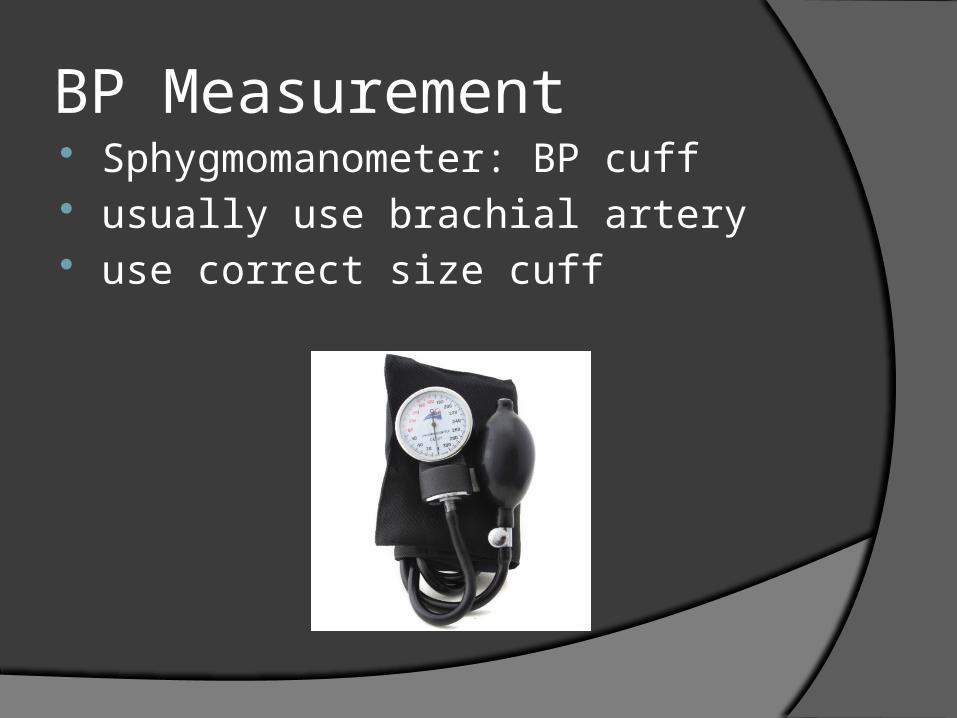
BP Measurement Sphygmomanometer: BP cuff usually use brachial artery use correct size cuff
BP
pump used to inflate cuff to a pressure > the systolic pressure:puts pressure on the artery, flattens it,
& stops blood flow in the arterypressure slowly released from cuff as
stethoscope used to auscultate over brachial artery
BP
reported in mm Hg as pressure in cuff becomes <
pressure in artery…examiner will hear a sound can be heard, caused by the turbulent flow of blood as artery goes from flattened normal