Embed Size (px)
Citation preview
CHAPTER 9 • CIRCULATION: THE CARDIOVASCULAR AND LYMPHATIC SYSTEMS 167
Blood circulates throughout the body in the cardiovascular system, which consists of the heart and theblood vessels (Fig. 9-1). This system forms a continuous circuit that delivers oxygen and nutrients toall cells and carries away waste products. Also functioning in circulation is the lymphatic system,
which drains fluid and proteins from the tissues and returns them to the bloodstream.
The Heart
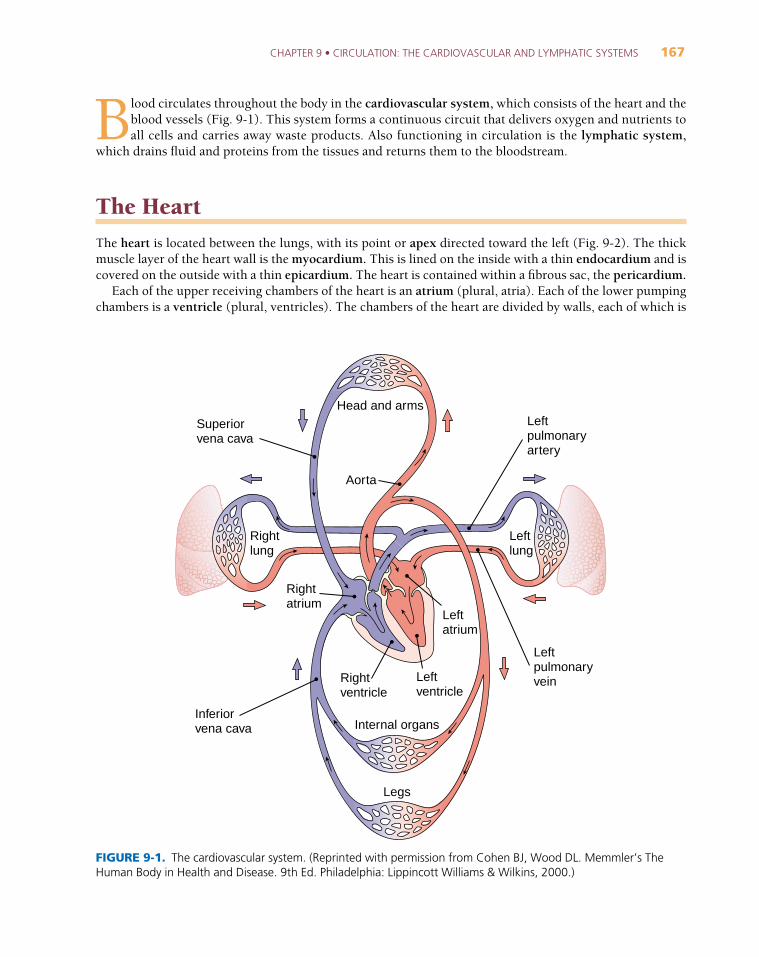
The heart is located between the lungs, with its point or apex directed toward the left (Fig. 9-2). The thickmuscle layer of the heart wall is the myocardium. This is lined on the inside with a thin endocardium and iscovered on the outside with a thin epicardium. The heart is contained within a fibrous sac, the pericardium.
Each of the upper receiving chambers of the heart is an atrium (plural, atria). Each of the lower pumpingchambers is a ventricle (plural, ventricles). The chambers of the heart are divided by walls, each of which is
Head and arms
Aorta
Superiorvena cava
Leftpulmonaryartery
Leftpulmonaryvein
Inferiorvena cava
Rightlung
Leftlung
Rightventricle
Leftventricle
Leftatrium
Rightatrium
Internal organs
Legs
FIGURE 9-1. The cardiovascular system. (Reprinted with permission from Cohen BJ, Wood DL. Memmler’s TheHuman Body in Health and Disease. 9th Ed. Philadelphia: Lippincott Williams & Wilkins, 2000.)
168 PART 3 • BODY SYSTEMS
called a septum. The interventricular septum separates the two ventricles; the interatrial septum divides thetwo atria. There is also a septum between the atrium and ventricle on each side.
The heart pumps blood through two circuits. The right side pumps blood to the lungs to be oxygenatedthrough the pulmonary circuit. The left side pumps to the remainder of the body through the systemic circuit.
Blood Flow Through the HeartThe pathway of blood through the heart is shown by the arrows in Figure 9-2. The right atrium receives bloodlow in oxygen from all body tissues through the superior vena cava and the inferior vena cava. The bloodthen enters the right ventricle and is pumped to the lungs through the pulmonary artery. Blood returns from
Brachiocephalic arteryLeft common carotid artery
Left subclavian artery
Right pulmonaryartery(branches)
Ascendingaorta
Superior vena cava
Rightpulmonaryveins
Rightatrium
Tricuspidvalve
Inferiorvena cava
Right ventricle
Aortic arch
Pulmonary artery
Left pulmonaryartery (branches)
Pulmonic valve
LeftpulmonaryveinsLeft atrium
Aortic valve
Mitral(bicuspid)valve
Endocardium
Leftventricle
MyocardiumBlood high in oxygen
Blood low in oxygen Epicardium
Apex
Interventricularseptum
FIGURE 9-2. The heart and great vessels. (Reprinted with permission from Cohen BJ, Wood DL. Memmler’s TheHuman Body in Health and Disease. 9th Ed. Philadelphia: Lippincott Williams & Wilkins, 2000.)
CHAPTER 9 • CIRCULATION: THE CARDIOVASCULAR AND LYMPHATIC SYSTEMS 169
the lungs high in oxygen and enters the left atrium through the pulmonary veins. From here it enters the leftventricle and is forcefully pumped into the aorta to be distributed to all tissues.
Blood is kept moving in a forward direction by one-way valves. The valve in the septum between the rightatrium and ventricle is the tricuspid valve (meaning three cusps or flaps); the valve in the septum betweenthe left atrium and ventricle is the bicuspid valve (having two cusps), usually called the mitral valve (sonamed because it resembles a bishop’s miter). The valves leading into the pulmonary artery and the aortahave three cusps. Each cusp is shaped like a half-moon, so these valves are described as semilunar valves. Thevalve at the entrance to the pulmonary artery is specifically named the pulmonic valve; the valve at the en-trance to the aorta is the aortic valve.
Heart sounds are produced as the heart functions. The loudest of these, the familiar lubb and dupp that canbe heard through the chest wall, are produced by alternate closing of the valves. The first heart sound (S1) isheard when the valves between the chambers close. The second heart sound (S2) is produced when the valvesleading into the aorta and pulmonary artery close. Any sound made as the heart functions normally is termeda functional murmur. (The word murmur used alone with regard to the heart describes an abnormal sound.)
The HeartbeatEach contraction of the heart, termed systole (SIS-to
_-le
_), is followed by a relaxation phase, diastole (di
_-AS-
to_-le
_), during which the chambers fill. Each time the heart beats, both atria contract and immediately there-
after both ventricles contract. The wave of increased pressure produced in the vessels each time the ventriclescontract is the pulse.
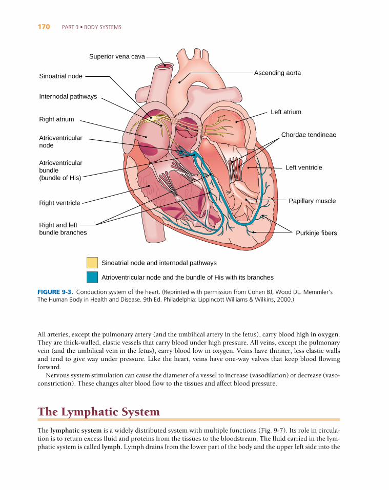
Contractions are stimulated by a built-in system that regularly transmits electrical impulses through theheart. The components of this conduction system are shown in Figure 9-3. They include the sinoatrial (SA)node, called the pacemaker because it sets the rate of the heartbeat, the atrioventricular (AV) node, the AVbundle (bundle of His), the left and right bundle branches, and Purkinje ( pur-KIN-je
_) fibers.
Although the heart itself generates the heartbeat, factors such as nervous system stimulation, hormones,and drugs can influence the rate and the force of heart contractions.
Blood Pressure
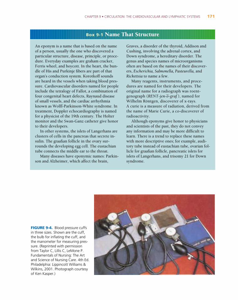
Blood pressure is the force exerted by blood against the wall of a blood vessel. It is commonly measured ina large artery with an inflatable cuff (Fig. 9-4) known as a blood pressure cuff or blood pressure apparatus,but technically called a sphygmomanometer. Both systolic and diastolic pressures are measured and reportedas systolic then diastolic separated by a slash, such as 120/80. Pressure is expressed as millimeters of mercury(mm Hg), that is, the height to which the pressure can push a column of mercury in a tube. Blood pressureis a valuable diagnostic measurement that is easily obtained.
The Vascular System
The vascular system consists of:
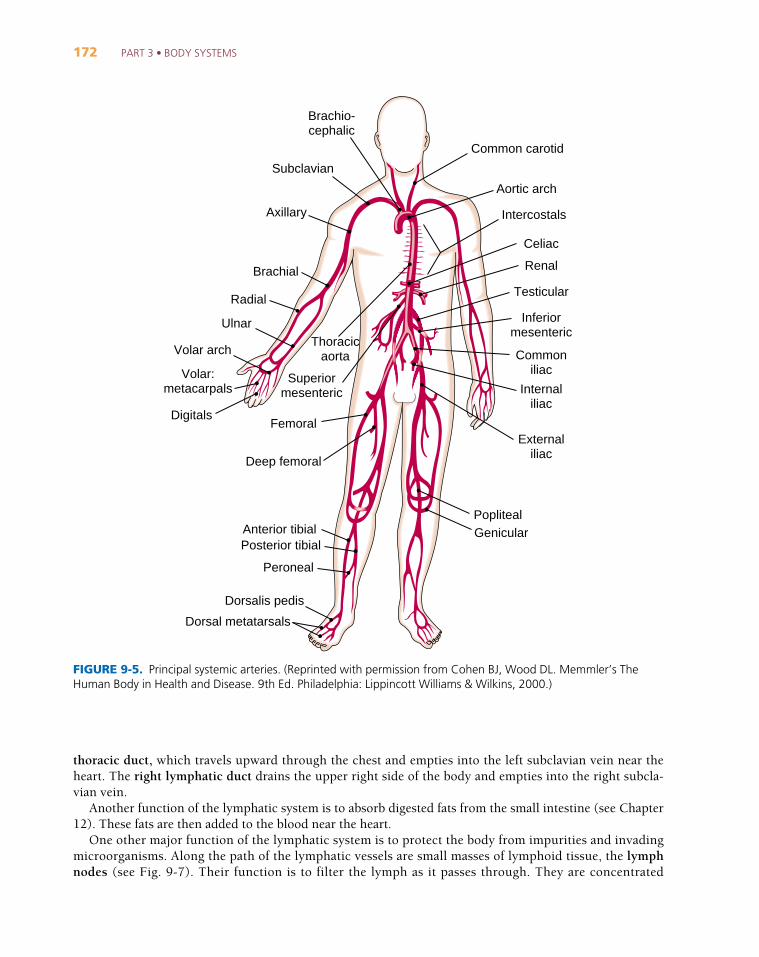
1. Arteries that carry blood away from the heart (Fig. 9-5). Arterioles are small arteries that lead into thecapillaries.
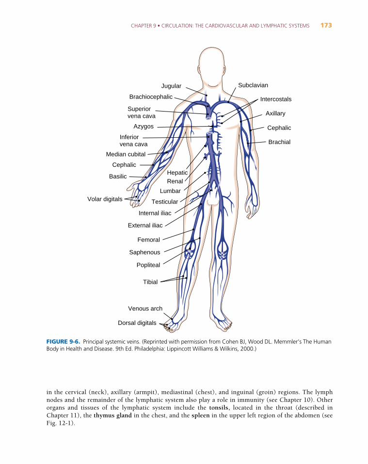
2. Capillaries, the smallest vessels, through which exchanges take place between the blood and the tissues.3. Veins that carry blood back to the heart (Fig. 9-6). The small veins that receive blood from the capillar-
ies and drain into the veins are venules.
170 PART 3 • BODY SYSTEMS
All arteries, except the pulmonary artery (and the umbilical artery in the fetus), carry blood high in oxygen.They are thick-walled, elastic vessels that carry blood under high pressure. All veins, except the pulmonaryvein (and the umbilical vein in the fetus), carry blood low in oxygen. Veins have thinner, less elastic wallsand tend to give way under pressure. Like the heart, veins have one-way valves that keep blood flowingforward.
Nervous system stimulation can cause the diameter of a vessel to increase (vasodilation) or decrease (vaso-constriction). These changes alter blood flow to the tissues and affect blood pressure.
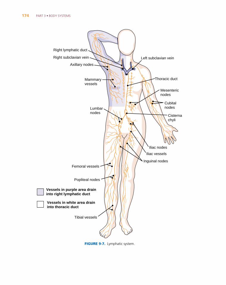
The Lymphatic System
The lymphatic system is a widely distributed system with multiple functions (Fig. 9-7). Its role in circula-tion is to return excess fluid and proteins from the tissues to the bloodstream. The fluid carried in the lym-phatic system is called lymph. Lymph drains from the lower part of the body and the upper left side into the
Sinoatrial node
Internodal pathways
Atrioventricularnode
Atrioventricularbundle(bundle of His)
Right and leftbundle branches
Sinoatrial node and internodal pathways
Purkinje fibers
Superior vena cava
Right atrium
Right ventricle
Ascending aorta
Left atrium
Papillary muscle
Left ventricle
Chordae tendineae
Atrioventricular node and the bundle of His with its branches
FIGURE 9-3. Conduction system of the heart. (Reprinted with permission from Cohen BJ, Wood DL. Memmler’sThe Human Body in Health and Disease. 9th Ed. Philadelphia: Lippincott Williams & Wilkins, 2000.)
CHAPTER 9 • CIRCULATION: THE CARDIOVASCULAR AND LYMPHATIC SYSTEMS 171
An eponym is a name that is based on the nameof a person, usually the one who discovered aparticular structure, disease, principle, or proce-dure. Everyday examples are graham cracker,Ferris wheel, and boycott. In the heart, the bun-dle of His and Purkinje fibers are part of thatorgan’s conduction system. Korotkoff soundsare heard in the vessels when taking blood pres-sure. Cardiovascular disorders named for peopleinclude the tetralogy of Fallot, a combination offour congenital heart defects, Raynaud diseaseof small vessels, and the cardiac arrhythmiaknown as Wolff-Parkinson-White syndrome. Intreatment, Doppler echocardiography is namedfor a physicist of the 19th century. The Holtermonitor and the Swan-Ganz catheter give honorto their developers.
In other systems, the islets of Langerhans areclusters of cells in the pancreas that secrete in-sulin. The graafian follicle in the ovary sur-rounds the developing egg cell. The eustachiantube connects the middle ear to the throat.
Many diseases have eponymic names: Parkin-son and Alzheimer, which affect the brain,
Graves, a disorder of the thyroid, Addison andCushing, involving the adrenal cortex, andDown syndrome, a hereditary disorder. Thegenus and species names of microorganismsoften are based on the names of their discover-ers, Escherichia, Salmonella, Pasteurella, andRickettsia to name a few.
Many reagents, instruments, and proce-dures are named for their developers. Theoriginal name for a radiograph was roent-genograph (RENT-jen-o
_-graf ), named for
Wilhelm Röntgen, discoverer of x-rays. A curie is a measure of radiation, derived fromthe name of Marie Curie, a co-discoverer ofradioactivity.
Although eponyms give honor to physiciansand scientists of the past, they do not conveyany information and may be more difficult tolearn. There is a trend to replace these nameswith more descriptive ones; for example, audi-tory tube instead of eustachian tube, ovarian fol-licle for graafian follicle, pancreatic islets forislets of Langerhans, and trisomy 21 for Downsyndrome.
Box 9-1 Name That Structure
FIGURE 9-4. Blood pressure cuffsin three sizes. Shown are the cuff,the bulb for inflating the cuff, andthe manometer for measuring pres-sure. (Reprinted with permissionfrom Taylor C, Lillis C, LeMone P.Fundamentals of Nursing: The Artand Science of Nursing Care. 4th Ed.Philadelphia: Lippincott Williams &Wilkins, 2001. Photograph courtesyof Ken Kasper.)
172 PART 3 • BODY SYSTEMS
thoracic duct, which travels upward through the chest and empties into the left subclavian vein near theheart. The right lymphatic duct drains the upper right side of the body and empties into the right subcla-vian vein.
Another function of the lymphatic system is to absorb digested fats from the small intestine (see Chapter12). These fats are then added to the blood near the heart.
One other major function of the lymphatic system is to protect the body from impurities and invadingmicroorganisms. Along the path of the lymphatic vessels are small masses of lymphoid tissue, the lymphnodes (see Fig. 9-7). Their function is to filter the lymph as it passes through. They are concentrated
Common carotid
Celiac
Renal
Subclavian
Aortic arch
Testicular
Inferiormesenteric
Commoniliac
Externaliliac
PoplitealGenicular
Dorsal metatarsals
Dorsalis pedis
Peroneal
Posterior tibialAnterior tibial
Deep femoral
FemoralDigitals
Volar:metacarpals
Volar arch
Ulnar
Radial
Brachial
Axillary
Brachio-cephalic
Superiormesenteric
Thoracicaorta
Internaliliac
Intercostals
FIGURE 9-5. Principal systemic arteries. (Reprinted with permission from Cohen BJ, Wood DL. Memmler’s TheHuman Body in Health and Disease. 9th Ed. Philadelphia: Lippincott Williams & Wilkins, 2000.)
CHAPTER 9 • CIRCULATION: THE CARDIOVASCULAR AND LYMPHATIC SYSTEMS 173
in the cervical (neck), axillary (armpit), mediastinal (chest), and inguinal (groin) regions. The lymphnodes and the remainder of the lymphatic system also play a role in immunity (see Chapter 10). Other organs and tissues of the lymphatic system include the tonsils, located in the throat (described in Chapter 11), the thymus gland in the chest, and the spleen in the upper left region of the abdomen (seeFig. 12-1).
Subclavian
Intercostals
Jugular
Azygos
Median cubital
Axillary
Cephalic
Basilic
Venous arch
Saphenous
Popliteal
Tibial
Femoral
External iliac
Internal iliac
Testicular
Lumbar
RenalHepatic
Volar digitals
Dorsal digitals
Superiorvena cava
Inferiorvena cava
Brachiocephalic
Brachial
Cephalic
FIGURE 9-6. Principal systemic veins. (Reprinted with permission from Cohen BJ, Wood DL. Memmler’s The HumanBody in Health and Disease. 9th Ed. Philadelphia: Lippincott Williams & Wilkins, 2000.)
174 PART 3 • BODY SYSTEMS
Lumbarnodes
Mammaryvessels
Axillary nodes
Right subclavian vein Left subclavian vein
Right lymphatic duct
Thoracic duct
Mesentericnodes
Cubitalnodes
Cisternachyli
Iliac nodes
Iliac vessels
Inguinal nodes
Tibial vessels
Popliteal nodes
Femoral vessels
Vessels in purple area draininto right lymphatic duct
Vessels in white area draininto thoracic duct
FIGURE 9-7. Lymphatic system.
CHAPTER 9 • CIRCULATION: THE CARDIOVASCULAR AND LYMPHATIC SYSTEMS 175
Key Terms
NORMAL STRUCTURE AND FUNCTION
Cardiovascular Systemaortaa_-OR-ta
aortic valvea_-OR-tik
apexA_-peks
artery
arteriolear-TE
_-re
_-o_l
atrioventricular (AV) nodea_-tre
_-o_-ven-TRIK-u
_-lar
AV bundle
atriumA_-tre
_-um
bicuspid valvebi_-KUS-pid
blood pressure
bundle branches
capillaryKAP-i-lar-e
_
cardiovascular systemkar-de
_-o_-VAS-ku
_-lar
diastoledi_-AS-to
_-le
_
endocardiumen-do
_-KAR-de
_-um
epicardiumep-i-KAR-de
_-um
functional murmur
The largest artery. It receives blood from the left ventricle andbranches to all parts of the body (root aort/o).
The semilunar valve at the entrance to the aorta
The point of a cone-shaped structure (adjective, apical). The apex ofthe heart is formed by the left ventricle. It is inferior and pointed to-ward the left (see Fig. 9-2).
A vessel that carries blood away from the heart. All except the pulmo-nary and umbilical arteries carry oxygenated blood (root arter, arteri/o).
A small artery (root arteriol/o)
A small mass in the lower septum of the right atrium that passes im-pulses from the sinoatrial (SA) node toward the ventricles
A band of fibers that transmits impulses from the atrioventricular(AV) node to the top of the interventricular septum. It divides intothe right and left bundle branches, which descend along the twosides of the septum; the bundle of His.
An entrance chamber, one of the two upper receiving chambers ofthe heart (root atri/o)
The valve between the left atrium and the left ventricle; the mitralvalve
The force exerted by blood against the wall of a vessel
Branches of the AV bundle that divide to the right and left sides ofthe interventricular septum
A microscopic blood vessel through which materials are exchangedbetween the blood and the tissues
The part of the circulatory system that consists of the heart and theblood vessels
The relaxation phase of the heartbeat cycle
The thin membrane that lines the chambers of the heart and coversthe valves
The thin outermost layer of the heart wall
Any sound produced as the heart functions normally
176 PART 3 • BODY SYSTEMS
Cardiovascular System, continuedhearthart
heart sounds
inferior vena cavaVE
_-na-KA
_-va
mitral valveMI
_-tral
myocardiummi
_-o_-KAR-de
_-um
pericardiumper-i-KAR-de
_-um
pulmonary arteryPUL-mo-nar-e
pulmonary circuit
pulmonary veins
pulmonic valvepul-MON-ik
pulse
Purkinje fiberspur-KIN-je
_
septumSEP-tum
sinoatrial (SA) nodesi_-no
_-A_-tre
_-al
sphygmomanometersf ig-mo
_-man-OM-e-ter
superior vena cavaVE
_-na-KA
_-va
systemic circuitsis-TEM-ik
systoleSIS-to
_-le
_
The muscular organ with four chambers that contracts rhythmicallyto propel blood through vessels to all parts of the body (root cardi/o)
Sounds produced as the heart functions. The two loudest sounds areproduced by alternate closing of the valves and are designated S1 and S2.
The large inferior vein that brings blood back to the right atrium ofthe heart from the lower part of the body
The valve between the left atrium and the left ventricle; the bicuspidvalve
The thick middle layer of the heart wall composed of cardiac muscle
The fibrous sac that surrounds the heart
The vessel that carries blood from the right side of the heart to thelungs
The system of vessels that carries blood from the right side of theheart to the lungs to be oxygenated and then back to the left side ofthe heart
The vessels that carry blood from the lungs to the left side of theheart
The semilunar valve at the entrance to the pulmonary artery
The wave of increased pressure produced in the vessels each time theventricles contract
The terminal fibers of the conducting system of the heart. They carryimpulses through the walls of the ventricles.
A wall dividing two cavities, such as the chambers of the heart
A small mass in the upper part of the right atrium that initiates theimpulse for each heartbeat; the pacemaker
An instrument for determining arterial blood pressure (root sphygm/omeans “pulse”); blood pressure apparatus or cuff (see Fig. 9-4)
The large superior vein that brings deoxygenated blood back to theright atrium from the upper part of the body
The system of vessels that carries oxygenated blood from the left sideof the heart to all tissues except the lungs and returns deoxygenatedblood to the right side of the heart
The contraction phase of the heartbeat cycle
CHAPTER 9 • CIRCULATION: THE CARDIOVASCULAR AND LYMPHATIC SYSTEMS 177
Cardiovascular System, continuedtricuspid valvetri
_-KUS-pid
valve
veinva_n
ventricleVEN-trik-l
venuleVEN-u
_l
vesselVES-el
Lymphatic Systemlymphlimf
lymph node
lymphatic systemlim-FAT-ik
right lymphatic duct
spleen
thoracic duct
thymus glandTHI
_-mus
tonsilsTON-silz
The valve between the right atrium and the right ventricle
A structure that keeps fluid flowing in a forward direction (rootvalv/o, valvul/o)
A vessel that carries blood back to the heart. All except the pulmonaryand umbilical veins carry blood low in oxygen (root ven, phleb/o).
A small cavity. One of the two lower pumping chambers of the heart(root ventricul/o).
A small vein
A tube or duct to transport fluid (root angi/o, vas/o, vascul/o)
The thin plasmalike fluid that drains from the tissues and is trans-ported in lymphatic vessels (root lymph/o)
A small mass of lymphoid tissue along the path of a lymphatic vesselthat filters lymph (root lymphaden/o)
The system that drains fluid and proteins from the tissues and re-turns them to the bloodstream. This system also aids in absorption offats from the digestive tract and participates in immunity.
The lymphatic duct that drains fluid from the upper right side of thebody
A large reddish-brown organ in the upper left region of the abdomen.It filters blood and destroys old red blood cells (root splen/o).
The lymphatic duct that drains fluid from the upper left side of thebody and all of the lower portion of the body
A gland in the upper part of the chest beneath the sternum. It func-tions in immunity (root thym/o).
Small masses of lymphoid tissue located in the region of the throat
178 PART 3 • BODY SYSTEMS
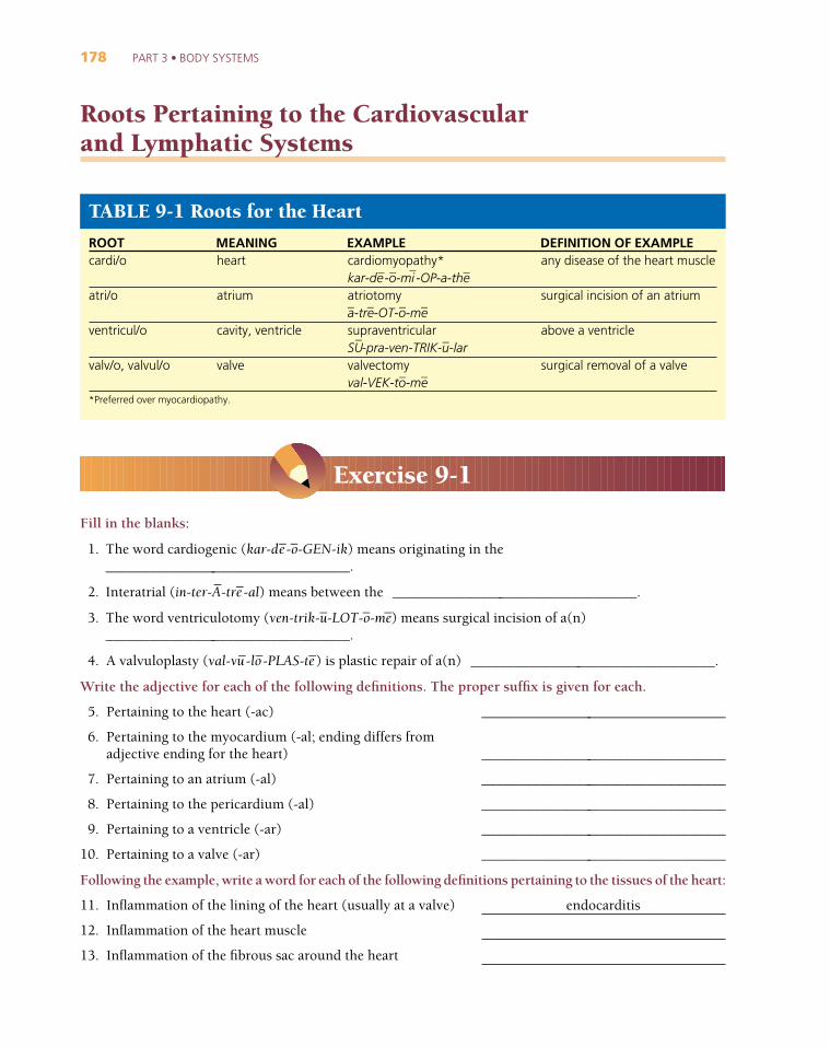
Roots Pertaining to the Cardiovascular and Lymphatic Systems
TABLE 9-1 Roots for the Heart
ROOT MEANING EXAMPLE DEFINITION OF EXAMPLEcardi/o
atri/o
ventricul/o
valv/o, valvul/o
*Preferred over myocardiopathy.
heart
atrium
cavity, ventricle
valve
any disease of the heart muscle
surgical incision of an atrium
above a ventricle
surgical removal of a valve
cardiomyopathy*kar-de
_-o_-mi
_-OP-a-th
_e
atriotomya_-tre
_-OT-o
_-me
_
supraventricularSU
_-pra-ven-TRIK-u
_-lar
valvectomyval-VEK-to
_-me
_
Fill in the blanks:
1. The word cardiogenic (kar-de_-o_-GEN-ik) means originating in the
__________________________________.
2. Interatrial (in-ter-A_-tre
_-al) means between the __________________________________.
3. The word ventriculotomy (ven-trik-u_-LOT-o
_-me
_) means surgical incision of a(n)
__________________________________.
4. A valvuloplasty (val-vu_-lo
_-PLAS-te
_) is plastic repair of a(n) __________________________________.
Write the adjective for each of the following definitions. The proper suffix is given for each.
5. Pertaining to the heart (-ac) __________________________________
6. Pertaining to the myocardium (-al; ending differs from adjective ending for the heart) __________________________________
7. Pertaining to an atrium (-al) __________________________________
8. Pertaining to the pericardium (-al) __________________________________
9. Pertaining to a ventricle (-ar) __________________________________
10. Pertaining to a valve (-ar) __________________________________
Following the example, write a word for each of the following definitions pertaining to the tissues of the heart:
11. Inflammation of the lining of the heart (usually at a valve) endocarditis
12. Inflammation of the heart muscle
13. Inflammation of the fibrous sac around the heart
Exercise 9-1
CHAPTER 9 • CIRCULATION: THE CARDIOVASCULAR AND LYMPHATIC SYSTEMS 179
Write a word for each of the following definitions:
14. Study (-logy) of the heart __________________________________
15. Enlargement (-megaly) of the heart __________________________________
16. Between (inter-) the ventricles __________________________________
17. Pertaining to an atrium and a ventricle __________________________________
18. Surgical incision of a valve __________________________________
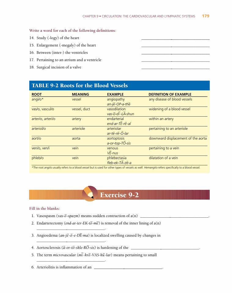
TABLE 9-2 Roots for the Blood Vessels
ROOT MEANING EXAMPLE DEFINITION OF EXAMPLEangi/o*
vas/o, vascul/o
arter/o, arteri/o
arteriol/o
aort/o
ven/o, ven/i
phleb/o
*The root angi/o usually refers to a blood vessel but is used for other types of vessels as well. Hemangi/o refers specifically to a blood vessel.
vessel
vessel, duct
artery
arteriole
aorta
vein
vein
any disease of blood vessels
widening of a blood vessel
within an artery
pertaining to an arteriole
downward displacement of the aorta
pertaining to a vein
dilatation of a vein
angiopathyan-je
_-OP-a-the
_
vasodilationvas-o
_-di
_-LA
_-shun
endarterialend-ar-TE
_-re
_-al
arteriolarar-te
_-re
_-O
_-lar
aortoptosisa-or-top-TO
_-sis
venousVE
_-nus
phlebectasiafleb-ek-TA
_-ze
_-a
Fill in the blanks:
1. Vasospasm (vas-o_-spazm) means sudden contraction of a(n) __________________________________.
2. Endarterectomy (end-ar-ter-EK-to_-me
_) is removal of the inner lining of a(n)
__________________________________.
3. Angioedema (an-je_-o_-e-DE
_-ma) is localized swelling caused by changes in
__________________________________.
4. Aortosclerosis (a_-or-to
_-skle-RO
_-sis) is hardening of the __________________________________.
5. The term microvascular (mi_-kro
_-VAS-ku
_-lar) means pertaining to small
__________________________________.
6. Arteriolitis is inflammation of an __________________________________.
Exercise 9-2
180 PART 3 • BODY SYSTEMS
Define the following words:
7. angiitis (an-je_-I_-tis) (note spelling); also angitis or vasculitis
8. cardiovascular (kar-de_-o_-VAS-ku
_-lar)
9. arteriorrhexis (ar-te_-re
_-o_-REK-sis)
10. intra-aortic (in-tra-a_-OR-tik)
11. phlebitis (fleb-I_-tis)
Use the ending -gram to form a word for a radiograph of each of the following:
12. vessels (use angi/o)
13. aorta
14. veins
Use the root angi/o to write a word with each of the following meanings:
15. Surgical removal (-ectomy) of a vessel
16. Dilatation (-ectasis) of a vessel
17. Formation (-genesis) of a vessel
18. Plastic repair of a vessel
Use the appropriate root to write a word with each of the following meanings:
19. Narrowing (-stenosis) of the aorta
20. Incision of an artery
21. Within (intra-) a vein
22. Excision of a vein
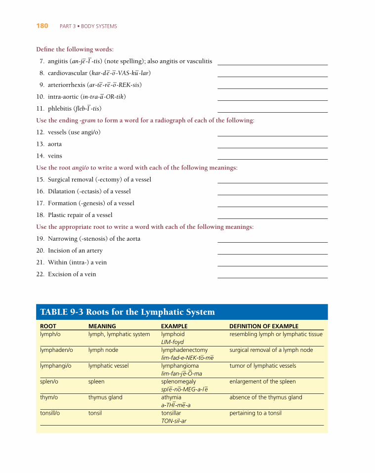
TABLE 9-3 Roots for the Lymphatic System
ROOT MEANING EXAMPLE DEFINITION OF EXAMPLElymph/o
lymphaden/o
lymphangi/o
splen/o
thym/o
tonsill/o
lymph, lymphatic system
lymph node
lymphatic vessel
spleen
thymus gland
tonsil
resembling lymph or lymphatic tissue
surgical removal of a lymph node
tumor of lymphatic vessels
enlargement of the spleen
absence of the thymus gland
pertaining to a tonsil
lymphoidLIM-foydlymphadenectomylim-fad-e-NEK-to
_-me
_
lymphangiomalim-fan-je
_-O_
-masplenomegalysple
_-no
_-MEG-a-l e
_
athymiaa-THI
_-me
_-a
tonsillarTON-sil-ar
CHAPTER 9 • CIRCULATION: THE CARDIOVASCULAR AND LYMPHATIC SYSTEMS 181
Fill in the blanks:
1. Lymphedema (limf-e-DE_-ma) means swelling caused by obstruction of the flow of
__________________________________.
2. Lymphadenitis (lim-fad-e-NI_-tis) is inflammation of a(n) __________________________________.
3. A lymphangiogram (lim-FAN-je_-o_-gram) is an x-ray image (radiograph) of
__________________________________.
4. The adjective splenic (SPLEN-ik) means pertaining to the __________________________________.
5. Thymectomy (thi_-MEK-to
_-me
_) is surgical removal of the __________________________________.
6. Tonsillopathy (ton-sil-OP-a-the_) is any disease of the __________________________________.
Identify and define the root in each of the following words:
Root Meaning of Root
7. lymphangial (lim-FAN-je_-al) lymphangi/o lymphatic vessel
8. lymphadenography (lim-fad-e-NOG-ra-f e_) _____________
9. perisplenitis (per-i-sple_-NI
_-tis) _____________
10. hypothymism (hi_-po
_-THI
_-mizm) _____________
11. tonsillectomy (ton-sil-EK-to_-me
_) _____________
Use the appropriate root to write a word with each of the following meanings:
12. Inflammation of lymphatic vessels
13. A tumor (-oma) of lymphatic tissue
14. Any disease (-pathy) of the lymph nodes
15. Pain (-algia) in the spleen
16. Inflammation of a tonsil
Clinical Aspects of the Circulatory System
AtherosclerosisThe accumulation of fatty deposits within the lining of an artery is termed atherosclerosis (Fig. 9-8). Thistype of deposit, called a plaque, begins to form when a vessel receives tiny injuries, usually at a point ofbranching. Plaques gradually thicken and harden with fibrous material, cells, and other deposits, restrictingthe lumen (opening) of the vessel and reducing blood flow to the tissues, a condition known as ischemia. Amajor risk factor for the development of atherosclerosis is dyslipidemia, abnormally high levels or imbalancein lipoproteins that are carried in the blood, especially high levels of cholesterol-containing low-densitylipoproteins (LDL). Other risk factors for atherosclerosis include smoking, high blood pressure, poor diet,inactivity, stress, and family history of the disorder. Atherosclerosis may involve any arteries, but most of its
Exercise 9-3

182 PART 3 • BODY SYSTEMS
effects are seen in the coronary vessels of the heart, the aorta, the carotid arteries in the neck, and vessels inthe brain.
Thrombosis and Embolism
Atherosclerosis predisposes a person to thrombosis, the formation of a blood clot within a vessel. The clot,called a thrombus, interrupts blood flow to the tissues supplied by that vessel, resulting in necrosis (tissuedeath). Blockage of a vessel by a thrombus or other mass carried in the bloodstream is an embolism, and themass itself is called an embolus. Usually the mass is a blood clot that breaks loose from the wall of a vessel, butit may also be air (as from injection or trauma), fat (as from marrow released after a bone break), bacteria, orother solid materials. Often a venous thrombus will travel through the heart and then lodge in an artery of thelungs, resulting in a life-threatening pulmonary embolism. An embolus from a carotid artery often blocks acerebral vessel, causing a cerebrovascular accident (CVA), commonly called stroke (see Chapter 17).
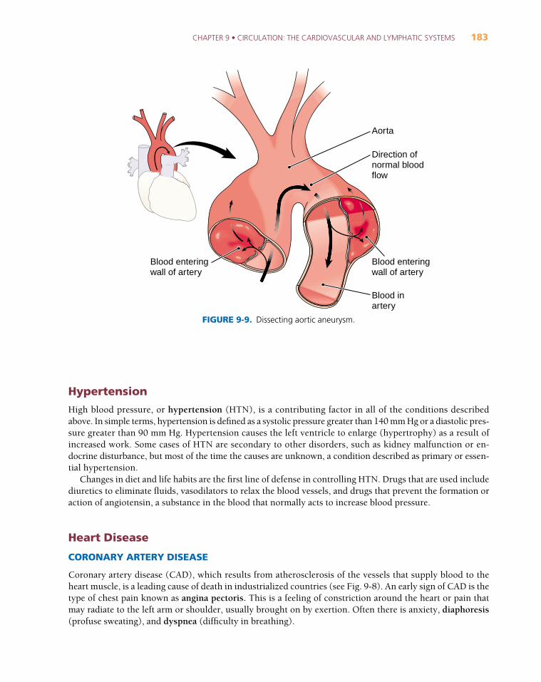
AneurysmAn arterial wall weakened by atherosclerosis, malformation, injury, or other causes may balloon out, form-ing an aneurysm. If an aneurysm ruptures, hemorrhage results. Rupture of a cerebral artery is another causeof stroke. The abdominal aorta and carotid arteries are also common sites of aneurysm. In a dissectinganeurysm (Fig. 9-9), blood hemorrhages into the thick middle layer of the artery wall, separating the mus-cle as it spreads and sometimes rupturing the vessel. The aorta is most commonly involved. It may be possi-ble to repair a dissecting aneurysm surgically with a graft.
Blood clotFat deposits Occlusion
FIGURE 9-8. Coronary atherosclerosis. (A) Fat de-posits narrow an artery leading to ischemia. (B) Block-age (occlusion) of a coronary artery. (C) Formation ofa blood clot (thrombus) leading to myocardial infarc-tion. (Adapted with permission from Cohen BJ, Wood DL. Memmler’s The Human Body in Health andDisease. 9th Ed. Philadelphia: Lippincott Williams &Wilkins, 2000.)A B C
CHAPTER 9 • CIRCULATION: THE CARDIOVASCULAR AND LYMPHATIC SYSTEMS 183
Hypertension
High blood pressure, or hypertension (HTN), is a contributing factor in all of the conditions describedabove. In simple terms, hypertension is defined as a systolic pressure greater than 140 mm Hg or a diastolic pres-sure greater than 90 mm Hg. Hypertension causes the left ventricle to enlarge (hypertrophy) as a result ofincreased work. Some cases of HTN are secondary to other disorders, such as kidney malfunction or en-docrine disturbance, but most of the time the causes are unknown, a condition described as primary or essen-tial hypertension.
Changes in diet and life habits are the first line of defense in controlling HTN. Drugs that are used includediuretics to eliminate fluids, vasodilators to relax the blood vessels, and drugs that prevent the formation oraction of angiotensin, a substance in the blood that normally acts to increase blood pressure.
Heart Disease
CORONARY ARTERY DISEASE
Coronary artery disease (CAD), which results from atherosclerosis of the vessels that supply blood to theheart muscle, is a leading cause of death in industrialized countries (see Fig. 9-8). An early sign of CAD is thetype of chest pain known as angina pectoris. This is a feeling of constriction around the heart or pain thatmay radiate to the left arm or shoulder, usually brought on by exertion. Often there is anxiety, diaphoresis(profuse sweating), and dyspnea (difficulty in breathing).
Aorta
Direction ofnormal blood flow
Blood inartery
Blood enteringwall of artery
Blood enteringwall of artery
FIGURE 9-9. Dissecting aortic aneurysm.
184 PART 3 • BODY SYSTEMS
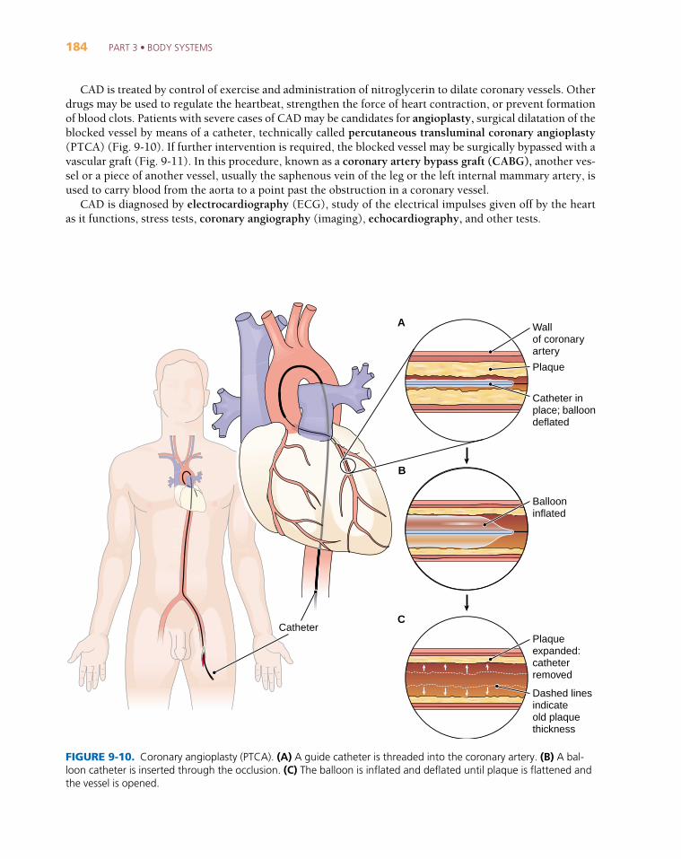
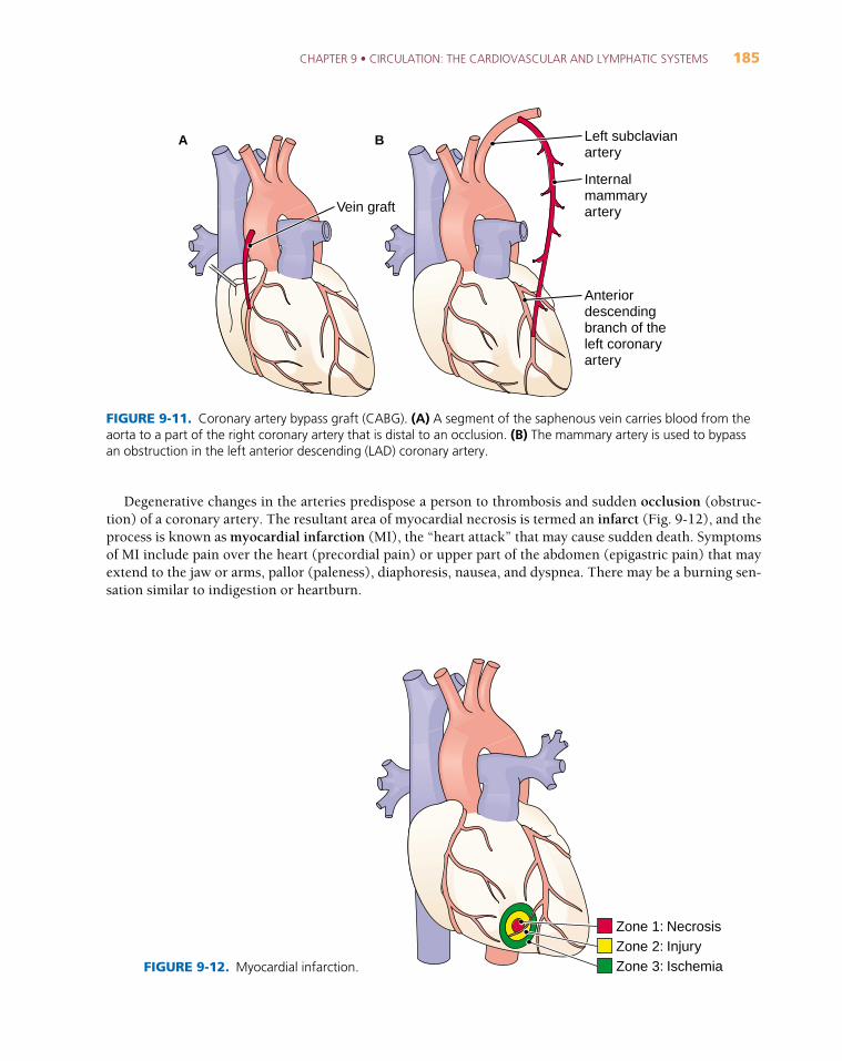
CAD is treated by control of exercise and administration of nitroglycerin to dilate coronary vessels. Otherdrugs may be used to regulate the heartbeat, strengthen the force of heart contraction, or prevent formationof blood clots. Patients with severe cases of CAD may be candidates for angioplasty, surgical dilatation of theblocked vessel by means of a catheter, technically called percutaneous transluminal coronary angioplasty(PTCA) (Fig. 9-10). If further intervention is required, the blocked vessel may be surgically bypassed with avascular graft (Fig. 9-11). In this procedure, known as a coronary artery bypass graft (CABG), another ves-sel or a piece of another vessel, usually the saphenous vein of the leg or the left internal mammary artery, isused to carry blood from the aorta to a point past the obstruction in a coronary vessel.
CAD is diagnosed by electrocardiography (ECG), study of the electrical impulses given off by the heartas it functions, stress tests, coronary angiography (imaging), echocardiography, and other tests.
Wallof coronaryartery
Plaque
Catheter in place; balloondeflated
Catheter
Ballooninflated
Plaqueexpanded:catheterremoved
Dashed lines indicateold plaquethickness
FIGURE 9-10. Coronary angioplasty (PTCA). (A) A guide catheter is threaded into the coronary artery. (B) A bal-loon catheter is inserted through the occlusion. (C) The balloon is inflated and deflated until plaque is flattened andthe vessel is opened.
A
B
C
CHAPTER 9 • CIRCULATION: THE CARDIOVASCULAR AND LYMPHATIC SYSTEMS 185
Degenerative changes in the arteries predispose a person to thrombosis and sudden occlusion (obstruc-tion) of a coronary artery. The resultant area of myocardial necrosis is termed an infarct (Fig. 9-12), and theprocess is known as myocardial infarction (MI), the “heart attack” that may cause sudden death. Symptomsof MI include pain over the heart (precordial pain) or upper part of the abdomen (epigastric pain) that mayextend to the jaw or arms, pallor (paleness), diaphoresis, nausea, and dyspnea. There may be a burning sen-sation similar to indigestion or heartburn.
Vein graft
Left subclavianartery
Internal mammaryartery
Anteriordescendingbranch of theleft coronaryartery
FIGURE 9-11. Coronary artery bypass graft (CABG). (A) A segment of the saphenous vein carries blood from theaorta to a part of the right coronary artery that is distal to an occlusion. (B) The mammary artery is used to bypassan obstruction in the left anterior descending (LAD) coronary artery.
Zone 1: NecrosisZone 2: InjuryZone 3: IschemiaFIGURE 9-12. Myocardial infarction.
A B
186 PART 3 • BODY SYSTEMS
MI is diagnosed by electrocardiography, by measurement of certain enzymes (CK, LDH, AST) released intothe blood from the damaged heart muscle and by a variety of other methods described later in this chapter.
Patient outcome is based on the degree of damage and early treatment to dissolve the clot and re-establishnormal heart rhythm.
ARRHYTHMIA
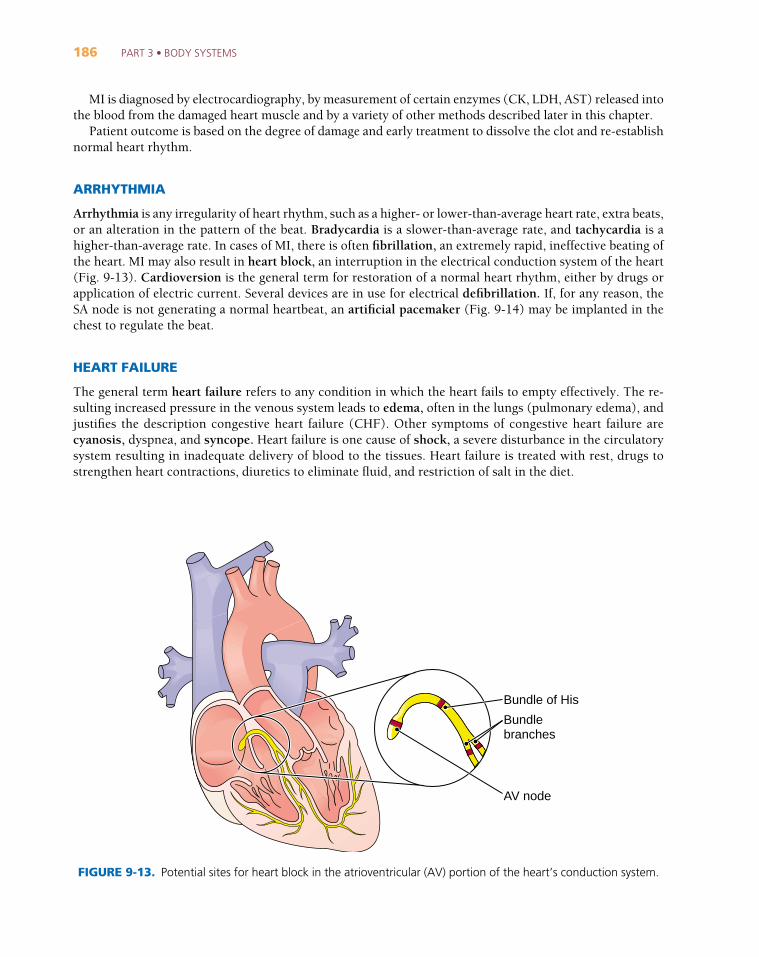
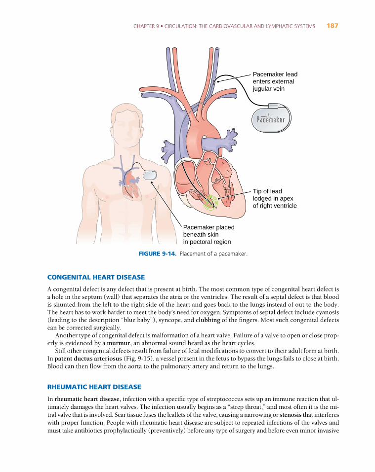
Arrhythmia is any irregularity of heart rhythm, such as a higher- or lower-than-average heart rate, extra beats,or an alteration in the pattern of the beat. Bradycardia is a slower-than-average rate, and tachycardia is ahigher-than-average rate. In cases of MI, there is often fibrillation, an extremely rapid, ineffective beating ofthe heart. MI may also result in heart block, an interruption in the electrical conduction system of the heart(Fig. 9-13). Cardioversion is the general term for restoration of a normal heart rhythm, either by drugs orapplication of electric current. Several devices are in use for electrical defibrillation. If, for any reason, theSA node is not generating a normal heartbeat, an artificial pacemaker (Fig. 9-14) may be implanted in thechest to regulate the beat.
HEART FAILURE
The general term heart failure refers to any condition in which the heart fails to empty effectively. The re-sulting increased pressure in the venous system leads to edema, often in the lungs (pulmonary edema), andjustifies the description congestive heart failure (CHF). Other symptoms of congestive heart failure arecyanosis, dyspnea, and syncope. Heart failure is one cause of shock, a severe disturbance in the circulatorysystem resulting in inadequate delivery of blood to the tissues. Heart failure is treated with rest, drugs tostrengthen heart contractions, diuretics to eliminate fluid, and restriction of salt in the diet.
AV node
Bundlebranches
Bundle of His
FIGURE 9-13. Potential sites for heart block in the atrioventricular (AV) portion of the heart’s conduction system.
CHAPTER 9 • CIRCULATION: THE CARDIOVASCULAR AND LYMPHATIC SYSTEMS 187
CONGENITAL HEART DISEASE
A congenital defect is any defect that is present at birth. The most common type of congenital heart defect isa hole in the septum (wall) that separates the atria or the ventricles. The result of a septal defect is that bloodis shunted from the left to the right side of the heart and goes back to the lungs instead of out to the body.The heart has to work harder to meet the body’s need for oxygen. Symptoms of septal defect include cyanosis(leading to the description “blue baby”), syncope, and clubbing of the fingers. Most such congenital defectscan be corrected surgically.
Another type of congenital defect is malformation of a heart valve. Failure of a valve to open or close prop-erly is evidenced by a murmur, an abnormal sound heard as the heart cycles.
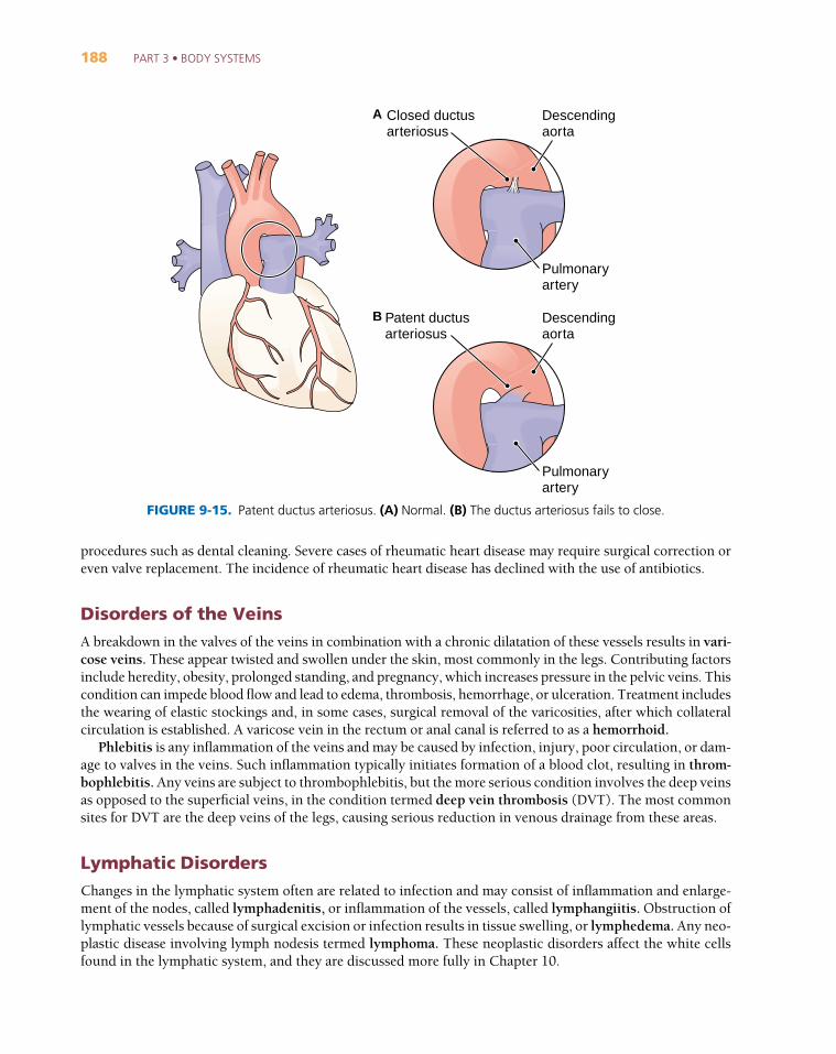
Still other congenital defects result from failure of fetal modifications to convert to their adult form at birth.In patent ductus arteriosus (Fig. 9-15), a vessel present in the fetus to bypass the lungs fails to close at birth.Blood can then flow from the aorta to the pulmonary artery and return to the lungs.
RHEUMATIC HEART DISEASE
In rheumatic heart disease, infection with a specific type of streptococcus sets up an immune reaction that ul-timately damages the heart valves. The infection usually begins as a “strep throat,” and most often it is the mi-tral valve that is involved. Scar tissue fuses the leaflets of the valve, causing a narrowing or stenosis that interfereswith proper function. People with rheumatic heart disease are subject to repeated infections of the valves andmust take antibiotics prophylactically (preventively) before any type of surgery and before even minor invasive
Pacemaker leadenters external jugular vein
Tip of leadlodged in apexof right ventricle
Pacemaker placedbeneath skinin pectoral region
FIGURE 9-14. Placement of a pacemaker.
188 PART 3 • BODY SYSTEMS
procedures such as dental cleaning. Severe cases of rheumatic heart disease may require surgical correction oreven valve replacement. The incidence of rheumatic heart disease has declined with the use of antibiotics.
Disorders of the VeinsA breakdown in the valves of the veins in combination with a chronic dilatation of these vessels results in vari-cose veins. These appear twisted and swollen under the skin, most commonly in the legs. Contributing factorsinclude heredity, obesity, prolonged standing, and pregnancy, which increases pressure in the pelvic veins. Thiscondition can impede blood flow and lead to edema, thrombosis, hemorrhage, or ulceration. Treatment includesthe wearing of elastic stockings and, in some cases, surgical removal of the varicosities, after which collateralcirculation is established. A varicose vein in the rectum or anal canal is referred to as a hemorrhoid.
Phlebitis is any inflammation of the veins and may be caused by infection, injury, poor circulation, or dam-age to valves in the veins. Such inflammation typically initiates formation of a blood clot, resulting in throm-bophlebitis. Any veins are subject to thrombophlebitis, but the more serious condition involves the deep veinsas opposed to the superficial veins, in the condition termed deep vein thrombosis (DVT). The most commonsites for DVT are the deep veins of the legs, causing serious reduction in venous drainage from these areas.
Lymphatic DisordersChanges in the lymphatic system often are related to infection and may consist of inflammation and enlarge-ment of the nodes, called lymphadenitis, or inflammation of the vessels, called lymphangiitis. Obstruction oflymphatic vessels because of surgical excision or infection results in tissue swelling, or lymphedema. Any neo-plastic disease involving lymph nodesis termed lymphoma. These neoplastic disorders affect the white cellsfound in the lymphatic system, and they are discussed more fully in Chapter 10.
Patent ductusarteriosus
Descendingaorta
Pulmonaryartery
Closed ductusarteriosus
Descendingaorta
Pulmonaryartery
FIGURE 9-15. Patent ductus arteriosus. (A) Normal. (B) The ductus arteriosus fails to close.
A
B
CHAPTER 9 • CIRCULATION: THE CARDIOVASCULAR AND LYMPHATIC SYSTEMS 189
Key Clinical Terms
CARDIOVASCULAR DISORDERSaneurysmAN-u
_-rizm
angina pectorisan-JI
_-na PEK-to
_-ris
arrhythmiaa-RITH-me
_-a
atherosclerosisath-er-o
_-skle-RO
_-sis
bradycardiabrad-e
_-KAR-de-a
cerebrovascularaccident (CVA)ser-e-bro
_-VAS-ku
_-lar
clubbingKLUB-ing
cyanosissi_-a-NO
_-sis
deep vein thrombosis (DVT)
diaphoresisdi_-a-f o
_-RE
_-sis
dissecting aneurysm
dyslipidemiadis-lip-i-DE
_-me
_-a
dyspneaDYSP-ne
_-a
edemae-DE
_-ma
embolismEM-bo
_-lizm
A localized abnormal dilation of a blood vessel, usually an artery,caused by weakness of the vessel wall; may eventually burst
A feeling of constriction around the heart or pain that may radiate tothe left arm or shoulder, usually brought on by exertion; caused byinsufficient blood supply to the heart
Any abnormality in the rate or rhythm of the heartbeat (literally“without rhythm”; note doubled r). Also called dysrhythmia.
The development of fatty, fibrous patches (plaques) in the lining ofarteries, causing narrowing of the lumen and hardening of the vesselwall. The most common form of arteriosclerosis (hardening of the arteries). Root ather/o means “porridge” or “gruel.”
A slow heart rate of less than 60 beats per minute
Sudden damage to the brain resulting from reduction of blood flow.Causes include atherosclerosis, embolism, thrombosis, or hemor-rhage from a ruptured aneurysm; commonly called stroke.
Enlargement of the ends of the fingers and toes caused by growth ofthe soft tissue around the nails (see Fig. 7-10). Seen in a variety of diseases in which there is poor peripheral circulation.
Bluish discoloration of the skin caused by lack of oxygen
Thrombophlebitis involving the deep veins
Profuse sweating
An aneurysm in which blood enters the arterial wall and separatesthe layers. Usually involves the aorta (see Fig. 9-9).
Disorder in serum lipid levels, which is an important factor in devel-opment of atherosclerosis. Includes hyperlipidemia (high lipids), hypercholesterolemia (high cholesterol), hypertriglyceridemia (hightriglycerides).
Difficult or labored breathing (-pnea)
Swelling of body tissues caused by the presence of excess fluid.Causes include cardiovascular disturbances, kidney failure, inflam-mation, and malnutrition.
Obstruction of a blood vessel by a blood clot or other matter carriedin the circulation
190 PART 3 • BODY SYSTEMS
Cardiovascular Disorders, continuedembolusEM-bo
_-lus
fibrillationfi-bri-LA
_-shun
heart block
heart failure
hemorrhoidHEM-o
_-royd
hypertensionhi_-per-TEN-shun
infarctin-FARKT
ischemiais-KE
_-me
_-a
murmur
myocardial infarction (MI)mi
_-o_-KAR-de
_-al
in-FARK-shun
occlusiono_-KLU
_-zhun
patent ductus arteriosusPA
_-tent DUK-tus
ar-te_r-e
_-O_-sus
phlebitisfle-BI
_-tis
plaqueplak
rheumatic heart diseaseru_-MAT-ik
A mass carried in the circulation. Usually a blood clot, but may alsobe air, fat, bacteria, or other solid matter from within or from outsidethe body.
Spontaneous, quivering, and ineffectual contraction of muscle fibers,as in the atria or the ventricles
An interference in the conduction system of the heart resulting in arrhythmia (see Fig. 9-13). The condition is classified in order ofincreasing severity as first-, second-, or third-degree heart block.Block in a bundle branch is designated as a left or right bundlebranch block (BBB).
A condition caused by the inability of the heart to maintain adequatecirculation of blood
A varicose vein in the rectum
A condition of higher-than-normal blood pressure. Essential (pri-mary, idiopathic) hypertension has no known cause.
An area of localized necrosis (death) of tissue resulting from a block-age or a narrowing of the artery that supplies the area
Local deficiency of blood supply caused by obstruction of the circula-tion (root hem/o)
An abnormal heart sound
Localized necrosis (death) of cardiac muscle tissue resulting fromblockage or narrowing of the coronary artery that supplies that area.Myocardial infarction is usually caused by formation of a thrombus(clot) in a vessel (see Fig. 9-12).
A closing off or obstruction, as of a vessel
Persistence of the ductus arteriosus after birth. The ductus arteriosusis a vessel that connects the pulmonary artery to the descending aortain the fetus to bypass the lungs.
Inflammation of a vein
A patch. With regard to the cardiovascular system, a deposit of fattymaterial and other substances on a vessel wall that impedes bloodflow and may block the vessel. Atheromatous plaque.
Damage to heart valves after infection with a type of streptococcus(group A hemolytic streptococcus). The antibodies produced in response to the infection produce scarring of the valves, usually themitral valve.
CHAPTER 9 • CIRCULATION: THE CARDIOVASCULAR AND LYMPHATIC SYSTEMS 191
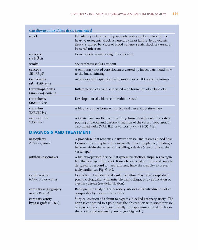
Cardiovascular Disorders, continuedshock
stenosisste-NO
_-sis
stroke
syncopeSIN-ko
_-pe
_
tachycardiatak-i-KAR-de
_-a
thrombophlebitisthrom-bo
_-f le-BI
_-tis
thrombosisthrom-BO
_-sis
thrombusTHROM-bus
varicose veinVAR-i-ko
_s
DIAGNOSIS AND TREATMENTangioplastyAN-je
_-o_-plas-te
_
artificial pacemaker
cardioversionKAR-de
_-o_-ver-zhun
coronary angiographyan-je
_-OG-ra-f e
_
coronary artery bypass graft (CABG)
Circulatory failure resulting in inadequate supply of blood to theheart. Cardiogenic shock is caused by heart failure; hypovolemicshock is caused by a loss of blood volume; septic shock is caused bybacterial infection.
Constriction or narrowing of an opening
See cerebrovascular accident
A temporary loss of consciousness caused by inadequate blood flowto the brain; fainting
An abnormally rapid heart rate, usually over 100 beats per minute
Inflammation of a vein associated with formation of a blood clot
Development of a blood clot within a vessel
A blood clot that forms within a blood vessel (root thromb/o)
A twisted and swollen vein resulting from breakdown of the valves,pooling of blood, and chronic dilatation of the vessel (root varic/o);also called varix (VAR-iks) or varicosity (var-i-KOS-i-te
_)
A procedure that reopens a narrowed vessel and restores blood flow.Commonly accomplished by surgically removing plaque, inflating aballoon within the vessel, or installing a device (stent) to keep thevessel open.
A battery-operated device that generates electrical impulses to regu-late the beating of the heart. It may be external or implanted, may bedesigned to respond to need, and may have the capacity to preventtachycardia (see Fig. 9-14).
Correction of an abnormal cardiac rhythm. May be accomplishedpharmacologically, with antiarrhythmic drugs, or by application ofelectric current (see defibrillation).
Radiographic study of the coronary arteries after introduction of anopaque dye by means of a catheter
Surgical creation of a shunt to bypass a blocked coronary artery. Theaorta is connected to a point past the obstruction with another vesselor a piece of another vessel, usually the saphenous vein of the leg orthe left internal mammary artery (see Fig. 9-11).
192 PART 3 • BODY SYSTEMS
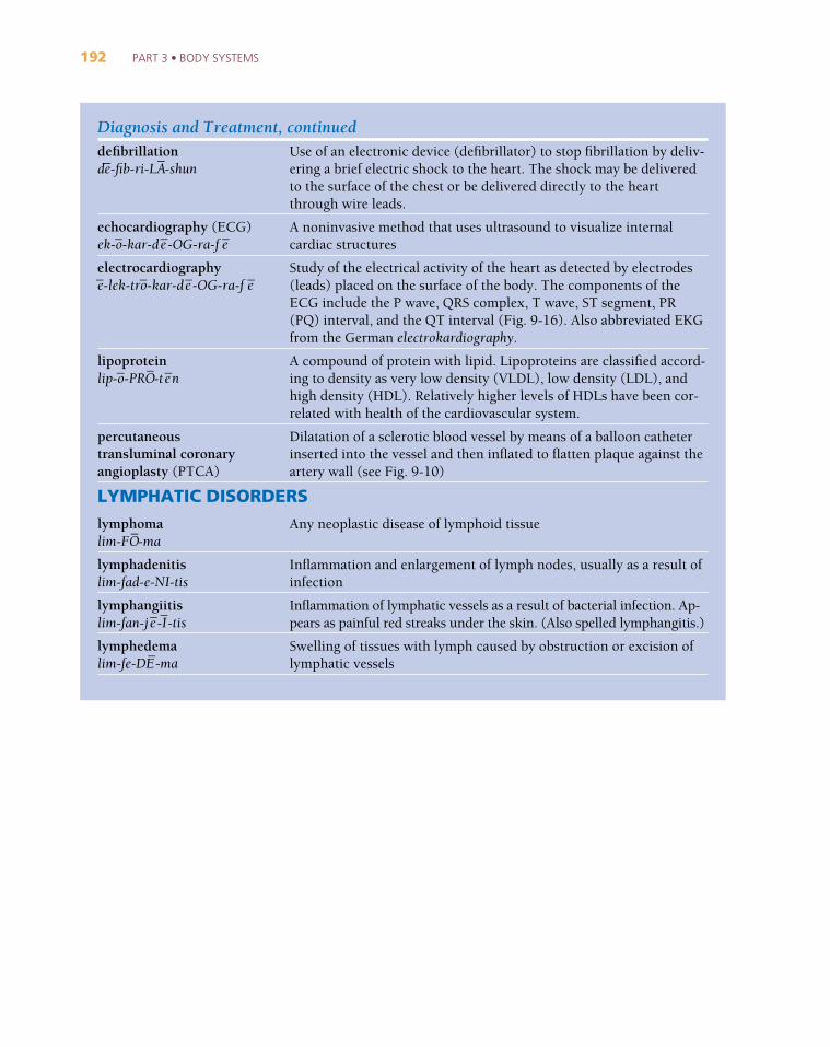
Diagnosis and Treatment, continueddefibrillationde_-fib-ri-LA
_-shun
echocardiography (ECG)ek-o
_-kar-de
_-OG-ra-f e
_
electrocardiographye_-lek-tro
_-kar-de
_-OG-ra-f e
_
lipoproteinlip-o
_-PRO
_-te
_n
percutaneoustransluminal coronary angioplasty (PTCA)
LYMPHATIC DISORDERSlymphomalim-FO
_-ma
lymphadenitislim-fad-e-NI-tis
lymphangiitislim-fan-je
_-I_-tis
lymphedemalim-fe-DE
_-ma
Use of an electronic device (defibrillator) to stop fibrillation by deliv-ering a brief electric shock to the heart. The shock may be deliveredto the surface of the chest or be delivered directly to the heartthrough wire leads.
A noninvasive method that uses ultrasound to visualize internalcardiac structures
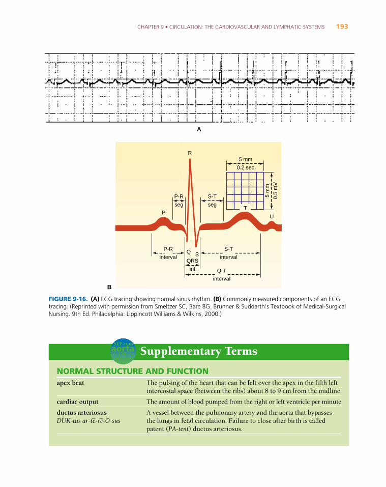
Study of the electrical activity of the heart as detected by electrodes(leads) placed on the surface of the body. The components of theECG include the P wave, QRS complex, T wave, ST segment, PR(PQ) interval, and the QT interval (Fig. 9-16). Also abbreviated EKGfrom the German electrokardiography.
A compound of protein with lipid. Lipoproteins are classified accord-ing to density as very low density (VLDL), low density (LDL), andhigh density (HDL). Relatively higher levels of HDLs have been cor-related with health of the cardiovascular system.
Dilatation of a sclerotic blood vessel by means of a balloon catheterinserted into the vessel and then inflated to flatten plaque against theartery wall (see Fig. 9-10)
Any neoplastic disease of lymphoid tissue
Inflammation and enlargement of lymph nodes, usually as a result ofinfection
Inflammation of lymphatic vessels as a result of bacterial infection. Ap-pears as painful red streaks under the skin. (Also spelled lymphangitis.)
Swelling of tissues with lymph caused by obstruction or excision oflymphatic vessels
CHAPTER 9 • CIRCULATION: THE CARDIOVASCULAR AND LYMPHATIC SYSTEMS 193
P-R
interval
P-R
seg
QRS
int.
Q S
S-T
seg
5 mm
0.2 sec
5 m
m
0.5
mV
P
R
U
Q-T
interval
T
S-T
interval
FIGURE 9-16. (A) ECG tracing showing normal sinus rhythm. (B) Commonly measured components of an ECGtracing. (Reprinted with permission from Smeltzer SC, Bare BG. Brunner & Suddarth’s Textbook of Medical-SurgicalNursing. 9th Ed. Philadelphia: Lippincott Williams & Wilkins, 2000.)
Supplementary Termsaortaa\-OR-ta
SEP-tum
NORMAL STRUCTURE AND FUNCTIONapex beat
cardiac output
ductus arteriosusDUK-tus ar-te
_-re
_-O-sus
The pulsing of the heart that can be felt over the apex in the fifth leftintercostal space (between the ribs) about 8 to 9 cm from the midline
The amount of blood pumped from the right or left ventricle per minute
A vessel between the pulmonary artery and the aorta that bypassesthe lungs in fetal circulation. Failure to close after birth is calledpatent (PA-tent) ductus arteriosus.
B
A
194 PART 3 • BODY SYSTEMS
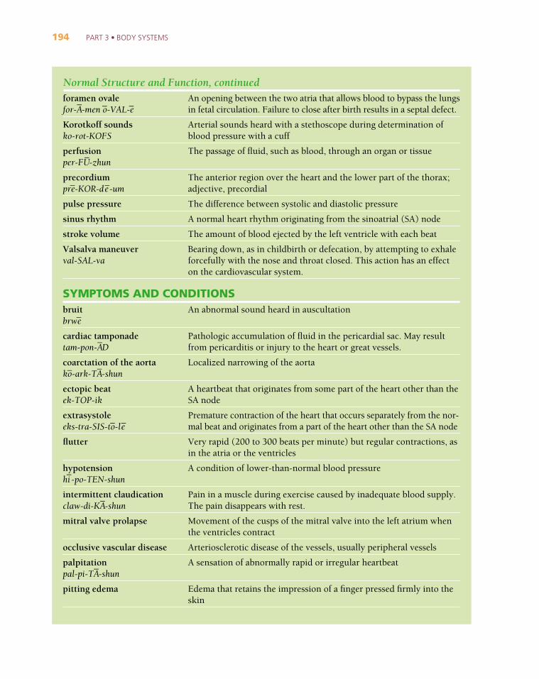
Normal Structure and Function, continuedforamen ovalefor-A
_-men o
_-VAL-e
_
Korotkoff soundsko-rot-KOFS
perfusionper-FU
_-zhun
precordiumpre
_-KOR-de
_-um
pulse pressure
sinus rhythm
stroke volume
Valsalva maneuverval-SAL-va
SYMPTOMS AND CONDITIONSbruitbrwe
_
cardiac tamponadetam-pon-A
_D
coarctation of the aortako_-ark-TA
_-shun
ectopic beatek-TOP-ik
extrasystoleeks-tra-SIS-to
_-le
_
flutter
hypotensionhi_-po-TEN-shun
intermittent claudicationclaw-di-KA
_-shun
mitral valve prolapse
occlusive vascular disease
palpitationpal-pi-TA
_-shun
pitting edema
An opening between the two atria that allows blood to bypass the lungsin fetal circulation. Failure to close after birth results in a septal defect.
Arterial sounds heard with a stethoscope during determination ofblood pressure with a cuff
The passage of fluid, such as blood, through an organ or tissue
The anterior region over the heart and the lower part of the thorax;adjective, precordial
The difference between systolic and diastolic pressure
A normal heart rhythm originating from the sinoatrial (SA) node
The amount of blood ejected by the left ventricle with each beat
Bearing down, as in childbirth or defecation, by attempting to exhaleforcefully with the nose and throat closed. This action has an effecton the cardiovascular system.
An abnormal sound heard in auscultation
Pathologic accumulation of fluid in the pericardial sac. May resultfrom pericarditis or injury to the heart or great vessels.
Localized narrowing of the aorta
A heartbeat that originates from some part of the heart other than theSA node
Premature contraction of the heart that occurs separately from the nor-mal beat and originates from a part of the heart other than the SA node
Very rapid (200 to 300 beats per minute) but regular contractions, asin the atria or the ventricles
A condition of lower-than-normal blood pressure
Pain in a muscle during exercise caused by inadequate blood supply.The pain disappears with rest.
Movement of the cusps of the mitral valve into the left atrium whenthe ventricles contract
Arteriosclerotic disease of the vessels, usually peripheral vessels
A sensation of abnormally rapid or irregular heartbeat
Edema that retains the impression of a finger pressed firmly into theskin
CHAPTER 9 • CIRCULATION: THE CARDIOVASCULAR AND LYMPHATIC SYSTEMS 195
Symptoms and Conditions, continuedpolyarteritis nodosano_-DO
_-sa
Raynaud diseasera_-NO
_
regurgitationre_-gur-ji-TA
_-shun
stasisST
_A-sis
subacute bacterial endocarditis (SBE)
tetralogy of Fallotfal-O
_
thromboangiitisobliterans
vegetation
Wolff-Parkinson-Whitesyndrome (WPW)
DIAGNOSIScardiac catheterization
central venous pressure (CVP)
cineangiocardiographysin-e-an-je
_-o_-kar-de
_-OG-ra-f e
_
Doppler echocardiography
enzyme studies
heart scan
Potentially fatal collagen disease causing inflammation of small vis-ceral arteries. Symptoms depend on the organ affected.
A disorder characterized by abnormal constriction of peripheral ves-sels in the arms and legs on exposure to cold
A backward flow, such as the backflow of blood through a defectivevalve
Stoppage of normal blood normal flow, as of blood or urine. Bloodstasis may lead to dermatitis and ulcer formation.
Growth of bacteria in a heart or valves previously damaged byrheumatic fever
A combination of four congenital heart abnormalities: pulmonaryartery stenosis, interventricular septal defect, displacement of theaorta to the right, right ventricular hypertrophy
Inflammation and thrombus formation resulting in occlusion of smallvessels, especially in the legs. Most common in young men and corre-lated with heavy smoking. Thrombotic occlusion of leg vessels inyoung men leading to gangrene of the feet. Patients show a hypersen-sitivity to tobacco. Also called Buerger disease.
Irregular outgrowths of bacteria on the heart valves; associated withrheumatic fever
A cardiac arrhythmia consisting of tachycardia and a premature ven-tricular beat caused by an alternate conduction pathway
Passage of a catheter into the heart through a vessel to inject a con-trast medium for imaging, diagnosing abnormalities, obtaining sam-ples, or measuring pressure
Pressure in the superior vena cava
The photographic recording of fluoroscopic images of the heart andlarge vessels using motion picture techniques
An imaging method used to study the rate and pattern of blood flow
Measurement of serum levels of enzymes that are released in increasedamounts from damaged heart tissue. These include CK (creatinekinase), LDH (lactate dehydrogenase), AST (aspartate amino-transferase), and ALT (alanine aminotransferase).
Imaging of the heart after injection of a radioactive isotope. The PYP(pyrophosphate) scan using technetium-99m (99mTc) is used to testfor myocardial infarction because the isotope is taken up by damagedtissue. The MUGA (multigated acquisition) scan gives informationon heart function.
196 PART 3 • BODY SYSTEMS
Diagnosis, continuedHolter monitor
homocysteineho_-mo
_-SIS-te
_n
phlebotomistfle-BOT-o
_-mist
phonocardiographyfo_-no
_-kar-de
_-OG-ra-f e
_
plethysmographyple-thiz-MOG-ra-f e
_
pulmonary wedge pressure (PWP)
stress test
Swan-Ganz catheter
transesophagealechocardiography (TEE)
triglyceridestri
_-GLIS-er-i
_dz
ventriculographyven-trik-u
_-LOG-ra-f e
_
TREATMENT AND SURGICAL PROCEDURESatherectomyath-er-EK-to
_-me
_
automated external defibrillator (AED)
commissurotomykom-i-shur-OT-o
_-me
_
embolectomyem-bo
_-LEK-to
_-me
_
implantable cardioverter defibrillator (ICD)
A portable device that can record up to 24 hours of an individual’sECG readings during normal activity
An amino acid that at higher-than-normal levels in the blood is asso-ciated with increased risk of cardiovascular disease
Technician who specializes in drawing blood
Electronic recording of heart sounds
Measurement of changes in the size of a part based on the amount ofblood contained in or passing through it. Impedance plethysmogra-phy measures changes in electrical resistance and is used in diagnosisof deep vein thrombosis.
Pressure measured by a catheter in a branch of the pulmonary artery.It is an indirect measure of pressure in the left atrium.
Evaluation of physical fitness by continuous ECG monitoring duringexercise. In a thallium stress test, a radioactive isotope of thallium isadministered to trace blood flow through the heart during exercise.
A cardiac catheter with a balloon at the tip that is used to measurepulmonary arterial pressure. It is flow-guided through a vein into theright side of the heart and then into the pulmonary artery.
Use of an ultrasound transducer placed endoscopically into theesophagus to obtain images of the heart
Simple fats that circulate in the bloodstream
X-ray study of the ventricles of the heart after introduction of anopaque dye by means of a catheter
Removal of atheromatous plaque from the lining of a vessel. May bedone by open surgery or through the lumen of the vessel.
Electronic device that detects arrhythmia and automatically delivers acorrect programmed shock. These devices, used on the scene of aheart attack, can prevent death.
Surgical incision of a scarred mitral valve to increase the size of thevalve opening
Surgical removal of an embolus
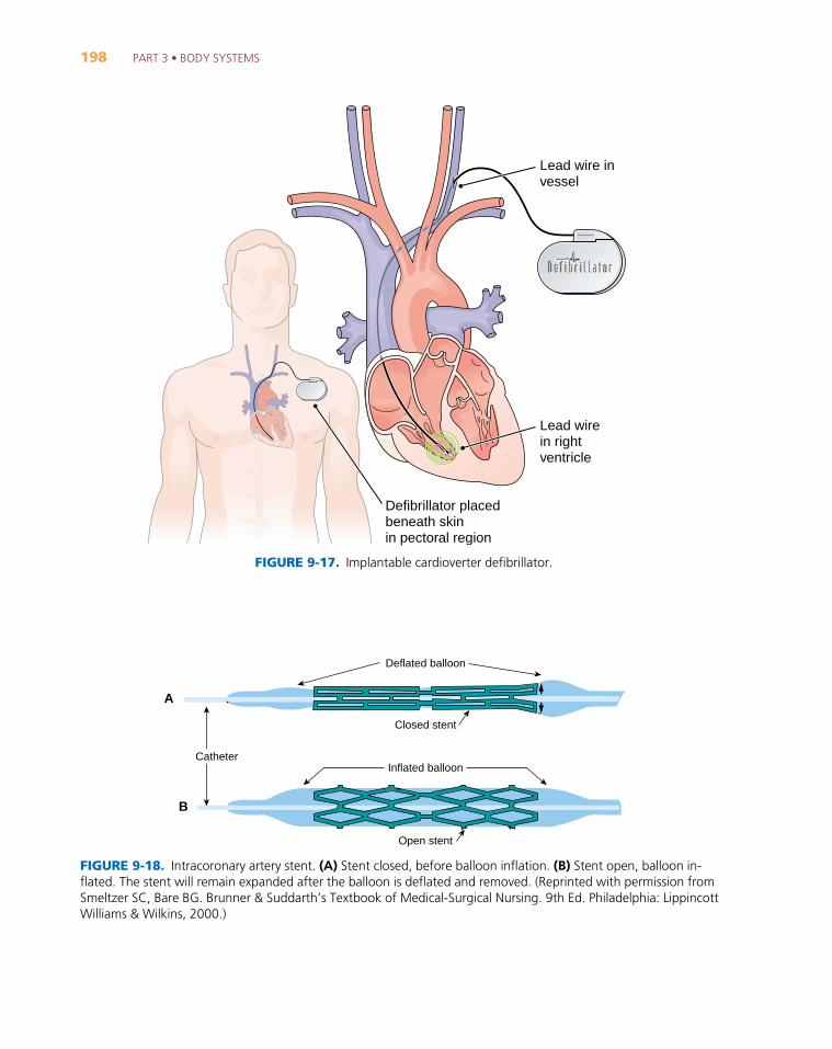
A battery-powered device that can shock the heart during fibrillationto restore a normal rhythm. The ICD is implanted under the collar-bone. A lead wire is threaded through the pulmonary artery into theright ventricle (Fig. 9-17).
CHAPTER 9 • CIRCULATION: THE CARDIOVASCULAR AND LYMPHATIC SYSTEMS 197
Treatment and Surgical Procedures, continuedintra-aortic balloon pump (IABP)
left ventricular assist device (LVAD)
stent
MEDICATIONSangiotensin-convertingenzyme (ACE) inhibitor
angiotensin II receptor antagonist
antiarrhythmic agent
beta-adrenergicblocking agent
calcium channel blocker
digitalisdij-i-TAL-is
diureticdi_-u_-RET-ik
hypolipidemic agenthi_-po
_-lip-i-DE
_-mik
lidocaineLI_-do
_-ka
_n
nitroglycerinni_-tro
_-GLIS-er-in
statins
streptokinase (SK)strep-to
_-KI
_-nas
tissue plasminogen activator (tPA)
vasodilatorvas-o
_-di
_-LA
_-tor
A mechanical-assist device that consists of an inflatable balloonpump inserted through the femoral artery into the thoracic aorta. Itinflates during diastole to improve coronary circulation and deflatesbefore systole to allow blood ejection from the heart.
A pump that takes over the function of the left ventricle in deliveringblood into the systemic circuit. These devices are used to assist patients awaiting heart transplantation or those who are recoveringfrom heart failure.
A small metal device in the shape of a coil or slotted tube that isplaced inside an artery to keep the vessel open after balloon angio-plasty (Fig. 9-18).
A drug that lowers blood pressure by blocking the formation in theblood of angiotensin II, a substance that normally acts to increaseblood pressure
A drug that blocks tissue receptors for angiotensin II
A drug that regulates the rate and rhythm of the heartbeat
Drug that decreases the rate and strength of heart contractions
Drug that controls the rate and force of heart contraction by regulat-ing calcium entrance into the cells
A drug that slows and strengthens heart muscle contractions
Drug that eliminates fluid by increasing the output of urine by thekidneys. Lowered blood volume decreases the workload of the heart.
Drug that lowers serum cholesterol
A local anesthetic that is used intravenously to treat cardiac arrhythmias
A drug used in the treatment of angina pectoris to dilate coronaryvessels
Drugs that act to lower lipids in the blood. The drug names end with-statin, such as lovastatin, pravastatin, atorvastatin.
An enzyme used to dissolve blood clots
A drug used to dissolve blood clots. It activates production of a sub-stance (plasmin) in the blood that normally dissolves clots.
A drug that widens blood vessels and improves blood flow
198 PART 3 • BODY SYSTEMS
Lead wire in vessel
Lead wirein right ventricle
Defibrillator placedbeneath skinin pectoral region
FIGURE 9-17. Implantable cardioverter defibrillator.
Catheter
Open stent
Inflated balloon
Deflated balloon
Closed stent
FIGURE 9-18. Intracoronary artery stent. (A) Stent closed, before balloon inflation. (B) Stent open, balloon in-flated. The stent will remain expanded after the balloon is deflated and removed. (Reprinted with permission fromSmeltzer SC, Bare BG. Brunner & Suddarth’s Textbook of Medical-Surgical Nursing. 9th Ed. Philadelphia: LippincottWilliams & Wilkins, 2000.)
A
B
CHAPTER 9 • CIRCULATION: THE CARDIOVASCULAR AND LYMPHATIC SYSTEMS 199
ABBREVIATIONS
ACE Angiotensin-converting enzymeAED Automated external defibrillatorAF Atrial fibrillationALT Alanine aminotransferase (SGPT)AMI Acute myocardial infarctionAPC Atrial premature complexAR Aortic regurgitationAS Aortic stenosis; arteriosclerosisASCVD Arteriosclerotic cardiovascular diseaseASD Atrial septal defectASHD Arteriosclerotic heart diseaseAST Aspartate aminotransferase (SGOT)AT Atrial tachycardiaAV AtrioventricularBBB Bundle branch block (left or right)BP Blood pressurebpm Beats per minuteCABG Coronary artery bypass graftCAD Coronary artery diseaseCCU Coronary/cardiac care unitCHD Coronary heart diseaseCHF Congestive heart failureC(P)K Creatine (phospho)kinaseCPR Cardiopulmonary resuscitationCVD Cardiovascular diseaseCVI Chronic venous insufficiencyCVP Central venous pressureDOE Dyspnea on exertionDVT Deep vein thrombosisECG (EKG) ElectrocardiogramHDL High-density lipoproteinHTN HypertensionIABP Intra-aortic balloon pumpICD Implantable cardioverter defibrillatorIVCD Intraventricular conduction delayJVP Jugular venous pulseLAD Left anterior descending (coronary artery)LAHB Left anterior hemiblockLDH Lactic dehydrogenaseLDL Low-density lipoproteinLV Left ventricle
LVAD Left ventricular assist deviceLVEDP Left ventricular end-diastolic pressureLVH Left ventricular hypertrophyMI Myocardial infarctionmm Hg Millimeters of mercuryMR Mitral regurgitation, refluxMS Mitral stenosisMUGA Multigated acquisition (scan)MVP Mitral valve prolapseMVR Mitral valve replacementNSR Normal sinus rhythmP PulsePAC Premature atrial contractionPAP Pulmonary arterial pressurePMI Point of maximal impulsePSVT Paroxysmal supraventricular tachycardiaPTCA Percutaneous transluminal coronary
angioplastyPVC Premature ventricular contractionPVD Peripheral vascular diseasePWP Pulmonary (artery) wedge pressurePYP Pyrophosphate (scan)S1 The first heart soundS2 The second heart soundSA SinoatrialSBE Subacute bacterial endocarditisSGOT Serum glutamic oxaloacetic trans-
aminase (AST)SK StreptokinaseSVT Supraventricular tachycardia99mTc Technetium-99mTEE Transesophageal echocardiographytPA Tissue plasminogen activatorVAD Ventricular assist deviceVF Ventricular fibrillationVLDL Very low density lipoproteinVPC Ventricular premature complexVSD Ventricular septal defectVT Ventricular tachycardiaVTE Venous thromboembolismWPW Wolff-Parkinson-White syndrome
200 PART 3 • BODY SYSTEMS
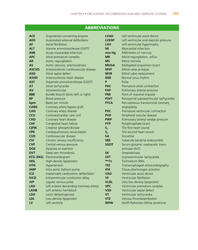
The Cardiovascular SystemWrite the name of each numbered part on the corresponding line of the answer sheet.
1.
2.
3.
4.
5.
6.
7.
8.
9.
10.
11.
12.
13.
14.
10
9
11 3
6
14
5 4
28
7
1
12
13
Labeling Exercise 9-1
AortaHead and armsInferior vena cavaInternal organsLeft atriumLeft lungLeft pulmonary arteryLeft pulmonary veinLeft ventricleLegsRight atriumRight lungRight ventricleSuperior vena cava
CHAPTER 9 • CIRCULATION: THE CARDIOVASCULAR AND LYMPHATIC SYSTEMS 201
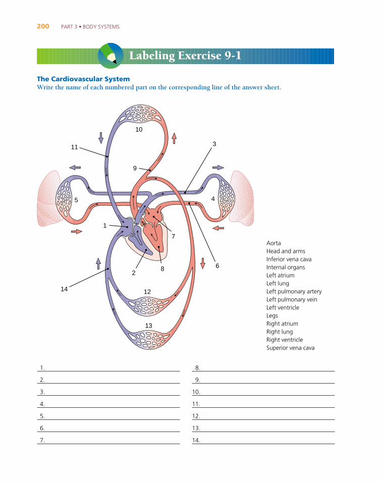
The Heart and Great VesselsWrite the name of each numbered part on the corresponding line of the answer sheet.
Aortic archAortic valveApexAscending aortaBrachiocephalic arteryEndocardiumEpicardium
Inferior vena cavaInterventricular septumLeft atriumLeft common carotid
arteryLeft pulmonary artery
(branches)
Left pulmonary veinsLeft subclavian arteryLeft ventricleMitral (bicuspid) valveMyocardiumPulmonary arteryPulmonic valve
Right atriumRight pulmonary artery
(branches)Right pulmonary veinsRight ventricleSuperior vena cavaTricuspid valve
1819
20
8
16
1
11
3
4
2
5
17
7
9
6
10
12
15
13
23
14
24Blood high in oxygen
Blood low in oxygen 25
2122
Labeling Exercise 9-2

Chapter Review 9-1Match the following terms and write the appropriate letter to the left of each number:
_____ 1. tricuspid a. central opening of a vessel
_____ 2. pericardium b. pacemaker of the heart
_____ 3. SA node c. fibrous sac around the heart
_____ 4. apex d. lower pointed region of the heart
_____ 5. lumen e. right atrioventricular valve
_____ 6. pulmonic valve a. lymphoid organ in the chest
_____ 7. vena cava b. vessel that empties into the right atrium
_____ 8. thymus c. part of the heart’s conduction system
_____ 9. mitral valve d. valve that regulates blood flow to the lungs
_____ 10. Purkinje fibers e. left atrioventricular valve
_____ 11. atherosclerosis a. absence of a heartbeat
_____ 12. aneurysm b. inflammation of the heart muscle
_____ 13. ischemia c. localized dilatation of a blood vessel
_____ 14. myocarditis d. local deficiency of blood
_____ 15. asystole e. accumulation of fatty deposits in the lining of a blood vessel
202 PART 3 • BODY SYSTEMS
1.
2.
3.
4.
5.
6.
7.
8.
9.
10.
11.
12.
13.
14.
15.
16.
17.
18.
19.
20.
21.
22.
23.
24.
25.
CHAPTER 9 • CIRCULATION: THE CARDIOVASCULAR AND LYMPHATIC SYSTEMS 203
_____ 16. thrombosis a. twisted and swollen vessel
_____ 17. occlusion b. ineffective quivering of muscle
_____ 18. varix c. local death of tissue
_____ 19. infarction d. blockage
_____ 20. fibrillation e. formation of a blood clot in a vessel
_____ 21. ECG a. disease of the heart’s vessels
_____ 22. CABG b. defibrillation device
_____ 23. PVC c. generation of an extra heartbeat
_____ 24. AED d. study of the electrical activity of the heart
_____ 25. CAD e. surgery to create a shunt around a blocked vessel
SUPPLEMENTARY TERMS
_____ 26. intermittent claudication a. drug that lowers serum cholesterol
_____ 27. precordium b. normal heart rhythm
_____ 28. statin c. accumulation of fluid in the pericardial sac
_____ 29. cardiac tamponade d. muscular pain during exercise
_____ 30. sinus rhythm e. anterior region over the heart
Fill in the blanks:
31. Each lower pumping chamber of the heart is a(n) __________________________________.
32. The heart muscle is the __________________________________.
33. The microscopic vessels through which materials are exchanged between the blood and the tissues arethe __________________________________.
34. The largest artery is the __________________________________.
35. Blood returning to the heart from the lungs enters the chamber called the__________________________________.
36. The lymphoid organ in the abdomen is the __________________________________.
37. At its termination in the abdomen, the aorta divides into the right and left (see Fig. 9-5)__________________________________.
38. The large vein that drains the head is the (see Fig. 9-6) __________________________________.
39. Microangiopathy (mi_-kro
_-an-je
_-OP-a-the
_) is disease of many small
__________________________________.
40. A phlebotomist (fle-BOT-o_-mist) is one who drains blood from a(n)
__________________________________.
41. The term varicoid pertains to a(n) __________________________________.
True-False. Examine each of the following statements. If the statement is true, write T in the first blank.If the statement is false, write F in the first blank and correct the statement by replacing the underlinedword in the second blank.
42. The systemic circuit pumps blood to the lungs. _____ __________________________________
43. An artery is a vessel that carries blood back to the heart. _____ __________________________________
44. Diastole is the relaxation phase of the heart cycle. _____ __________________________________
45. The left ventricle pumps blood into the aorta. _____ __________________________________
46. The brachial artery supplies blood to the leg. _____ __________________________________
47. The bicuspid valve is also called the mitral valve. _____ __________________________________
48. Bradycardia is a lower than average heart rate. _____ __________________________________
Define each of the following terms:
49. Interatrial (in-ter-A_-tre
_-al)
50. Avascular (a_-VAS-ku
_-lar)
51. Atriotomy (a_-tre
_-OT-o
_-me
_)
52. Angiostenosis (an-je_-o_-ste-NO
_-sis)
53. Thymectomy (thi_-MEK-to
_-me
_)
54. Lymphangitis (lim-fan-JI_-tis)
Word building. Write a word for each of the following definitions:
55. Physician who specializes in study and treatment of the heart
56. Suture (-rhaphy) of an artery
57. Radiographic study of the ventricles
58. Stoppage (-stasis) of lymph flow
59. An instrument (-tome) for incising a valve
60. Incision of a lymph node
61. Surgical fixation (-pexy) of the spleen
Word building. Use the root aort/o to write a word with each of the following meanings:
62. Radiograph (-gram) of the aorta
63. Before or in front of (pre-) the aorta
64. Narrowing (-stenosis) of the aorta
65. Any disease (-pathy) of the aorta
66. Downward displacement (-ptosis) of the aorta
Adjectives. Write the adjective form of each of the following words:
67. vein
68. septum
204 PART 3 • BODY SYSTEMS
CHAPTER 9 • CIRCULATION: THE CARDIOVASCULAR AND LYMPHATIC SYSTEMS 205
69. atrium
70. varix
71. spleen
72. sclerosis
Plurals. Write the plural form of each of the following words:
73. embolus
74. stenosis
75. apex
76. varix
77. septum
Write the meaning of the following abbreviations as they apply to the cardiovascular system:
78. BBB
79. PCTA
80. LVH
81. BP
82. NSR
83. CVI
Word analysis. Define each of the following words, and give the meaning of the word parts in each. Use adictionary if necessary.
84. Endarterectomy (end-ar-ter-EK-to_-me
_) _____________________________________________________
a. end/o- _____________________b. arteri/o _____________________c. ecto- _____________________d. -tomy _____________________
85. Telangiectasia (tel-an-je_-e_k-TA
_-ze
_-a) _______________________________________________________
a. tel- _____________________b. angi/o _____________________c. -ectasia _____________________
86. Lymphangiophlebitis (lim-fan-je_-o_-fle-BI
_-tis) ________________________________________________
a. lymph/o _____________________b. angi/o _____________________c. phleb/o _____________________d. -itis _____________________
206 PART 3 • BODY SYSTEMS
Case Studies
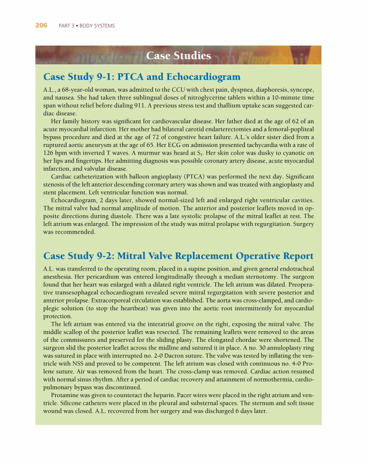
Case Study 9-1: PTCA and EchocardiogramA.L., a 68-year-old woman, was admitted to the CCU with chest pain, dyspnea, diaphoresis, syncope,and nausea. She had taken three sublingual doses of nitroglycerine tablets within a 10-minute timespan without relief before dialing 911. A previous stress test and thallium uptake scan suggested car-diac disease.
Her family history was significant for cardiovascular disease. Her father died at the age of 62 of anacute myocardial infarction. Her mother had bilateral carotid endarterectomies and a femoral-poplitealbypass procedure and died at the age of 72 of congestive heart failure. A.L.’s older sister died from aruptured aortic aneurysm at the age of 65. Her ECG on admission presented tachycardia with a rate of126 bpm with inverted T waves. A murmur was heard at S1. Her skin color was dusky to cyanotic onher lips and fingertips. Her admitting diagnosis was possible coronary artery disease, acute myocardialinfarction, and valvular disease.
Cardiac catheterization with balloon angioplasty (PTCA) was performed the next day. Significantstenosis of the left anterior descending coronary artery was shown and was treated with angioplasty andstent placement. Left ventricular function was normal.
Echocardiogram, 2 days later, showed normal-sized left and enlarged right ventricular cavities.The mitral valve had normal amplitude of motion. The anterior and posterior leaflets moved in op-posite directions during diastole. There was a late systolic prolapse of the mitral leaflet at rest. Theleft atrium was enlarged. The impression of the study was mitral prolapse with regurgitation. Surgerywas recommended.
Case Study 9-2: Mitral Valve Replacement Operative ReportA.L. was transferred to the operating room, placed in a supine position, and given general endotrachealanesthesia. Her pericardium was entered longitudinally through a median sternotomy. The surgeonfound that her heart was enlarged with a dilated right ventricle. The left atrium was dilated. Preopera-tive transesophageal echocardiogram revealed severe mitral regurgitation with severe posterior andanterior prolapse. Extracorporeal circulation was established. The aorta was cross-clamped, and cardio-plegic solution (to stop the heartbeat) was given into the aortic root intermittently for myocardialprotection.
The left atrium was entered via the interatrial groove on the right, exposing the mitral valve. Themiddle scallop of the posterior leaflet was resected. The remaining leaflets were removed to the areasof the commissures and preserved for the sliding plasty. The elongated chordae were shortened. Thesurgeon slid the posterior leaflet across the midline and sutured it in place. A no. 30 annuloplasty ringwas sutured in place with interrupted no. 2-0 Dacron suture. The valve was tested by inflating the ven-tricle with NSS and proved to be competent. The left atrium was closed with continuous no. 4-0 Pro-lene suture. Air was removed from the heart. The cross-clamp was removed. Cardiac action resumedwith normal sinus rhythm. After a period of cardiac recovery and attainment of normothermia, cardio-pulmonary bypass was discontinued.
Protamine was given to counteract the heparin. Pacer wires were placed in the right atrium and ven-tricle. Silicone catheters were placed in the pleural and substernal spaces. The sternum and soft tissuewound was closed. A.L. recovered from her surgery and was discharged 6 days later.
CHAPTER 9 • CIRCULATION: THE CARDIOVASCULAR AND LYMPHATIC SYSTEMS 207
Case Studies, continued
CASE STUDY QUESTIONS
Write the word or phrase from the case study that has the same meaning as each of the followingwords or phrases:
1. The state of profuse perspiration __________________________________
2. Under the tongue __________________________________
3. Test of cardiac function during physical exertion __________________________________
4. Disease that includes both heart and blood vessel pathology __________________________________
5. Excision of the inner lining along with atherosclerotic plaque from an artery (plural) __________________________________
6. An abnormal heart sound __________________________________
7. Bluish discoloration of the skin; sign of anoxia __________________________________
8. The noun form of stenotic __________________________________
9. Between the atria __________________________________
10. Below the sternum __________________________________
Multiple choice: Select the best answer and write the letter of your choice to the left of each number.
_____ 11. The word transluminal means:a. across a wallb. between branchesc. through an outer layerd. through a central openinge. across a valve
_____ 12. The term that means backflow, as of blood, is:a. infarctionb. regurgitationc. amplituded. prolapsee. tourniquet
_____ 13. The term for a narrowing of the bicuspid valve is:a. atrial prolapseb. pulmonic stenosisc. mitral stenosisd. mitral prolapsee. atrial stenosis
_____ 14. Blowout of a dilated segment of the main artery is:a. left anterior diastoleb. peritoneal infarction
208 PART 3 • BODY SYSTEMS
Case Studies, continued
c. coarctation of the aortad. cardiac tamponadee. ruptured aortic aneurysm
_____ 15. Sternotomy is:a. incision into the sternumb. removal of the sternumc. narrowing of the sternumd. plastic repair of the sternume. surgical fixation of the sternum
_____ 16. Extracorporeal circulation occurs:a. within the brainb. within the pericardiumc. within the bodyd. in the legse. outside the body
_____ 17. Protamine was given to counteract the action of the heparin. This drug action is described as:a. antagonisticb. synergyc. potentiatingd. similatione. addiction
Abbreviations. Define the following abbreviations:
18. CCU __________________________________
19. AMI __________________________________
20. CAD __________________________________
21. LAD __________________________________
22. CHF __________________________________
23. TEE __________________________________
24. MVR __________________________________

CHAPTER 9 • CIRCULATION: THE CARDIOVASCULAR AND LYMPHATIC SYSTEMS 209
ACROSS1. A microscopic vessel6. Pacemaker of the heart: ____ ____ node7. A route for injection (abbreviation)8. The right AV valve
10. Vein: combining form12. Relaxation phase of the heart cycle15. Hardening of the arteries (abbreviation)16. Substance used to dissolve blood clots
(abbreviation)18. Pulse: combining form, as in the name of blood
pressure apparatus20. Form of lipoprotein (abbreviation)21. Heart disease associated with pulmonary edema
(abbreviation)22. Part of the heart’s conduction system; it receives
impulses from the AV node
DOWN1. Main artery in the neck2. Hospital unit that cares for the critically ill
(abbreviation)3. Category of compounds that includes fats:
combining form4. Heart attack (abbreviation)5. Obstruction circulating in the bloodstream6. Segment of the ECG tracing9. Thrombotic condition of the veins (abbreviation)
11. Procedure for dilating an obstructed vessel (abbreviation)
13. Fluid that circulates in the lymphatic system14. Lymphoid organ in the chest: root17. Vein: root19. Units in which blood pressure is measured
(abbreviation)20. Mechanical device to assist the heart, ___ ___ ___
D (abbreviation)
Chapter 9 CrosswordCirculation
* 1 2 3 4 * 5
6 * * * 7 * * *
8 9 * *
* * * * * * 10
* * * 11 * * * * *
12 13 * * *
* * * * * 14 * 15
* * 16 17 * * * * *
* * * * 18 19
* 20 * * * * *
* * * * * * * 21
* 22 * *
![SummaryMap ward2 [Converted] · 2019-10-01 · MU-2 MU-6 MU-16 MU-14 MU-6 MU-2 MU-20 MU-9 MU-4 MU-13 MU-15 MU-13 MU-16 MU-18 MU-22 MU-19 MU-16 MU-27 MU-4 MU-3A MU-17 MU-13 MU-4](https://img.pdfslide.us/doc/110x75/5f5e4f591750d150e9633369/summarymap-ward2-converted-2019-10-01-mu-2-mu-6-mu-16-mu-14-mu-6-mu-2-mu-20.jpg)