Embed Size (px)
Citation preview
© 2017 Ebneshahidi
THE HEART
Dr. Ali Ebneshahidi
© 2017 Ebneshahidi
Functions is of the heart & blood vessels
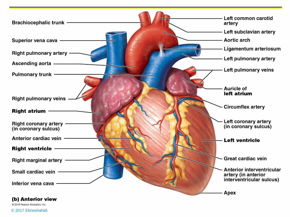
1. The heart is an essential pumping organ in the cardiovascular
system where the right heart pumps deoxygenated blood
(returned from body tissues) to the lungs for gas exchange,
while the left heart pumps oxygenated blood (returned from
the lungs) to tissues cells for sustaining cellular respiration.
2. Attached to the heart is blood vessels that transport blood in
various circulation pathways - pulmonary blood vessels
transport blood between the heart and the lungs, and systemic
blood vessels transport blood between the heart and body
tissues.
© 2017 Ebneshahidi
© 2017 Ebneshahidi
Heart chambers
Hollow cavities within the heart for containing blood.
Two smaller chambers called atrium are near the base, and two larger
chambers called ventricle are close to the apex.
Right atrium (RA) after receiving deoxygenated blood from body
tissues through the superior and inferior vena cava, pumps the blood
into the right ventricle (RV) via the right atria ventricular orifice. RV
then pumps the blood to the lungs for gas exchange, through the
pulmonary trunk and arteries.
Left atrium (LA) after receiving oxygenated blood from the lungs
through the pulmonary veins, pumps the blood into the left ventricle
(LV) via the left atria ventricular orifice.
© 2017 Ebneshahidi
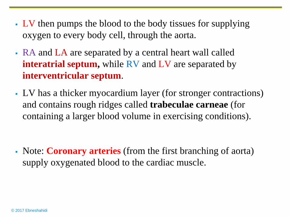
LV then pumps the blood to the body tissues for supplying
oxygen to every body cell, through the aorta.
RA and LA are separated by a central heart wall called
interatrial septum, while RV and LV are separated by
interventricular septum.
LV has a thicker myocardium layer (for stronger contractions)
and contains rough ridges called trabeculae carneae (for
containing a larger blood volume in exercising conditions).
Note: Coronary arteries (from the first branching of aorta)
supply oxygenated blood to the cardiac muscle.
© 2017 Ebneshahidi
© 2017 Ebneshahidi
Heart Valves
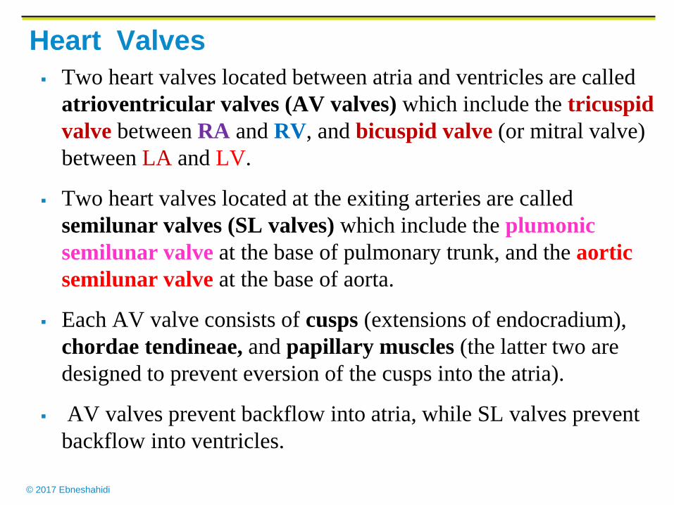
Two heart valves located between atria and ventricles are called
atrioventricular valves (AV valves) which include the tricuspid
valve between RA and RV, and bicuspid valve (or mitral valve)
between LA and LV.
Two heart valves located at the exiting arteries are called
semilunar valves (SL valves) which include the plumonic
semilunar valve at the base of pulmonary trunk, and the aortic
semilunar valve at the base of aorta.
Each AV valve consists of cusps (extensions of endocradium),
chordae tendineae, and papillary muscles (the latter two are
designed to prevent eversion of the cusps into the atria).
AV valves prevent backflow into atria, while SL valves prevent
backflow into ventricles.
© 2017 Ebneshahidi
© 2017 Ebneshahidi
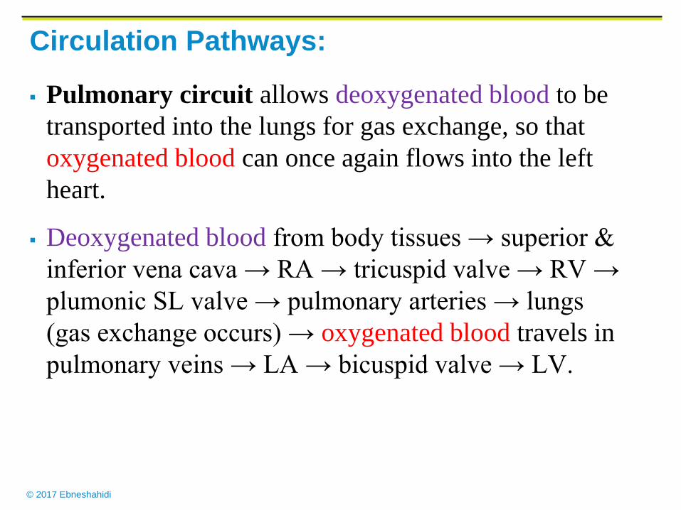
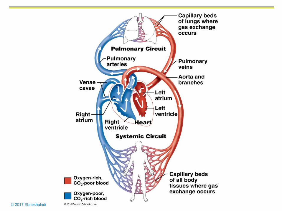
Circulation Pathways:
Pulmonary circuit allows deoxygenated blood to be
transported into the lungs for gas exchange, so that
oxygenated blood can once again flows into the left
heart.
Deoxygenated blood from body tissues → superior &
inferior vena cava → RA → tricuspid valve → RV →
plumonic SL valve → pulmonary arteries → lungs
(gas exchange occurs) → oxygenated blood travels in
pulmonary veins → LA → bicuspid valve → LV.
© 2017 Ebneshahidi
© 2017 Ebneshahidi
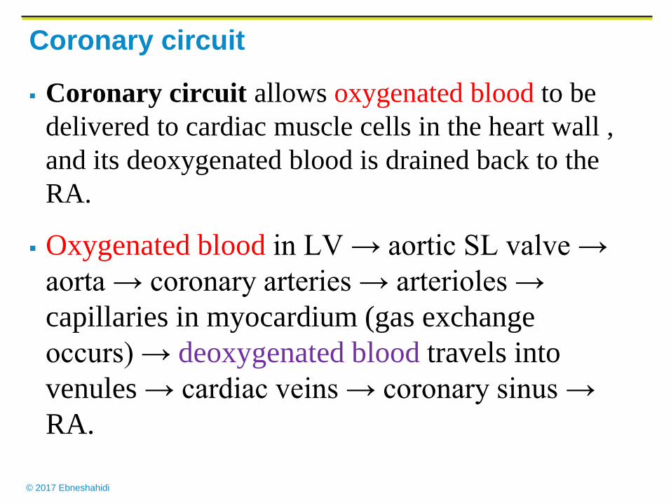
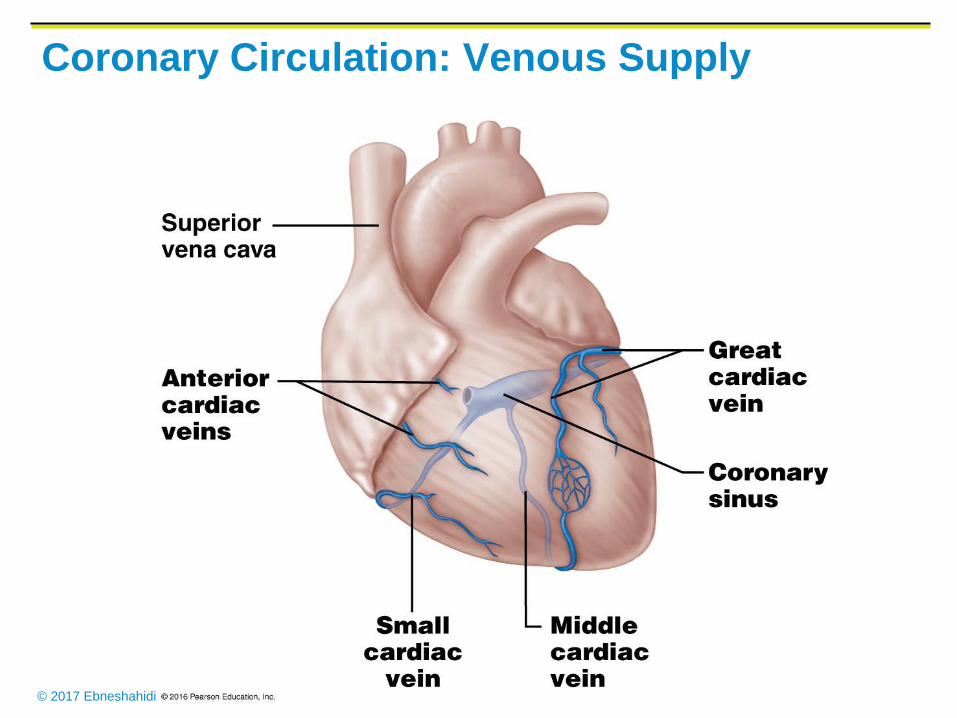
Coronary circuit
Coronary circuit allows oxygenated blood to be
delivered to cardiac muscle cells in the heart wall ,
and its deoxygenated blood is drained back to the
RA.
Oxygenated blood in LV → aortic SL valve →
aorta → coronary arteries → arterioles →
capillaries in myocardium (gas exchange
occurs) → deoxygenated blood travels into
venules → cardiac veins → coronary sinus →
RA.
© 2017 Ebneshahidi
Coronary Circulation: Arterial Supply
© 2017 Ebneshahidi
Coronary Circulation: Venous Supply
© 2017 Ebneshahidi
Systemic Circuit
Systemic circuit allows oxygenated blood from
the left heart to be delivered to tissue cells through
arteries and arterioles , and deoxygenated blood is
transported back to the right heart through veins
and venues.
Oxygenated blood in LV → aortic SL valve →
aorta → arteries → arterioles → capillaries in
tissues (gas exchange occurs) → deoxygenated
blood travels in venules → veins → superior &
inferior vena cava → RA.
© 2017 Ebneshahidi
Cardiac cycles are mainly controlled by nerve
impulse, while hormones only can influence the
heart rate.
Two mechanisms to regulate cardiac cycles -
intrinsic control (consists of pacemakers and a
conduction system) and extrinsic control (consists
of sympathetic and parasympathetic nerves, and
hormones, that influence the pacemakers and affect
the heart rate).
© 2017 Ebneshahidi
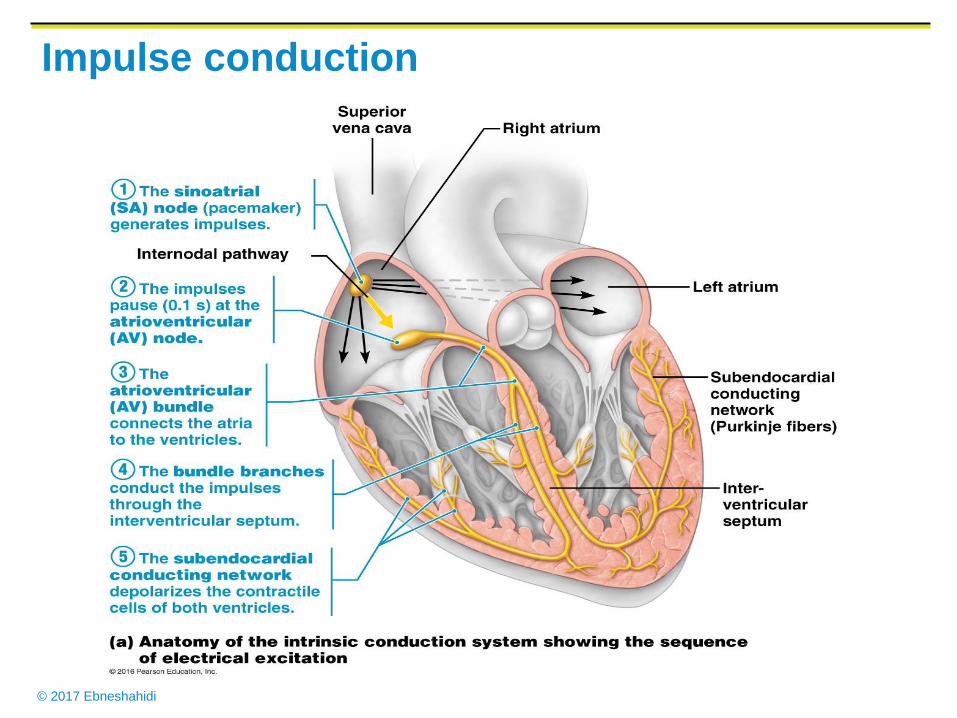
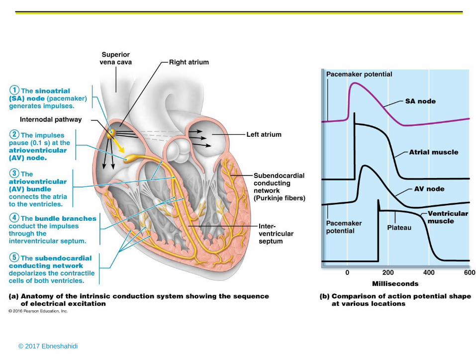
Impulse conducting system of the heart (Intrinsic control)
Group of muscle cells specialized for conduction rather than
contraction. Innervated by both the sympathetic and
parasympathetic nervous system. Regulate cardiac rhythm, and
adapts cardiac output to the physiologic needs of the body.
Components:
1. Sinoatrial node (pace maker), cluster of excitable cells
which sets cardiac rhythm. Located near the inlet of the
superior vena cava.
2. Atrial syncytium, a mass of merging cells that act as a unit.
Cardiac muscle fibers function like those of skeletal muscles,
but the fibers connect in branching networks. Stimulation of
any part of the network send impulses throughout the heart,
which contracts as a unit.
© 2017 Ebneshahidi
Impulse conduction
© 2017 Ebneshahidi
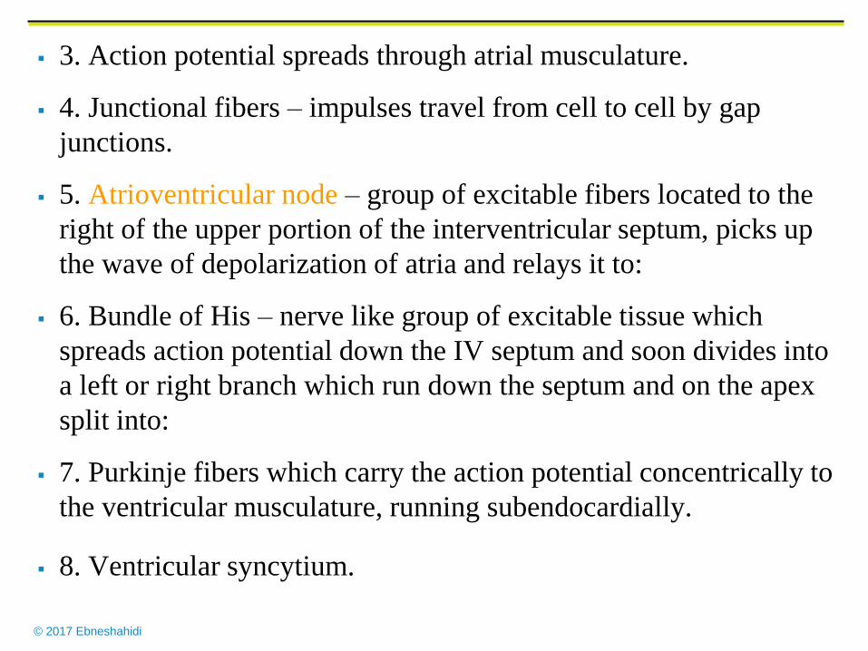
3. Action potential spreads through atrial musculature.
4. Junctional fibers – impulses travel from cell to cell by gap
junctions.
5. Atrioventricular node – group of excitable fibers located to the
right of the upper portion of the interventricular septum, picks up
the wave of depolarization of atria and relays it to:
6. Bundle of His – nerve like group of excitable tissue which
spreads action potential down the IV septum and soon divides into
a left or right branch which run down the septum and on the apex
split into:
7. Purkinje fibers which carry the action potential concentrically to
the ventricular musculature, running subendocardially.
8. Ventricular syncytium.
© 2017 Ebneshahidi
© 2017 Ebneshahidi
Functions of the cardiac conduction system
1. Causes both atria to contract simultaneously and
generate enough hydrostatic pressure to open both
AV valves so that blood flows to ventricles.
2. Causes the ventricles to contract a few
milliseconds later, overcome the resistance of the
semilunar valves and generate enough hydrostatic
pressure to circulate the blood through the
pulmonary and systemic pathways.
3. Adapts the heart to beat under different
functional demands. Sympathetic nervous system
increases rate and output, parasympathetic
decreases.
© 2017 Ebneshahidi
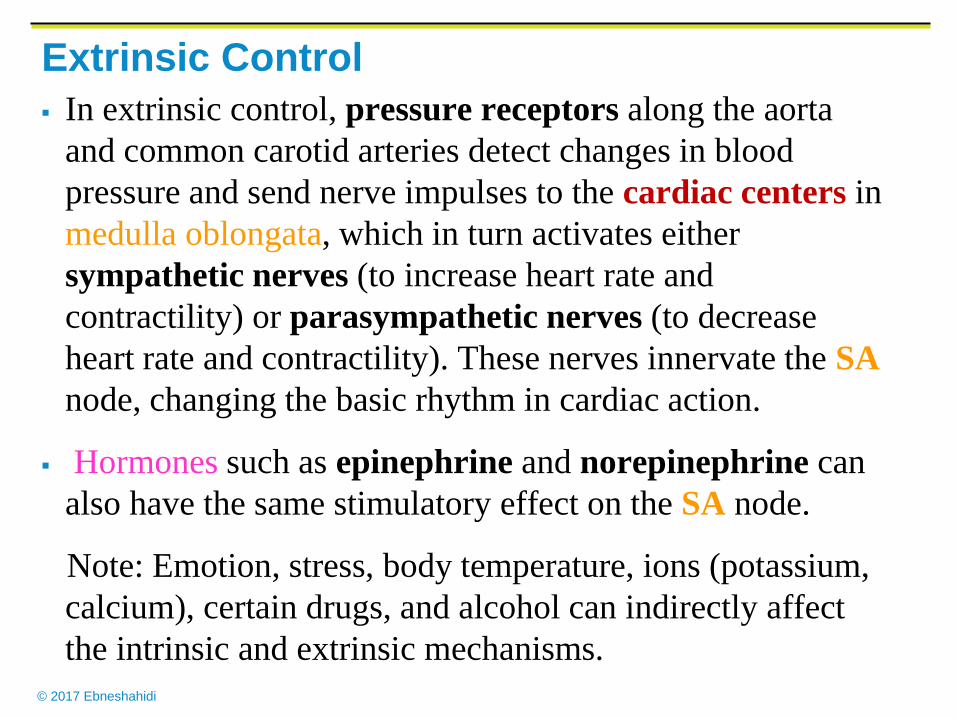
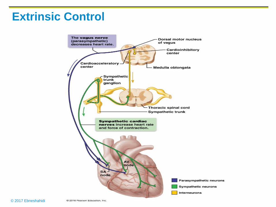
Extrinsic Control
In extrinsic control, pressure receptors along the aorta
and common carotid arteries detect changes in blood
pressure and send nerve impulses to the cardiac centers in
medulla oblongata, which in turn activates either
sympathetic nerves (to increase heart rate and
contractility) or parasympathetic nerves (to decrease
heart rate and contractility). These nerves innervate the SA
node, changing the basic rhythm in cardiac action.
Hormones such as epinephrine and norepinephrine can
also have the same stimulatory effect on the SA node.
Note: Emotion, stress, body temperature, ions (potassium,
calcium), certain drugs, and alcohol can indirectly affect
the intrinsic and extrinsic mechanisms.
© 2017 Ebneshahidi
Extrinsic Control
© 2017 Ebneshahidi
Cardiac Excitability & Contractility
1. The duration of the cardiac action potential is very long, lasting
throughout contraction. Action potentials in cardiac muscle is 100
times longer than in skeletal muscles.
2. There are prolonged refractory periods.
3. Cardiac muscle contractions are always brief twitches. In skeletal
muscles, contractions resulting from rapid repetitive stimulation can
summate to provide sustained contraction. This can not happen in
cardiac muscle because the long refractory period cancels any
stimulus that occurs before the heart has a chance to relax.
- Relaxtion between beats is essential for the heart to fill with blood,
to be pumped at the next beat. Summation of contractions (tetanus)
does not occur due to long refractory periods.
Note Ca++ entry prolongs the period of depolarization.
© 2017 Ebneshahidi
4. Cardiac muscles are interconnected by gap junctions – the
heart beat is All – or – None, and spreads.
5. Cardiac muscles excites itself. Normally skeletal muscles will
contract only if it receives a nerve impulse. Nerves that carry
impulses to the heart influence the rate and strength of
contraction, but they do not initiate the primitive heartbeat.
6. Excitation for the heart beat arises from within the cardiac
muscle itself; spontaneous depolarization of the SA node cell
which spread to other cells. Syncytium refers to cells that
contract as a unit.
- SA and AV node cells are pacemaker cells; they have intrinsic
automaticity characterized by spontaneous depolarization which
perhaps may be due to a decreased membrane permeability to
K+.
© 2017 Ebneshahidi
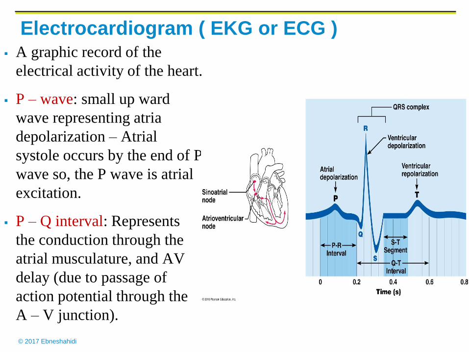
Electrocardiogram ( EKG or ECG ) A graphic record of the
electrical activity of the heart.
P – wave: small up ward
wave representing atria
depolarization – Atrial
systole occurs by the end of P
wave so, the P wave is atrial
excitation.
P – Q interval: Represents
the conduction through the
atrial musculature, and AV
delay (due to passage of
action potential through the
A – V junction).
© 2017 Ebneshahidi
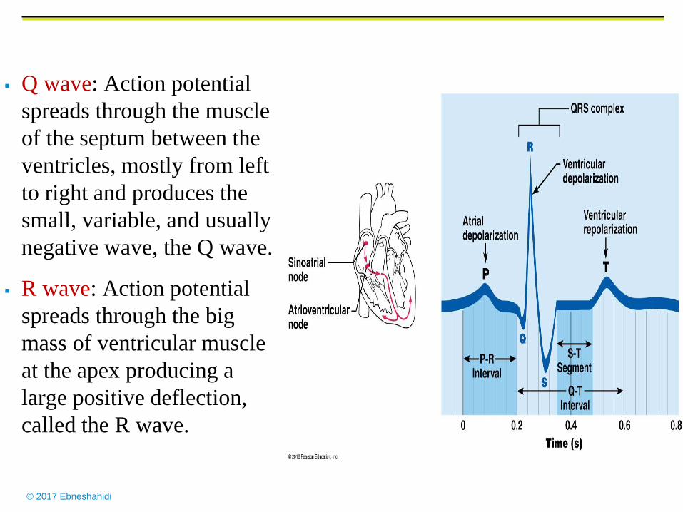
Q wave: Action potential
spreads through the muscle
of the septum between the
ventricles, mostly from left
to right and produces the
small, variable, and usually
negative wave, the Q wave.
R wave: Action potential
spreads through the big
mass of ventricular muscle
at the apex producing a
large positive deflection,
called the R wave.
© 2017 Ebneshahidi
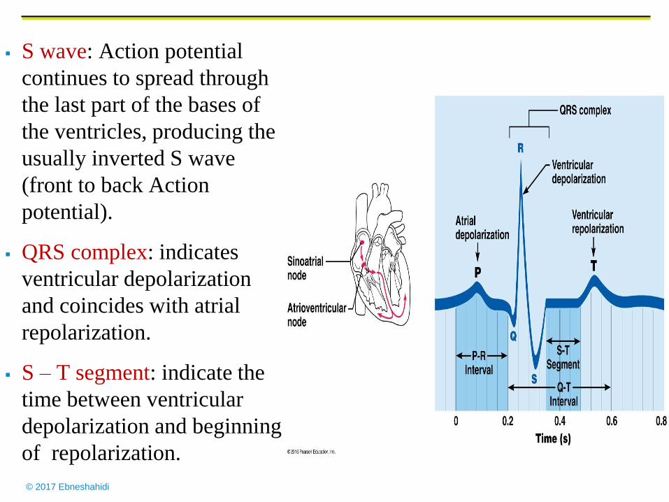
S wave: Action potential
continues to spread through
the last part of the bases of
the ventricles, producing the
usually inverted S wave
(front to back Action
potential).
QRS complex: indicates
ventricular depolarization
and coincides with atrial
repolarization.
S – T segment: indicate the
time between ventricular
depolarization and beginning
of repolarization.
© 2017 Ebneshahidi
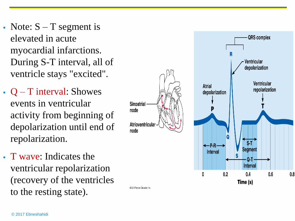
Note: S – T segment is
elevated in acute
myocardial infarctions.
During S-T interval, all of
ventricle stays "excited".
Q – T interval: Showes
events in ventricular
activity from beginning of
depolarization until end of
repolarization.
T wave: Indicates the
ventricular repolarization
(recovery of the ventricles
to the resting state).
© 2017 Ebneshahidi
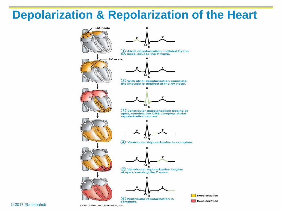
Depolarization & Repolarization of the Heart
© 2017 Ebneshahidi
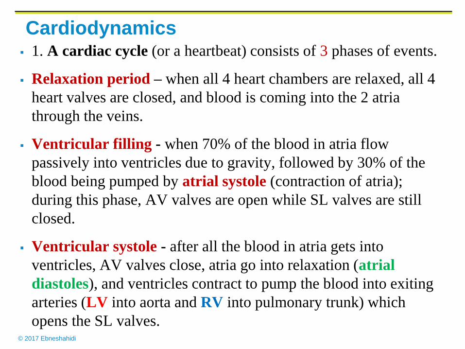
Cardiodynamics 1. A cardiac cycle (or a heartbeat) consists of 3 phases of events.
Relaxation period – when all 4 heart chambers are relaxed, all 4
heart valves are closed, and blood is coming into the 2 atria
through the veins.
Ventricular filling - when 70% of the blood in atria flow
passively into ventricles due to gravity, followed by 30% of the
blood being pumped by atrial systole (contraction of atria);
during this phase, AV valves are open while SL valves are still
closed.
Ventricular systole - after all the blood in atria gets into
ventricles, AV valves close, atria go into relaxation (atrial
diastoles), and ventricles contract to pump the blood into exiting
arteries (LV into aorta and RV into pulmonary trunk) which
opens the SL valves.
© 2017 Ebneshahidi
SL valves will close after almost all ventricular blood is ejected (there
is always 60 ml blood remained in each ventricle, a volume called end-
systolic volume or ESV). Then the ventricles relax (ventricular
diastole), and a new cardiac cycle will follow.
2. Each cardiac cycle takes about 0.8 second to complete.
3.Toward the end of ventricular filling, when pressure builds up in the
ventricles AV valves begin to close. Now papillary muscles contract,
pulling the chordae and cusps, to prevent eversion (overbulging) of the
cusps into the atria.
4. In each cardiac cycle, the two atria contract and relax simultaneously
- a phenomenon called atrial syncytum. The same is true for the
ventricles, which is known as ventricular syncytum.
Note: when cardiac muscles in heart chambers lose this syncytum
ability, fibrillation occurs which can be fatally out of control.
© 2017 Ebneshahidi
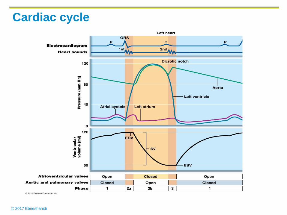
Cardiac cycle
© 2017 Ebneshahidi
Cardiac output: volume of blood pumped by the heart each minute =
stroke volume (volume of each beat) × Heart rate C.H.R) [# beats /
min].
Stroke volume: determined by the volume of blood in the heart at the
beginning of systole (end diastolic volume) minus the amount of blood
remaining in the ventricles when the valves close at the end of systole
(end systolic volume).
SV (ml / beat) = EDV (120 ml) – ESV (50 ml) = 70 ml / beat
Note: End diastolic volume is the amount of blood that collects in a
ventricle during diastole.
End systolic volume is the amount of blood remaining in the ventricle
after it has contracted.
© 2017 Ebneshahidi
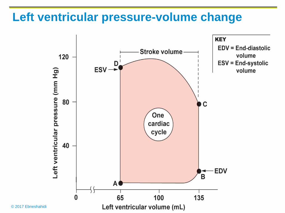
Left ventricular pressure-volume change
© 2017 Ebneshahidi
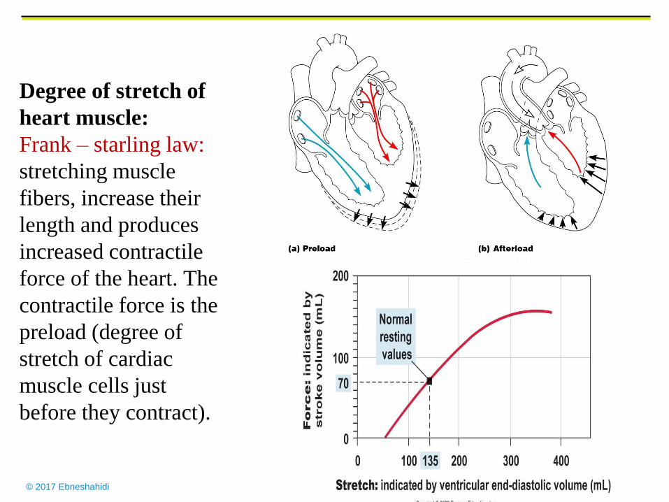
Degree of stretch of
heart muscle:
Frank – starling law:
stretching muscle
fibers, increase their
length and produces
increased contractile
force of the heart. The
contractile force is the
preload (degree of
stretch of cardiac
muscle cells just
before they contract).
© 2017 Ebneshahidi
© 2017 Ebneshahidi
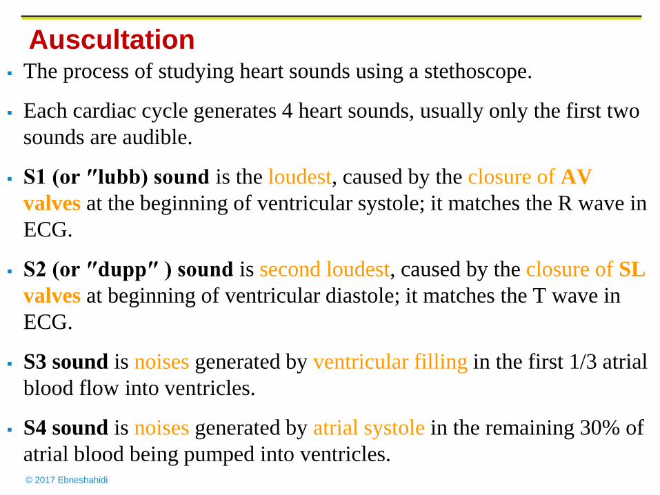
Auscultation
The process of studying heart sounds using a stethoscope.
Each cardiac cycle generates 4 heart sounds, usually only the first two
sounds are audible.
S1 (or ″lubb) sound is the loudest, caused by the closure of AV
valves at the beginning of ventricular systole; it matches the R wave in
ECG.
S2 (or ″dupp″ ) sound is second loudest, caused by the closure of SL
valves at beginning of ventricular diastole; it matches the T wave in
ECG.
S3 sound is noises generated by ventricular filling in the first 1/3 atrial
blood flow into ventricles.
S4 sound is noises generated by atrial systole in the remaining 30% of
atrial blood being pumped into ventricles.
© 2017 Ebneshahidi
Heart Sounds
© 2017 Ebneshahidi
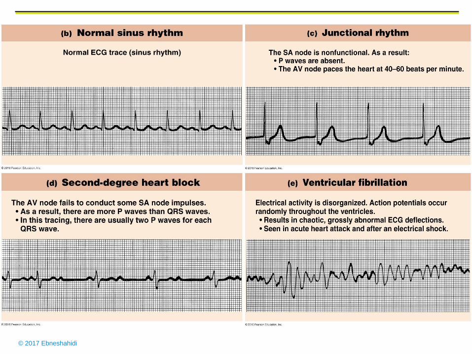
Sinus rhythm
Normal cardiac action or heart rate for female is about 72-80
beats/min, for male, about 64-72 beats/ min., and for fetus, about 140 -
160 beats/min.
Arrhythmias are any abnormal hear beat.
Bradycardia is slow heart rate (<60 beats/min) which is normal during
sleep, but can be induced by low body temperature, parasympathetic
stimulation, and certain drugs. It is indicated by a Short PQ interval
and a flat T wave on the ECG.
Tachycardia is fast heart rate (> 100 beats/min) which is normal
during exercising or excitement, but can be induced by high body
temperature, sympathetic stimulation, drugs, heart diseases, anemia, or
shock. It is indicated by a lack of P,Q, S, or T wave, with only high-
frequency of upward and downward deflections on the ECG.
© 2017 Ebneshahidi
Flutter is very high heart rate (>250 beats/min) which is
usually pathological (e.g. bacterial infection or inflammation
of myocardium). It is indicated by many small, unrecognized
waves, then a big upward/downward wave on the ECG.
Fibrillation is high but uncoordinated heart rate caused by
regions of myocardium contracting and relaxing
independently (lack of syncytum). Atrial fibrillation is not
very serious if ventricles are functioning normally.
Ventricular fibrillation is usually fatal (the most common
cause of sudden death) where blood cannot be pumped
properly into the lunges and body tissues. It is indicated by
extremely irregular waves on the ECG, and usually Q and S
waves are absent.
© 2017 Ebneshahidi
© 2017 Ebneshahidi
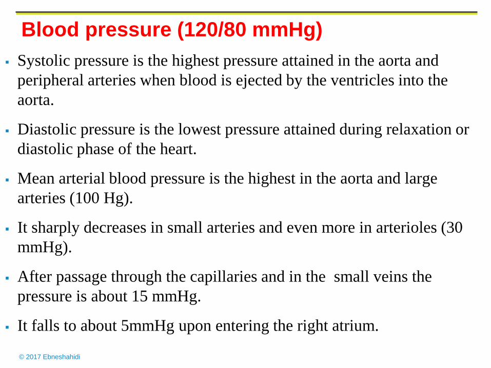
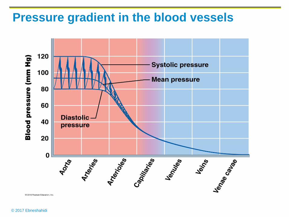
Blood pressure (120/80 mmHg)
Systolic pressure is the highest pressure attained in the aorta and
peripheral arteries when blood is ejected by the ventricles into the
aorta.
Diastolic pressure is the lowest pressure attained during relaxation or
diastolic phase of the heart.
Mean arterial blood pressure is the highest in the aorta and large
arteries (100 Hg).
It sharply decreases in small arteries and even more in arterioles (30
mmHg).
After passage through the capillaries and in the small veins the
pressure is about 15 mmHg.
It falls to about 5mmHg upon entering the right atrium.
© 2017 Ebneshahidi
Pressure gradient in the blood vessels
© 2017 Ebneshahidi
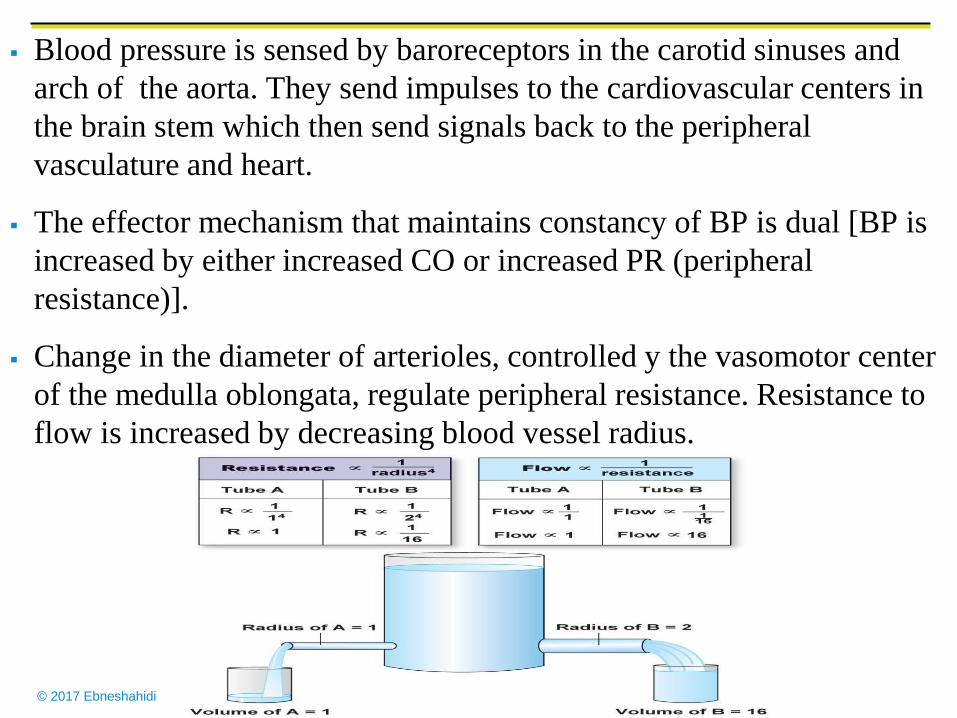
Blood pressure is sensed by baroreceptors in the carotid sinuses and
arch of the aorta. They send impulses to the cardiovascular centers in
the brain stem which then send signals back to the peripheral
vasculature and heart.
The effector mechanism that maintains constancy of BP is dual [BP is
increased by either increased CO or increased PR (peripheral
resistance)].
Change in the diameter of arterioles, controlled y the vasomotor center
of the medulla oblongata, regulate peripheral resistance. Resistance to
flow is increased by decreasing blood vessel radius.
© 2017 Ebneshahidi
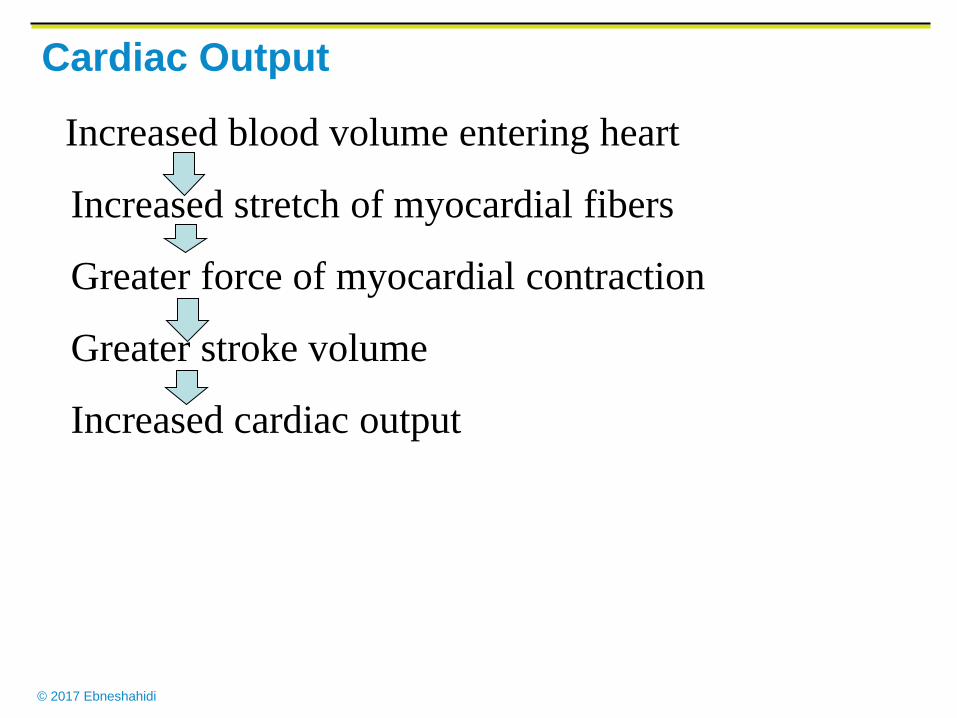
Cardiac Output
Increased blood volume entering heart
Increased stretch of myocardial fibers
Greater force of myocardial contraction
Greater stroke volume
Increased cardiac output
© 2017 Ebneshahidi
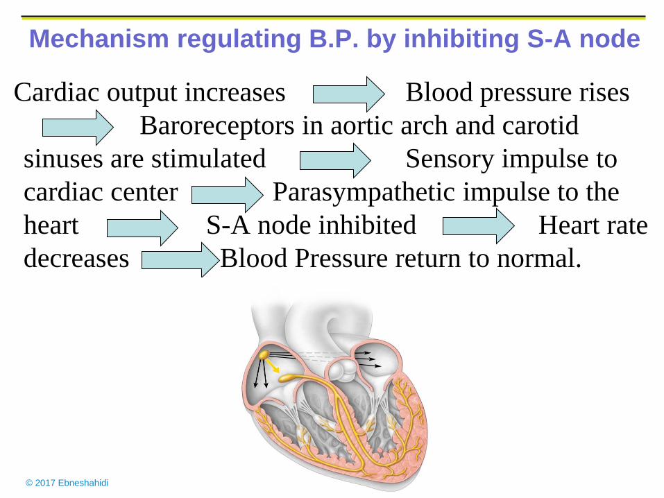
Mechanism regulating B.P. by inhibiting S-A node
Cardiac output increases Blood pressure rises
Baroreceptors in aortic arch and carotid
sinuses are stimulated Sensory impulse to
cardiac center Parasympathetic impulse to the
heart S-A node inhibited Heart rate
decreases Blood Pressure return to normal.
© 2017 Ebneshahidi
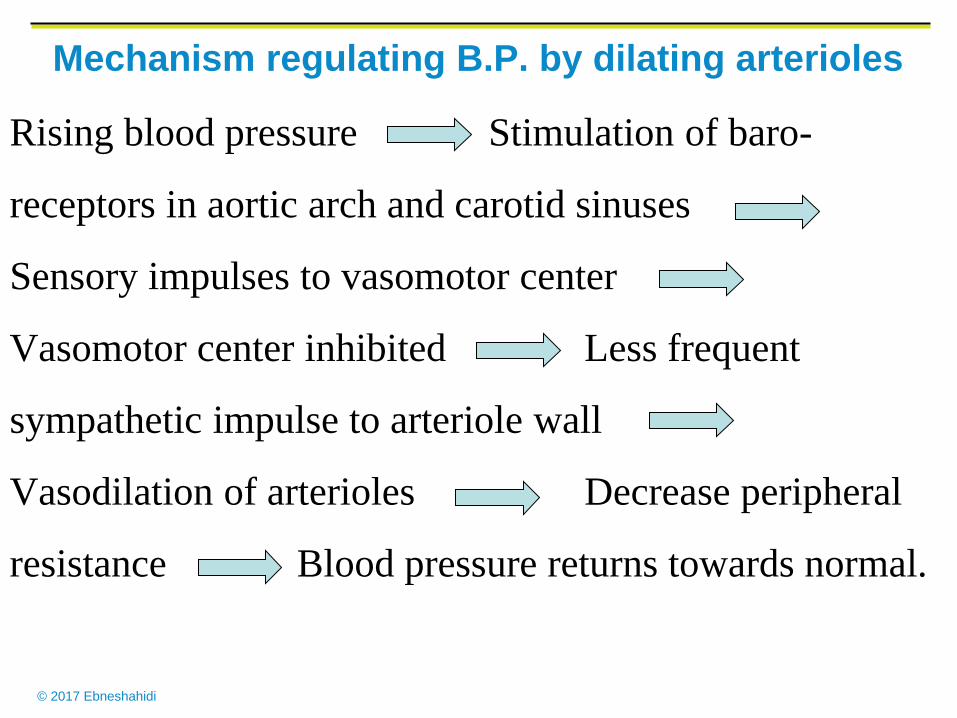
Mechanism regulating B.P. by dilating arterioles
Rising blood pressure Stimulation of baro-
receptors in aortic arch and carotid sinuses
Sensory impulses to vasomotor center
Vasomotor center inhibited Less frequent
sympathetic impulse to arteriole wall
Vasodilation of arterioles Decrease peripheral
resistance Blood pressure returns towards normal.
© 2017 Ebneshahidi
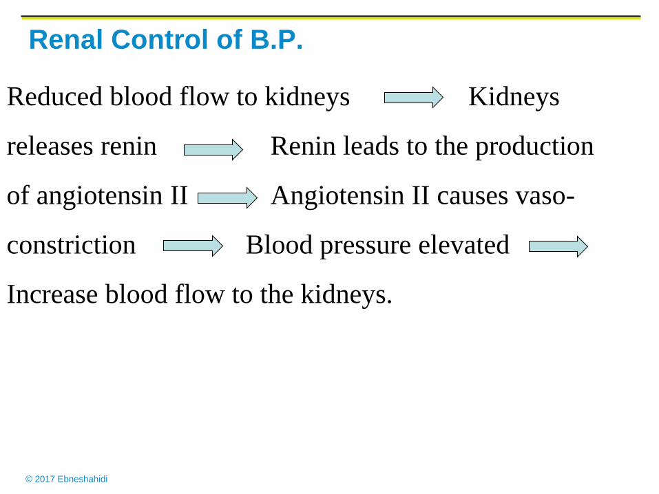
Renal Control of B.P.
Reduced blood flow to kidneys Kidneys
releases renin Renin leads to the production
of angiotensin II Angiotensin II causes vaso-
constriction Blood pressure elevated
Increase blood flow to the kidneys.
© 2017 Ebneshahidi
Risk factors for stroke
1. Alcohol consumption
2. Diabetes
3. Elevated serum cholesterol
4. Family history of cardiovascular disease
5. Hypertension
6. Smoking
7. Transient ischemic attacks
© 2017 Ebneshahidi
Drugs to treat hypertension
Angiotensin – converting enzyme inhibitors:
Block formation of Angiotensin II, preventing
vasoconstriction.
Beta blockers: lower heart rate.
Ca++ channel blockers: Dilate blood vessels by
keeping Ca ++ ions out of muscle cells in vessel
walls.
Diuretics: increase urine output, lowering blood
volume.
© 2017 Ebneshahidi
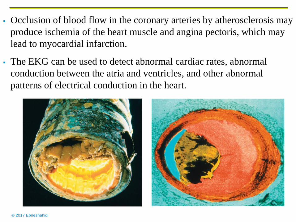
Atheroscleroses
Atherosclerosis of arteries can occlude blood flow to the heart and
brain and is a causative factor in up to 50% of all deaths in U.S.A.,
Europe, and Japan.
A. Atherosclerosis begins with injury to the endothelium, the
movement of monocytes and lymphocytes into the tunica interna
and the conversion of monocytes into macrophages that engulf
lipids. Smooth muscle cells then proliferate and secrete extra
cellular matrix.
B. Atherosclerosis is promoted by such risk factors as smoking,
hypertension, and high plasma cholesterol concentration.
LDLs which carry cholesterol into the artery wall, are oxidized by
the endothelium and are a major contributor to atherosclerosis.
© 2017 Ebneshahidi
Occlusion of blood flow in the coronary arteries by atherosclerosis may
produce ischemia of the heart muscle and angina pectoris, which may
lead to myocardial infarction.
The EKG can be used to detect abnormal cardiac rates, abnormal
conduction between the atria and ventricles, and other abnormal
patterns of electrical conduction in the heart.


![Heart-Muscle Fiber Reconstruction from Diffusion Tensor MRI · ventricles at the bottom of the heart, and two atria above [Streeter 1979]. The heart also contains other structures,](https://img.pdfslide.us/doc/110x75/60cde7c9647ab0358f659874/heart-muscle-fiber-reconstruction-from-diffusion-tensor-mri-ventricles-at-the-bottom.jpg)