Embed Size (px)
Citation preview

1
The Gleason Score and Beyond
Growth Patterns in Prostate Cancer
Eva Hollemans

2
The Gleason Score and Beyond
Growth Patterns in Prostate Cancer
E. Hollemans

3
E. Hollemans The Gleason Score and Beyond: Growth Patterns in Prostate Cancer Printed on recycled paper by Gildeprint
Designed by E. Hollemans. Chapter image ‘Jupiter Blues’ by Gerald Eichstädt and Sean Doran, based on photographs made by spacecraft Juno, provided courtesy of NASA/JPL-Calltech/SwRI/MSSS.
Copyright © E. Hollemans 2021, Rotterdam, The Netherlands
The research described in this thesis was conducted at the Department of Pathology, Erasmus MC Cancer Institute, University Medical Center, Rotterdam, the Netherlands.
Printing of this thesis was kindly supported by the Jaap Schouten Foundation, Brompton Bicycle and Erasmus Medical Center.
All rights reserved. No part of this thesis may be reproduced, stored in a retrieval system of any nature, or transmitted on any form by any means, electronic, mechanical, photocopying, recording or otherwise, including in a complete or partial transcription without permission of the author.

4
The Gleason Score and Beyond Growth Patterns in Prostate Cancer
De Gleason score en daar voorbij Groeipatronen in prostaatkanker
Proefschrift
ter verkrijging van de graad van doctor aan de
Erasmus Universiteit Rotterdam
Op gezag van de rector magnificus
Prof. Dr. F.A. van der Duijn Schouten
En volgens besluit van het College voor Promoties.
De openbare verdediging zal plaatsvinden op
11 mei 2021
om 15.30 uur
door
Eva Hollemans
geboren te Dordrecht.
5
Promotie commissie
Promotor: Prof. dr. F.J. van Kemenade
Overige leden: Prof. dr. M.J. Roobol – Bouts
Prof. dr. S. Osanto
Prof. dr. R.J.A. van Moorselaar
Co-promotor: Dr. G.J.L.H. van Leenders

6
Contents
Chapter I General introduction and aims of the thesis
7
Chapter II Large cribriform growth pattern identifies Grade Group
2 prostate cancer at high risk for recurrence and
metastasis
15
Chapter III Concordance of cribriform architecture in matched
prostate cancer biopsy and radical prostatectomies
31
Chapter IV Clinical outcome comparison of Grade Group 1 and
Grade Group 2 prostate cancer with and without
cribriform architecture
47
Chapter V Clinicopathological characteristics of glomeruloid
architecture in prostate cancer
61
Chapter VI Cribriform architecture in radical prostatectomies
predicts oncological outcome in Grade Group 4 prostate
cancer patients
77
Chapter VII Discussion
95
Addendum Summary | Samenvatting
List of publications
List of abbreviations
Curriculum vitae
PhD portfolio
Dankwoord
References
108
112
114
115
116
118
120

7
8
Chapter I General introduction and aims of the thesis.

9
The prostate gland
The prostate gland is an organ of the male reproductive system. It is located just below the bladder,
around the urethra. In adolescents, the prostate is the size of a walnut and grows larger with age.
The prostate fluid consists of enzymes that together with fluid from the seminal vesicles on the
upper side of the prostate, forms an alkaline liquid to aid motility of spermatozoa.
Histologically, the prostate consists of glands covered by two layers of epithelium, lying
within fibromuscular stroma. The secretory luminal cells are inner, cuboidal to columnar cells
with small round nuclei an no or inconspicuous nucleoli. Among the secretory products is the
prostate specific antigen (PSA). The basal cells are the outer, flattened cells surrounded by a
basement membrane. The prostatic and ejaculatory ducts flow into the urethra, running through
the centre of the prostate. Neurovascular bundles run from apex to base at the lateral edges of the
prostate.1
Brief history of prostate cancer
Cancer represents a paradox in our modern age.
Although there is remarkable faith in our
biomedical capability, we fail to comprehend the
nature of cancer. The rise of morbid anatomy in
the 16th century and postmortem pathology in the
18th century was the first leap forward in
understanding the aspects of death and disease.
Andreas Vesalius was a physician from
the Habsburg Netherlands, who was determined
to investigate the human body based on careful
observation, since medical knowledge at that
time was not sufficiently based on human
dissection. He wanted to open and read the body
for himself. His tremendous objective was
finalized in 1543 with the publication of De
Humani Corporis Fabricia. The books became well-
known. Herein the prostate gland is illustrated
for the first time. Vesalius had a crucial influence
on the interest in postmortem anatomy. During
the following centuries many tried to correlate
clinical manifestations and postmortem
Figure 1.1. Male genitalia, anterior view.
Vesalius, A. De Humani Corporis Fabricia,
fig. XXIII, 1543.

10
observations. However, descriptions of the prostate were not sufficient enough to determine the
nature of disease in men with urinary symptoms.
Change came in the 19th century. When in 1853 dr. Adams wrote a letter about how his
colleagues and he had come across a “very rare, scirrous disease” of the prostate, it must have
been implausible to him that over 150 years later, it would be among the most common male
malignancies.2 Earlier possible cases of prostate cancer were described, however unfortunately,
the true origin of anomalies could not be determined by gross inspection alone.3 Examination of
the body in dr. Adam’s case showed a scirrous tumour, meaning of firm and fibrous
consistency, in the left lobe of the prostate. Dr. Adams was able to consult a microscopist, who
had declared it to be ‘true scirrous in every particular’. When he showed his colleagues the growth
was also present in iliac glands, all uncertainty regarding its nature was brought to an end.
Clinical approach
In the Netherlands, the incidence of prostate cancer is rising. Prostate cancer affected 12.646 men
in 2018.4 The lifetime chance of prostate cancer diagnosis is 11%.5 Usually prostate cancer is
asymptomatic, rarely patients present with urinary symptoms or hematuria. The cause of these
symptoms, however, often lies with benign prostatic hyperplasia (BPH). BPH is a common disease
among elderly men, affecting 50% of men at age 50 and 80% of men at age 80.6 Elevated PSA
levels and abnormal findings on digital rectal examination raise clinical suspicion for prostate
cancer. Currently, prostate cancer is diagnosed using biopsies taken with multiresonance imaging
(MRI) and/or ultrasound. When the pathologist confirms the diagnosis of cancer on biopsies,
patients become eligible for either surveillance or active treatment.
Although prostate cancer has a high prevalence and it is the secondary cause of death
among males, most men will not die from their disease. Low risk prostate cancer is a slow growing
malignancy, unlikely to decrease life expectancy. Therefore, active surveillance has become a
widely acceptable alternative for surgery in patients with low risk prostate cancer. With the use of
risk calculation models, patients who will not benefit from therapy are identified and monitored
instead.7 A bi-annual urologists’ appointment with measurement of PSA levels and a digital rectal
examination are used to monitor and detect disease progression. Also, repeat prostate biopsies are
taken when PSA levels rise.
Surgery is a common treatment for intermediate to high risk prostate cancer.
Laparoscopic robot-assisted radical prostatectomy is a curative strategy, however considerable
potential side effects are urinary incontinence and erectile dysfunction. Other treatment strategies
include radiation or hormone therapy. Radiation therapy can be used as initial treatment for low
grade tumours as well as high grade tumours, and for tumours growing outside the prostate. When

11
prostate cancer surgery did not succeed in complete removal of the malignancy, or in case of
disease progression, men are treated with radiation therapy. Radiation affects the surrounding
tissues as well and may cause symptoms of bladder and bowel injury, sexual dysfunction, skin
irritation and fatigue. Hormone therapy interferes with the need of prostate cancer cells for male
hormones. Androgens are growth stimuli for prostate cancer cells. Deprivation of these hormones
causes reduced growth and even shrinkage of the malignancy. Both radiation and hormone
therapy are often used without curative intent, when the prostate cancer is too widespread, to
alleviate symptoms and prolong life expectancy.
Figure 1.2. A. Modified Gleason grading system based on the ISUP 2014 consensus meeting. B-E. Gleason
grade 4 growth patterns with ill-formed (B), fused (C), glomeruloid (D) and cribriform glands (E). F-G.
Intraductal carcinoma can mimic invasive cribriform carcinoma on HE staining (F), however, basal cell
immunohistochemistry using 34BE12 shows presence of basal cells (G).

12
Pathology
For decades, the Gleason grading system has been the fundamental way for pathologists to classify
prostate cancer. In 1966, dr. Donald Gleason developed the histological classification based solely
on architectural growth patterns of prostate cancer, rather than by cytological nuclear atypia, to
bring order into the morphological heterogeneity.8 Within the same tumour, different architectural
growth patterns can be identified. Dr. Gleason distinguished five elementary patterns and
suggested each tumour would be assigned two patterns: the primary, most common architectural
pattern, followed by the secondary pattern. After validation, the Gleason grading system was
incorporated in pathology departments worldwide and is one of the most important predictive
parameters in prostate cancer outcome.9, 10 The International Society for Urological Pathology
(ISUP) modified the grading system during consensus meetings in 2005 and 2014, leading to the
Gleason score as known today.11, 12
• Gleason grade pattern 1 and 2 were initially described as well differentiated glands, with
little variation in size, forming a circumscribed tumour mass. Some cases later appeared
to be mimickers of cancer. This, together with histomorphological similarity to pattern 3
and the inability to distinguish these patterns on biopsies, made the patterns obsolete.
Therefore, nowadays the lowest possible score of prostate cancer according to the
Gleason grading system is 3 + 3 = 6.
• Gleason grade pattern 3 consist of well delineated invading glands, with marked
variation in size and shape and lined with one layer of epithelial cells. The epithelial cells
show slightly pale or basophilic cytoplasm and mild to moderate atypia with enlarged
nuclei and visible nucleoli.
• Gleason grade pattern 4 comprises four major growth patterns. Ill-defined glands are
irregular with poorly formed lumina. Fused glands form interconnecting structures with
increased complexity. Cribriform growth shows a field of glands with punched out
lumina without intervening stroma. Glomeruloid glands resemble the glomerulus of the
kidney. They are dilated glands wherein a proliferation of tumour cells is present,
attached to one side of the gland wall. The proliferation might have a cribriform aspect.
• Gleason grade pattern 5 is devoid of glandular differentiation and is composed of single
cells, cords or sheets of cells. Pattern 5 may also show solid or cribriform fields with
comedonecrosis.
In 2016, the World Health Organization (WHO) and ISUP proposed a grouping system, based on
the modified Gleason grading system, in order to distinguish clinically significant patient groups.12

13
The Grade Groups comprise Grade Group 1 (Gleason score ≤6), Grade Group 2 (Gleason score
3 + 4 = 7), Grade Group 3 (Gleason score 4 + 3 = 7), Grade Group 4 (Gleason score 8) and Grade
Group 5 (Gleason score 9 and 10).
Cribriform prostate cancer
Last decade, cribriform growth in prostate cancer has been recognized as highly
significant subtype of Gleason grade pattern 4. In 2011, Iczkowski et al. were the first to relate
cribriform growth to adverse outcome.13 They reported worse biochemical recurrence-free survival
in patients with cribriform prostate cancer compared to patients without the pattern. To date,
cribriform growth has been linked to advanced stage, worse biochemical recurrence-free survival,
metastasis and disease-specific death in biopsies as well as in radical prostatectomy specimens.14-
17 Cribriform carcinoma is associated with increased genomic instability and harbours distinct
genomic alterations.18 Remarkably, cribriform growth pattern used to belong in the Gleason grade
3 group, but was reassigned to grade 4 in 2005. Hitherto, the clinical significance of cribriform
growth has been acknowledged. However, there is a need to elucidate heterogeneity among
cribriform growth patterns and their individual prognostic value.
Although cribriform growth is a high risk pattern compared to ill-defined and fused
growth patterns, little is known about the clinical significance of glomeruloid growth. Few studies
reported on the glomeruloid pattern and they show contradictory results.16, 19 Lotan et al. reported
an association between glomerulations and concurrent high grade carcinoma and cribriform
growth on prostate biopsies.19 Others could not find an association with glomeruloid growth and
worse outcome.20, 21 The study of Kweldam et al. even showed a trend towards favourable outcome
in Gleason score 7 prostate cancer on radical prostatectomy.16
The clinical significance of intraductal carcinoma of the prostate (IDC-P) has been
acknowledged as well, although this lesion is not incorporated in the 2014 Gleason grading system.
Intraductal carcinoma is defined as an expansile proliferation of atypical secretory epithelial cells
within pre-existent prostatic ducts.22, 23 Intraductal carcinoma often shows cribriform architecture,
but may also have solid or papillary appearance. Invasive cribriform carcinoma and cribriform
intraductal carcinoma of the prostate can be distinguished by the presence of basal cells
surrounding intraductal carcinoma, since they belong in the normal gland epithelium. Presence of
intraductal carcinoma has been associated with advanced tumour stage, concurrent high grade
invasive carcinoma and worse outcome, including biochemical recurrence and metastasis.24-28 The
origin of intraductal carcinoma is yet unclear, as proposed concepts include retrograde glandular
colonization from a common denominator with invasive cribriform growth as well as a
premalignant precursor lesion 29, 30.
14
Aims of the thesis
The general scope of this thesis is to study histomorphological growth patterns in prostate cancer
and to identify favourable parameters for intermediate and high grade prostate cancer. In more
detail, the aims of this thesis are
• To study variants of cribriform growth, especially small and large invasive cribriform
prostate cancer and intraductal cribriform prostate cancer in radical prostatectomy
specimens. (Chapter 2)
• To investigate concordance of cribriform growth on biopsies and radical prostatectomy
specimens and to identify predictive parameters for presence of cribriform growth.
(Chapter 3)
• To study the prognostic value of cribriform-negative prostate cancer in intermediate risk
cancer in radical prostatectomy specimens. (Chapter 4)
• To elucidate the morphology and effect on clinicopathological outcome of glomeruloid
Gleason pattern 4. (Chapter 5)
• To stratify high risk prostate cancer according to presence of cribriform growth in radical
prostatectomy specimens and investigate its impact on clinical outcome. (Chapter 6)

15

16
Chapter II
Large cribriform growth pattern identifies Grade Group 2 prostate cancer at high risk for recurrence and metastasis.
Hollemans E, Verhoef EI, Bangma CH, Rietbergen J, Helleman J, Roobol MJ, van Leenders GJHL.
Mod Path. 2019 Jan; 32: 139-146.

17
Abstract
Invasive cribriform and intraductal carcinoma are associated with adverse clinical outcome in
patients with Gleason score 7 prostate cancer. It is yet unclear whether invasive cribriform and
intraductal carcinoma of the prostate both have independent prognostic value, or whether field
size of invasive cribriform carcinoma has impact on disease outcome. Our objective was to
determine the prognostic impact of intraductal and invasive cribriform prostate cancer histological
subtypes in radical prostatectomies.
We reviewed 420 prostatectomy specimens with Grade Group 2 prostate cancer,
assessed the percentages of Gleason grade 4 and tertiary 5, and performed immunohistochemistry
for basal cells to discriminate intraductal from invasive cribriform growth. Small and large invasive
cribriform fields were distinguished based on a diameter of at least twice the size of adjacent pre-
existent normal glands. Clinicopathological parameters and biochemical recurrence-free survival
were used as endpoints.
Cribriform architecture was observed in 228 (54.3%) men, 103 (24.5%) of whom had
intraductal, 194 (46.2%) small invasive and 34 (8.1%) large invasive cribriform growth. Large
invasive cribriform architecture was associated with older age (P<0.001), higher percentage
Gleason grade 4 (P=0.001), extraprostatic expansion (P<0.001) and more frequent lymph node
metastases (P=0.002), when compared with small invasive cribriform and/or intraductal
carcinoma. Univariate analysis identified PSA, pT-stage, surgical margin status, intraductal and
invasive cribriform growth as significant predictors for biochemical recurrence-free survival. In
multivariable Cox regression analysis, pT-stage (hazard ratio 1.64, 95%CI 1.02-2.63, P=0.04),
positive surgical margins (hazard ratio 3.28, 95%CI 2.06-5.23, P<0.001) and large cribriform
growth (hazard ratio 4.36, 95%CI 2.08-9.17, P<0.001) were independent predictors for
biochemical recurrence-free survival, while intraductal carcinoma, small cribriform growth, and
percentage of Gleason grade 4 were not.
In conclusion, large cribriform fields represent an aggressive sub-pattern of invasive
cribriform prostate cancer and are an independent predictive factor for biochemical recurrence-
free survival in Grade Group 2 prostate cancer patients.

18
Introduction
The Gleason score is one of the most important parameters for clinical decision-making in men
with prostate cancer. The Gleason grading system is entirely based on tumour architectural growth
patterns which are classified into five different grades. While men with biopsy Gleason score 6 are
frequently eligible for active surveillance, treatment is warranted in patients with Gleason score 8-
10. The optimal therapeutic strategy for individual patients with Gleason score 7 is not yet clear.
While most patients with Gleason score 7 undergo radical prostatectomy or radiation therapy,
active surveillance is increasingly being considered in this large group of men. Therefore, there is
an urgent need for additional parameters to aid therapeutic decision-making in men with Gleason
score 7 prostate cancer.
Gleason score 7 prostate cancer is composed of well-delineated Gleason grade 3 glands
along with Gleason grade 4 structures. Gleason grade 4 prostate cancer is heterogeneous,
comprising a range of growth patterns, categorised as poorly formed, fused, glomeruloid and
cribriform.12, 31 These individual growth patterns are generally not specified in pathology reports,
however several studies have found that patients with invasive cribriform growth have a worse
outcome than men without this pattern.13-17, 32, 33 Among cribriform prostate cancers heterogeneity
of architectural pattern is still present, with some areas being round and small, while others are
large and confluent vastly exceeding pre-existent gland diameter.11, 34
Intraductal carcinoma of the prostate is characterised by either cribriform or solid
malignant epithelial proliferation, or loose cribriform and micropapillary formations of severely
atypical cells, in pre-existent large acini and prostatic ducts, with preservation of basal cells. 12
Although intraductal carcinoma is formally not included in the Gleason score, numerous studies
have linked intraductal carcinoma to more aggressive disease.25, 28, 34-37 The presence of intraductal
carcinoma ought thus to be routinely noted in pathology reports.12, 38
Invasive cribriform Gleason grade 4 prostate cancer and intraductal carcinoma often
coexist, and can be difficult to distinguish without the use of immunohistochemical staining of
basal cells. At present, it is not clear whether invasive cribriform carcinoma and intraductal
carcinoma both have independent prognostic value for prostate cancer, or whether invasive
cribriform sub-patterns have additional prognostic value. 16, 39 The objective of this study is to
determine the outcome of invasive cribriform sub-patterns and intraductal carcinoma in patients
with Grade Group 2 after radical prostatectomy.

19
Materials and methods
Patient selection
In total 854 patients were identified who had undergone radical prostatectomy for prostate
adenocarcinoma at the Erasmus MC, Rotterdam, The Netherlands between 2000 and 2017. Men
who had undergone hormonal, radiation or viral therapy (n=19) prior to operation were excluded
from this study.40 After fixation in neutral-buffered formalin, the radical prostatectomy specimens
were sectioned transversely and entirely embedded for diagnostic purposes. All slides and blocks
were available for pathology review. The use of tissue samples for scientific purposes was approved
by the institutional Medical Research Ethics Committee (MEC-2011-295, MEC-2011-296).
Samples were used in accordance with the “Code for Proper Secondary Use of Human Tissue in
The Netherlands” as developed by the Dutch Federation of Medical Scientific Societies (FMWV,
version 2002, update 2011).
Pathologic evaluation
Two investigators blinded to clinical outcome (EH, GvL) reviewed all radical prostatectomy
specimens (n=854). The following features were recorded: Gleason score according to the
WHO/ISUP 2014 guidelines, pT-stage according to the AJCC TNM 8th edition, surgical margin
status, presence of intraductal carcinoma, percentage Gleason grade 4, including specific growth
patterns, and presence of tertiary Gleason grade 5.12, 41 Based on this revision, Grade Group 2
specimens were identified. The following Gleason grade 4 growth patterns were recognised: poorly
formed, fused, glomeruloid and cribriform glands.12, 38 In addition, we distinguished small and
large cribriform growth patterns. Small cribriform structures had a diameter less than twice the
size of adjacent benign glands. Large cribriform pattern was defined as having a diameter of at
least twice the size of adjacent pre-existent normal glands, and could either represent one large
well defined cribriform field or a confluent cribriform area (Figure 2.1). Invasive cribriform
Gleason grade 4 was morphologically distinguished from intraductal carcinoma based on the
following features: invasive cribriform prostate cancer had irregular outline, showed anastomosing
fields beyond pre-existent gland architecture or extension into periprostatic adipose tissue,
ejaculatory ducts or seminal vesicles. Intraductal carcinoma was morphologically identified if
cribriform structures were clearly continuous with pre-existent glands lined by normal basal
epithelium, or containing corpora amylacea. Where invasive cribriform carcinoma and intraductal
carcinoma could not be differentiated by morphological criteria alone, additional
immunohistochemical staining for the presence of basal cells was performed; in total 261 slides

20
from 156 Grade Group 2 patients were stained. Gleason grade 5 was considered as a tertiary
pattern if it occupied less than 5% of the total tumour area.12, 31, 38
Immunohistochemistry
Four micrometer thick tissue sections were cut from selected paraffin-embedded blocks and
mounted on slides (Superfrost Microscopic Slides, ThermoFisher Scientific, Bleiswijk). Slides
were deparaffinised and rehydrated with xylene and ethanol. Endogenous peroxidase was blocked
using 0.3% H2O2 in PBS. Heat-induced antigen retrieval was accomplished by 15 min in Tris-
EDTA buffer (pH 9; Klinipath, Duiven, The Netherlands). Mouse monoclonal high molecular
weight cytokeratin (clone 34BE12; 1:200; DAKO; Heverlee, Belgium) diluted in normal antibody
diluent (APG-500; ScyTek Laboratories, West Logan, USA) was incubated for 2h at room
temperature. Antibody visualization was performed with Envision kit (DAKO) and slide
counterstaining with hematoxylin. When basal cell staining was completely absent around a
cribriform gland, it was categorised as invasive cribriform carcinoma; if sporadic, scattered or
continuous basal cells were identified the structure was classified as intraductal carcinoma.
Clinical follow-up
Clinical follow-up after radical prostatectomy consisted of six-monthly, and later annual
monitoring of serum PSA levels. Biochemical recurrence was defined as PSA level ≥ 0.2 ng/ml
measured at two separate time points at least three months apart when PSA had been undetectable
after operation, or as PSA increase of > 2.0 ng/ml if serum PSA had not declined to zero after
operation. Post-operative lymph node and distant metastases were confirmed by biopsy or
multidisciplinary consensus. Biochemical recurrence-free survival was defined as time in months
from radical prostatectomy to biochemical recurrence.
Statistical analysis
Normally distributed, continuous variables were analysed using the independent sample Student’s
t-test, whereas variables without normal distribution were analysed using the Mann-Whitney U
test. Pearson’s chi squared (χ2) test was used for categorical parameters. Percentage Gleason grade
4 was analysed both as continuous and dichotomous parameter (≥5% and <25% versus ≥25% and
<50%). Missing PSA values (n=27) were imputated using the median PSA value. Biochemical
recurrence-free survival was analysed using Cox proportional hazards regression and visualised by
Kaplan-Meier curves, excluding patients with lymph node metastases at time of operation.
Statistics were performed using SPSS version 24 (IBM, Chicago, IL, USA). Results were
considered significant when the two-sided P-value was <0.05.

21
Figure 2.1. Gleason grade 4 cribriform growth patterns and intraductal carcinoma. A. Small invasive
cribriform carcinoma, 10x. B. Large invasive cribriform carcinoma, 10x. C-D. Intraductal cribriform
carcinoma, with presence of basal cells, 10x.

22
Results
Patient characteristics
Out of the 854 revised patients, 420 showed Grade Group 2 at radical prostatectomy and were
included in this study. Median age at radical prostatectomy was 64.6 years (interquartile range
59.8-68.1) and median PSA level was 8.2 ng/ml (interquartile range 5.9-12.6). The tumour stage
was distributed as follows: pT2 (n=234; 55.7%), pT3a (n=153; 36.4%) and pT3b (n=33; 7.9%). A
positive surgical margin was present in 142 cases (33.8%). In total 241 men (57.4%) had undergone
pelvic lymph node dissection at the time of radical prostatectomy; in 12 patients (2.9%) one or
more lymph node metastases were present.
Poorly formed glands (n=325; 77.4%) were the most common Gleason grade 4 pattern
followed by fused (n=290; 69.0%), cribriform (n=204; 48.6%) and glomeruloid (n=194; 46.2%)
glands. Seventy-five patients (17.9%) had one Gleason grade 4 pattern, 152 (36.2%) two, 133
(31.6%) three and 60 (14.3%) four growth patterns. Tertiary grade 5 was present in 49 (11.6%)
men.
In total, 228 (54.3%) patients showed either invasive or intraductal cribriform carcinoma.
These patients had higher PSA levels (mean 12.2 ng/ml versus 9.4 ng/ml; P=0.006) than those
without cribriform architecture. They also more frequently had extraprostatic extension (51.8%
versus 35.4%; P<0.0001) and positive surgical margins (39.5% versus 27.1%; P=0.007). One
hundred and fifty (65.8%) patients with cribriform architecture and 91 (47.4%) patients without
cribriform architecture had undergone pelvic lymph node dissection at the time of radical
prostatectomy. Twelve (8.0%) of the patients with cribriform architecture were found to have
lymph node metastasis at time of radical prostatectomy, compared to none in the group without
cribriform architecture (P=0.006).
Comparison of invasive cribriform and intraductal carcinoma
Detailed histopathological and immunohistochemical analysis revealed that invasive cribriform
carcinoma was present in 204 (48.6%), and intraductal carcinoma in 103 (24.5%) cases. Solid and
loose papillary morphological variants of intraductal carcinoma were rarely observed, and co-
existed with cribriform intraductal carcinoma in each case. Seventy-nine (18.8%) men had both
intraductal carcinoma and invasive cribriform carcinoma, while 24 (5.7%) patients had intraductal
carcinoma without invasive cribriform growth. Invasive cribriform growth without intraductal
carcinoma was present in 125 men (29.8%). PSA levels (P=0.06), pT stage (P=0.32), surgical
margin status (P=0.36) and occurrence of lymph node metastasis (P=0.39) were not significantly
different between patients with invasive cribriform carcinoma without intraductal carcinoma

23
(n=125) and men with intraductal carcinoma only (n=24). Patients with both invasive cribriform
and intraductal carcinoma (n=79) more frequently had extraprostatic extension (60.8% versus
46.4%; P=0.02) and lymph node metastasis (11.4% versus 1.6%; P=0.003) than those with invasive
cribriform growth without intraductal carcinoma; there was no statistically significant difference
in PSA level (P=0.07), pT stage (P=0.64), surgical margin status (P=0.20) and lymph node
metastasis (P=0.32) between men with combined invasive cribriform and intraductal carcinoma,
and intraductal carcinoma only.
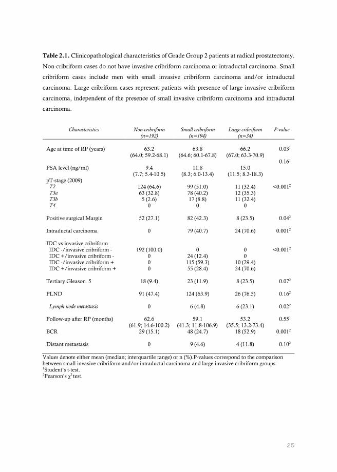
Large invasive cribriform carcinoma
Large invasive cribriform growth was observed in 34 (8.1%) patients. All of these men (100%) had
concomitant small invasive cribriform growth and 24 (70.6%) had intraductal carcinoma (Table
1.1). We compared patients with invasive large cribriform growth with men who had either small
invasive cribriform growth and/ or intraductal carcinoma (n=194). The age of patients with large
cribriform architecture (66.2 years; interquartile range 63.3-70.9) was higher (P=0.03) than of men
with small cribriform architecture (63.8 years; interquartile range 60.1-67.8). Albeit PSA levels of
men with large cribriform architecture were higher (15.0 ng/ml; interquartile range 8.3-18.3) than
in those with small cribriform architecture (11.8 ng/ml; interquartile range 6.0-13.4), this did not
reach significance in this cohort (P=0.16). In total 23/34 (67.7%) patients with large cribriform
pattern had extraprostatic extension (pT3) as compared to 96/194 (49.0%) with small cribriform
pattern (P<0.001), although positive surgical margins were more frequently observed in the latter
group (23.5% versus 42.3%; P=0.04). The total percentage of Gleason grade 4 was 30.0%
(interquartile range 20%-40%) in large and 23.3% (interquartile range 15%-30%) in small
cribriform pattern (P=0.001). Tertiary Gleason grade 5 was observed in 8/34 (23.5%) patients with
large and 23/194 (11.9%) with small cribriform architecture, but this difference did not reach
conventional measures of significance (P=0.07). Lymph node metastases were observed in 6/26
(23.1%) men with large cribriform architecture and in 6/124 (4.8%) with small cribriform
architecture (P=0.002).
Clinical outcome of invasive and intraductal carcinoma
The median follow-up of Grade Group 2 patients without positive lymph node dissection at time
of radical prostatectomy (n=408) was 53 months (interquartile range 12.7-99.1). During follow-up
86 men experienced biochemical recurrence after a median of 26 (interquartile range 10.7-47.6)
months. Biochemical recurrence occurred more frequently (χ2, P=0.01) in the large invasive
cribriform (13/28; 46.4%) than in the small invasive cribriform and/or intraductal group (44/188;
23.4%), and was lowest in Grade Group 2 patients without any cribriform growth (29/192; 15.1%,

24
P=0.04). The median time to biochemical recurrence was significantly shorter (log rank, P<0.001)
in patients with large invasive cribriform growth (11 months; interquartile range 2.6-37.2) than in
patients with small cribriform growth (25 months; interquartile range 11.3-39.3) and no cribriform
architecture (43 months, interquartile range 15.4-73.8) (Figure 2.2).
Univariate analysis showed that PSA (hazard ratio 1.02, 95% CI 1.01-1.04; P=0.0001),
pT3a (hazard ratio 2.00, 95% CI 1.27-3.14; P=0.003), pT3b (hazard ratio 4.42, 95% CI 2.24-8.72;
P<0.001), positive surgical margins (hazard ratio 3.24, 95% CI 2.11-4.97; P<0.0001), intraductal
carcinoma (hazard ratio 2.13, 95% CI 1.36-3.36; P=0.001) and any invasive cribriform growth
(hazard ratio 1.78, 1.16-2.74; P=0.008) were all significant predictors for biochemical recurrence-
free survival (Table 2.2). Percentage Gleason grade 4 was neither predictive as a continuous
(hazard ratio 1.01, 95% CI 0.99-1.03, P=0.076) nor as a dichotomised parameter (hazard ratio
1.26, 95% CI 0.82-1.93, P=0.29). Tertiary Gleason grade 5 (hazard ratio 1.29, 95% CI 0.66-2.50,
P=0.46) did not have predictive value for biochemical recurrence in this cohort. In multivariable
analysis, extraprostatic extension (pT3a, hazard ratio 1.64, 95% CI 1.02-2.63, P=0.04), seminal
vesicle invasion (pT3b, hazard ratio 3.00, 95% CI 1.42-6.34, P=0.004), positive surgical margins
(hazard ratio 3.28, 95% CI 2.06-5.23, P<0.0001) and invasive large cribriform architecture (hazard
ratio 4.36, 95% CI 2.08-9.17, P=0.0001) were independent predictors for biochemical recurrence-
free survival, while small invasive cribriform growth pattern and intraductal carcinoma were not.
To determine whether the difference in prognostic value between invasive small and large
cribriform growth could be explained by an overall higher percentage of cribriform growth, we
compared the outcome of patients with ≥5% invasive cribriform growth and those with <5%.
When invasive cribriform growth was present, no statistical difference existed between low and
high cribriform percentage (log rank; P=0.087).
During follow-up 13 patients developed bone metastases. Nine of these patients had
small invasive cribriform or intraductal carcinoma (4.6%) and four had invasive large cribriform
carcinoma (11.8%) at radical prostatectomy. The median time to bone metastasis was 138 months
(interquartile range 109.4-172.6) for small invasive and intraductal cribriform carcinoma and 59
months (interquartile range 17.9-114.8) for invasive large cribriform carcinoma. Due to the low
number of events we were not able to perform further statistical analysis.
25
Table 2.1. Clinicopathological characteristics of Grade Group 2 patients at radical prostatectomy.
Non-cribriform cases do not have invasive cribriform carcinoma or intraductal carcinoma. Small
cribriform cases include men with small invasive cribriform carcinoma and/or intraductal
carcinoma. Large cribriform cases represent patients with presence of large invasive cribriform
carcinoma, independent of the presence of small invasive cribriform carcinoma and intraductal
carcinoma.
Characteristics Non-cribriform (n=192)
Small cribriform (n=194)
Large cribriform (n=34)
P-value
Age at time of RP (years) PSA level (ng/ml)
63.2
(64.0; 59.2-68.1)
9.4 (7.7; 5.4-10.5)
63.8
(64.6; 60.1-67.8)
11.8 (8.3; 6.0-13.4)
66.2
(67.0; 63.3-70.9)
15.0 (11.5; 8.3-18.3)
0.031
0.161
pT-stage (2009) T2 T3a T3b T4
124 (64.6) 63 (32.8) 5 (2.6)
0
99 (51.0) 78 (40.2) 17 (8.8)
0
11 (32.4) 12 (35.3) 11 (32.4)
0
<0.0012
Positive surgical Margin
52 (27.1) 82 (42.3) 8 (23.5) 0.042
Intraductal carcinoma
0 79 (40.7) 24 (70.6) 0.0012
IDC vs invasive cribriform IDC -/invasive cribriform - IDC +/invasive cribriform - IDC -/invasive cribriform + IDC +/invasive cribriform +
192 (100.0)
0 0 0
0
24 (12.4) 115 (59.3) 55 (28.4)
0 0
10 (29.4) 24 (70.6)
<0.0012
Tertiary Gleason 5
18 (9.4) 23 (11.9) 8 (23.5) 0.072
PLND Lymph node metastasis
91 (47.4) 0
124 (63.9)
6 (4.8)
26 (76.5)
6 (23.1)
0.162
0.022
Follow-up after RP (months)
62.6 (61.9; 14.6-100.2)
59.1 (41.3; 11.8-106.9)
53.2 (35.5; 13.2-73.4)
0.551
BCR
29 (15.1) 48 (24.7) 18 (52.9) 0.0012
Distant metastasis
0 9 (4.6) 4 (11.8) 0.102
Values denote either mean (median; interquartile range) or n (%).P-values correspond to the comparison between small invasive cribriform and/or intraductal carcinoma and large invasive cribriform groups. 1Student’s t-test. 2Pearson’s χ2 test.

26
Figure 2.2. Biochemical recurrence-free survival of Grade Group 2 patients, stratified for absent,
small invasive and/or intraductal carcinoma, and large invasive cribriform architecture growth (P-
value <0.001).
Table 2.2. Cox regression analysis of biochemical recurrence-free survival in Grade Group 2
prostate cancer patients without lymph node metastasis at time of operation (n=408).
Univariate analysis Multivariable analysis HR 95% CI P-value HR 95% CI P-value Age 0.99 0.96 - 1.03 0.56 0.99 0.95 - 1.03 0.57 PSA 1.02 1.01 - 1.04 <0.001 1.01 0.99 - 1.02 0.34 pT-stage T2 T3a T3b
ref
2.00 4.42
1.27 - 3.14 2.24 - 8.72
0.003 <0.001
ref
1.64 3.00
1.02 - 2.63 1.42 - 6.34
0.04 0.004
Positive surgical margin
3.24 2.11 - 4.97 <0.001 3.28 2.06 - 5.23 <0.001
Percentage Gleason 4
1.26 0.82 - 1.93 0.29 0.94 0.59 - 1.51 0.80
Tertiary Gleason 5 1.29 0.66 - 2.50 0.46 0.95 0.44 - 2.06 0.90 Intraductal carcinoma
2.13 1.36 - 3.36 0.001 1.32 0.77 - 2.25 0.31
Invasive cribriform Small Large
1.50 3.98
0.95 - 2.37 2.10 - 7.57
0.09
<0.001
1.07 4.36
0.65 - 1.75 2.08 - 9.17
0.80
<0.001 HR = hazard ratio, CI = confidence interval.

27
Discussion
While most patients with Grade Group 2 prostate cancer are treated with radiotherapy and/or
surgery, active surveillance is increasingly being considered as alternative strategy for these men.42-
46 Further risk stratification in this large group of patients is necessary to support therapeutic
decision-making. Recently, invasive cribriform carcinoma and intraductal carcinoma have been
recognised as promising additional predictive parameters for men with Grade Group 2 prostate
cancer.14, 16, 32, 34, 47 In the current study, invasive and/or intraductal cribriform carcinoma was
present in 54.3% of radical prostatectomies with Grade Group 2 prostate cancer. While the
clinicopathological features of men with invasive cribriform carcinoma without cribriform
intraductal carcinoma were not statistically significant from those with cribriform intraductal
carcinoma only, patients with both invasive and intraductal cribriform carcinoma more often had
extraprostatic extension and lymph node metastasis than those with invasive cribriform carcinoma
only. Furthermore, we found that patients with large invasive cribriform growth had higher pT-
stage and more frequent positive lymph nodes than those with small invasive and/or intraductal
cribriform carcinoma. In multivariable analysis, large invasive cribriform carcinoma was an
independent predictor for biochemical recurrence-free survival, while small invasive carcinoma
and intraductal cribriform carcinoma were not.
Various studies have addressed the association of either invasive cribriform carcinoma
or intraductal carcinoma with adverse features at prostatectomy and with clinical outcome. 14-16, 20,
34 We observed that invasive and intraductal cribriform carcinoma were present in respectively
48.6% and 24.5% of prostatectomy specimens with Grade Group 2 prostate cancer. These rates
are comparable to those found by others. Trudel et al. for instance found intraductal carcinoma in
17.5%, invasive cribriform carcinoma in 45.6% and both invasive and intraductal cribriform
carcinoma in 36.8% of 57 prostate specimens.34 In a cohort of 286 Grade Group 2 prostate cancer
patients, Choy et al. demonstrated intraductal carcinoma in 26.5% and invasive cribriform growth
in 38.7%.20 Two studies took into account large cribriform architecture, however these used
different thresholds.13, 34 Iczkowski et al. defined large cribriform pattern as having more than 12
luminal spaces, while area size exceeding the size of an average benign gland was used by Trudel
et al. Our threshold of large cribriform fields as at least twice the size of normal adjacent glands,
exceeds that of previous studies. For instance, in the study of Iczkowski et al. no cases were present
with small cribriform pattern only, while small invasive cribriform carcinoma was present in 40%
of our cases. To elucidate the clinical and biologic relevance of invasive cribriform and intraductal
carcinoma in prostate cancer, it is crucial that it is clear how both entities are defined.

28
In a previous case-control study of 161 men with Gleason score 7 at radical
prostatectomy, we found invasive cribriform but not intraductal carcinoma to be a significant
predictive marker for metastasis- and disease specific-free survival in multivariate analysis.16 In a
subsequent analysis of prostate biopsies with long term follow-up, both invasive and intraductal
carcinoma had predictive value for disease-specific death, and combining both lesions had the
strongest prognostic value.32 The prognostic value of invasive and intraductal carcinomas at
biopsies does not always correspond with the prognostic value at radical prostatectomies.
Sampling artifacts inherently associated with diagnostic biopsies are likely the cause of
discrepancies between biopsies and radical prostatectomy specimens. This is for instance reflected
by the frequency of cribriform growth in biopsies and resection specimens; while invasive and/or
intraductal cribriform architecture was found in 17% of sextant biopsies with Grade Group 2, it
was present in 54.3% of radical prostatectomy specimens in the current study.32 Since most biopsy
schedules currently include between 8 and 16 biopsies, and biopsies are increasingly being targeted
by Magnetic Resonance Imaging (MRI), the frequency of invasive cribriform and/or intraductal
carcinoma is higher with fewer sampling artifacts.48, 49 Since both small cribriform growth and
intraductal carcinoma are often associated with large cribriform growth, these patterns should still
be reported.
The outcome of this study may have important implications. First, we propose the
inclusion of the presence of large invasive cribriform in pathology reports. Of 26 men with large
cribriform architecture who had undergone pelvic lymph node dissection at time of radical
prostatectomy, 23% (n=6) had lymph node metastasis. Men with large cribriform architecture
should therefore not be considered for surveillance but instead be offered active treatment with
lymph node dissection. On the other hand, the absence of metastasis and low risk of biochemical
recurrence in Grade Group 2 patients with no cribriform architecture might indicate that active
surveillance can be considered in these men, and that pelvic lymph node dissection might be
omitted when treatment is offered. However, it is important to note that the current results were
obtained after studying radical prostatectomy specimens, while treatment decisions are made
based on diagnostic biopsies. An urgent need exists to incorporate pathological features such as
small and large invasive cribriform growth, as well as intraductal carcinoma, into clinical
nomograms and prediction tools.
Strong points of this study are the detailed histological review and the extensive
immunohistochemical staining for classification of cribriform architecture. Although large
cribriform growth is an adverse predictive parameter for Grade Group 2 prostate cancer patients,
the stringent cut-off used in this study resulted in the inclusion of a relatively small number of cases

29
and must be validated. Finally, the retrospective study design and relatively short median follow-
up of 53 months possibly gave rise to a selection bias.
In conclusion, we demonstrate that patients with large invasive cribriform growth
represent a more aggressive subgroup of cribriform Grade Group 2 prostate cancer. Men with large
invasive cribriform carcinoma should be actively treated since they are at increased risk for
biochemical recurrence and metastasis.

30

31
32
Chapter III
Concordance of cribriform architecture in matched prostate cancer biopsy and radical prostatectomies.
Hollemans E, Verhoef EI, Bangma CH, Schoots I, Rietbergen J, Helleman J, Roobol MJ, van Leenders GJLH.
Histopathology. 2019 Sept; 75: 338-345.
33
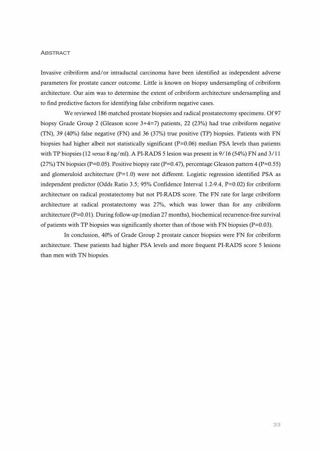
Abstract
Invasive cribriform and/or intraductal carcinoma have been identified as independent adverse
parameters for prostate cancer outcome. Little is known on biopsy undersampling of cribriform
architecture. Our aim was to determine the extent of cribriform architecture undersampling and
to find predictive factors for identifying false cribriform negative cases.
We reviewed 186 matched prostate biopsies and radical prostatectomy specimens. Of 97
biopsy Grade Group 2 (Gleason score 3+4=7) patients, 22 (23%) had true cribriform negative
(TN), 39 (40%) false negative (FN) and 36 (37%) true positive (TP) biopsies. Patients with FN
biopsies had higher albeit not statistically significant (P=0.06) median PSA levels than patients
with TP biopsies (12 versus 8 ng/ml). A PI-RADS 5 lesion was present in 9/16 (54%) FN and 3/11
(27%) TN biopsies (P=0.05). Positive biopsy rate (P=0.47), percentage Gleason pattern 4 (P=0.55)
and glomeruloid architecture (P=1.0) were not different. Logistic regression identified PSA as
independent predictor (Odds Ratio 3.5; 95% Confidence Interval 1.2-9.4, P=0.02) for cribriform
architecture on radical prostatectomy but not PI-RADS score. The FN rate for large cribriform
architecture at radical prostatectomy was 27%, which was lower than for any cribriform
architecture (P=0.01). During follow-up (median 27 months), biochemical recurrence-free survival
of patients with TP biopsies was significantly shorter than of those with FN biopsies (P=0.03).
In conclusion, 40% of Grade Group 2 prostate cancer biopsies were FN for cribriform
architecture. These patients had higher PSA levels and more frequent PI-RADS score 5 lesions
than men with TN biopsies.

34
Introduction
Risk stratification and therapeutic decision-making in prostate cancer patients is affected by
potential biopsy undersampling. The Gleason score is one of the most important parameters for
predicting disease outcome and guiding individual treatment. Men with Gleason score 3+3=6
(International Society of Urological Pathology (ISUP) Grade Group 1) prostate cancer are eligible
for active surveillance, whereas men with Gleason score ≥ 4+3=7 (Grade Group 3-5) are usually
treated with radical prostatectomy, radiation therapy and/or hormonal therapy. The optimal
therapeutic strategy for men with Gleason score 3+4=7 (Grade Group 2) still is a matter of debate.
While most of these patients will undergo active treatment, surveillance is increasingly being
considered in this subgroup. Incorporation of additional clinicopathological and molecular
parameters might be able to support optimal decision-making in this large prostate cancer
subpopulation.
Grade Group 2 prostate cancer is a heterogeneous disease with variable architectural
growth patterns and Gleason pattern 4 quantities. While individual growth patterns are not
routinely mentioned in pathology reports, recent studies have shown that patients with cribriform
architecture have adverse outcome as compared to those without.15, 39, 50 Both invasive and
intraductal cribriform architecture have been associated with adverse clinicopathological
characteristics, post-operative recurrence rates, metastasis and disease-specific death.14, 16, 32, 34, 51
On the other hand, biopsy Grade Group 2 prostate cancer patients without cribriform architecture
have comparable disease-specific survival and post-operative biochemical recurrence rates as men
with Grade Group 1 disease.39, 52 Quantification of Gleason pattern 4 can further add in risk
stratification since post-operative biochemical recurrence rates increment with higher Gleason
pattern 4 tumour percentage.53 Cribriform architecture and Gleason pattern 4 quantification might
therefore be important adjuncts in risk stratification of Grade Group 2 prostate cancer patients.
While pathological tumour characteristics are important for clinical decision-making,
prostate biopsies are prone to undersampling. Prostate cancer is upgraded in up to 40% of
subsequent radical prostatectomy specimens.54, 55 At present, little is known on the extent of
undersampling in detection of cribriform architecture or Gleason pattern 4 percentage. The aim of
our study is to determine the extent of undersampling for the detection of cribriform architecture
in matched prostate biopsy and radical prostatectomy specimens, and to identify potential factors
for discriminating true from false cribriform negative prostate biopsies.

35
Materials and Methods
Patient selection
We identified 186 patients who had undergone both biopsy and subsequent radical prostatectomy
at Erasmus MC University Medical Center, Rotterdam, The Netherlands between 2010 and 2017.
Biopsies were prompted by elevated Prostate Specific Antigen (PSA) levels or obtained in the
scope of active surveillance. The Prostate Imaging Reporting and Data System (PI-RADS) score
was annotated by an expert uroradiologist, when patients had received multiparametric magnetic
resonance imaging (MRI).56 When suspicious lesions (PI-RADS 3 to 5) were visible on MRI,
targeted MRI-ultrasound fusion biopsies were taken. Individual biopsy cores were enclosed in
separate containers and radical prostatectomy specimens were completely embedded for
diagnostic purposes. All slides of both biopsies and radical prostatectomies were available for
pathologic review. This study was approved by the institutional Medical Research Ethics
Committee (MEC-2018-1614).
Pathologic evaluation
All biopsies were reviewed by three investigators, who were blinded to clinical outcome and
radical prostatectomy characteristics. For each biopsy core the following features were recorded:
Gleason score, Grade Groups according to the WHO/ISUP 2014 guidelines, maximal single
biopsy tumour length (mm), overall estimated percentage Gleason pattern 4 and individual
tumour growth patterns.12 Invasive cribriform Gleason pattern 4 was not distinguished from
intraductal carcinoma because of their significant morphological overlap, which would require
extensive immunohistochemical staining for further discrimination.39 In case targeted biopsies
were obtained, these were considered as separate biopsies and not as one single biopsy. Matching
radical prostatectomy specimens were evaluated as described previously.51 We recorded Gleason
score, Grade Group, pT-stage according to the AJCC TNM 8th edition, surgical margin status,
percentage Gleason pattern 4 and individual growth patterns.41 Furthermore, we distinguished
small and large expansive cribriform growth pattern based on a cut-off of two times the size of
adjacent pre-existent normal glands.51
Clinical follow-up
After radical prostatectomy, clinical follow-up consisted of bi-annual, and later annual monitoring
of serum PSA levels. Biochemical recurrence was defined as PSA levels ≥ 0.2 ng/ml measured at
two consecutive points in time, at least three months apart with undetectable PSA levels after
operation, or as PSA increase of > 2.0 ng/ml when serum PSA had not declined to zero after

36
operation. Survival was defined as time in months from radical prostatectomy to biochemical
recurrence or last follow-up.
Statistical analysis
Continuous variables with normal distribution were compared by Student’s t-test and One-way
ANOVA analysis, those without normal distribution with the Mann-Whitney U test. For
categorical parameters Chi-square or Fishers exact were used. Correlation between continuous
variables was analysed using Pearson’s correlation coefficient. Dichotomous outcome variables
were analysed using logistic regression. Survival was visualised by Kaplan-Meier curves. Statistics
were performed using R version 3.2.2 (R, Vienna, Austria) and results were considered significant
when the two-sided P-value was <0.05.

37
Results
Clinicopathological characteristics
The entire cohort consisted of 186 patients with matched biopsy and radical prostatectomy
specimens. The mean age at time of operation was 65 years (interquartile range (IQR) 62-70) and
the mean PSA level was 12 ng/ml (IQR 6-15). In total 144 (77%) patients underwent systematic
biopsies, 26 (14%) received systematic and targeted biopsies, and 16 (9%) had targeted biopsies
only. The mean number of biopsies taken was 9 (IQR 8-10) with 4 (IQR 3-5) biopsies containing
adenocarcinoma, representing 49% (IQR 30-66) of the total number of biopsy cores. Fifty (27%)
patients had overall biopsy Grade Group 1, 99 (53%) Grade Group 2, 11 (6%) Grade Group 3, 15
(8%) Grade Group 4 and 11 (6%) Grade Group 5.
On radical prostatectomy, 87 (47%) adenocarcinomas were pT2, 76 (41%) pT3a and 23
(12%) pT3b. Distribution of the Grade Groups on radical prostatectomy was as follows: 19 (10%)
Grade Group 1, 108 (58%) Grade Group 2, 25 (14%) Grade Group 3, 17 (9%) Grade Group 4 and
17 (9%) Grade Group 5. Tumour upgrading occurred in 65 (35%) and down-grading in 14 (8%)
radical prostatectomies, while 107 (57%) cases had concordant tumour grades. Positive surgical
margins were present in 63 (34%) patients. Eighty patients had simultaneously undergone pelvic
lymph node dissection, of which 18 (23%) contained lymph node metastasis. The mean post-
operative follow-up was 32 months (median 22, IQR 8-51).
Invasive cribriform and/or intraductal carcinoma was observed in 57 (31%) diagnostic
biopsies and in 128 (69%) radical prostatectomy specimens (Table 3.1). Cribriform architecture
was present in both matched biopsy and radical prostatectomy specimens in 55 (30%), and absent
in 56 (30%) cases. In 73 (39%) men cribriform architecture was observed in the radical
prostatectomy specimen, but not in preceding biopsies. Two cases (1%) with cribriform
architecture at biopsy but not at subsequent radical prostatectomy, probably due to sampling error,
were excluded from further analyses. Therefore, sensitivity for cribriform architecture on biopsies
was 43%, while specificity was 97%. Cribriform architecture was observed more frequently in
targeted (19/40; 48%) than systematic biopsies (36/144; 25%, P=0.01).
Table 3.1. Prevalence of invasive cribriform and/or intraductal carcinoma (CR/IDC) in biopsies
and matched radical prostatectomies.
Radical prostatectomy
Prostate biopsy CR/IDC- CR/IDC+
CR/IDC- 56 (30%) 73 (39%)
CR/IDC+ 2 (1%) 55 (30%)

38
Concordance of cribriform architecture in Grade Group 2 prostate cancer biopsies
Since cribriform architecture might be most relevant for treatment decisions in patients with biopsy
Grade Group 2 prostate cancer, we performed further analyses within this subgroup (n=97). Thirty
six (37%) patients with biopsy Grade Group 2 demonstrated cribriform architecture on both
matched biopsy and radical prostatectomy specimen (true cribriform positive, CR+/CR+), while
cribriform architecture was absent in both specimens in 22 (23%) cases (true cribriform negative,
CR-/CR-). In 39 (40%) patients cribriform architecture was present on radical prostatectomy but
not on preceding biopsy; these patients were considered as having false cribriform negative (CR-
/CR+) biopsies. None of the patients with biopsy Grade Group 2 had cribriform architecture on
biopsy while radical prostatectomy was negative for cribriform architecture.
Identification of predictors in true and false cribriform negative Grade Group 2 prostate cancer biopsies
Patients with true negative biopsies were slightly younger (62 versus 65 years, P=0.06) and had
lower PSA levels (8 ng/ml versus 12 ng/ml, P=0.06) than men with false negative biopsies,
however these differences were not significant (Table 3.2). In total, 51 patients (53%) had
undergone multiparametric MRI prior to biopsy. Out of 11 patients with true negative biopsies, 3
(27%) had a PI-RADS 5 lesion as compared to 9/16 (56%) of false negative and 17/24 (71%) of
true positive biopsy patients (P=0.05). The number of biopsies (P=0.53), percentage of positive
biopsies (P=0.47) and maximal tumour length (P=0.44) were not different between true and false
negative biopsies.
Since Gleason pattern 4 percentage and glomeruloid architecture have both been
associated with cribriform architecture, we assessed the predictive value of these pathologic
parameters.16, 19 Mean percentage of Gleason pattern 4 was 12% (IQR 5-10%) in true negative
biopsies and 11% (IQR 5-16%) in false negative biopsies (P=0.55). There was only a weak
correlation between percentage Gleason pattern 4 on biopsies (mean 13%, IQR 5-20%) and
matched radical prostatectomies (mean 31%, IQR 10-40%, R2=0.093; P=0.001). Glomeruloid
growth pattern was encountered in 6/22 (27%) true negative and 11/39 (28%) false negative
biopsies (P=1.0).
Logistic regression analysis on cribriform negative biopsy patients showed that age (odds
ratio (OR) 1.1, 95% confidence interval (CI) 1.0-1.3, P=0.02) and PSA (OR 3.3, 95% CI 1.2-9.1,
P=0.02) were independent predictive parameters for presence of cribriform architecture on radical
prostatectomy in multivariable analysis, whereas PI-RADS score, number and percentage of
positive biopsies, maximal tumour length, presence of targeted biopsies and percentage Gleason
grade 4 were not (Table 3.3).

39
Table 3.2. Characteristics of biopsy Grade Group 2 prostate cancer (PCa) patients stratified for
true cribriform negative (CR-/CR-), false cribriform negative (CR-/CR+) and true cribriform
positive (CR+/CR+) biopsies.
CR-/CR- (n=22) CR-/CR+ (n=39) CR+/CR+ (n=36) P-value
Age 62 (63, 58-65) 65 (66, 62-71) 66 (66, 62-71) 0.06a
PSA 8 (8, 6-10) 12 (10, 6-17) 16 (13, 9-19) 0.06b
PI-RADS score: no MRI 11 (50%) 23 (59%) 12 (33%) 0.10c
1-2 3 (14%) 0 (0%) 0 (0%)
3 1 (5%) 1 (3%) 2 (6%)
4 4 (18%) 6 (15%) 5 (14%)
5 3 (14%) 9 (23%) 17 (47%)
Number of biopsies 9 (9, 8-10) 8 (8, 7-10) 10 (10, 8-12) 0.53d
# PCa positive biopsies 4 (3, 2-6) 4 (4, 3-5) 6 (5, 4-8) 0.64d
% PCa positive biopsies 47 (38, 25-71) 52 (50, 31-73) 59 (61, 40-76) 0.47d
Max tumour length (mm) 7 (7, 5-8) 8 (7, 5-10) 9 (10, 7-12) 0.44d
% Gleason pattern 4 12 (8, 5-10) 11 (8, 5-16) 17 (15, 7-23) 0.55a
Glomeruloid growth 6 (27%) 11 (28%) 12 (33%) 1.0e
Large cribriform growth 0 6 (15%) 16 (44%) N/A
Targeted biopsies 2 (9%) 8 (20%) 13 (36%) 0.30e
Grade Group (RP): 1 2 (9%) 1 (3%) 1 (3%) 0.01e
2 18 (82%) 29 (74%) 26 (72%)
3 0 (0%) 8 (20%) 7 (19%)
4 0 (0%) 1 (3%) 1 (3%)
5 2 (9%) 0 (0%) 1 (3%)
Positive surgical margins 8 (36%) 12 (31%) 12 (33%) 0.78c
pT stage (TNM 8th ): 2 11 (50%) 15 (38%) 17 (47%) 0.66c
3a 10 (45%) 20 (51%) 12 (33%)
3b 1 (5%) 4 (11%) 7 (20%)
Biochemical recurrence 2 (9%) 6 (15%) 13 (36%) 0.69e
Metastasis 0 (0%) 1 (3%) 4 (11%) N/A
Mean (median, IQR) or n (%). a Wilcox-test, b t-test (log2 values were used for this test), c Chi-square (χ2), d
One-Way Anova, e Fisher test. P-values resemble comparison between CR-/CR- and CR-/CR+.

40
Table 3.3. Logistic regression analysis of biopsy Grade Group 2 cribriform negative prostate
cancer (PCa) patients (n=61), predicting cribriform architecture on radical prostatectomy.
Univariate Multivariable
OR 95% CI P-value OR 95% CI P-value
Age 1.1 1.0-1.2 0.06 1.1 1.0-1.3 0.02
PSA (log2) 2.2a 1.0-4.8 0.04 3.3a 1.2-9.1 0.02
PI-RADS score
<5 ref
5 1.9 0.5-7.9 0.38 1.8 0.3-9.1 0.49
Number of biopsies 0.9 0.8-1.1 0.53 0.8 0.6-1.1 0.21
Percentage PCa positive biopsies 2.1 0.3-15 0.47 0.2 0.0-5.5 0.35
Maximal tumour length (mm) 1.1 0.9-1.2 0.43 1.0 0.9-1.3 0.70
Percentage Gleason pattern 4 1.0 0.9-1.0 0.70 1.0 0.9-1.0 0.36
Presence of targeted biopsies
No ref
Yes 2.6 0.5-13 0.26 1.1 0.1-10 0.91 a Per doubling unit. OR = odds ratio, CI = confidence interval.
Figure 3.1. Biochemical recurrence-free survival of biopsy Grade Group 2 prostate cancer
patients, stratified for the presence of cribriform architecture on biopsies and subsequent radical
prostatectomies (log rank over all groups, P-value = 0.02).

41
Comparison of false negative and true cribriform positive Grade Group 2 biopsies
PSA levels of men with true positive biopsies were slightly higher than of those with false negative
biopsies, but this was not statistically significant (16 ng/ml versus 12 ng/ml, P=0.13). Patients with
true positive biopsies had a significantly higher total number of biopsies (10 versus 8, P=0.02) and
number of tumour positive biopsies (6 versus 4, P=0.001), however no differences were seen in
percentage positive biopsies (59% versus 52%, P=0.19) when compared to patients with false
negative biopsies. Percentage Gleason pattern 4 was higher in patients with cribriform positive
biopsies than in those with false negative biopsies (17% versus 11%, P=0.03). Final Grade Group
(P=0.97), pT stage (P=0.27) and surgical margin status (P=0.24) of the radical prostatectomy
specimens were not different between these two groups. The tumour volume percentage of
cribriform growth at radical prostatectomy was higher in patients with true positive biopsies than
in those with false negative biopsies, but this did not meet conventional measures of significance
(13% versus 6%, P=0.06).
Large expansile cribriform architecture, which represents an aggressive subtype of
invasive cribriform carcinoma, was present in 22/97 (23%) radical prostatectomy specimens.51
Sixteen of these 22 (73%) patients had any size cribriform fields on biopsy, while biopsies were
false negative in 6 (27%) men. The false negative rate for more aggressive large cribriform
architecture (6/22; 27%) was lower than for any cribriform architecture (39/75; 52%, P=0.01). In
case large cribriform carcinoma was present at radical prostatectomy, the tumour volume
percentage of any cribriform growth at the operation specimens did not differ between men with
false cribriform negative and true positive biopsies (P=0.5). This indicates that the lower false
negative rate of large cribriform growth was not merely due to larger total cribriform tumour
percentage at radical prostatectomy.
Clinicopathological outcome in Grade Group 2 patients
Of 97 patients with biopsy Grade Group 2 prostate cancer, 73 (75%) had concordant Grade Group
at radical prostatectomy, 20 (21%) were upgraded to Grade Group 3 to 5, and 4 (4%) down-graded
to Grade Group 1. Upgrading occurred in 9/36 (25%) true positive and in 9/39 (23%) false
negative biopsies, and was significantly lower (P=0.01) in true negative biopsies (2/22, 9%). Extra-
prostatic expansion and surgical margins status were not significantly different between the three
groups.
Biochemical recurrence occurred in 21 (22%) patients and was significantly more
frequent in the true positive (13/36, 36%) than in the false negative group (6/39, 15%, P=0.03).
The true negative group (2/22, 9%) showed the lowest incidence of biochemical recurrence,
however this difference was not significant (P=0.13) when compared to the false negative group.
42
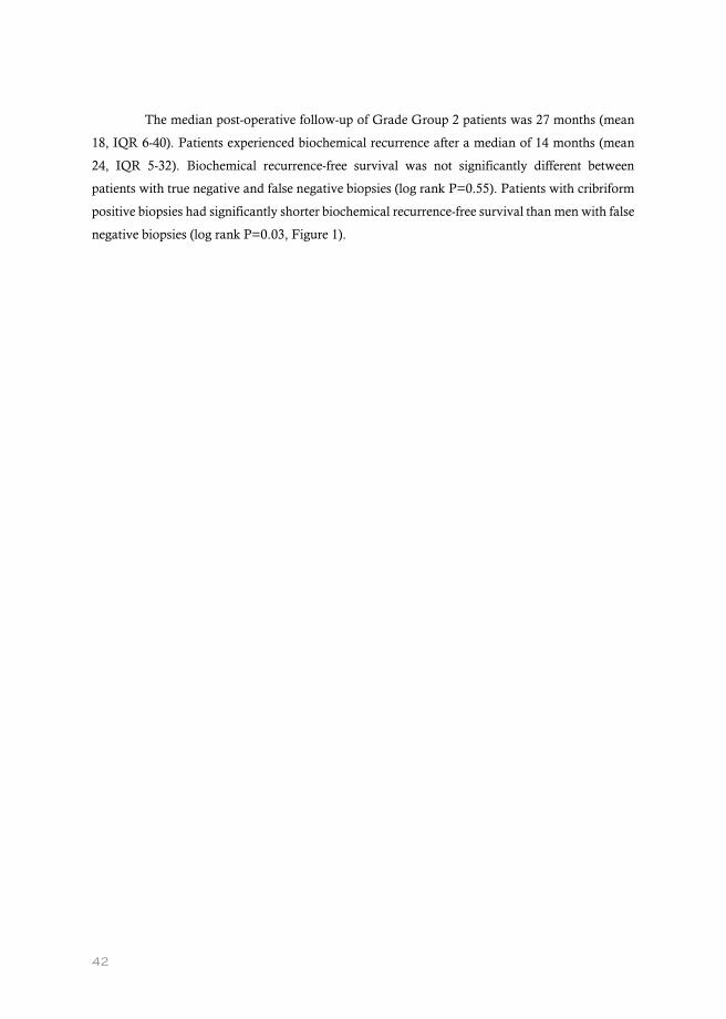
The median post-operative follow-up of Grade Group 2 patients was 27 months (mean
18, IQR 6-40). Patients experienced biochemical recurrence after a median of 14 months (mean
24, IQR 5-32). Biochemical recurrence-free survival was not significantly different between
patients with true negative and false negative biopsies (log rank P=0.55). Patients with cribriform
positive biopsies had significantly shorter biochemical recurrence-free survival than men with false
negative biopsies (log rank P=0.03, Figure 1).

43
Discussion
Identification and pathologic reporting of invasive cribriform and/or intraductal carcinoma of the
prostate are increasingly important since they are both associated with adverse clinical outcome.14,
34, 39, 50 Biopsy undersampling is a well-known problem which might have significant impact on
individual patient management.54, 57, 58 Hitherto, little is known about biopsy undersampling in
identifying cribriform architecture. In this study we demonstrated that biopsies were false negative
for cribriform architecture in 39% of all cases and in 40% of patients with biopsy Grade Group 2
prostate cancer. In false negative Grade Group 2 patients, age and PSA level were independent
predictive parameters for presence of cribriform architecture on subsequent radical prostatectomy,
while percentage of positive biopsies, maximal biopsy tumour length, percentage Gleason pattern
4 and glomeruloid growth were not. Patients with the more aggressive large cribriform growth
pattern on radical prostatectomy were, however, less likely to have cribriform negative biopsies.51
Biopsy Grade Group 2 patients with false cribriform negative biopsies showed better biochemical
recurrence-free survival rates than men with true cribriform positive biopsies albeit follow-up was
relatively short.
Masoomian et al. studied concordance rates of cribriform architecture in 245 matched
biopsies and operation specimens, and found a relatively low sensitivity of 47%, corresponding
well with the 43% sensitivity in our study.59 In their subset of Grade Group 2 biopsy patients, false
negative and true positive biopsies both had more advanced stage as compared to true negative
biopsies on radical prostatectomy suggesting men with false negative and true positive biopsies
have comparable outcome. This contrasts with our study as we found that post-operative
biochemical recurrence-free survival of men with true positive biopsies was significantly shorter
than of those with false negative biopsies. The difference might be explained by the different and
relatively small cohorts of both studies.
While most patients with biopsy Grade Group 2 prostate cancer undergo active
treatment, the question is rising whether surveillance could be a safe alternative for subgroups of
this large patient population. It has for instance been proposed that patients with biopsy Grade
Group 2 prostate cancer and low Gleason pattern 4 percentage should be considered for
surveillance.44, 60 Others have suggested that biopsy Grade Group 2 prostate cancer patients
without invasive cribriform and/or intraductal carcinoma might be eligible for surveillance.32, 52
To further support clinical decision tools, it is important to get insight in the false negative rate of
potentially aggressive disease parameters and to determine how this rate can be minimised to an
acceptable level. In the current study, we showed that consideration of PSA level, which is an
important parameter for active surveillance, might prevent men with potentially aggressive false
44
negative biopsies from being abstained from immediate treatment. Furthermore, presence of a PI-
RADS 5 lesion on multiparametric MRI might also be indicative of more aggressive disease.
Truong et al. identified cribriform morphology in combined systematic and targeted biopsies in
37% of PI-RADS 5, 24% of PI-RADS 4 and 6% of PI-RADS 2 lesions, suggesting that high-grade
MRI lesions are related to more aggressive tumours with cribriform morphology.48 Prendeville et
al. identified cribriform morphology in 8% of PI-RADS 3/4 lesions and in 39% of PI-RADS 5
lesions, indicating that PI-RADS score might be a predictor for cribriform positive prostate
cancer.49 Here we showed that 56% of false negative biopsies had a PI-RADS 5 lesion as compared
to 27% of true negative biopsies. However, due to the small number of patients that had undergone
MRI, PI-RADS score was not a predictor for cribriform architecture in logistic regression analysis.
We were not able to find any predictive value of biopsy percentage Gleason pattern 4 or
glomeruloid growth pattern for cribriform architecture on radical prostatectomy. Presence of
cribriform architecture has been associated with higher percentage Gleason pattern 4 on biopsies.
In a cohort of 370 biopsy Grade Group 2 prostate cancer patients, we found cribriform architecture
in 6% of men with <10% Gleason pattern 4, in 22% of men with 10-25% pattern 4, and in 44% of
men with 25-50% pattern 4.32 Nevertheless, biopsy percentage Gleason pattern 4 was not
predictive for cribriform architecture in false negative biopsies. This paradoxical outcome could
be explained by the low level of concordance between percentage Gleason pattern 4 on biopsy and
matched radical prostatectomy specimens in this study. Similarly, glomeruloid Gleason pattern 4
which has been hypothesised to represent a precursor lesion of cribriform growth, was not
associated with cribriform architecture in false negative biopsies.19
Amongst patients with cribriform architecture, those with large expansive cribriform
fields have the worst outcome.51 The false negative rate of 27% for large cribriform pattern is
significantly less than the rate of 52% for overall cribriform morphology. Since 44% of true positive
biopsies had large cribriform fields on radical prostatectomy as compared to only 15% of false
negative biopsies, this might explain the significantly better biochemical recurrence-free survival
of false negative biopsies as compared to true positive biopsies, in addition to other
clinicopathological confounding factors.
The strong points of this study are the detailed histological review of matched biopsies
and radical prostatectomies. The study is however limited by its low number of patients, the
heterogeneity of the study population including both patients with first-time diagnosis and
progression during active surveillance, and variability of diagnostic work-up encompassing
systematic and/or targeted biopsies as well as multiparametric MRI assessment. Finally, follow-
up is relatively short with a median of 27 months.

45
In conclusion, we demonstrate that 40% of men with biopsy Grade Group 2 prostate
cancer were false negative for invasive cribriform and/or intraductal carcinoma. Age and PSA
were independent predictors for cribriform architecture in false negative biopsies, while patients
with false negative biopsies more frequently had PI-RADS score 5 lesions than men with true
negative biopsies. Multimodal evaluation of biopsy Grade Group 2 prostate cancer patients could
therefore identify men with true cribriform negative biopsies who might become eligible for active
surveillance.

46

47

48
Chapter IV
Clinical outcome comparison of Grade Group 1 and Grade Group 2 prostate cancer with and without cribriform architecture.
Hollemans E, Verhoef EI, Bangma CH, Rietbergen J, Roobol MJ, Helleman J, van Leenders GJLH.
Histopathology. 2020 Apr; 76: 755-762.
49
Abstract
Invasive cribriform and intraductal carcinoma are associated with aggressive disease in Grade
Group 2 (GG2) prostate cancer patients. However, the characteristics and clinical outcome of
Grade Group 2 patients without cribriform architecture (GG2-) compared to those with Grade
Group 1 (GG1) disease are unknown. The aim of this study was to investigate the clinical and
pathological characteristics of GG1 and GG2- prostate cancer in radical prostatectomy specimens.
We reviewed 835 radical prostatectomy specimens for Grade Group, pT-stage, surgical margin
status and presence of cribriform architecture. Biochemical recurrence-free survival and metastasis
were used as clinical outcomes. GG1 prostate cancer was seen in 207 and GG2 in 420 patients, of
whom 228 (54%) showed cribriform architecture (GG2+) and 192 (46%) did not. Patients with
GG2- disease had higher Prostate Specific Antigen levels (9.4 versus 7.0 ng/ml; P<0.001), more
often extra-prostatic extension (36% versus 11%; P<0.001) and more frequent positive surgical
margins (27% versus 17%; P=0.01) than those with GG1. GG2- patients had shorter biochemical
recurrence-free survival (Hazard Ratio (HR) 2.7, 95% Confidence Interval (CI) 1.4-4.9; P=0.002)
than those with GG1. Lymph node and distant metastasis were neither observed in GG2- nor in
GG1 patients, but occurred in 22/228 (10%) of GG2+ patients.
In conclusion, patients with GG2- prostate cancer at radical prostatectomy have more advanced
disease and shorter biochemical recurrence-free survival than men with GG1, but both groups
have very low risk of developing metastasis.
50
Introduction
Active surveillance is increasingly applied in men with prostate cancer. Whereas most men with
biopsy Grade Group 1 (Gleason score 3+3=6, GG1) prostate cancer are eligible for active
surveillance, inclusion of favourable Grade Group 2 (Gleason score 3+4=7, GG2) patients with
limited Gleason pattern 4 is gradually accepted.45, 61-64 In general, these patients have Prostate
Specific Antigen (PSA) levels of <10 ng/ml, present with organ-confined disease and have <10%
Gleason pattern 4 in their diagnostic biopsies.7
Gleason pattern 4 prostate cancer is a heterogeneous disease encompassing various
histopathological growth patterns. Invasive and/or intraductal cribriform carcinoma, both also
referred to as cribriform architecture, have been identified as pathological parameters for worse
outcome in both biopsy as well as radical prostatectomy specimens.13-17, 34 Cribriform architecture
has been associated with advanced tumour stage, biochemical recurrence, metastasis and disease-
specific death in GG2 prostate cancer.32, 50, 51 While patients with GG2 prostate cancer without
cribriform architecture (GG2-) have favourable outcome compared to those with invasive and/or
intraductal cribriform carcinoma (GG2+), it is unclear to what extent GG2- differs from GG1
disease.
In previous sextant biopsy studies with long-term follow-up, patients with biopsy GG2-
prostate cancer had similar biochemical recurrence-free and disease-specific survival as men with
GG1 disease.39, 52 Therefore, it has been proposed that patients without cribriform architecture
might be eligible for active surveillance.32, 39, 44, 52, 60, 65 Prostate biopsies are, however, subject to
significant sampling errors with tumour undergrading in up to 40% and there is low sensitivity for
detection of cribriform architecture.58, 59, 66 Moreover, in contrast to radical prostatectomy
specimens, minor high-grade patterns are always taken into account when grading prostate cancer
biopsies. To elucidate on its clinical and biological features, GG2- prostate cancer should be
investigated on radical prostatectomy specimens, which excludes study bias by biopsy sampling
artefacts. The aim of this study was to compare the clinicopathological characteristics and
biochemical recurrence-free survival of GG1 and GG2- prostate cancer patients in radical
prostatectomy specimens.

51
Materials and methods
Patient selection
In total, 854 men who had undergone radical prostatectomy for prostate adenocarcinoma at
Erasmus MC, University Medical Center, Rotterdam, The Netherlands between 2000 and 2017
were included. Men who had received hormonal, radiation or viral therapy (n=19) prior to
operation were excluded from this study.40 After fixation in neutral-buffered formalin, radical
prostatectomy specimens were sectioned transversely and totally embedded for diagnostic
purposes. All slides were available for pathology review. The use of tissue samples for scientific
purposes was approved by the institutional Medical Research Ethics Committee (MEC-2018-
1614).
Pathologic evaluation
All 835 radical prostatectomy specimens were reviewed by two investigators (EH, GvL), who were
blinded to clinical outcome. The following features were recorded: Gleason score and Grade
Group according to the World Health Organization (WHO) 2016 guidelines, pT-stage according
to the American Joint Committee on cancer (AJCC) TNM 8th edition, surgical margin status,
Gleason pattern 3 to 5 percentages, Gleason 4 growth patterns and presence of intraductal
carcinoma.12, 38, 41 Intraductal carcinoma was not incorporated in the Gleason score. Tertiary
Gleason patterns occupied less than 5% of the total tumour area and were not incorporated in the
Gleason score.12, 38
In order to most accurately distinguish intraductal form invasive cribriform carcinoma, the
following criteria were used. Invasive cribriform Gleason grade 4 was morphologically
distinguished from intraductal carcinoma when it had an irregular outline, anastomosing fields
beyond pre-existent gland architecture or extension into periprostatic fat tissue, ejaculatory ducts
or seminal vesicles. Intraductal carcinoma was morphologically identified if cribriform structures
were clearly continuous with pre-existent glands lined by normal basal epithelium, or containing
corpora amylacea. When invasive cribriform carcinoma and intraductal carcinoma could not be
differentiated by morphological criteria alone, additional immunohistochemical staining for the
presence of basal cells was performed.
Immunohistochemistry
Four micrometer thick tissue sections were cut from selected paraffin-embedded blocks (Superfrost
Microscopic Slides, ThermoFisher Scientific, Bleiswijk). Slides were deparaffinized and
rehydrated with xylene and ethanol. Endogenous peroxidase was blocked using 0.3% H2O2 in PBS

52
and heat-induced antigen retrieval accomplished by 15 min in Tris-EDTA buffer (pH 9; Klinipath,
Duiven, The Netherlands). Mouse monoclonal high molecular weight cytokeratin (clone 34BE12;
1:200; DAKO; Heverlee, Belgium) diluted in normal antibody diluent (APG-500; ScyTek
Laboratories, West Logan, USA) was incubated for 2 hours at room temperature. Antibody
visualization was performed using the Envision kit (DAKO) and slide counterstaining with
hematoxylin. When basal cell staining was absent, the cribriform structure was categorized as
invasive carcinoma; if sporadic, scattered or continuous basal cells were identified the growth
pattern was classified as intraductal carcinoma.
Clinical follow-up
Clinical follow-up after radical prostatectomy consisted of six-monthly, and later annual
monitoring of serum PSA levels. Biochemical recurrence was defined as PSA level ≥ 0.2 ng/ml
measured at two separate points in time at least three months apart when PSA had been
undetectable after operation, or as PSA increase of > 2.0 ng/ml whenever serum PSA had not
declined to zero after operation. Post-operative lymph node and distant metastases were confirmed
by biopsy or multidisciplinary consensus. Biochemical recurrence-free survival was defined as time
in months from radical prostatectomy to biochemical recurrence.
Statistical analysis
Normally distributed, continuous variables were analysed using the independent sample Student’s
t-test. Pearson’s chi squared (χ2) test was used for categorical parameters. Missing PSA values
(n=27) were imputed using the median PSA value. Biochemical recurrence-free survival was
analysed using Cox proportional hazards regression and visualized by Kaplan-Meier curves.
Statistics were performed using SPSS version 24 (IBM, Chicago, IL, USA). Results were
considered significant when the two-sided P-value was <0.05.

53
Results
Patient characteristics
Out of 835 radical prostatectomy specimens, 207 had GG1 and 420 GG2 prostate cancer. The
median age of these 627 patients at time of operation was 64.1 years (interquartile range (IQR)
59.8-67.6 years) and the median PSA level was 7.6 ng/ml (IQR 5.4-10.8 ng/ml). Pathologic
tumour stage was distributed as follows: 419 (66%) pT2, 173 (28%) pT3a and 35 (6%) pT3b
tumours. Positive surgical margins were present in 177 (28%) cases. Pelvic lymph node dissection
was performed in 375 (60%) men, of whom 12 (3%) had lymph node metastasis.
Invasive cribriform and/or intraductal carcinoma
Among men with GG2 prostate cancer, 228 (54%) had invasive cribriform and/or intraductal
carcinoma (GG2+) and 192 (46%) did not (GG2-). GG2+ patients had higher PSA levels (12.2
ng/ml versus 9.4 ng/ml; P=0.006), higher percentage Gleason pattern 4 (24% versus 18%; P<0.001),
more frequent extra-prostatic extension (pT3; 52% versus 36%; P<0.001), positive surgical margins
(40% versus 27%; P=0.007) and lymph node metastases (8% versus 0%; P=0.001) than GG2-
patients. Patients with GG2- presented with higher median PSA levels (9.4 versus 7.0 ng/ml;
P<0.001), more frequent extra-prostatic extension (36% versus 11%; P<0.001) and positive surgical
margins (27% versus 17%; P=0.01) than men with GG1 prostate cancer (Table 4.1). None of the
patients with GG1 or GG2- tumours had metastasis at lymph node dissection.
Tertiary Gleason pattern 4 in Grade Group 1 prostate cancer
GG1 prostate cancer in radical prostatectomy specimens might by definition contain tertiary high-
grade patterns. To investigate to what extent GG2- disease differed from GG1 with tertiary pattern
and/or pure GG1, we analysed both GG1 subgroups separately. Tertiary Gleason pattern 4 was
present in 42 out of 207 (20%) GG1 patients, of whom 9 (4%) had cribriform architecture. Tertiary
Gleason pattern 5 was observed in only one (0.5%) patient. Men with tertiary Gleason pattern 4
had higher median PSA levels (8.4 versus 6.6 ng/ml; P=0.01), more frequent extra-prostatic
extension (41% versus 3%; P<0.001) and positive surgical margins (43% versus 10%; P<0.001) than
GG1 men without a tertiary pattern. Although GG2- patients had higher percentage Gleason
pattern 4 (18% versus 3%; P<0.001) and more often tertiary Gleason pattern 5 (9% versus 0.5%;
P=0.04) than GG1 men with tertiary Gleason pattern 4, PSA levels (9.4 ng/ml versus 8.4 ng/ml;
P=0.4) and extra-prostatic extension (36% versus 41%; P=0.7) were not statistically different.

54
Clinical outcome
The median follow-up of the entire cohort was 59.6 months (IQR 17.5-113.9). Biochemical
recurrence occurred in 112 (18%) men after a median of 29.9 months (IQR 11.6-55.5). GG2-
patients had shorter biochemical recurrence-free survival than GG1 patients, while those with
cribriform and/or intraductal carcinoma had the worst survival outcome (overall log rank,
P<0.001, Figure 4.1). Biochemical recurrence-free survival of men with GG1 disease with tertiary
Gleason pattern 4 and GG2- was similar (log rank, P=0.4).
In univariate Cox regression analysis, PSA level (Hazard Ratio (HR) 1.03, 95%
Confidence Interval (CI) 1.02-1.04; P<0.001), pT-stage (HR 2.7, 95% CI 1.8-4.1; P<0.001),
percentage Gleason pattern 4 (HR 1.03, 95% CI 1.02-1.04; P<0.001), tertiary Gleason pattern 5
(HR 2.4, 95% CI 1.4-4.2; P=0.002), positive surgical margins (HR 3.5, 95% CI 2.4-5.1; P<0.001),
positive lymph nodes (HR 20.1, 95% CI 9.8-41.5; P<0.001) and Grade Groups were all
significantly associated with biochemical recurrence-free survival (Table 4.2). In multivariable
analysis, GG2+ (HR 3.0, 95% CI 1.4-6.3; P=0.004), pT3-stage (HR 1.6, 95% CI 1.0-2.4; P=0.05),
positive surgical margins (HR 2.3, 95% CI 1.6-3.5; P<0.001) and positive lymph nodes (HR 7.2,
95% CI 3.0-17.2; P<0.001) had independent predictive value for biochemical recurrence-free
survival. Although GG2- patients had shorter biochemical recurrence-free survival (HR 1.9, 95%
CI 0.9-3.8) than GG1 patients, this did not meet conventional measures of significance (P=0.08)
in multivariate analysis.
During follow-up, 13 (6%) patients with GG2+ developed distant metastasis, of whom 3
had positive lymph nodes at time of radical prostatectomy. While in total 22 (10%) patients with
GG2+ had developed either lymph node or distant metastasis, no metastases were identified in
any men with GG2- or GG1 prostate cancer at time of operation or during follow-up. Three men
deceased from prostate cancer, all having GG2+ disease.

55
Table 4.1. Clinicopathological characteristics of prostate cancer patients with Grade Group 1
(GG1), Grade Group 2 without cribriform architecture (GG2-) and Grade Group 2 patients with
cribriform architecture (GG2+).
GG1 (n=207)
GG2- (n=192)
P-value* GG2+ (n=228)
Age (years)
62.5 (63.2; 59.8-66.7)
63.2 (64.0; 59.2-68.1)
0.26
64.2 (64.9; 60.3-67.9)
PSA (ng/ml) 7.0 (6.3; 4.0-9.2) 9.4 (7.7; 5.4-10.5) <0.001 12.2 (8.3; 6.3-14.0)
pT-stage T2 T3a T3b
185 (89%) 20 (10%) 2 (1%)
124 (64%) 63 (33%) 5 (3%)
<0.001
110 (48%) 90 (40%) 28 (12%)
Gleason pattern 4 (%) 0.6 (0; 0-0) 18 (15; 10-25) <0.001
24 (20; 15-30)
Invasive cribriform carcinoma
5 (2%)** 0 0.03 204 (90%)
Intraductal carcinoma
4 (2%)** 0 0.05 103 (45%)
Tertiary Gleason pattern 5
1 (0.5%) 18 (9%) <0.001 31 (14%)
Positive surgical margin status
35 (17%) 52 (27%) 0.014 90 (40%)
Pelvic lymph node dissection Lymph node metastasis
134 (65%) 0
91 (47%) 0
<0.001 -
150 (66%) 12 (8%)
Biochemical recurrence
16 (8%) 29 (15%) 0.02 67 (29%)
Metastasis
0 0 - 13 (6%)
Disease-specific death
0 0 - 3 (1%)
Values represent either mean (median; IQR) or n (%). *P-values represent statistical comparison of GG1 and
GG2-. ** Invasive cribriform and intraductal carcinoma as tertiary component in GG1.
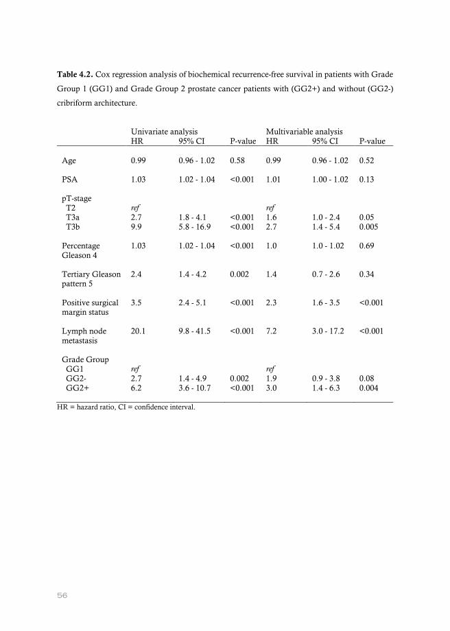
56
Table 4.2. Cox regression analysis of biochemical recurrence-free survival in patients with Grade
Group 1 (GG1) and Grade Group 2 prostate cancer patients with (GG2+) and without (GG2-)
cribriform architecture.
Univariate analysis Multivariable analysis HR 95% CI P-value HR 95% CI P-value Age
0.99
0.96 - 1.02
0.58
0.99
0.96 - 1.02
0.52
PSA
1.03 1.02 - 1.04 <0.001 1.01 1.00 - 1.02 0.13
pT-stage T2 T3a T3b
ref 2.7 9.9
1.8 - 4.1 5.8 - 16.9
<0.001 <0.001
ref 1.6 2.7
1.0 - 2.4 1.4 - 5.4
0.05 0.005
Percentage Gleason 4
1.03 1.02 - 1.04 <0.001 1.0 1.0 - 1.02 0.69
Tertiary Gleason pattern 5
2.4 1.4 - 4.2 0.002 1.4 0.7 - 2.6 0.34
Positive surgical margin status
3.5 2.4 - 5.1 <0.001 2.3 1.6 - 3.5 <0.001
Lymph node metastasis
20.1 9.8 - 41.5 <0.001 7.2 3.0 - 17.2 <0.001
Grade Group GG1 GG2- GG2+
ref 2.7 6.2
1.4 - 4.9 3.6 - 10.7
0.002 <0.001
ref 1.9 3.0
0.9 - 3.8 1.4 - 6.3
0.08 0.004
HR = hazard ratio, CI = confidence interval.
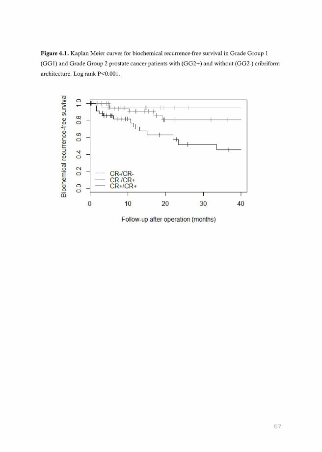
57
Figure 4.1. Kaplan Meier curves for biochemical recurrence-free survival in Grade Group 1
(GG1) and Grade Group 2 prostate cancer patients with (GG2+) and without (GG2-) cribriform
architecture. Log rank P<0.001.
58
Discussion
During the last decade, various studies demonstrated that GG2 prostate cancer patients with
invasive cribriform and/or intraductal carcinoma have worse disease outcome than those
without.13, 15, 24, 39, 51, 67 Although it is generally accepted that GG2 prostate cancer patients have
more aggressive disease than men with GG1, it is unclear whether this is still the case when those
with aggressive cribriform pathology are excluded. In this study, we found that 46% of men with
GG2 prostate cancer at radical prostatectomy had neither invasive cribriform nor intraductal
carcinoma. These men had significantly higher PSA levels, pT-stage, positive surgical margin rates
and shorter biochemical recurrence-free survival than men with GG1 disease. However, none of
the 399 men with GG1 or GG2- prostate cancer had metastasis at time of operation or during
follow-up, while metastases were identified in 10% of GG2+ patients. These findings indicate that
invasive cribriform and/or intraductal carcinoma might have most impact on metastatic disease
progression.
Although both invasive cribriform and intraductal carcinoma are pathological features
associated with tumour aggressiveness, it is yet unclear how to incorporate these parameters in
clinical risk stratification. For instance, Iczkowski et al. proposed to modify current Grade Groups
2 to 4 by reporting the presence of invasive cribriform and intraductal carcinoma denoted with a
“C”, which would increase the risk groups from 5 to 8.68 However, it is not yet evident whether
clinically relevant differences exist between each of these subgroups or if they partially overlap.
Previously, our group found that men with GG2- at biopsy had similar biochemical recurrence
rates and disease-specific survival to men with GG1.32, 39, 52 We currently demonstrate that GG2-
is associated with significantly worse clinicopathological characteristics and outcome than GG1
disease. These findings at radical prostatectomy specimens slightly differ from our previous study
on biopsy specimens52. This might be explained by biopsy sampling artefacts, as upgrading occurs
in up to 40% of biopsy GG1 and GG2 prostate cancer.61, 69. Furthermore, recent studies indicate
that biopsies have a moderate sensitivity of 43 to 47% for detecting cribriform architecture.59, 66
Despite the moderate concordance of growth patterns between biopsy and radical prostatectomy
evaluation, incorporation of cribriform architecture into the Grade Groups has better
discriminative value for disease-specific survival and metastasis-free survival.65 In biopsies, we
previously demonstrated that subtraction of one Grade Group, when cribriform architecture is not
present, is a simple and valuable modification of the current prostate cancer grading.65
Metastasis-free survival of GG2- and GG1 disease was similar in both biopsy and radical
prostatectomy studies.52 GG1 prostate cancer at radical prostatectomy is known to have very low
if any risk of metastasis and disease-specific death.45, 70-77 Since no metastases were identified in
59
pelvic lymph node dissection or during follow-up of GG2- men, this population seems to have a
low risk of metastatic progression as well. This indicates that invasive cribriform and intraductal
carcinoma in particular might have impact on the biological potential of developing metastatic
disease. In contrast, postoperative biochemical recurrence-free survival is also related to tumour
volume parameters and surgical technique, which do not necessarily reflect biological
derangement of the disease. Cribriform architecture has been associated with genomic instability
and has clonally been related to lymph node metastasis, which might provide a rationale for its
aggressive biological behaviour.18, 78-80
Forty-two out of 207 (20%) GG1 patients had a tertiary Gleason pattern 4. These patients
had worse clinicopathological features than men with pure GG1 disease and were more
comparable to GG2- patients. This finding is in line with others reporting on the clinical relevance
of tertiary patterns and underlines the importance of reporting them.81-90
The strong point of this study is the detailed histological evaluation of the radical
prostatectomy specimens, including recently identified clinically relevant pathologic parameters.
Limitations of this retrospective investigation are its relatively low number of patients and limited
follow-up time of median 59.6 months. Identification of small differences in metastasis-free
survival would need a large number of low-risk patients with long-term follow-up.
In conclusion, patients with GG2- prostate cancer at radical prostatectomy have more
advanced disease and shorter biochemical recurrence-free survival than men with GG1. However,
both groups have very low risk of developing metastatic disease.

60

61
62
Chapter V
Clinicopathological characteristics of glomeruloid architecture in prostate cancer.
Hollemans E, Verhoef EI, Bangma CH, Rietbergen J, Osanto S, Pelger RCM, van Wezel T, van der Poel H, Bekers E, Helleman J, Roobol MJ, van Leenders GJLH.
Mod Pathol. 2020 Feb; 33: 1618-1625.

63
Abstract
Glomeruloid architecture is the least common Gleason 4 growth pattern in prostate
adenocarcinoma. Its clinicopathological features and relation with cribriform architecture, which
has been recognized as an adverse feature, remains to be established. Our objective was to
investigate clinicopathological features of glomeruloid architecture in radical prostatectomies.
We reviewed 1064 radical prostatectomy specimens and recorded Grade Group, pT-
stage, margin status, Gleason pattern percentages and growth patterns. Simple and complex
glomerulations were distinguished by gland size and intraluminal cribriform protrusions. Clinical
endpoint was biochemical recurrence-free survival.
Glomerulations were identified in 365 (34%) specimens. In 472 Grade Group 2 patients,
210 (44%) had simple and 92 (19%) complex glomerulations. Complex glomerulations coincided
with cribriform architecture more often than simple glomerulations (67% versus 52%; P=0.01).
Men with simple glomerulations had significantly lower Prostate Specific Antigen (PSA) levels
(9.7 versus 12.1 ng/ml; P=0.03), percentage Gleason pattern 4 (19% versus 25%; P=0.001), extra-
prostatic extension (34% versus 50%; P=0.01) and positive surgical margins (25% versus 39%;
P=0.04) than those with cribriform architecture. Extra-prostatic extension (37%) and positive
surgical margins (30%) in men with complex glomerulations resembled those with simple
glomeruloid rather than those with cribriform architecture. In multivariate Cox regression analysis
adjusted for PSA, pT-stage, margin status and lymph node metastases, cribriform architecture had
independent predictive value for biochemical recurrence-free survival (Hazard Ratio (HR) 1.9;
95% Confidence Interval (CI) 1.2-2.9; P=0.004), while simple (HR 0.8; 95% CI 0.5-1.2; P=0.26)
and complex (HR 0.9; 95% CI 0.5-1.6; P=0.67) glomerulations did not.
Both simple and complex glomeruloid architecture are associated with better outcome
than cribriform architecture in Grade Group 2 prostate cancer patients. Therefore, glomeruloid
pattern and particularly complex glomerulations should not be classified as a cribriform growth
pattern variant in radical prostatectomy specimens.

64
Introduction
The Gleason score and Grade Group are the most important parameters for clinical outcome in
prostate cancer patients.8, 12 Gleason pattern 4 is a heterogeneous group of growth patterns
including poorly formed, fused, glomeruloid and cribriform structures. The clinical importance of
cribriform architecture in prostate cancer has been well-established in recent years, as it is
independently associated with disease progression and disease-specific death.13, 15, 16, 28, 34, 51, 91
Glomeruloid growth pattern consists of dilated malignant glands with intraluminal cribriform
protrusions, attached to one side of the gland wall, resembling a renal glomerulus.12 In 1998,
Pacelli et al. first described this growth pattern in relation to tumour grade and stage.92 Cribriform
and glomeruloid growth patterns are often observed together and some have hypothesized that
glomeruloid morphology might be a precursor of cribriform architecture.19, 93 However, more
recent studies indicate that glomeruloid pattern is associated with beneficial histopathological
features and longer biochemical recurrence-free survival among Gleason score 7 prostate cancer
patients.16, 20
Interobserver studies have shown that glomeruloid architecture is one of the most
reproducible growth patterns in prostate cancer grading.94, 95 While interobserver agreement is
excellent for small glomeruloid protrusions, no consensus exists on the classification of large
glomeruloid structures as either glomeruloid or cribriform growth pattern.95 Since some institutes
use cribriform architecture as threshold for active surveillance in Grade Group 2 prostate cancer,
distinction between glomeruloid and cribriform growth patterns might have major implications
for patient management. Therefore, the aim of this study was to investigate the clinicopathological
features and biochemical recurrence-free survival of prostate cancer patients with glomeruloid
growth pattern who had undergone radical prostatectomy.

65
Materials and methods
Patient selection
Patients who had undergone radical prostatectomy for prostate adenocarcinoma in three medical
centers in The Netherlands between 2000 and 2017 were included in this study. In total, 854
patients were operated at Erasmus MC, University Medical Center, Rotterdam. In addition,
patients from Leiden University Medical Center, Leiden (n=96), and the Netherlands Cancer
Institute, Amsterdam (n=137) were selected for high-grade morphology (Grade Group 3-5). We
excluded men who had undergone hormonal, radiation or viral therapy (n=23) prior to
operation.40 Radical prostatectomy specimens were fixed in neutral-buffered formalin, after which
they were sectioned transversely and completely embedded for diagnostic evaluation. All slides
were available for pathology review. This study was approved by the institutional Medical
Research Ethics Committee (MEC-2018-1614).
Pathologic evaluation
Radical prostatectomy specimens were reviewed by two investigators (EH, GvL) in common
sessions, blinded to clinical outcome. For each specimen the following features were recorded:
Gleason score and Grade Group according to the World Health Organization (WHO) and
International Society of Urological Pathology (ISUP) 2014 guidelines, pT-stage according to the
American Joint Committee on Cancer (AJCC) TNM 8th edition, surgical margin status, presence
of individual growth patterns and intraductal carcinoma, and percentage of Gleason pattern 4 and
5.12, 41 The following Gleason 4 growth patterns were recognized: poorly formed, fused,
glomeruloid and cribriform glands.12, 38 Furthermore, we distinguished two subgroups of
glomeruloid growth pattern based on the architecture of intraluminal protrusions (Figure 5.1).
Simple glomeruloid architecture was defined as malignant glands with small to medium-sized
solid intraluminal cell clusters with unilocular connection to the gland wall. Complex glomeruloid
growth pattern had medium to large-sized intraluminal cribriform protrusions with unilocular
connection to the gland wall; in some cases the gland wall connection was more extensive. The
distinction between glomeruloid and cribriform architecture was arbitrarily made by the extent of
gland wall connection, which occupied at least half of the inner glandular surface in cribriform
growth and less than half in the glomeruloid pattern. In addition, we distinguished small and large
cribriform growth patterns, the latter being defined as cribriform structures with a diameter more
than twice the size of adjacent benign glands. Invasive cribriform Gleason pattern 4 was
morphologically distinguished from intraductal carcinoma based on the following features:
invasive cribriform prostate cancer had an irregular outline, interconnecting fields beyond pre-

66
existent gland architecture or extension into extra-prostatic tissue. Intraductal carcinoma was
morphologically identified if cribriform structures were continuous with pre-existent glands or
contained corpora amylacea. If invasive cribriform carcinoma and intraductal carcinoma could
not be distinguished by morphological criteria alone, additional basal cell immunohistochemistry
was performed. Cribriform glands completely lacking basal cell staining were categorized as
invasive cribriform carcinoma. If basal cells were present sporadically, scattered or continuously,
the cribriform structures were classified as intraductal carcinoma. Gleason pattern 5 was
considered as a tertiary pattern if it occupied less than 5% of the total tumour area.12, 31, 38
Intraductal carcinoma and tertiary patterns were not incorporated in the Gleason score.
Clinical follow-up
Post-operative clinical follow-up consisted of six-monthly, and later annual monitoring of serum
Prostate Specific Antigen (PSA) levels. Biochemical recurrence was defined as PSA levels ≥ 0.2
ng/ml measured at two consecutive points in time, at least three months apart with undetectable
PSA levels after operation, or as PSA increase of > 2.0 ng/ml when serum PSA had not declined
to zero after the operation. Post-operative lymph node and distant metastases were confirmed by
biopsy or multidisciplinary consensus.
Statistical analysis
Continuous variables with normal distribution were analysed using the independent sample
Student’s t-test. Pearson’s chi squared (χ2) test was used for categorical parameters. Missing PSA
values (n=27) were imputed using the median PSA value. Biochemical recurrence-free survival
and metastasis-free survival were analysed using Cox proportional hazards regression and
visualized by Kaplan-Meier curves. Statistics were performed using SPSS version 24 (IBM,
Chicago, IL, USA). Results were considered significant when the two-sided P-value was <0.05.

67
Figure 5.1. Gleason grade 4 cribriform and glomeruloid growth patterns. A. Simple glomeruloid architecture
with intraluminal cell clusters, 10x. B. Complex glomeruloid architecture with large cribriform proliferations
protruding into the lumen, 5x. C. Simple glomeruloid architecture with vacuolated cytoplasm, therefore not
classified as complex glomeruloid architecture, 20x. D. Complex glomeruloid architecture (asterisk) based on
a gland wall connection that occupies less than half of the inner gland surface, and simple glomeruloid
architecture (arrows) with a small intraluminal protrusion, 15x. E. Small invasive cribriform carcinoma, 10x.
F. Large invasive cribriform carcinoma showing expansive confluent fields, 5x.
68
Results
General patients characteristics
The cohort consisted of 1064 men with a median age of 65 years (interquartile range (IQR) 60-68)
and median serum PSA level of 8.3 ng/ml (IQR 6.0-13.2). Median follow-up was 61 months (IQR
20-104). The cohort included 207 (20%) men with Grade Group 1, 472 (44%) with Grade Group
2, 126 (12%) with Grade Group 3, 140 (13%) with Grade Group 4 and 119 (11%) with Grade
Group 5 prostate cancer. Pathological stage was distributed as follows: 582 (55%) pT2, 334 (31%)
pT3a, 145 (14%) pT3b and 3 (0.3%) pT4 tumours. Surgical margins were positive in 389 (37%)
patients. Pelvic lymph node dissection was performed in 664 (62%) patients, of whom 64 (10%)
had lymph node metastases.
Gleason 4 growth patterns
Poorly formed and fused glands were observed in 691 (65%) and 613 (58%) men, respectively.
Invasive cribriform pattern was present in 519 (49%) men, 189 (18%) of whom had large expansive
growth. Intraductal carcinoma was identified in 314 (30%) specimens. In total, 569 (54%) men
had invasive cribriform and/or intraductal carcinoma, 190 (33%) of whom had large invasive
cribriform growth. Glomeruloid growth was the least frequent Gleason 4 pattern, being present in
365 (34%) men. It was the single Gleason 4 pattern in only 10 (1%) men. Simple glomeruloid
glands were present in 352 (33%) men and complex glomeruloid glands in 154 (15%) men. Among
patients with glomeruloid architecture, 211 (58%) had simple glomerulations only, 13 (4%) had
complex glomerulations only, and 141 (38%) had both simple and complex glomeruloid glands.
Simple and complex glomeruloid patterns had concomitant invasive and/or intraductal cribriform
carcinoma in 128/211 (61%) and 121/154 (79%) cases (P<0.001), respectively. Complex
glomeruloid growth did not coincide more often with large compared to small cribriform growth
(P=0.26).
Simple glomerulations were present in 212 (45%) Grade Group 2, 56 (44%) Grade Group
3, 47 (34%) Grade Group 4 and 28 (24%) Grade Group 5 tumours. In 9 (4%) men with Grade
Group 1 tumours, simple glomerulations were present as tertiary Gleason pattern 4. Complex
glomerulations were observed in 92 (19%) men with Grade Group 2, 34 (27%) with Grade Group
3, 15 (11%) with Grade Group 4 and 13 (11%) with Grade Group 5 tumours. In none of the cases
with Grade Group 1, complex glomerulations were present as tertiary pattern. Simple and
complex glomerulations were observed significantly more often in Grade Group 2 and 3 patients
compared to Grade Group 4 and 5 patients (P=0.05).

69
Table 5.1. Clinicopathological characteristics of Grade Group 2 prostate cancer patients
categorized for cribriform and/or glomeruloid architecture: men with neither glomeruloid nor
cribriform architecture (group A), men with simple glomeruloid pattern without complex and/or
cribriform architecture (group B), men with complex glomeruloid pattern without invasive and/or
intraductal cribriform carcinoma (group C), and men with invasive and/or intraductal cribriform
carcinoma regardless of presence of glomerulations (group D).
Group A n=130
Group B n=60
Group C n=30
Group D n=252
Age PSA
63.3 (64.1; 59.1-68.3) 9.1 (7.6; 5.0-11.1)
63.4 (64.6; 59.0-69.1) 9.7 (8.2; 6.2-11.0)
63.4 (63.6; 60.1-67.2) 13.1 (9.3; 6.5-13.7)
64.2 (64.7; 60.3-67.9) 12.1 (8.3; 6.3-13.5)
pT-stage T2 T3a T3b
83 (64%) 43 (33%) 4 (3%)
40 (66%) 19 (32%) 1 (2%)
19 (64%) 10 (33%) 1 (3%)
126 (50%) 97 (38%) 29 (12%)
Gleason pattern 4 (%) Cribriform Fused Ill-defined Glomeruloid simple complex
18 (15; 10-26) 0 81 (62%) 103 (79%) 0 0 0
19 (15; 10-30) 0 42 (70%) 44 (73%) 60 (100%) 60 (100%) 0
20 (20; 10-30) 0 22 (73%) 19 (63%) 30 (100%) 28 (93%) 30 (100%)
25 (25; 20-34) 223 (89%) 180 (71%) 198 (79%) 126 (50%) 124 (49%) 62 (25%)
Intraductal carcinoma
0 0 0 113 (45%)
Tertiary Gleason 5
13 (10%) 6 (10%) 3 (10%) 35 (14%)
PSM
35 (27%) 15 (25%) 9 (30%) 97 (39%)
PLND Lymph node metastasis
64 (49%) 0
22 (37%) 0
16 (53%) 0
160 (64%) 13 (5%)
Biochemical recurrence
22 (17%) 8 (13%) 4 (13%) 73 (29%)
Metastasis
0 0 0 17 (7%)
Disease-specific death
0 0 0 3 (1%)
*Values denote either mean (median; IQR) or n (%). PSM = positive surgical margin.
70
Glomeruloid architecture in Grade Group 2 prostate cancer
Since clinical impact of glomeruloid growth pattern classification is most relevant for Grade Group
2 prostate cancer patients, we performed further analyses in this subpopulation of 472 men. Of
these, 216 (46%) had glomerulations: 212 (45%) had simple and 92 (20%) complex glomeruloid
structures. Simple glomerulations only were present in 124 (57%) men, complex glomerulations
only in 4 (2%) men, and both patterns occurred concurrently in 88 (41%) cases. Invasive and/or
intraductal cribriform carcinoma was present in 252 (53%) men with Grade Group 2 prostate
cancer, 34 (13%) of whom had large invasive cribriform carcinoma. Glomeruloid architecture was
associated with presence of invasive and/or intraductal cribriform carcinoma, as 126/216 (58%)
men with glomerulations had coexistent cribriform architecture compared to 126/256 (49%) men
without glomerulations (P=0.05). Complex glomeruloid structures (62/92, 67%) were more
frequently concomitant with invasive and/or intraductal cribriform carcinoma than simple
glomerulations (64/124, 52%, P=0.01).
Further analyses on glomeruloid and cribriform growth patterns were performed in four subgroups
(Table 5.1), to investigate the relation between glomeruloid and cribriform architecture. These
subgroups consisted of men with neither glomeruloid nor cribriform architecture, thus with poorly
formed and fused glands only (n=130, group A), men with simple glomeruloid pattern without
complex and/or cribriform architecture (n=60, group B), men with complex glomeruloid pattern
without invasive and/or intraductal cribriform carcinoma (n=30, group C), and men with invasive
and/or intraductal cribriform carcinoma regardless of presence of glomerulations (n=252, group
D). Patients with invasive cribriform and/or intraductal carcinoma (group D) had significantly
higher percentage Gleason pattern 4 (25% versus 18%; P<0.001), pT-stage (50% versus 36% pT3;
P=0.003) and positive surgical margin rates (39% versus 27%; P=0.02) than those with poorly
formed and fused glands only (group A). Men with simple glomerulations (group B) had similar
PSA levels (9.7 versus 9.1 ng/ml; P=0.6), percentage Gleason pattern 4 (19% versus 18%; P=0.6),
pT-stage (34% versus 36% pT3; P=0.8), and positive surgical margins (25% versus 27%; P=0.7) to
men with poorly formed and fused glands only (group A). Compared to men with invasive
cribriform and/or intraductal carcinoma (group D), men with simple glomerulations (group B)
had significantly lower PSA levels (9.7 versus 12.1 ng/ml; P=0.03), percentage Gleason pattern 4
(19% versus 25%; P=0.001), pT-stage (34% versus 50% pT3; P=0.01), and positive surgical margins
(25% versus 39%; P=0.04). Although men with complex glomeruloid glands only (group C) had
higher PSA levels (13.1 versus 9.7 ng/ml; P=0.05) than men with simple glomeruloid growth
pattern (group B), percentage Gleason pattern 4 (20% versus 19%; P=0.6), presence of tertiary
Gleason pattern 5 (both 10%; P=1.0), pT-stage (37% versus 34% pT3; P=0.8) and surgical margin
status (30% versus 25%; P=0.6) were similar. Patients with complex glomeruloid glands (group C)

71
had similar PSA levels (13.1 versus 12.1 ng/ml; P=0.7) to those with invasive cribriform and/or
intraductal carcinoma (group D), but its percentage Gleason pattern 4 was significantly lower (20%
versus 25%; P=0.04). Although the complex glomeruloid sample size (n=30) was too low for
reliable statistical analysis, percentage of Gleason pattern 4, presence of tertiary pattern 5, pT-stage
and positive surgical margin status resembled simple glomerulations (group B) rather than
cribriform architecture (group D). Among patients with invasive and/or intraductal cribriform
carcinoma (group D), no significant differences were found in PSA, pT-stage, percentage Gleason
pattern 4, presence of tertiary Gleason pattern 5 and surgical margin status between those with
and without glomeruloid architecture (data not shown).
Clinical outcome of Grade Group 2 patients
Median follow-up of Grade Group 2 patients was 57 months (IQR 14-99). Biochemical recurrence
occurred in 22/130 (17%) men with poorly formed and fused glands only (group A), in 8/60 (13%)
men with simple glomerulations (group B), in 4/30 (13%) men with complex glomerulations
(group C) and in 73/252 (29%) men with cribriform architecture (group D). Survival curves are
shown in Figure 5.2. No statistically significant difference in biochemical recurrence-free survival
was found between men with poorly formed and fused glands only (group A), simple glomeruloid
glands (group B) or complex glomeruloid glands (group C; log rank P=0.38). Patients with
invasive and/or intraductal cribriform architecture (group D) had significantly shorter biochemical
recurrence-free survival (log rank P<0.001) than patients with simple (group B, P=0.006) and
complex glomeruloid glands (group C, P=0.05).
Cox regression analysis showed that intraductal carcinoma (Hazard ratio (HR) 2.7, 95%
CI 1.8-4.0, P<0.001), small invasive cribriform carcinoma (HR 1.9, 95% CI 1.3-3.0, P=0.002) and
large invasive cribriform carcinoma (HR 6.3, 95% CI 3.6-11.1, P<0.001) were associated with
shorter biochemical recurrence-free survival in univariate analysis (Table 5.2). Adjusted for PSA
level, pT-stage, surgical margin and pelvic lymph node metastases, large invasive cribriform
carcinoma remained an independent predictor for biochemical recurrence (HR 3.8, 95% CI 2.1-
6.8, P<0.001) in multivariable analysis. Simple (HR 0.8; 95% CI 0.6-1.4; P=0.64) and complex
(HR 0.6; 95% CI 0.4-1.2; P=0.19) glomeruloid growth patterns were not associated with
biochemical recurrence-free survival in univariate or multivariable analysis. Metastases (n=17)
and disease-specific death (n=3) only occurred in patients with presence of invasive cribriform
and/or intraductal carcinoma, and not in other subgroups.

72
Figure 5.2. Kaplan-Meier curves of biochemical recurrence-free survival in Grade Group 2
patients with cribriform and/or glomeruloid architecture, stratified for subgroups A to D.
Table 5.2. Cox regression analysis for biochemical recurrence-free survival in Grade Group 2
prostate cancer patients.
Univariate Multivariable
HR 95% CI P-value HR 95% CI P-value
PSA 1.03 1.01-1.04 <0.001 1.01 1.00-1.02 0.06
pT-stage
T2 ref ref
T3a 1.8 1.2-2.7 0.008 1.5 1.0-2.3 0.07
T3b 4.9 2.8-8.5 <0.001 1.9 1.0-3.7 0.07
Positive surgical margins 2.5 1.7-3.7 <0.001 2.4 1.6-3.7 <0.001
Pelvic lymph node metastases 15.2 7.6-30.3 <0.001 7.3 3.2-16.8 <0.001
Intraductal carcinoma 2.7 1.8-4.0 <0.001 1.4 0.9-2.3 0.15
Invasive cribriform carcinoma
Small cribriform 1.9 1.3-3.0 0.002 1.2 0.7-1.8 0.48
Large cribriform 6.3 3.6-11.1 <0.001 3.8 2.1-6.8 <0.001
Glomeruloid growth pattern
Simple 0.8 0.5-1.2 0.20 0.8 0.6-1.4 0.64
Complex 1.0 0.6-1.6 0.86 0.6 0.4-1.2 0.19 *HR = hazard ratio, CI = confidence interval.

73
Discussion
In this study we investigated the clinicopathological features of glomeruloid Gleason pattern 4
architecture. Overall glomeruloid architecture was present in 34% of radical prostatectomy
specimens. In Grade Group 2 patients, simple glomeruloid was seen in 45% and complex
glomeruloid growth in 20% of men. Men with simple glomeruloid glands only had similar
clinicopathological characteristics to those with poorly formed and fused glands. Although
patients with complex glomeruloid glands had higher PSA levels than men with poorly formed,
fused or simple glomeruloid glands, no significant difference was observed for pT-stage,
percentage Gleason pattern 4, or biochemical recurrence-free survival. Biochemical recurrence
and metastasis occurred significantly more often in patients with invasive cribriform and/or
intraductal carcinoma. Despite their morphological resemblance, men with complex
glomerulations had better outcome than those with cribriform architecture. Therefore, complex
glomeruloid pattern should not be classified as a variant of the more aggressive cribriform growth
pattern in radical prostatectomy specimens.
Pathological grading of the glomeruloid growth pattern has been uncertain for a long
time. At the 2005 ISUP conference, no consensus was reached on grading glomeruloid pattern
due to lack of scientific evidence for its prognostic value.11 In 2009, Lotan and Epstein studied 45
prostate cancer biopsies with glomeruloid features and found they were surrounded by Gleason
pattern 4 structures in 80% and Gleason pattern 5 in 4% of cases.19 The authors noted that half of
the glomeruloid structures were accompanied by cribriform pattern, which is in concordance with
our findings. For this reason it was unanimously consented at the 2014 ISUP conference that
glomeruloid glands should be assigned Gleason pattern 4, regardless of morphology.12
Since the increased awareness of the dismal outcome of cribriform Gleason pattern 4,
glomeruloid growth pattern has also been included in clinicopathological studies. Among 350
radical prostatectomies with Gleason score 7 prostate cancer, Choy et al. found that patients with
glomeruloid pattern had improved five-year biochemical recurrence-free survival in multivariable
analysis, but had shorter survival than those with Gleason score 6.20 Glomeruloid growth pattern
was associated with improved, although not statistically significant, metastasis-free survival of
Gleason score 7 men in our previous radical prostatectomy study.16 However, both studies did not
focus on glomeruloid architecture specifically and were performed on a Gleason score 7 cohort.
The study of Kweldam et al. had a case-control study design and used metastasis-free survival as
endpoint. We investigated glomeruloid architecture in a larger cohort of Grade Group 1 to 5
prostate cancer and distinguished simple and complex glomerulations, which was not done in
previous manuscripts. In the current study, clinical outcome of patients with glomeruloid

74
architecture was better than of those with cribriform architecture, and did not differ from those
with poorly formed and fused glands only. Statistical analyses were hampered by the fact that
glomeruloid architecture is rarely observed as single Gleason pattern 4, mostly coexisting with
other growth patterns.
Glomeruloid architecture is not a homogeneous Gleason pattern 4 subgroup. As
previously reported by Lotan and Epstein, most glomerulations consisted of relatively small
dilated glands, but some contain larger glomeruloid protrusions.19 Furthermore, a subset of
glomerulations has fibrovascular cores. This is in line with three-dimensional renderings of
glomeruloid structures which revealed an interconnecting network of tubules resembling Gleason
pattern 3 glands with nodular epithelial proliferations near tubular branching points, or markedly
curved tubules with small fibrovascular cores.96 While glomeruloid architecture is the most
reproducible Gleason 4 growth pattern, interobserver variability exists for classifying glomeruloid
structures with larger intraluminal cribriform protrusions.95 In this case, classification as either
glomeruloid or cribriform architecture is uncertain, because the extent of cribriform intraluminal
gland wall attachment has not been defined. Among Grade Group 2 prostate cancer patients, two-
thirds of specimens with complex glomeruloid pattern had concomitant cribriform pattern which
might suggest that both patterns are related. Complex cribriform pattern for instance could be a
tangential sectioning of cribriform architecture expanding into malignant tubules or might
represent a precursor lesion. While designation of these structures as either cribriform or
glomeruloid does not have clinical relevance if they coexist with cribriform architecture, their
distinction might be important in diagnostic biopsies without cribriform structures. Active
surveillance is mostly offered to men with biopsy Grade Group 1 disease, but some surveillance
protocols are also including men with Grade Group 2 prostate cancer without cribriform
architecture and/or low Gleason pattern 4 percentages.45, 61-64, 97 Therefore, classifying complex
glomeruloid structures might directly affect clinical decision-making. Our current findings in
radical prostatectomy specimens indicate that men with complex glomeruloid structures without
concomitant cribriform growth have better outcome than those with cribriform architecture.
Therefore, at this moment insufficient evidence exists for classifying complex glomeruloid pattern
as cribriform Gleason pattern 4.
The strong point of this study was the detailed histological review of a large cohort of
radical prostatectomy specimens. In this study the distinction between glomeruloid and cribriform
architecture was arbitrarily made by the extent of gland wall connection, which occupied at least
half of the inner glandular surface in cribriform growth and less in the glomeruloid pattern. No
standard definition of complex glomeruloid pattern has been formulated yet. Therefore, it is
important that future studies define the morphological criteria for distinguishing complex
75
glomeruloid and cribriform growth patterns. Some pathologists might have interpreted the
complex glomeruloid cases as cribriform, however our current results do not provide evidence for
this classification. Finally, this study was limited by its retrospective design and relatively short
follow-up of 57 months.
In conclusion, glomeruloid architecture is observed in 34% of radical prostatectomy
specimens. Patients with simple glomeruloid glands have similar clinicopathological
characteristics and biochemical recurrence-free survival as poorly formed and fused glands in
Grade Group 2 prostate cancer. Although complex glomeruloid glands are more often coexistent
with cribriform growth pattern, biochemical recurrence and metastasis occurred significantly less
frequently in this subgroup. Therefore, complex glomeruloid architecture does not classify as a
cribriform Gleason 4 growth pattern in radical prostatectomy specimens. Further studies are
needed to address the significance of glomeruloid architecture in prostate biopsies.

76

77

78
Chapter VI
Cribriform architecture in radical prostatectomies predicts oncological outcome in Grade Group 4 prostate cancer patients.
Hollemans E, Verhoef EI, Bangma CH, Rietbergen J, Osanto S, Pelger RCM, van Wezel T, van der Poel H, Bekers E, Helleman J, Roobol MJ, van Leenders GJLH.
Mod Pathol. 2021 Jan; 34: 184-193.

79
Abstract
The Gleason score is an important parameter for clinical outcome in prostate cancer patients.
Gleason score 8 is a heterogeneous disease including Gleason score 3+5, 4+4, and 5+3 tumours,
and encompasses a broad range of tumour growth patterns. Our objective was to characterize
individual growth patterns and identify prognostic parameters in Gleason score 8 prostate cancer
patients.
We reviewed 1,064 radical prostatectomy specimens, recorded individual Gleason 4 and 5 growth
patterns as well as presence of intraductal carcinoma, and evaluated biochemical recurrence- and
metastasis-free survival.
Gleason score 8 disease was identified in 140 (13%) patients, of whom 76 (54%) had Gleason score
3+5, 46 (33%) 4+4, and 18 (13%) 5+3 disease. Invasive cribriform and/or intraductal carcinoma
(n=87, 62%) was observed more frequently in Gleason score 4+4 (93%) than 3+5 (47%; P<0.001)
and 5+3 (44%; P<0.001) patients. Gleason pattern 5 was present in 110 (79%) men: as single cells
and/or cords in 99 (90%) and solid fields in 32 (29%) cases. Solid field pattern 5 coexisted with
cribriform architecture (23/32, 72%) more frequently than non-solid pattern 5 cases (36/78, 46%,
P=0.02). In multivariable analysis including age, Prostate-Specific Antigen, pT-stage, surgical
margin status and lymph node metastases, presence of cribriform architecture was an independent
parameter for biochemical recurrence-free (Hazard Ratio (HR) 2.0, 95% Confidence Interval (CI)
1.0-3.7; P=0.04) and metastasis-free (HR 3.5, 95% CI 1.0-12.3; P=0.05) survival.
In conclusion, invasive cribriform and/or intraductal carcinoma occurs more frequently in
Gleason score 4+4 prostate cancer patients than in Gleason score 3+5 and 5+3, and is an
independent parameter for biochemical recurrence and metastasis. Therefore, cribriform
architecture has added value in risk stratification of Gleason score 8 prostate cancer patients.

80
Introduction
The Gleason grading system for prostate cancer is based on classification of histomorphological
growth patterns.8 At the 2014 meeting of the International Society of Urological Pathology (ISUP),
consent was reached that a Grade Group should be reported in conjunction with the Gleason
score, based on the initial work of Pierorazio et al., which was endorsed by the World Health
Organization (WHO) in 2016.12, 38, 98. The Grade Group system is comprehensive and facilitates
patient communication as it labels Gleason score 2-6 as Grade Group 1 and emphasizes the
important distinction between Gleason score 3+4=7 (Grade Group 2) and 4+3=7 (Grade Group
3) prostate cancer. Grade Group 4 prostate cancer encompasses Gleason score 8 tumours,
including Gleason score 3+5, 5+3, and 4+4.58, 99 However, it is not yet clear whether these three
Gleason score 8 subgroups have similar clinical outcome.
The importance of distinguishing individual prostate cancer growth patterns is
increasingly being acknowledged. Gleason pattern 4 encompasses four major growth patterns,
including poorly formed, fused, glomeruloid and cribriform glands.12 The clinical relevance of
cribriform architecture in prostate cancer has been well established in recent years, as it is
associated with biochemical recurrence, metastasis and disease-specific death.13, 15, 16, 28, 34, 51, 91
Intraductal carcinoma is characterized by a proliferation of malignant epithelial cells with
cribriform or solid architecture distending pre-existent acini and prostatic ducts with preservation
of basal cells.12 Although not incorporated in the Gleason score or Grade Group, intraductal
carcinoma is independently associated with adverse oncological outcome.28, 35, 39 The adverse
impact of invasive cribriform and intraductal carcinoma has mainly been studied in Gleason score
3+4 prostate cancer, as it might affect clinical decision-making in this patient population in
particular. Some studies indicate that presence of invasive cribriform and intraductal carcinoma
also has independent predictive value in Gleason score 8 prostate cancer patients.39, 100
While the impact of cribriform architecture is well recognized, little is known about the
clinical relevance of individual Gleason 5 growth patterns.13 Gleason pattern 5 encompasses
tumour growth in single cells, cords and solid fields.12 Furthermore, presence of comedonecrosis
is considered Gleason pattern 5, whether it is present within papillary, cribriform or solid fields.
Of notice, recent studies have shown that comedonecrosis more commonly occurs in intraductal
carcinoma than in invasive carcinoma, requiring basal cell immunohistochemistry for their
distinction.101-103 While Gleason score 8 prostate cancer is generally considered a high-risk disease
requiring immediate therapeutic intervention, analysis of individual Gleason 4 and 5 growth
patterns might attribute to risk stratification and optimize personalized treatment decisions. The
objective of this study is to compare the clinical characteristics and outcome of Gleason score 3+5,

81
5+3, and 4+4 subgroups and to investigate the impact of invasive cribriform and/or intraductal
carcinoma in Gleason score 8 radical prostatectomy specimens.
Figure 6.1. Gleason pattern 4 and pattern 5 tumor morphology. A. Gleason pattern 4, small invasive
cribriform structures, 15x. B. Gleason pattern 4, large invasive cribriform structures, 10x. C. Gleason pattern
5, cords, 20x. D. Gleason pattern 5, small solid nests with subtle intervening stroma, 20x. E. Gleason pattern
5, medium to large sized solid fields, 15x. F. Gleason pattern 5, comedonecrosis in a solid field, 15x.

82
Materials and methods
Patient selection
Patients who had undergone radical prostatectomy for prostatic adenocarcinoma from three
university medical centers in The Netherlands between 2000 and 2017 were included in this study;
854 patients were operated at Erasmus MC, University Medical Center, Rotterdam; 96 at Leiden
University Medical Center (LUMC), Leiden; and 137 at Antoni van Leeuwenhoek Hospital, the
Netherlands Cancer Institute (NKI), Amsterdam. Whereas the radical prostatectomies from
Erasmus MC were consecutive, those from LUMC and NKI were selected for presence of Gleason
score 4+3 to 10 in the original pathology report. We excluded men who had undergone hormonal,
radiation and/or viral therapy (n=23) prior to operation.40 Radical prostatectomy specimens were
fixed in neutral-buffered formalin, after which they were sectioned transversely and embedded
entirely for diagnostic purposes. All slides were available for pathology review. This study was
approved by the institutional Medical Research Ethics Committee (MEC-2018-1614).
Pathologic evaluation
All 1,064 radical prostatectomy specimens were reviewed in common sessions by two investigators
(EH, GvL), blinded to clinical outcome. For each specimen the following features were recorded:
Gleason score and Grade Group according to the 2014 ISUP/2016 WHO guidelines, pT-stage
according to the American Joint Committee on Cancer (AJCC) TNM 8th edition, surgical margin
status, presence of intraductal carcinoma, and percent Gleason 4 and 5 growth patterns.12, 41 In
case of multifocality we only monitored the characteristics of the index tumour defined as the
tumour with the highest grade, stage or volume.
The following Gleason 4 growth patterns were recognized: poorly formed, fused,
glomeruloid and cribriform glands.12, 38 Furthermore, we distinguished small and large cribriform
gland architecture (Figure 6.1, A-B), since the latter is associated with more aggressive behaviour.51
Large cribriform structures were defined as having a diameter more than twice the size of adjacent
benign glands. We examined the following Gleason 5 growth patterns: single cells, cords, and
solid fields (Figure 6.1, C-F). Single cells and cords were grouped for analysis. Solid fields were
divided into those with small solid nests containing 10 to 30 cells, and those consisting of medium
to large solid fields with more than 30 cells. In case comedonecrosis was present in invasive
cribriform or solid fields, this was considered Gleason pattern 5. Invasive cribriform Gleason
pattern 4 and solid pattern 5 either with or without comedonecrosis were morphologically
distinguished from intraductal carcinoma based on the following features: invasive cribriform and
solid prostate cancer had irregular borders or formed interconnecting fields, well exceeding the

83
outline of distended pre-existent glands, or extended into periprostatic adipose tissue, ejaculatory
ducts or seminal vesicles. Intraductal carcinoma was continuous with pre-existent glands lined by
basal cells, or contained corpora amylacea. In case invasive cribriform or solid carcinoma and
intraductal carcinoma could not be differentiated by morphological criteria alone, additional basal
cell immunohistochemistry was performed. Basal cell immunohistochemistry (34BE12) was
performed in 189/854 (22%) radical prostatectomy specimens from Erasmus MC, including 14/31
(45%) Gleason score 8 tumours with cribriform or solid architecture; no paraffin blocks were
available from the other hospitals. If basal cells were completely absent, the lesion was classified
as either invasive cribriform Gleason pattern 4 or solid pattern 5 carcinoma. When sporadic,
scattered or continuous basal cells were identified, the lesion was considered intraductal
carcinoma. Intraductal carcinoma and tertiary patterns were not incorporated in the Gleason
score.12, 31, 38 Minor high-grade components occupying <5% of the tumour volume were considered
as tertiary pattern. The Grade Group concordance rate at revision was 88/135 (65%) for radical
prostatectomies from NKI and 39/94 (41%) for specimens from LUMC.
Clinical follow-up
Clinical follow-up after radical prostatectomy consisted of six-monthly, and later annual
monitoring of serum Prostate-Specific Antigen (PSA) levels. Biochemical recurrence was defined
as PSA levels ≥ 0.2 ng/ml measured at two consecutive points in time, at least three months apart
with undetectable PSA levels after operation, or as PSA increase of > 2.0 ng/ml when serum PSA
had not declined to zero after operation. Post-operative lymph node and distant metastases were
confirmed by biopsy or multidisciplinary consensus.
Statistical analysis
Continuous variables with normal distribution were analysed using the independent sample
Student’s t-test for two groups, or One-way ANOVA for ≥3 groups. Variables without normal
distribution were analysed using the Mann-Whitney U test for two groups, or Kruskal-Wallis test
for ≥3 groups. For comparison of categorical parameters Pearson’s chi squared (χ2) test was used,
and Fisher’s exact test in case of small numbers (n≤20). Missing PSA values (n=27) were imputed
using the median PSA value. Biochemical recurrence-free survival and metastasis-free survival
were analysed using Cox proportional hazards model and visualized by Kaplan-Meier curves.
Statistics were performed using SPSS version 24 (IBM, Chicago, IL, USA). Results were
considered significant when the two-sided P-value was <0.05.

84
Results
Characteristics of Gleason score 8 prostate cancer patients
Out of 1,064 radical prostatectomy specimens, 140 (13%) had Gleason score 8 prostate cancer.
The median age of Gleason score 8 patients was 65.3 years (interquartile range (IQR) 61.4-68.5
years) and median serum PSA level was 10.0 ng/ml (IQR 7.2-16.0 ng/ml). Gleason scores were
distributed as follows: 76 (54%) men had Gleason score 3+5, 46 (33%) Gleason score 4+4, and 18
(13%) Gleason score 5+3. Pathologic tumour stage was T2 in 67 (48%) men, T3a in 44 (31%) and
T3b in 28 (20%). One (1%) patient had a T4 tumour and was grouped with T3b tumours for further
analysis. Positive surgical margins were present in 68 (49%) cases. Pelvic lymph node dissection
was performed in 91 (65%) men, 12 (9%) of whom had lymph node metastasis. Median follow-up
time was 68.7 months (IQR 36.7-102.8).
Clinicopathological features and outcome of Gleason score 3+5, 4+4 and 5+3
The clinicopathological features of Gleason score 8 patients stratified for Gleason score are shown
in Table 6.1. The median PSA level of patients with Gleason score 5+3 prostate cancer was 13.4
ng/ml (IQR 8.8-26.8 ng/ml), significantly higher than for men with Gleason score 3+5 (10.0
ng/ml; IQR 7.4-15.0 ng/ml; P=0.05) and Gleason score 4+4 (8.9 ng/ml; IQR 6.9-16.0 ng/ml;
P=0.03). PSA levels of Gleason score 3+5 and 4+4 were comparable (P=0.45). Age, pT-stage,
surgical margin status and lymph node metastases were not significantly different between groups.
While Gleason pattern 4 constituted ≥95% of the tumour volume in Gleason score 4+4 by
definition, it was present in 73/76 (96%) Gleason score 3+5 and 12/18 (67%) 5+3 tumours. The
median percentage of Gleason pattern 4 was 30% (IQR 20%-35%) in 3+5 tumours and 18% (IQR
0%-21%) in 5+3 tumours (P<0.001). Tertiary (<5%) Gleason pattern 5 was observed in 16/46
(35%) Gleason score 4+4 tumours.
Biochemical recurrence and post-operative distant metastasis were observed in 68 (49%)
and 36 (26%) patients, respectively. Twenty-nine (63%) men with Gleason score 4+4 tumours
experienced biochemical recurrence compared to 31 (41%, P=0.02) with Gleason score 3+5 and
8 (44%, P=0.78) with 5+3. Biochemical recurrence-free survival was significantly shorter for
patients with Gleason score 4+4 than Gleason score 3+5 (log rank P=0.02) prostate cancer.
Gleason score 5+3 had the lowest absolute number of events and did not significantly differ from
Gleason score 3+5 (log rank P=0.82) and Gleason score 4+4 (log rank P=0.26, Figure 6.2). A
similar trend was found for post-operative metastasis. Metastases occurred in 18 (39%) men with
Gleason score 4+4 compared to 14 (18%, P=0.01) with Gleason score 3+5 and 4 (22%, P=0.20)
men with Gleason score 5+3. Metastasis-free survival was significantly shorter

85
T
able
6.1
. G
leas
on s
core
8 p
atie
nts
stra
tifi
ed fo
r in
divi
dual
Gle
ason
sco
re (
GS)
.
P-va
lue
0.46
0.07
0.35
<0.
001
<0.
001
<0.
001
0.02
<0.
001
0.00
3 0.
006
0.02
0.58
0.22
0.
54
0.05
0.04
0.17
Val
ues
deno
te e
ithe
r m
ean
(med
ian;
IQ
R)
or n
(%
). P
SA =
Pro
stat
e Sp
ecif
ic A
ntig
en.
GS
5+3
(n=
18)
64.5
(65
.2; 6
0.4-
67.2
)
18.5
(13
.4; 8
.8-2
6.8)
7 (3
9%)
6 (3
3%)
5 (2
8%)
8 (4
4%)
8 (4
4%)
4 (2
2%)
4 (2
2%)
17 (
94%
) 6
(33%
) 6
(33%
) 0
8 (4
4%)
15 (
83%
) 3
(20%
)
8 (4
4%)
4 (2
2%)
1 (6
%)
GS
4+4
(n=
46)
65.6
(66
.2; 6
2.4-
69.2
)
11.5
(8.
9; 6
.9-1
6.0)
19 (
41%
) 19
(41
%)
8 (1
8%)
43 (
93%
)
42 (9
1%)
28 (6
1%)
23 (
50%
)
10 (
22%
) 1
(2%
) 5
(11%
) 7
(15%
)
20 (
44%
)
29 (
63%
) 4
(14%
)
29 (
63%
)
18 (
39%
)
7 (1
5%)
GS
3+5
(n=
76)
64.3
(64
.5; 6
0.7-
68.2
)
12.5
(10
.0; 7
.4-1
5.0)
41 (
54%
) 19
(25
%)
16 (
21%
)
36 (
47%
)
33 (4
3%)
6 (8
%)
21 (
28%
)
72 (
95%
) 16
(21
%)
4 (5
%)
2 (3
%)
40 (
53%
)
47 (
62%
) 5
(11%
)
31 (
41%
)
14 (
18%
)
4 (5
%)
All
n=14
0
64.7
(65
.3; 6
1.4-
68.5
)
12.9
(10
.0; 7
.2-1
6.0)
67 (
48%
) 44
(31
%)
29 (
21%
)
87 (
62%
)
83 (5
9%)
38 (2
7%)
48 (
34%
)
99 (
71%
) 23
(16
%)
15 (
11%
) 9
(6%
)
68 (
49%
)
91 (
65%
) 12
(13
%)
68 (
49%
)
36 (
26%
)
12 (
9%)
Age
(yea
rs)
PSA
(ng
/ml)
pT-s
tage
T
2 T
3a
T3b
/T4
Ove
rall
crib
rifo
rm
Gle
ason
pat
tern
4
Sm
all c
ribr
iform
Lar
ge c
ribr
iform
Intr
aduc
tal c
arci
nom
a G
leas
on p
atte
rn 5
Sing
le ce
lls a
nd/o
r cor
ds
Sm
all s
olid
nes
ts
M
ediu
m to
larg
e so
lid fi
elds
Com
edon
ecro
sis
Pos
itiv
e su
rgic
al m
argi
ns
Pel
vic
lym
ph n
ode
diss
ecti
on
Lym
ph n
ode
met
asta
sis
Bio
chem
ical
rec
urre
nce
Met
asta
sis
Dis
ease
-spe
cific
dea
th

86
Figure 6.2. Kaplan-Meier curves of A. biochemical recurrence-free survival (log rank P=0.001),
B. metastasis-free survival (log rank P<0.001) and C. disease-specific survival (log rank P=0.01) in
Gleason score 8 patients stratified for Gleason score.
Figure 6.3. Kaplan-Meier curves of A. biochemical recurrence-free survival (log rank P=0.001),
B. metastasis-free survival (log rank P<0.001) and C. disease-specific survival (log rank P=0.01) in
Gleason score 8 patients with invasive and/or intraductal cribriform carcinoma (CR/IDC+) and
without invasive and/or intraductal cribriform carcinoma (CR/IDC-).
87
for patients with Gleason score 4+4 than Gleason score 3+5 (log rank P=0.006) prostate cancer.
Gleason score 5+3 did not significantly differ from Gleason score 3+5 (log rank P=0.63) and
Gleason score 4+4 (log rank P=0.25). The number of disease-specific deaths (n=12, 9%) was too
low for subgroup analysis.
Cribriform architecture in Gleason score 8 prostate cancer
Invasive and/or intraductal cribriform carcinoma was present in 87 (62%) men, of whom 36 (41%)
had Gleason score 3+5, 43 (49%) Gleason score 4+4, and 8 (10%) Gleason score 5+3 tumours. Of
these, 83 (95%) had invasive and 48 (55%) had intraductal cribriform carcinoma. Both patterns
were concurrently present in 44 (51%) men. Invasive cribriform carcinoma only was seen in 39
(44%) men and intraductal cribriform carcinoma only in 4 (5%) men. Large cribriform carcinoma
was present in 37 (43%) men with cribriform architecture and was always accompanied by small
cribriform carcinoma. Invasive and/or intraductal cribriform carcinoma was observed more
frequently in Gleason score 4+4 than in Gleason score 3+5 (93% versus 47%, P<0.001) and 5+3
(93% versus 44%, P<0.001) tumours. Large invasive cribriform carcinoma also occurred more often
in Gleason score 4+4 than in 3+5 (61% versus 8%, P<0.001) or 5+3 (61% versus 22%, P<0.001)
tumours, while its appearance in Gleason score 3+5 and 5+3 was not significantly different
(P=0.08) in this cohort.
Gleason score 8 prostate cancer was stratified based on presence of invasive and/or
intraductal cribriform carcinoma (Table 6.2). Non-organ confined disease (63% versus 34%,
≥pT3a, P=0.003) and positive pelvic lymph nodes (19% versus 3%, P=0.05) were more common
in patients with cribriform architecture. Age, PSA levels and surgical margin status were not
significantly different between Gleason score 8 patients with or without cribriform architecture.
Patients with cribriform architecture had significantly shorter biochemical recurrence-free (log-
rank P=0.001), metastasis-free (log-rank P<0.001) and disease specific (log-rank P=0.01) survival
than those without (Figure 6.3).
Histomorphology of Gleason pattern 5
Gleason pattern 5 was observed in 110 (79%) Gleason score 8 tumours. In addition to men with
Gleason score 3+5 and 5+3, 16 (35%) men with Gleason score 4+4 had tertiary Gleason pattern
5. Single cells and/or cords were present in 99/110 (90%) and solid fields in 32/110 (29%)
tumours. All Gleason 5 patterns were simultaneously present in 26 (24%) cases. Invasive and/or
intraductal cribriform carcinoma was present in 23/32 (72%) cases with solid pattern 5 and in
36/78 (46%) cases with non-solid pattern 5 (P=0.02). Of interest, the nine solid field cases without
associated cribriform architecture all were of the small nested type (Figure 6.1, D).
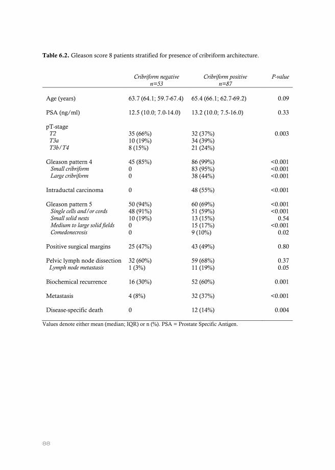
88
Table 6.2. Gleason score 8 patients stratified for presence of cribriform architecture.
Cribriform negative
n=53 Cribriform positive
n=87 P-value
Age (years)
63.7 (64.1; 59.7-67.4)
65.4 (66.1; 62.7-69.2)
0.09
PSA (ng/ml)
12.5 (10.0; 7.0-14.0) 13.2 (10.0; 7.5-16.0) 0.33
pT-stage T2 T3a T3b/T4
35 (66%) 10 (19%) 8 (15%)
32 (37%) 34 (39%) 21 (24%)
0.003
Gleason pattern 4 Small cribriform Large cribriform
45 (85%) 0 0
86 (99%) 83 (95%) 38 (44%)
<0.001 <0.001 <0.001
Intraductal carcinoma
0 48 (55%) <0.001
Gleason pattern 5 Single cells and/or cords Small solid nests Medium to large solid fields Comedonecrosis
50 (94%) 48 (91%) 10 (19%) 0 0
60 (69%) 51 (59%) 13 (15%) 15 (17%) 9 (10%)
<0.001 <0.001
0.54 <0.001
0.02
Positive surgical margins
25 (47%) 43 (49%) 0.80
Pelvic lymph node dissection Lymph node metastasis
32 (60%) 1 (3%)
59 (68%) 11 (19%)
0.37 0.05
Biochemical recurrence
16 (30%) 52 (60%) 0.001
Metastasis
4 (8%) 32 (37%) <0.001
Disease-specific death
0 12 (14%) 0.004
Values denote either mean (median; IQR) or n (%). PSA = Prostate Specific Antigen.

89
Comedonecrosis was present in 9 cases, 7 (78%) of which were present in Gleason score 4+4
tumours. Comedonecrosis was accompanied by cribriform architecture in all 9/9 (100%) cases
and by solid fields in 5/9 (56%) cases. Moreover, comedonecrosis was observed more often in
patients with large cribriform fields (7/20, 35%) than in those without (2/90, 2%, P<0.001).
Multivariable analysis of clinical outcome in Gleason score 8 patients
In univariate Cox regression analysis, pT3a (Hazard Ratio (HR) 2.1, 95% Confidence Interval (CI)
1.1-3.8, P=0.02), pT3b/4 (HR 4.6, 95% CI 2.5-8.5, P<0.001), Gleason score 4+4 (HR 1.9, 95% CI
1.1-3.1, P=0.02), positive lymph nodes at time of operation (HR 11.8, 95% CI 5.6-25.2, P<0.001)
and overall presence of invasive and/or intraductal cribriform carcinoma (HR 2.4, 95% CI 1.4-
4.1, P=0.003) were significantly associated with shorter biochemical recurrence-free survival,
while age (P=0.18), PSA level (P=0.43), Gleason score 5+3 (P=0.78) and surgical margin status
(P=0.21) were not (Table 6.3). In multivariable analysis, pT3b/4-stage (HR 4.4, 95% CI 2.1-9.3,
P<0.001), positive lymph nodes (HR 9.9, 95% CI 4.2-23.5, P<0.001) and overall cribriform
architecture (HR 2.0, 95% CI 1.0-3.7, P=0.04) had independent predictive value for biochemical
recurrence-free survival, while Gleason score 4+4 (HR 1.7, 95% CI 1.0-2.9, P=0.07) did not meet
conventional measures of significance in this cohort. In case individual cribriform growth patterns
were included in multivariable analysis instead of overall cribriform architecture, large invasive
cribriform carcinoma (HR 2.0, 95% CI 1.0-4.1, P=0.05) had independent predictive value for
biochemical recurrence-free survival, whereas intraductal cribriform carcinoma (HR 1.3, 95% CI
0.8-3.5, P=0.4) and small invasive cribriform carcinoma (HR1.6, 95% CI 0.8-3.5, P=0.2) did not
(data not shown).
Similar trends were observed for metastasis as pT3a (HR 2.7, 95% CI 1.2-6.2, P=0.02),
Gleason score 4+4 (HR 3.8, 95% CI 1.8-8.1, P=0.001), positive lymph nodes (HR 11.5, 95% CI
5.2-25.9, P<0.001) and overall cribriform architecture (HR 6.7, 95% CI 2.0-21.9, P=0.002) were
significantly associated with shorter metastasis-free survival, whereas age (P=0.80), PSA level
(P=0.96), pT3b/4 (P=0.06), Gleason score 5+3 (P=0.85) and positive surgical margins (P=0.95)
were not (Table 6.4). In multivariable analysis, Gleason score 4+4 (HR 2.4, 95% CI 1.0-5.9,
P=0.05), positive lymph nodes (HR 15.0, 95% CI 5.6-40.0, P<0.001) and overall cribriform
architecture (HR 3.5, 95% CI 1.0-12.3, P=0.05) had independent predictive value for metastasis-
free survival. Due to the low number of events and risk of model overfitting we were not able to
include individual cribriform or Gleason 5 growth patterns in multivariable analysis.

90
Table 6.3. Cox regression analysis for biochemical recurrence-free survival in Gleason score 8
patients.
Univariate Multivariable
HR* 95% CI P-value HR 95% CI P-value
Age (years) 1.0 0.9-1.0 0.18 1.0 0.9-1.0 0.05
PSA (ng/ml) 1.0 1.0-1.0 0.43 1.0 1.0-1.0 0.23
pT-stage
T2 ref ref
T3a 2.1 1.1-3.8 0.02 1.8 1.0-3.4 0.06
T3b/T4 4.6 2.5-8.5 <0.001 4.4 2.1-9.3 <0.001
Gleason score
3 + 5 ref ref
4 + 4 1.9 1.1-3.1 0.02 1.7 1.0-2.9 0.07
5 + 3 1.1 0.5-2.4 0.78 1.5 0.6-3.6 0.37
Positive surgical margins 1.4 0.8-2.2 0.21 0.8 0.5-1.5 0.55
Pelvic lymph node metastasis 11.8 5.6-25.2 <0.001 9.9 4.2-23.5 <0.001
Cribriform architecture 2.4 1.4-4.1 0.003 2.0 1.0-3.7 0.04 *HR = hazard ratio, CI = confidence interval.

91
Table 6.4. Cox regression analysis for metastasis-free survival in Gleason score 8 patients.
Univariate Multivariable
HR* 95% CI P-value HR 95% CI P-value
Age (years) 1.0 0.9-1.1 0.80 1.0 0.9-1.1 0.52
PSA (ng/ml) 1.0 1.0-1.0 0.96 1.0 0.9-1.0 0.50
pT-stage
T2 ref ref
T3a 2.7 1.2-6.2 0.02 2.5 1.0-6.7 0.06
T3b/T4 2.4 1.0-6.0 0.06 1.3 0.4-3.8 0.67
Gleason score
3 + 5 ref ref
4 + 4 3.8 1.8-8.1 0.001 2.4 1.0-5.9 0.05
5 + 3 1.1 0.32-4.0 0.85 0.7 0.2-3.0 0.65
Positive surgical margins 1.0 0.5-2.0 0.95 1.1 0.5-2.4 0.84
Pelvic lymph node metastasis 11.5 5.2-25.9 <0.001 15.0 5.6-40.0 <0.001
Cribriform architecture 6.7 2.0-21.9 0.002 3.5 1.0-12.3 0.05 *HR = hazard ratio, CI = confidence interval.

92
Discussion
Our study demonstrates that among Gleason score 8 prostate cancer patients on radical
prostatectomy, biochemical recurrence and metastases occur more often in Gleason score 4+4
than in Gleason score 3+5 or 5+3 tumours. Invasive and/or intraductal cribriform carcinoma was
observed in 62% of tumours and was associated with adverse pathological features and clinical
outcome. Cribriform architecture occurred more frequently in men with Gleason score 4+4 (93%)
than in those with Gleason score 3+5 (47%) and 5+3 (44%). In multivariable analysis, cribriform
architecture was an independent parameter for biochemical recurrence- and metastasis-free
survival, while Gleason score was not. Therefore, cribriform architecture also has important value
for risk stratification among Gleason score 8 prostate cancer patients.
Since the introduction of Grade Groups several reports have analysed the clinical
outcome of Gleason score 3+5, 4+4 and 5+3 prostate cancer.98 Some of these studies found that
men with Gleason score 3+5 at radical prostatectomy had reduced risk of biochemical recurrence
among Gleason score 8 patients. 104, 105 Others did not find a difference among Gleason score 8
subgroups or concluded that men with primary Gleason pattern 5 had worse outcome.100, 106-109
This variability of results might be explained by the use of different specimen types and clinical
outcome measures.110 Furthermore, Gleason score 8 is relatively uncommon, hampering statistical
analysis on large numbers of patients or resulting in clustering of Gleason score 5+3 and 5+3
tumours.109, 111 Finally, from a morphologically point of view, Gleason score 8 prostate cancer is a
very heterogeneous disease including highly variable quantities of Gleason 3, 4 and 5 growth
patterns. This heterogeneity might lead to significant interobserver variability in tumour grading.
For instance, Shah et al. only found fair interobserver reproducibility for Gleason pattern 5
assignment among 16 international expert genitourinary pathologists.112 Upon re-review of 40
archival cases with Gleason score 5+3 prostate cancer, Kryvenko et al. assigned the same score in
only 4 (10%) specimens, but upgraded 57.5% and downgraded 17.5% of cases.113
Many studies demonstrated worse clinical outcome for patients with cribriform
architecture.13, 15, 16, 28, 34 Most of these studies investigated cribriform architecture in intermediate
grade prostate cancer, while the impact of cribriform architecture in Gleason score ≥4+3 is less
well established. In Gleason score 7-10 prostate cancer biopsy patients, presence of cribriform
architecture has been associated with advanced pathological stage and worse disease-specific
survival compared to those with cribriform-negative biopsies.39, 59 Harding-Jackson et al. found
cribriform architecture, but not Gleason score, to have independent predictive value for cancer-
specific survival in Gleason score 8 patients.100 In the current study, we confirmed that cribriform
architecture had strong discriminative value, even in aggressive Gleason score 8 prostate cancer.

93
Both overall invasive cribriform and/or intraductal carcinoma as well as its more aggressive large
cribriform variant were significantly more often observed in Gleason score 4+4, than 3+5 and 5+3
disease. Since its association with adverse outcome, the high frequency of cribriform architecture
might well explain the worse outcome of Gleason score 4+4 prostate cancer compared to those
with 3+5 in our study, while the low number of 5+3 patients hampered powerful statistical
analysis. Of interest, however, is that in multivariable analysis not only cribriform architecture but
also Gleason score 4+4 had independent prognostic value for metastasis-free survival. A similar
trend was observed for biochemical recurrence-free survival although the predictive value of
Gleason score 4+4 did not reach conventional measures of significance (P=0.07). This implicates
that other grading factors apart from cribriform architecture contribute to the worse outcome in
Gleason score 4+4 patients. A possible explanation could be that Gleason score 4+4 disease has
the lowest percent of Gleason 3 growth pattern, which is by definition present in less than 5% of
the tumour volume. In 3+5 disease, percent Gleason pattern 3 theoretically varies from 50% to
95%, while it occupies 5% to 50% in Gleason score 5+3 tumours. Some groups have shown
independent prognostic value for percent Gleason pattern 4 and 5, which outperformed Gleason
score.114 The inverse could well be true for percent Gleason pattern 3; if a tumour still has the
biological capacity to mature into well-delineated glandular structures it is associated with better
outcome.
Little is known about the predictive value of individual Gleason grade 5 growth patterns,
which have been reported as either single cells, cords, small solid cylinders, solid fields, and
presence of comedonecrosis.12 Single cells and/or cords are the most common Gleason pattern
5.115, 116 Flood et al. found that presence of solid fields and number of different Gleason 5 growth
patterns were associated with shorter biochemical recurrence-free survival in Gleason score 9-10
prostatectomies.115 Compared to other Gleason 5 patterns, comedonecrosis was associated with
non-organ confined disease and biochemical recurrence.103, 117 While individual Gleason 4 growth
patterns have increasingly been subject to clinicopathological analysis, information on the clinical
relevance of Gleason 5 patterns is still scarce. Our group recently performed in-depth three-
dimensional visualization of prostate adenocarcinoma architectural growth patterns and revealed
two separate morphological groups.96 The first group consists of a tubular network in which the
vast majority if not all tumour cells are in direct contact with surrounding stroma. This group
encompasses the morphological continuum of Gleason pattern 3, poorly formed and fused pattern
4, and single cells and cords pattern 5. The second group has contiguous epithelial proliferations
in which the majority of tumour cells are not in contact with surrounding stroma and consists of
cribriform pattern 4 and solid pattern 5 with or without comedonecrosis. Our current finding that
solid fields mostly coexisted with cribriform structures is reflective of this continuum. In the

94
current study, we distinguished between small nested cylinders consisting up to 30 tumour cells
and larger solid fields. While the latter was continuous with cribriform growth, small nested
cylinders were not. This suggests that both have different biological and possibly clinical relevance.
However, larger studies are required to perform statistical analysis on the clinical relevance of
individual Gleason 5 growth patterns.
Strong points of this study are the detailed histological review of radical prostatectomy
specimens and the classification of cribriform architecture with the use of strict morphological
criteria and additional immunohistochemistry. The study is limited by the retrospective study
design. The inclusion of high-grade samples from two participating centers could have resulted in
a selection bias. Furthermore, the relatively short follow-up of 59 months and limited number of
patients restricted robust statistical analysis.
In conclusion, Gleason score 8 is a heterogeneous group of prostate cancers. Although
clinicopathological characteristics of Gleason score 3+5, 4+4 and 5+3 are mostly similar, Gleason
score 4+4 patients have a higher risk of adverse events. Cribriform architecture is an independent
predictor for metastasis-free survival and has better discriminative value for clinicopathological
outcome than Gleason score. Therefore, reporting cribriform architecture might add value in risk
stratification of Gleason score 8 prostate cancer patients.

95

96
Chapter VII
Discussion.
Adapted from ‘Prostate cancer growth patterns beyond Gleason score: entering a new era of comprehensive tumour grading’
Van Leenders GJLH, Hollemans E, Verhoef EI.
Histopathology. 2020 Dec; 77: 850-861.

97
Introduction
Prostate cancer management depends on a multitude of parameters for optimal risk stratification.
Prostate-Specific Antigen (PSA) level, clinical stage and histology of prostate biopsies are key
components in predictive risk models and subsequent clinical decision-making. The Gleason
grading system has important value for determining prostate cancer prognosis and clinical
decision-making. The Gleason score is entirely based on classification of architectural growth
patterns. These basic patterns are assigned a Gleason grade from 1 to 5. Since prostate cancer is
such a heterogeneous disease, the Gleason score is determined by adding to the most common
and highest grade in biopsies, or two most predominant grades in radical prostatectomy (RP)
specimens.11, 118 Gleason pattern 1 to 3 encompass well-delineated glandular structures with
variable inter-glandular distance and nodular circumscription. As no practical nor prognostic
differences exist between these three Gleason grades, the International Society of Urological
Pathology (ISUP) recommended that Gleason scores 2-4 should rarely, if ever, be used on biopsy
specimens.11 Gleason pattern 4 includes poorly formed, fused, glomeruloid and cribriform
glandular structures, whereas growth patterns with essentially no glandular differentiation such as
single cells, cords, solid fields and presence of comedonecrosis classify as Gleason pattern 5 (Figure
1).11, 12 While individual growth patterns have not been specified in pathology reporting, clinical
decision-making or molecular-biological investigations, recent studies indicate that individual
growth patterns have independent predictive value for clinical outcome and facilitate more
comprehensive interpretation of molecular-biological findings. The aim of this Discussion is to
summarize the clinicopathological impact of individual prostate cancer growth patterns beyond
Gleason score and to investigate their molecular-biological background. Moreover, we aim to give
recommendations on the incorporation of architectural growth patterns in order to optimize
decision-making in clinical practice.
Clinicopathological impact of cribriform growth patterns and their variants
Individual tumour growth patterns have mainly been analysed in Gleason score 3+4=7 (Grade
Group 2) prostate cancer, which is composed of variable quantities of well-delineated Gleason
pattern 3 glands and Gleason pattern 4 structures. In 2011, Iczkowski et al. were the first to report
that cribriform growth pattern had independent prognostic value for post-operative PSA failure.13
Many groups have thereafter confirmed the independent predictive value of cribriform pattern for
adverse pathological features, biochemical recurrence- and disease specific-free survival in biopsy
and RP specimens.14, 15, 20, 21, 34, 39, 52, 67, 71, 119, 120 However, a caveat of many studies on cribriform
growth pattern is that it is not entirely clear whether, and if so how, invasive Gleason pattern 4
pattern was distinguished from intraductal carcinoma (IDC) of the prostate. IDC is characterized

98
by a cribriform or solid proliferation of atypical epithelial cells within distended pre-existent
prostate acini either with or without comedonecrosis, and has also been associated with adverse
clinical outcome.28, 36, 39, 52, 121-123 The aetiology of IDC is not yet elucidated. While IDC is thought
to represent cancerisation of pre-existent glands by an invasive carcinoma, some data suggests that
IDC arises as a precursor lesion that has progressed beyond high grade prostatic intraepithelial
neoplasia (hgPIN).25 Invasive cribriform carcinoma and IDC are often difficult, if not impossible,
to distinguish without the application of basal cell immunohistochemistry. If no basal cells are
present, a cribriform lesion is generally considered as invasive Gleason pattern 4; if continuous,
scattered or sporadic basal cells are observed, cribriform architecture is mostly regarded as IDC.
Only few studies have attempted to investigate invasive cribriform and intraductal carcinoma
separately by using extensive immunohistochemistry.34, 39, 51 In a prostate biopsy screening cohort
of 1,031 men, Kweldam et al. found that presence of invasive cribriform carcinoma and IDC were
both associated with worse disease-specific survival in univariate analyses. The combination of
either invasive cribriform or intraductal carcinoma showed the strongest association with outcome
in this study. In the vast majority of patients, IDC occurs intermixed with invasive carcinoma, but
rare cases of isolated IDC without invasive disease have been described.24
Grading of IDC intermixed with invasive carcinoma has been controversial. While the
2014 ISUP meeting did not make a recommendation on this issue, the 2016 WHO states it should
not be factored into grading.12, 118 A consequence of the WHO recommendation is that basal cell
immunohistochemistry should be performed in every case where IDC cannot be distinguished
from invasive disease and classification of atypical lesion as either IDC or invasive carcinoma
would alter the final Gleason score. Apart from additional run-around time and costs, basal cell
immunohistochemistry does not distinguish between invasive cribriform or solid carcinoma, and
IDC in every case. It is well-known that foci of hgPIN can lack basal cells probably due to sampling
artifact. As IDC glands are by definition distended, the chance of false-negative basal cell
immunohistochemistry seems even larger, resulting in erroneous classification as invasive
cribriform pattern. On the other hand, large irregular cribriform tumour fields well exceeding pre-
existent gland architecture might have rare basal cells, as has also rarely been reported for low-
grade invasive adenocarcinoma.124 Since IDC is an adverse pathological parameter and difficult
or even impossible to distinguish from invasive carcinoma, even with the use of basal cell
immunohistochemistry, it is recommended by the latest 2019 ISUP consensus meeting that IDC
intermixed with invasive carcinoma should be assigned a Gleason grade based on its underlying
growth pattern, as if it were invasive carcinoma.125 This incorporation of intraductal carcinoma
into the tumour grade results in a Grade Group change in less than 2% of prostate cancer

99
biopsies.126 Although the modification might affect decision-making in individual patients, it has
minimal impact on overall prostate cancer management.
With increasing awareness of the their clinical impact, proposals for subclassification of
cribriform growth patterns have been made. More detailed analysis of cribriform pattern has
shown that the number of cribriform fields does not seem to affect clinical outcome in a negative
way, while their maximal individual size does.34, 39, 51 In the current thesis, , we found in 420 Grade
Group 2 RPs that large expansile cribriform fields, arbitrarily defined as exceeding at least two
times the size of adjacent benign glandular structures, had seminal vesicle invasion in 32% and
pelvic node metastasis in 23% of cases, which was significantly higher than 9% and 5% found,
respectively, for small invasive cribriform carcinoma.51 Other groups have also separated both
cribriform patterns, but are difficult to compare as they applied other size criteria such as presence
of at least 12 intercellular lumens or exceeding average benign gland diameter.13, 34, 127 Therefore,
we concluded that large cribriform carcinoma was an even more aggressive subtype.
Clinical implications of incorporating cribriform growth patterns
Since they both have independent predictive value, the presence of either invasive cribriform or
intraductal cribriform carcinoma should routinely be reported as “cribriform carcinoma”.125 The
question arises as to what extent incorporation of cribriform carcinoma can lead to optimization
of therapeutic decision-making in individual prostate cancer patients. Patients with biopsy
Gleason score 3+4=7 (Grade Group 2) prostate cancer will generally be offered definitive
treatment, while those with Gleason score 3+3=6 (Grade Group 1) are often eligible for active
surveillance. Recent identification of additional prognostic pathological parameters such as
presence of invasive and/or intraductal cribriform carcinoma and Gleason pattern 4 quantity,
allows for more detailed risk stratification of men with Grade Group 2 disease. Men with low
biopsy Gleason pattern 4 quantity might be eligible for active surveillance, firstly because their
outcome is comparable to those with Grade group 1 tumours, secondly due to the substantial inter-
observer variability for grade assignment to small foci of poorly formed and fused glands.43, 44, 128-
131 Since disease-specific and biochemical recurrence-free survival are not statistically significantly
different among men with biopsy Grade Group 2 without cribriform carcinoma and those with
Grade Group 1, absence of both invasive cribriform and intraductal carcinoma has also been
proposed as an eligible criterion for active surveillance for men with Grade Group 2 biopsies.39, 48,
52, 132 If the safety of this eligibility approach is shown in prospective studies, this will have major
impact on individual Grade Group 2 patient management. Presence of invasive carcinoma might
also affect other aspects of clinical decision-making. Absence of cribriform architecture has been
associated with a low-risk of pelvic lymph node metastasis.33, 120, 133 In our series of 627 RP’s,
100
22/228 (10%) Grade Group 2 men with cribriform carcinoma developed metastases, while no
metastases were observed among 192 cribriform-negative Grade Group 2 and 207 Grade Group 1
patients (this thesis).133 Although higher biochemical recurrence rates were seen in men with Grade
Group 2 prostate cancer, this did not affect long term outcome in our cohort. This indicates that
invasive cribriform and intraductal carcinoma, in particular, might have an impact on the
biological potential for metastatic disease to develop. In contrast, postoperative biochemical
recurrence-free survival is also related to tumour volume parameters and surgical technique, which
do not necessarily reflect biological derangement caused by the disease.
Current guidelines on performing of pelvic lymph node dissections (PLND) are based on
clinicopathological nomograms which do not consider cribriform architecture, but future inclusion
of invasive cribriform and intraductal carcinoma might result in optimization of these nomograms.
Few studies also have found independent value of cribriform carcinoma for response to radiation
therapy and docetaxel, but the definitive clinical impact on these treatment modalities remains to
be determined.121, 134, 135
Detection of cribriform growth patterns with MRI and prostate biopsies
Since invasive cribriform and intraductal carcinoma might increasingly affect clinical decision-
making, the sensitivity for detection of these adverse features on biopsy should ideally be high.
Concordance between Grade Group at RP specimens and matched biopsies is relatively low, with
tumour up-grading occurring in up to 40% of cases.94, 136 The sensitivity and specificity for
identification of invasive and/or intraductal cribriform carcinoma at biopsy specimens varies from
43-56% and 87-95%, respectively.59, 137, 138 This indicates that about half of cribriform carcinoma
lesions are missed at diagnostic biopsies. Detailed analysis of features potentially associated with
cribriform false-negative biopsies, did not reveal predictive value for number of positive biopsies,
percent Gleason pattern 4, or presence of glomeruloid Gleason pattern 4 architecture in a relatively
small series performed in this thesis.138 On the other hand, multiparametric magnetic resonance
imaging (MRI) might have added value in identification of men with prostatic invasive cribriform
and intraductal carcinoma with false-negative biopsies, since these more frequently present with
PI-RADS score 5 lesions.49, 138, 139 Finally, commercially available molecular tests might also play
a role in identification of men with these adverse features as discussed in more detail later.140-142
101
Invasive cribriform growth pattern delineation
As invasive cribriform carcinoma should be separately commented on in pathology reports and
might increasingly affect individual therapeutic decision-making, it is important to gain broad
consensus on the definition of cribriform morphology and to delineate this growth pattern from its
microscopic mimickers.65 Recognition and Gleason pattern 4 assignment is better for cribriform
and glomeruloid growth pattern than for poorly-formed and fused glands.94, 95, 131 Nevertheless,
tangentially sectioned tumour glands, complex fused glands, large glomeruloid structures and
solid Gleason pattern 5 might all be confused with invasive cribriform pattern.95 Our group has
defined cribriform architecture as an epithelial sheet (a) where the majority of tumour cells do not
contact surrounding stroma, (b) with a polarized gland-like structure being present at less than half
of the sheet circumference and (c) with regular intercellular lumens clearly visible on HE section
(Figure 2).65, 143 The first criterion (a) distinguishes cribriform from complex fused glands where
most if not all tumour cells are still in direct contact with subtle connective tissue cores being
present within the lesion. The second criterion (b) arbitrarily distinguishes cribriform from large
glomeruloid pattern where polarized gland-like spaces are circumventing more than half of the
central protrusion. The validity of this criterion is supported by the fact that clinicopathological
features and biochemical recurrence-free survival of large glomeruloid structures at RP were more
comparable to those of small glomeruloid than cribriform Gleason pattern 4 in our study (this
thesis).144 The third criterion (c) distinguishes cribriform from solid Gleason pattern 5 where
essentially no glandular differentiation is visible on HE sections.
Clinicopathological relevance of other growth patterns
Whereas invasive cribriform and intraductal carcinoma have independent value for clinical
outcome in men with Grade Group 2 prostate cancer, it is not entirely clear yet to what extent
outcome of Grade Group 2 men without cribriform architecture differs from those with Grade
group 1 tumours. With a median follow-up of 13 years, Kweldam et al. did not find statistically
different disease-specific survival for 256 patients with biopsy cribriform-negative Grade group 2
and 486 Grade group 1 prostate cancer.52 In our RP cohort, we did not find any metastasis at
PLND or during follow-up in207 men with Grade Group 1 and 197 Grade Group 2 cancer without
invasive cribriform or intraductal carcinoma.133 The latter group, however, had significantly
shorter biochemical recurrence-free survival than men with Grade Group 1. These data suggest
that cribriform architecture is reflective of intrinsic capacity for development of metastasis, while
the risk of biochemical recurrence also more depends on other factors such as tumour volume,
positive surgical margins on non-cribriform Gleason patterns. In a detailed study of 1275 RPs,
McKenney et al. distinguished 20 different growth pattern features.21 Apart from the adverse

102
outcome of cribriform architecture compared to poorly-formed glands, they revealed reactive
stroma response to be associated with worse and mucin extravasation with better prognosis.
Hitherto, the glomeruloid growth pattern has not yet been adequately addressed in
literature. In 1995, glomerulations were described for the first time in prostate needle biopsies.145
Glomeruloid growth consists of carcinoma glands with intraluminal cell clusters or cribriform
structures, attached to one side of the gland wall, resembling a glomerulus.12 Pacelli et al. were the
first to describe the glomeruloid growth pattern in relation to higher tumour grade and pathological
stage.92 Lotan et al. observed the glomeruloid growth pattern often simultaneously with high
tumour grade and especially cribriform growth.19 Because of its morphological reminiscence and
frequent co-occurrence with cribriform pattern, glomeruloid architecture has been classified as
Gleason pattern 4 since the 2014 ISUP consensus meeting, and some authors have postulated it
to represent a precursor of cribriform morphology.12, 19, 20 In 350 RP specimens with Gleason score
7, Choy et al. however found that cribriform morphology independently increased the risk of
biochemical recurrence, while glomeruloid architecture was significantly associated with a
reduced risk.20 The previously mentioned study by McKenney et al. could not associate
glomerulations with adverse recurrence-free survival.21 In our study among 472 Grade group 2
RPs, we distinguished small simple glomerulations and complex glomerulations with cribriform
protrusions (this thesis).144 Complex glomerulations coincided with cribriform architecture more
often than simple glomerulations. Men with cribriform morphology had significantly worse
clinicopathological features and biochemical recurrence-free survival than those with glomeruloid
pattern, irrespective of the size of the glomerulations. These findings seem counterintuitive with
the hypothesis that glomeruloid glands are precursors of cribriform architecture.
Still little is known on the clinical relevance of Gleason 5 growth patterns, which have
been classified as single cells, cords, solid fields, or presence of comedonecrosis.118 This is mostly
due to the fact that tumours with primary, secondary or tertiary Gleason pattern 5 are very
heterogeneous with variable quantities of Gleason pattern 3 , 4 and 5, several different growth
patterns and occurrence of IDC. Meaningful statistical analysis including all relevant covariates
requires inclusion of a large number of these patients. In that sense, it is of interest that even in
Grade Group 4 patients, presence of cribriform architecture has independent predictive value for
biochemical recurrence-free and disease-specific survival.39, 100, 146 In our cohort, presence of
cribriform architecture outperformed Gleason score in predicting outcome regarding biochemical
recurrence- and metastasis-free survival (this thesis).146 Nevertheless, presence of comedonecrosis
and solid sheets were found to be adverse parameters amongst Gleason 5 patterns in two relatively
small series.115, 117

103
Molecular aberrations in cribriform architecture
Genomic features of prostate cancer differ from other cancers, firstly due to the relatively low
mutation rate in prostate cancer. It features copy number variations involving prostate oncogenes
and tumour suppressor genes, and continuous, accumulating genomic and epigenetic
alterations.147, 148 The TMPRSS2-ERG fusion is one of the most common alterations, seen in 50%
of prostate cancers, and is associated with low-grade tumours.149 The androgen receptor pathway
is a key element in prostate cancer progression and harbours alterations in most castration-resistant
prostate cancers.150 In addition, gains of oncogene c-MYC, loss of tumour suppressor gene PTEN
and mutations in the SPOP gene have been associated with aggressive disease.151-155
Molecular alterations in prostate cancer have mostly been examined by Gleason score
without taking underlying growth patterns into account. Recently, few groups have aimed to
identify molecular characteristics of invasive cribriform and intraductal carcinoma.18, 78, 79 Since
these bioinformatic analysis were performed retrospectively on publically available databases with
digitally scanned HE reference slides, no reliable distinction between invasive and intraductal
cribriform carcinoma could be made. Cribriform carcinoma is characterised by genomic instability
and has been posited to be the result of an accumulation of aggressive features above early phase
tumour alterations within the prostate epithelium.18, 78 Amongst others, deletions of 8p, 10q and
amplification of 8q24 corresponding to PTEN loss and c-MYC gain were significantly enriched in
cribriform carcinoma, together with SPOP point-mutations.18, 78, 79, 156, 157 Molecular profiling and
RNA in situ hybridization revealed that the long noncoding RNA SChLAP1 had more than 3-
fold higher levels in cribriform architecture, and could serve as a potential marker for its detection
in clinical practice.78, 158 Epigenetic alternations have been detected in cribriform prostate cancer
as well. The hypermethylated clusters found in prostate cancer are enriched by cribriform patterns,
while non-cribriform patterns dominate the hypomethylated cluster.79, 159, 160 Interestingly, various
of the molecular aberrations associated with cribriform carcinoma have been linked to aggressive
clinical behaviour of prostate cancer before.161-166 Moreover, distant metastasis were found to be
partially comparable to intraductal cribriform carcinoma.80 Together these data indicate that
cribriform carcinoma is a morphological substrate of increased genomic instability, and brings
histopathology, molecular aberrations and adverse clinical outcome comprehensively together.
Molecular identification of a “cribriform signature” is of importance, especially since the
high rate of false cribriform-negative biopsies is a limitation for developing clinical decision models
incorporating cribriform carcinoma. A clinically applicable molecular urine, serum or tissue test
might identify men at risk for unsampled cribriform carcinoma. Recently, studies have shown that
higher risk scores using RNA expression-based tissue arrays (Decipher, Oncotype Dx and Prolaris)
were significantly associated with presence of cribriform carcinoma in the tissue sample

104
analysed.140, 141, 167 It remains to be determined whether these molecular assays still have added
clinical value if cribriform carcinoma and percent Gleason pattern 4 are accounted for, and if they
can support identification of men with false cribriform-negative prostate cancer biopsies.
Three-dimensional architecture of prostate cancer growth patterns
Microscopic diagnostic pathology in everyday practice is performed using thin tissue slides
representing two-dimensional cross-sections of a three-dimensional (3D) structure. Little is known
on the actual 3D architecture of prostate carcinoma growth patterns. Registration of hundreds of
consecutive slides have shown that poorly-formed Gleason pattern 4 is continuous with Gleason
pattern 3.168, 169 Recent improvements in tissue clearing techniques, long-distance confocal laser
scanning and light-sheet microscopy have enabled imaging of intact 1 mm thick prostate tissues.170-
172 By detailed 3D analysis of formalin-fixed paraffin-embedded RP specimens, we were able to
get comprehensive insight of the 3D architecture of prostate adenocarcinoma growth patterns.143
This revealed that Gleason pattern 3 three-dimensionally represented tubules with local
interconnections. This pattern was continuous with both poorly formed Gleason pattern 4 where
tubular size and lumen diameter was smaller and tubular interconnections occurred more
frequently, and fused pattern 4 where interconnections often occurred at distances smaller than
the tubular diameter. In fact, single cells and cords Gleason pattern 5 formed a continuum with
poorly formed glands, in which lumen size further decreased until its disappearance. On the other
hand cribriform Gleason pattern 4 and solid pattern 5 either without or with comedonecrosis
consisted of serpentine fields of epithelial cells with the majority of tumour cells not being in direct
contact with surrounding stroma. Both patterns formed a continuum with or without the presence
of recognizable intercellular lumens. Based on these three-dimensional features, we classified the
growth patterns in two distinct subgroups which both formed continua. The first group consisted
of Gleason pattern 3 tubules, Gleason pattern 4 poorly formed and fused glands, and Gleason
pattern 5 single cells and cords, which all consisted of cells directly contacting surrounding stroma,
but with variable gland size, lumen size and number of interconnections. The second group
encompassed cribriform Gleason pattern 4 and solid pattern 5 without or with comedonecrosis
consisting of epithelial cells, where the majority of cells did not connect to adjacent stroma and
with variable intercellular lumen frequency and size. Glomeruloid structures formed a 3D
intermediate between both subgroups. They represented intraluminal protrusions of epithelial cells
appearing within a background of Gleason pattern 3 tubules and were mostly present at the side
of tubular interconnections. The 3D architectural continuity and transitions between growth
patterns in both subgroups well explains Gleason grading inter-observer variability.94, 136 Whereas
growth patterns are ordinally classified in Gleason grades in clinical practice, they gradually
105
transition to each other without presence of clearly identifiable cut-offs. Furthermore, the 3D
architectural relationships support the definition and delineating characteristics of cribriform
growth pattern as mentioned previously. Future studies need to determine whether the 3D
dichotomization is also reflected by clinical and molecular observations.
Digital image analysis for automated detection of cribriform growth pattern
Digitalization in the field of pathology offers advantages in cancer diagnosis with the use of deep
learning algorithms.173-175 In radiology, computer-aided detection of prostate cancer in
multiparametric MRI images has similar performance scores as a radiologist, including to
differentiate between low- and high-grade prostate cancer.176, 177 For histological evaluation, deep
learning systems can be designed to segment epithelial tissue and recognize malignant glands. In
prostate needle biopsies, cancer likelihood maps have been computed with high sensitivity for
carcinoma.178 Hence, up to a third of slides not containing prostate cancer could be identified
reliably and would not have to be evaluated by pathologists. Based on unforcedly generated
features, deep learning systems are able to compute boundaries and cut-off points, thus enabling
automated identification of histomorphological growth patterns.178-180 As mentioned before,
prostate cancer grading is prone to intra- and interobserver variability, while treatment options and
outcome are highly dependent on assignment of patterns. By standardization, image analysis is
able to reduce the variability and prioritize cases for the pathologist. In a collaborative study we
focused on automated detection of cribriform growth in prostate cancer based on convolutional
neural networks.181 Original annotations performed by pathologists in prostate cancer biopsies
showed that sensitive cribriform region detection can be reached, but at the expense of a high
number of false positives.
Future perspectives
Last decade the growing interest in individual growth patterns in prostate cancer has resulted in
identification of cribriform architecture as pathological factor with independent prognostic value.65
However, the morphogenetic background of cribriform and other growth patterns remains to be
elucidated. Furthermore, the relation of intraductal carcinoma and invasive cribriform carcinoma
with show significant morphological overlap is yet unclear. To solve this issue, detailed molecular
analyses of individually dissected growth patterns is mandatory. Furthermore cell culture and
xenograft studies are subsequently important for investigating morphogenetic effects of specific
molecular alterations.
In turn, molecular markers might contribute to detection and diagnosis of cribriform
growth pattern in false negative biopsies. At this moment sensitivity of prostate cancer biopsies to

106
detect cribriform architecture is unsatisfactory, although the false negative rate is lower for the
large cribriform subtype. In future studies it should be determined how multiparametric MRI and
possibly commercially available molecular urine- or tissue-based tests could optimize the
representability of prostate biopsies for the entire tumour.
Future multivariable analyses need to elucidate on the mutual interaction and
independent value of the recently explored pathological parameters such as cribriform patterns
and percentage Gleason pattern 4 and 5. After identification of the independent, most influential
and reproducible factors, modification to the current Gleason grading/Grade group systems could
even be made to increase the discriminative value of tumour grading.53, 65 Furthermore, prospective
studies including cribriform architecture in clinical decision-making for instance on eligibility for
active surveillance, should indicate to what extent growth pattern specification can optimize
patient management.
Conclusion
Presence of cribriform architecture is associated with dismal outcome with large cribriform
architecture representing an even more aggressive subtype in prostate cancer. While
glomerulations may harbour cribriform intraluminal protrusions, they had favourable outcome
and should not be classified as a cribriform growth pattern variant. Treatment of patients with
cribriform architecture is essential, but the sensitivity of cribriform growth pattern detection at
biopsies should be improved. Incorporation of cribriform growth patterns beyond heterogeneous
Gleason groups in pathology reporting and clinical decision-making will improve personalized
treatment of prostate cancer patients.

107

108
Addendum
Summary | Samenvatting
List of publications
List of abbreviations
Curriculum vitae
PhD portfolio
Dankwoord
References
109
Summary
Prostate cancer is the most common cancer among men with great variability regarding outcome.
Clinical decision-making depends on Prostate-Specific Antigen (PSA) levels, clinical tumour stage
and Gleason score. The Gleason score is the most important parameter given by the clinical
pathologist and is entirely based on tumour architectural growth patterns, which are classified into
five different grades. The tumour is assigned a primary grade followed by a highest grade in needle
biopsy or secondary grade in radical prostatectomy specimens. Men with Gleason score 3+3=6
(Grade Group 1) prostate cancer have excellent outcome and are frequently eligible for active
surveillance. Men with Gleason score 3+4=7 (Grade Group 2) or higher generally receive
radiation therapy or radical prostatectomy surgery. Despite the urge for active treatment,
differences in clinical outcome are observed in intermediate to high risk prostate cancer patients.
There is a need for additional parameters to aid risk stratification in these men. Incorporation of
individual histopathological growth patterns might be able to support optimal decision-making in
this large prostate cancer subpopulation.
Last decade the cribriform growth pattern has been recognized as independent prognostic
pathological feature for prostate cancer outcome. Cribriform architecture however does not
comprise one entity. Chapter 2 elaborated on the prognostic impact of invasive and intraductal
cribriform prostate cancer subtypes in Grade Group 2 radical prostatectomies. Cribriform
architecture was associated with worse clinicopathological parameters such as tumour stage and
lymph node metastasis. Of the 420 radical prostatectomies, 8% revealed invasive large cribriform
architecture, based on a diameter of at least twice the size of adjacent pre-existent normal glands.
Large cribriform architecture represented an aggressive subtype of invasive cribriform prostate
cancer as it was an independent predictive factor for biochemical recurrence-free survival in
patients with Grade Group 2 prostate cancer. Furthermore, metastases were more often seen in
this cribriform subtype. If validated in biopsy cohorts, large cribriform pattern therefore could serve
as a new predictive parameter for optimisation of clinical decision-making.
In order to incorporate cribriform architecture as predictive parameter, it is essential to detect it in
diagnostic biopsies. In Chapter 3, we compared presence of cribriform architecture in biopsies and
following radical prostatectomies. Patients had cribriform architecture on radical prostatectomy
in 69% of cases, while on biopsies only 30% revealed this pattern. Of patients with Grade Group
2 prostate cancer, 40% had false negative biopsies for cribriform growth. These patients presented
with higher PSA levels than men with true negative biopsies for cribriform growth and more often
had radiological high grade lesions on magnetic resonance imaging. The false negative rate for
large cribriform prostate cancer, the more aggressive cribriform subtype, was lower than for any
110
cribriform architecture. Also, biochemical recurrence-free was significantly worse for patients with
true positive biopsies than false negative biopsies.
It has been well established that cribriform architecture is associated with dismal outcome,
however absence of this pattern was less well studied. Chapter 4 compared features and outcome
of Grade Group 1 and Grade Group 2 prostate cancer without cribriform architecture. Although
patients with cribriform-negative Grade Group 2 prostate cancer presented with higher PSA levels
and higher frequency of extra-prostatic extension and positive surgical margins, this did not affect
long-term outcome. Metastasis and disease-specific death occurred neither in patients with Grade
Group 1 nor in cribriform-negative Grade Group 2. Biochemical recurrence-free survival was
shortened in cribriform-negative Grade Group 2 patients, however this is also related to tumour
volume parameters and surgical technique, which do not necessarily reflect biological
derangement caused by the disease.
Glomeruloid growth is a hitherto poorly understood histomorphological pattern that bears
similarities to cribriform architecture. Glomerulations with small intraluminal protrusions have
been associated with favourable outcome. It is yet unclear what is the clinical behaviour of
complex glomeruloid patterns, which resemble cribriform architecture. In Chapter 5 we
specifically aimed at characterization of glomeruloid architecture in radical prostatectomy
specimens. Complex glomeruloid often coincided with other cribriform patterns. Although
complex glomerulations were associated with slightly worse clinicopathological features, no
independent prognostic value was seen for neither simple nor complex cribriform growth.
Therefore, glomeruloid pattern and particularly complex glomerulations should not be classified
as a cribriform growth pattern variant.
Chapter 6 focused on the effect of growth patterns in Grade Group 4 prostate cancer. Although
the incorporation of growth patterns in prostate cancer diagnosis particularly affects clinical
decision making in Grade Group 2 prostate cancer, it might also have predictive value in high-
grade prostate cancer. We investigated presence of cribriform architecture in Grade Group 4 and
found that it was present in 62% of cases. It was more frequently present in Gleason score 4 + 4
compared to 3 + 5 or 5 + 3 prostate cancer, and solid field pattern 5 often coexisted with cribriform
architecture. Presence of cribriform architecture was an independent parameter for biochemical
recurrence- and metastasis-free survival. Therefore, cribriform architecture also has added value
in risk stratification of Gleason score 8 prostate cancer patients.
Finally, in Chapter 7 the main findings of this thesis are summarized and reviewed together with
current available knowledge on prostate cancer growth patterns. We also postulate how
incorporation of cribriform architecture can optimize personalized clinical decision-making in
prostate cancer patients.

111
Samenvatting
Prostaatkanker is de meest voorkomende kanker onder mannen, met grote variabiliteit in
uitkomst. Klinische besluitvorming is afhankelijk van Prostaat-Specifieke Antigeen (PSA) levels,
klinisch tumorstadium en Gleason score. De Gleason score is de belangrijkste door de klinisch
patholoog afgegeven parameter en is gebaseerd op groeipatronen binnen de tumor, welke worden
ingedeeld in vijf graden. Aan iedere tumor wordt een primaire graad toegekend, gevolgd door een
hoogste graad in prostaatbiopten en secundaire graad in radicale prostatectomieën. Mannen met
Gleason score 3+3 (Grade Group 1) prostaatkanker tonen excellente overleving en komen meestal
in aanmerking voor actief vervolgen. Mannen met Gleason score 3+4 (Grade Group 2) of hoger
ontvangen meestal behandeling in de vorm van radiotherapie of chirurgie. Ondanks de indicatie
voor behandeling zijn er grote verschillen in klinische uitkomst tussen prostaatkanker patiënten
uit deze intermediair tot hoog risico groep. Incorporatie van individuele histopathologische
groeipatronen kan een additionele parameter in risicostratificatie zijn en optimale klinische
besluitvorming faciliteren in deze grote prostaatkanker subpopulatie.
In het afgelopen decennium is cribriforme groeiwijze geïdentificeerd als onafhankelijke
prognostische parameter bij prostaatkanker. Cribriforme architectuur omvat echter niet slechts één
entiteit. Hoofdstuk 2 onderzocht de prognostische waarde van subtypes van invasieve en
intraductale cribriforme prostaatkanker in radicale prostatectomieën met Grade Group 2.
Cribriforme architectuur was geassocieerd met ongunstige clinicopathologische parameters zoals
hoog tumorstadium en lymfekliermetastasen. Van de 420 radicale prostatectomieën toonde 8%
groot cribriforme groei, gebaseerd op een diameter van ten minste tweemaal die van een pre-
existente buis. Groot cribriforme groei representeerde een agressief subtype cribriforme
architectuur, daar het een onafhankelijke predictieve waarde had aangaande biochemisch recidief-
vrije overleving in patiënten met Grade Group 2 prostaatkanker. Metastasen werden eveneens
vaker gezien in dit cribriforme subtype. Wanneer deze parameter gevalideerd zou worden in een
groot cohort, kan groot cribriforme groei een nieuwe predictieve parameter worden ter
optimalisatie van klinische besluitvorming.
Om cribriforme architectuur en subtypes te incorporeren als predictieve marker, is het essentieel
deze in diagnostische biopten op te sporen. In hoofdstuk 3 vergeleken we de aanwezigheid van
cribriforme architectuur op biopten en opvolgende radicale prostatectomieën. Patiënten toonden
cribriforme architectuur in 69% van de radicale prostatectomieën, echter slechts 30% van de
biopten bevatten dit patroon. Patiënten met Grade Group 2 prostaatkanker hadden fout-negatieve
biopten voor cribriforme groei in 40% van de gevallen. Deze patiënten presenteerden zich met
hoger PSA en hadden vaker een radiologisch hooggradige laesie op magnetic resonance imaging.

112
Het aantal fout-negatieve biopten lag echter lager voor patiënten met groot cribriforme groei, het
agressieve subtype van cribriforme architectuur. Daarnaast was biochemisch recidief-vrije
overleving significant slechter voor patiënten met waar-positieve dan fout-negatieve biopten.
Hoewel cribriforme architectuur geassocieerd is met een somberdere uitkomst, is de afwezigheid
van dit patroon minder goed onderzocht. Hoofdstuk 4 vergeleek de kenmerken en uitkomsten van
Grade Group 1 en Grade Group 2 prostaatkanker zonder cribriforme architectuur. Ondanks het
feit dat patiënten met cribriform-negatieve Grade Group 2 prostaatkanker hogere PSA levels en
tumorstadium toonden, alsmede dat zij vaker positieve snijvlakken hadden, beïnvloedde dit niet
de lange termijn uitkomsten. Metastasen en ziekte-specifiek overlijden kwamen niet voor in Grade
Group 1 noch in cribriform-negatieve Grade Group 2 prostaatkanker. Biochemisch recidief-vrije
overleving was korter in cribriform-negatieve Grade Group 2 patiënten, echter deze overleving is
mede gerelateerd aan tumorvolume en chirurgische techniek, welke niet noodzakelijk
samenhangen met biologisch gedrag van de tumor.
Glomeruloïde groei toont gelijkenissen met cribriforme architectuur en is tot dusverre weinig
onderzocht. Glomerulaties met kleine intraluminale protusies zijn geassocieerd met betere
prostaatkanker overleving. Het klinische gedrag van complexe glomeruloïde patronen, welke
cribriform aspect kunnen tonen, is nog onduidelijk. Hoofdstuk 5 richtte zich op karakterisatie van
glomerulaties in radicale prostatectomieën. Complexe glomeruloïde groei werd vaak samen gezien
met andere cribriforme patronen en was geassocieerd met enigszins minder gunstigere
clinicopathologische kenmerken. Desalniettemin werd er geen onafhankelijke prognostische
waarde gevonden voor simpele noch complexe glomeruloïde groei. Daarom dient glomeruloïde
groei niet gekenmerkt te worden als subtype van cribriforme groei.
Hoofdstuk 6 onderzocht de groeipatronen in Grade Group 4 prostaatkanker. Het meewegen van
groeipatronen in de prostaatkanker diagnose heeft misschien de meeste waarde voor klinische
besluitvorming in Grade Group 2 patiënten, het kan eveneens aanvullende waarde tonen voor
hooggradige prostaatkanker. Cribriforme architectuur werd onderzocht in Grade Group 4
prostaatkanker waar het in 62% van de gevallen voorkwam. Het werd vaker gezien in Gleason
score 4+4 dan 3+5 of 5+3 prostaatkanker en vaak samen met solide groeipatronen. De
aanwezigheid van cribriforme architectuur was een onafhankelijke parameter voor biochemisch
recidief- en metastase-vrije overleving. Daarom heeft cribriforme architectuur toegevoegde waarde
in de risicostratificatie van Gleason score 8 prostaatkanker patiënten.
Tenslotte worden in hoofdstuk 7 de bevindingen van dit proefschrift uiteengezet en beoordeeld in
het licht van huidige kennis over prostaatkanker groeipatronen. We betogen dat incorporatie van
cribriforme architectuur de gepersonaliseerde klinische besluitvorming van prostaatkanker
patiënten kan verbeteren.
113
List of Publications
Vos MC, Hollemans E, Ezendam N, Feijen H, Boll D, Pijlman B, van der Putten H,
Klinkhamer P, van Kuppevelt TH, van der Wurff AA, Massuger LF. MMP-14 and CD44 in
Epithelial-to-Mesenchymal Transition (EMT) in ovarian cancer. J Ovarian Res. 2016 Sep; 9: 53.
doi: 10.1186/s13048-016-0262-7.
Hollemans E, Verhoef EI, Bangma CH, Rietbergen J, Helleman J, Roobol MJ, van Leenders
GJHL. Large cribriform growth pattern identifies ISUP grade 2 prostate cancer at high risk for
recurrence and metastasis. Mod Path. 2019 Jan; 32: 139-146. doi: 10.1038/s41379-018-0157-9.
Hollemans E, Verhoef EI, Bangma CH, Schoots I, Rietbergen J, Helleman J, Roobol MJ, van
Leenders GJLH. Concordance of cribriform architecture in matched prostate cancer biopsy and
radical prostatectomy specimens. Histopathology. 2019 Sept; 75: 338-345. doi: 10.1111/his.13893.
Van Leenders GJLH, Kweldam CF, Hollemans E, Kümmerlin IP, Nieboer D, Verhoef EI,
Remmers S, Incrocci L, Bangma CH, van der Kwast, TH, Roobol MJ. Improved prostate cancer
biopsy grading by incorporation of invasive cribriform and intraductal carcinoma in the 2014
Grade Groups. Eur Urol. 2020 Feb; 77: 191-198 doi: 10.1016/j.eururo.2019.07.051.
Hollemans E, Verhoef EI, Bangma CH, Schoots I, Rietbergen J, Helleman J, Roobol MJ, van
Leenders GJLH. Prostate carcinoma grade and length but not cribriform architecture at positive
surgical margins are predictive for biochemical recurrence after radical prostatectomy. Am J Surg
Pathol. 2020 Feb; 44: 191-197 doi10.1097/PAS. 0000000000001384.
Hollemans E, Verhoef EI, Bangma CH, Rietbergen J, Osanto S, Pelger RCM, van Wezel T, van
der Poel H, Bekers E, Helleman J, Roobol MJ, van Leenders GJLH. Clinicopathological
characteristics of glomeruloid architecture in prostate cancer. Mod Pathol. 2020 Feb; 33: 1618-
1625. doi: 10.1038/s41379-020-0507-2.
Vos MC, Hollemans E, van der Steen SCHA, van Kuppevelt TH, van der Wurff AAM,
Massuger LFAG. Primary ovarian tumors with lymphogenic and hematogenic metastasis
express high MMP-14, which colocalizes with highly sulfated chondroitin sulfate in the stroma.
Int J Gynecol Pathol. 2020 Mar; 39: 184-192. doi: 10.1097/PGP.0000000000000587.

114
Hollemans E, Verhoef EI, Bangma CH, Rietbergen J, Roobol MJ, Helleman J, van Leenders
GJLH. Clinical outcome comparison of Grade Group 1 and Grade Group 2 prostate cancer with
and without cribriform architecture at the time of radical prostatectomy. Histopathology. 2020
Apr; 76: 755-762. doi: 10.1111/his.13893.
Rijstenberg LL, Hansum T, Hollemans E, Kweldam CF, Kümmerlin IP, Bangma CH, van der
Kwast TH, Roobol MJ, van Leenders GJLH. Intraductal carcinoma has minimal impact on
Grade Group assignment in prostate cancer biopsy and radical prostatectomy specimens.
Histopathology. 2020 Jun; 77: 742-748. doi: 10.1111/his.14179
Hollemans E, Verhoef EI, Bangma CH, Rietbergen J, Osanto S, Pelger RCM, van Wezel T, van
der Poel H, Bekers E, Helleman J, Roobol MJ, van Leenders GJLH. Cribriform architecture in
radical prostatectomies predicts oncological outcome in Gleason score 8 prostate cancer patients.
Mod Pathol. 2021 Jan; 34: 184-193. doi: 10.1038/s41379-020-0625-x.
Van der Slot MA, Hollemans E, den Bakker MA, Hoedemaeker R, Kliffen M, Budel LM,
Goemaere NNT, van Leenders GJLH. Inter-observer variability of cribriform architecture and
percent Gleason pattern 4 in prostate cancer: relation to clinical outcome. Virchow’s Archive. 2020
Aug. doi: 10.1007/s00428-020-02902-9. Epub ahead of print.
Ambrosini P, Hollemans E, Kweldam CF, van Leenders GJLH, Stallinga S, Vos F. Automated
Detection of Cribriform Growth Patterns in Prostate Histology Images. Scientific Reports. 2020
Sep; 10: 14904. doi: 10.1038/s41598-020-71942-7. Epub ahead of print.
Remmers S, Hollemans E, Nieboer D, Luiting HB, van Leenders GJLH, Helleman J, Roobol
MJ. Improving the prediction of biochemical recurrence after radical prostatectomy with the
addition of detailed pathology of the positive surgical margin and cribriform growth. Submitted to
BJU International..
Hansum T, Hollemans E, Verhoef EI, Bangma CH, Rietbergen J, Osanto S, Pelger RCM, van
Wezel T, van der Poel H, Bekers E, Remmers S, van Leenders GJLH. Comedonecrosis Gleason
pattern 5 is associated with worse clinical outcome in operated prostate cancer patients.
Submitted to Modern Pathology.

115
List of abbreviations
BCR biochemical recurrence
BCRFS biochemical recurrence-free survival
CI confidence interval
CR cribriform
DRE digital rectal examination
DSD disease-specific death
GG Grade Group
GS Gleason score
HR hazard ratio
IDC(-P) intraductal carcinoma of the prostate
ISUP International Society of Urological Pathology
NSM Negative surgical margin
OR odds ratio
PI-RADS Prostate Imaging Reporting and Data System
PLND post-operative lymph node dissection
PSA prostate-specific antigen
PSM positive surgical margin
RP radical prostatectomy
WHO World Health Organization

116
Curriculum vitae
Eva Hollemans was born on the 9th of December
1990 in Dordrecht. She was always interested in
nature and technology and loved her side job as
assistant nurse in home care. At high school, she
wanted to become a pathologist, the perfect
amalgamation between healthcare and science. She
graduated high school at the Stedelijk Dalton
Lyceum, Dordrecht in 2009 and got accepted for
medical school at the Erasmus University in
Rotterdam. During her studies, she explored matrix
metalloproteinases in ovarian cancer with dr. Vos, which sparked her interest for research. She
enrolled the dedicated internship at the Erasmus MC Pathology Department in 2015. Her
medical degree was received in January, 2016. After a year of residency, she interrupted her
training to fully focus on her PhD. In October 2019 she continued her residency, which she
expects to finish in December 2022.
Eva lives in Dordrecht. She likes cats, whisky and ballet.

117
PhD portfolio
Name PhD student: E. Hollemans Erasmus MC Department: Pathology Research School: Mol Med
PhD period: 01-04-2017 – 31-10-2020 Promotor(s): Prof. Dr. F. van Kemenade Supervisor: Dr. G.J.L.H van Leenders
1. PhD training
Year
ECTS
General courses - Statistics: Basic course on ‘R’ - Basic and Translational Oncology - Research Integrity - BROK (‘Basiscursus Regelgeving Klinisch Onderzoek’)
2017 2017 2017 2018
1.8 1.8 0.3 1.5
Pathology courses - LPAV Cursus (Utrecht, Dermatopathologie) - LPAV Cursus (Utrecht, Nefropathologie) - LPAV Cursus (Utrecht, Schildklierpathologie) - LPAV Cursus (Utrecht, Melanocytaire pathologie) - Utrechtse Mamma Cursus
2018 2019 2019 2019 2019
0.5 0.5 0.5 0.5 0.3
Seminars and workshops - Annual PhD Day
2017-2019
1.2
Oral presentations - JNI Labmeetings / Presentations - Urology Labmeetings - Urology Journal Clubs - Molecular Meetings - 25th Meeting EAU Section Urology Research (Athens)
2017-2019 2017-2019 2017-2019 2017-2019 2018
1 5 2 1 1
Poster presentations - European Congress of Pathology (Bilbao) - European Congress of Pathology (Glasgow) - 10th European Congress on Urol. Cancers (Amsterdam)
2018 2020 2018
1 0,5 1
(Inter)national conferences - European Congress of Pathology (Amsterdam) - European Congress of Pathology (Bilbao) - 25th Meeting EAU Section Urology Research (Athens) - 10th European Multidisciplinary Congress on Urological Cancers
(Amsterdam)
2017 2018 2018 2018
1.0 1.5 2.0 1.0
Other Rosai session: prostate
2017
0.2

118
2. Teaching
Year ECTS
Lectures and tutoring - VO Microscopie van nieren en urinewegen - VO Microscopie van endocriene organen - VO Diagnose en stadiumbepaling van kanker - VO Microscopie van mnl genitaliën en spermatogenese - VO Histopathologie van epitheliale tumoren - VO Microscopie van het maagdarmstelsel - Clinical Trial Center: Inleiding in de Pathologie
2017-2020 2019
5 0.5
Other - Supervising pathology residents in research
2020
1
Total: 33.6 ECTS

119
120

121
References
1. Bostwick DG, Cheng L. Urologic Surgical Pathology London: Elsevier Health Sciences; 2014 2. Adams J. The case of scirrhous of the prostate gland with corresponding affliction of the lymphatic glands in the lumbar region and in the pelvis. Lancet. 1853;61:393-395. 3. Langstaff G. Cases of Fungus Haematodes Cancer, and Tuberculated Sarcoma, with Observations. Med Chir Trans. 1818;9:297-353. 4. Nederland IK. Nederlandse Kankerregistratie 2018 [Available from: https://www.cijfersoverkanker.nl/. 5. Siegel RL, Miller KD, Jemal A. Cancer statistics, 2018. CA Cancer J Clin. 2018;68:7-30. 6. Egan KB. The Epidemiology of Benign Prostatic Hyperplasia Associated with Lower Urinary Tract Symptoms: Prevalence and Incident Rates. Urol Clin North Am. 2016;43:289-297. 7. Bul M, Zhu X, Valdagni R, Pickles T, Kakehi Y, Rannikko A, et al. Active surveillance for low-risk prostate cancer worldwide: the PRIAS study. Eur Urol. 2013;63:597-603. 8. Gleason DF. Classification of prostatic carcinomas. Cancer Chemother Rep. 1966;50:125-128. 9. Gleason DF, Mellinger GT. Prediction of prognosis for prostatic adenocarcinoma by combined histological grading and clinical staging. J Urol. 1974;111:58-64. 10. Brimo F, Montironi R, Egevad L, Erbersdobler A, Lin DW, Nelson JB, et al. Contemporary grading for prostate cancer: implications for patient care. Eur Urol. 2013;63:892-901. 11. Epstein JI, Allsbrook WC, Jr., Amin MB, Egevad LL, Committee IG. The 2005 International Society of Urological Pathology (ISUP) Consensus Conference on Gleason Grading of Prostatic Carcinoma. Am J Surg Pathol. 2005;29:1228-1242. 12. Epstein JI, Egevad L, Amin MB, Delahunt B, Srigley JR, Humphrey PA, et al. The 2014 International Society of Urological Pathology (ISUP) Consensus Conference on Gleason Grading of Prostatic Carcinoma: Definition of Grading Patterns and Proposal for a New Grading System. Am J Surg Pathol. 2016;40:244-252. 13. Iczkowski KA, Torkko KC, Kotnis GR, Wilson RS, Huang W, Wheeler TM, et al. Digital quantification of five high-grade prostate cancer patterns, including the cribriform pattern, and their association with adverse outcome. Am J Clin Pathol. 2011;136:98-107. 14. Dong F, Yang P, Wang C, Wu S, Xiao Y, McDougal WS, et al. Architectural heterogeneity and cribriform pattern predict adverse clinical outcome for Gleason grade 4 prostatic adenocarcinoma. Am J Surg Pathol. 2013;37:1855-1861. 15. Kir G, Sarbay BC, Gumus E, Topal CS. The association of the cribriform pattern with outcome for prostatic adenocarcinomas. Pathol Res Pract. 2014;210:640-644. 16. Kweldam CF, Wildhagen MF, Steyerberg EW, Bangma CH, van der Kwast TH, van Leenders GJ. Cribriform growth is highly predictive for postoperative metastasis and disease-specific death in Gleason score 7 prostate cancer. Mod Pathol. 2015;28:457-464. 17. Sarbay BC, Kir G, Topal CS, Gumus E. Significance of the cribriform pattern in prostatic adenocarcinomas. Pathol Res Pract. 2014;210:554-557. 18. Bottcher R, Kweldam CF, Livingstone J, Lalonde E, Yamaguchi TN, Huang V, et al. Cribriform and intraductal prostate cancer are associated with increased genomic instability and distinct genomic alterations. BMC Cancer. 2018;18:8. 19. Lotan TL, Epstein JI. Gleason grading of prostatic adenocarcinoma with glomeruloid features on needle biopsy. Hum Pathol. 2009;40:471-477. 20. Choy B, Pearce SM, Anderson BB, Shalhav AL, Zagaja G, Eggener SE, et al. Prognostic Significance of Percentage and Architectural Types of Contemporary Gleason Pattern 4 Prostate Cancer in Radical Prostatectomy. Am J Surg Pathol. 2016;40:1400-1406. 21. McKenney JK, Wei W, Hawley S, Auman H, Newcomb LF, Boyer HD, et al. Histologic Grading of Prostatic Adenocarcinoma Can Be Further Optimized: Analysis of the Relative Prognostic Strength of Individual Architectural Patterns in 1275 Patients From the Canary Retrospective Cohort. Am J Surg Pathol. 2016;40:1439-1456. 22. Rhamy RK, Buchanan RD, Spalding MJ. Intraductal carcinoma of the prostate gland. J Urol. 1973;109:457-460. 23. Epstein JI, Netto GJ, Epstein JI. Biopsy interpretation of the prostate 2015 [Available from: http://ovidsp.ovid.com/ovidweb.cgi?T=JS&NEWS=n&CSC=Y&PAGE=booktext&D=books&AN=01787268$&XPATH=/PG(0). 24. Guo CC, Epstein JI. Intraductal carcinoma of the prostate on needle biopsy: Histologic features and clinical significance. Mod Pathol. 2006;19:1528-1535.

122
25. Magers M, Kunju LP, Wu A. Intraductal Carcinoma of the Prostate: Morphologic Features, Differential Diagnoses, Significance, and Reporting Practices. Arch Pathol Lab Med. 2015;139:1234-1241. 26. Robinson BD, Epstein JI. Intraductal carcinoma of the prostate without invasive carcinoma on needle biopsy: emphasis on radical prostatectomy findings. J Urol. 2010;184:1328-1333. 27. Zhou M. Intraductal carcinoma of the prostate: the whole story. Pathology. 2013;45:533-539. 28. Kimura K, Tsuzuki T, Kato M, Saito AM, Sassa N, Ishida R, et al. Prognostic value of intraductal carcinoma of the prostate in radical prostatectomy specimens. Prostate. 2014;74:680-687. 29. Haffner MC, Weier C, Xu MM, Vaghasia A, Gurel B, Gumuskaya B, et al. Molecular evidence that invasive adenocarcinoma can mimic prostatic intraepithelial neoplasia (PIN) and intraductal carcinoma through retrograde glandular colonization. J Pathol. 2016;238:31-41. 30. Taylor RA, Fraser M, Livingstone J, Espiritu SM, Thorne H, Huang V, et al. Germline BRCA2 mutations drive prostate cancers with distinct evolutionary trajectories. Nat Commun. 2017;8:13671. 31. Epstein JI, Amin MB, Reuter VE, Humphrey PA. Contemporary Gleason Grading of Prostatic Carcinoma: An Update With Discussion on Practical Issues to Implement the 2014 International Society of Urological Pathology (ISUP) Consensus Conference on Gleason Grading of Prostatic Carcinoma. Am J Surg Pathol. 2017;41:e1-e7. 32. Kweldam CF, Kummerlin IP, Nieboer D, Steyerberg EW, Bangma CH, Incrocci L, et al. Presence of invasive cribriform or intraductal growth at biopsy outperforms percentage grade 4 in predicting outcome of Gleason score 3+4=7 prostate cancer. Mod Pathol. 2017;30:1126-1132. 33. Kryvenko ON, Gupta NS, Virani N, Schultz D, Gomez J, Amin A, et al. Gleason score 7 adenocarcinoma of the prostate with lymph node metastases: analysis of 184 radical prostatectomy specimens. Arch Pathol Lab Med. 2013;137:610-617. 34. Trudel D, Downes MR, Sykes J, Kron KJ, Trachtenberg J, van der Kwast TH. Prognostic impact of intraductal carcinoma and large cribriform carcinoma architecture after prostatectomy in a contemporary cohort. Eur J Cancer. 2014;50:1610-1616. 35. Robinson B, Magi-Galluzzi C, Zhou M. Intraductal carcinoma of the prostate. Arch Pathol Lab Med. 2012;136:418-425. 36. Van der Kwast T, Al Daoud N, Collette L, Sykes J, Thoms J, Milosevic M, et al. Biopsy diagnosis of intraductal carcinoma is prognostic in intermediate and high risk prostate cancer patients treated by radiotherapy. Eur J Cancer. 2012;48:1318-1325. 37. Watts K, Li J, Magi-Galluzzi C, Zhou M. Incidence and clinicopathological characteristics of intraductal carcinoma detected in prostate biopsies: a prospective cohort study. Histopathology. 2013;63:574-579. 38. Humphrey PA, Moch H, Cubilla AL, Ulbright TM, Reuter VE. The 2016 WHO Classification of Tumours of the Urinary System and Male Genital Organs-Part B: Prostate and Bladder Tumours. Eur Urol. 2016;70:106-119. 39. Kweldam CF, Kummerlin IP, Nieboer D, Verhoef EI, Steyerberg EW, van der Kwast TH, et al. Disease-specific survival of patients with invasive cribriform and intraductal prostate cancer at diagnostic biopsy. Mod Pathol. 2016;29:630-636. 40. van der Linden RR, Haagmans BL, Mongiat-Artus P, van Doornum GJ, Kraaij R, Kadmon D, et al. Virus specific immune responses after human neoadjuvant adenovirus-mediated suicide gene therapy for prostate cancer. Eur Urol. 2005;48:153-161. 41. Buyyounouski MK, Choyke PL, McKenney JK, Sartor O, Sandler HM, Amin MB, et al. Prostate cancer - major changes in the American Joint Committee on Cancer eighth edition cancer staging manual. CA Cancer J Clin. 2017;67:245-253. 42. Chen RC, Rumble RB, Loblaw DA, Finelli A, Ehdaie B, Cooperberg MR, et al. Active Surveillance for the Management of Localized Prostate Cancer (Cancer Care Ontario Guideline): American Society of Clinical Oncology Clinical Practice Guideline Endorsement. J Clin Oncol. 2016;34:2182-2190. 43. Morash C, Tey R, Agbassi C, Klotz L, McGowan T, Srigley J, et al. Active surveillance for the management of localized prostate cancer: Guideline recommendations. Can Urol Assoc J. 2015;9:171-178. 44. Amin MB, Lin DW, Gore JL, Srigley JR, Samaratunga H, Egevad L, et al. The critical role of the pathologist in determining eligibility for active surveillance as a management option in patients with prostate cancer: consensus statement with recommendations supported by the College of American Pathologists, International Society of Urological Pathology, Association of Directors of Anatomic and Surgical Pathology, the New Zealand Society of Pathologists, and the Prostate Cancer Foundation. Arch Pathol Lab Med. 2014;138:1387-1405. 45. Klotz L, Vesprini D, Sethukavalan P, Jethava V, Zhang L, Jain S, et al. Long-term follow-up of a large active surveillance cohort of patients with prostate cancer. J Clin Oncol. 2015;33:272-277.

123
46. Wong LM, Tang V, Peters J, Costello A, Corcoran N. Feasibility for active surveillance in biopsy Gleason 3 + 4 prostate cancer: an Australian radical prostatectomy cohort. BJU Int. 2016;117 Suppl 4:82-87. 47. Chen Z, Chen N, Shen P, Gong J, Li X, Zhao T, et al. The presence and clinical implication of intraductal carcinoma of prostate in metastatic castration resistant prostate cancer. Prostate. 2015;75:1247-1254. 48. Truong M, Feng C, Hollenberg G, Weinberg E, Messing EM, Miyamoto H, et al. A Comprehensive Analysis of Cribriform Morphology on Magnetic Resonance Imaging/Ultrasound Fusion Biopsy Correlated with Radical Prostatectomy Specimens. J Urol. 2018;199:106-113. 49. Prendeville S, Gertner M, Maganti M, Pintilie M, Perlis N, Toi A, et al. Role of Magnetic Resonance Imaging Targeted Biopsy in Detection of Prostate Cancer Harboring Adverse Pathological Features of Intraductal Carcinoma and Invasive Cribriform Carcinoma. J Urol. 2018;200:104-113. 50. Truong M, Frye T, Messing E, Miyamoto H. Historical and contemporary perspectives on cribriform morphology in prostate cancer. Nat Rev Urol. 2018;15:475-482. 51. Hollemans E, Verhoef EI, Bangma CH, Rietbergen J, Helleman J, Roobol MJ, et al. Large cribriform growth pattern identifies ISUP grade 2 prostate cancer at high risk for recurrence and metastasis. Mod Pathol. 2019;32:139-146. 52. Kweldam CF, Kummerlin IP, Nieboer D, Verhoef EI, Steyerberg EW, Incrocci L, et al. Prostate cancer outcomes of men with biopsy Gleason score 6 and 7 without cribriform or intraductal carcinoma. Eur J Cancer. 2016;66:26-33. 53. Sauter G, Steurer S, Clauditz TS, Krech T, Wittmer C, Lutz F, et al. Clinical Utility of Quantitative Gleason Grading in Prostate Biopsies and Prostatectomy Specimens. Eur Urol. 2016;69:592-598. 54. Corcoran NM, Hovens CM, Hong MK, Pedersen J, Casey RG, Connolly S, et al. Underestimation of Gleason score at prostate biopsy reflects sampling error in lower volume tumours. BJU Int. 2012;109:660-664. 55. Kim KH, Lim SK, Shin TY, Lee JY, Chung BH, Rha KH, et al. Upgrading of Gleason score and prostate volume: a clinicopathological analysis. BJU Int. 2013;111:1310-1316. 56. Weinreb JC, Barentsz JO, Choyke PL, Cornud F, Haider MA, Macura KJ, et al. PI-RADS Prostate Imaging - Reporting and Data System: 2015, Version 2. Eur Urol. 2016;69:16-40. 57. Corcoran NM, Hong MK, Casey RG, Hurtado-Coll A, Peters J, Harewood L, et al. Upgrade in Gleason score between prostate biopsies and pathology following radical prostatectomy significantly impacts upon the risk of biochemical recurrence. BJU Int. 2011;108:E202-210. 58. Epstein JI, Feng Z, Trock BJ, Pierorazio PM. Upgrading and downgrading of prostate cancer from biopsy to radical prostatectomy: incidence and predictive factors using the modified Gleason grading system and factoring in tertiary grades. Eur Urol. 2012;61:1019-1024. 59. Masoomian M, Downes MR, Sweet J, Cheung C, Evans AJ, Fleshner N, et al. Concordance of biopsy and prostatectomy diagnosis of intraductal and cribriform carcinoma in a prospectively collected data set. Histopathology. 2019;74:474-482. 60. Montironi R, Hammond EH, Lin DW, Gore JL, Srigley JR, Samaratunga H, et al. Consensus statement with recommendations on active surveillance inclusion criteria and definition of progression in men with localized prostate cancer: the critical role of the pathologist. Virchows Arch. 2014;465:623-628. 61. Cooperberg MR, Cowan JE, Hilton JF, Reese AC, Zaid HB, Porten SP, et al. Outcomes of active surveillance for men with intermediate-risk prostate cancer. J Clin Oncol. 2011;29:228-234. 62. Gearman DJ, Morlacco A, Cheville JC, Rangel LJ, Karnes RJ. Comparison of Pathological and Oncologic Outcomes of Favorable Risk Gleason Score 3 + 4 and Low Risk Gleason Score 6 Prostate Cancer: Considerations for Active Surveillance. J Urol. 2018;199:1188-1195. 63. Morlacco A, Cheville JC, Rangel LJ, Gearman DJ, Karnes RJ. Adverse Disease Features in Gleason Score 3 + 4 "Favorable Intermediate-Risk" Prostate Cancer: Implications for Active Surveillance. Eur Urol. 2017;72:442-447. 64. Lee H, Lee IJ, Byun SS, Lee SE, Hong SK. Favorable Gleason 3 + 4 Prostate Cancer Shows Comparable Outcomes With Gleason 3 + 3 Prostate Cancer: Implications for the Expansion of Selection Criteria for Active Surveillance. Clin Genitourin Cancer. 2017;15:e1117-e1122. 65. van Leenders G, Kweldam CF, Hollemans E, Kummerlin IP, Nieboer D, Verhoef EI, et al. Improved Prostate Cancer Biopsy Grading by Incorporation of Invasive Cribriform and Intraductal Carcinoma in the 2014 Grade Groups. Eur Urol. 2019:Epub ahead of print. 66. Hollemans E, Verhoef EI, Bangma CH, Schoots I, Rietbergen J, Helleman J, et al. Concordance of cribriform architecture in matched prostate cancer biopsy and radical prostatectomy specimens. Histopathology. 2019;75:338-345.
124
67. Keefe DT, Schieda N, El Hallani S, Breau RH, Morash C, Robertson SJ, et al. Cribriform morphology predicts upstaging after radical prostatectomy in patients with Gleason score 3 + 4 = 7 prostate cancer at transrectal ultrasound (TRUS)-guided needle biopsy. Virchows Arch. 2015;467:437-442. 68. Iczkowski KA, Paner GP, Van der Kwast T. The New Realization About Cribriform Prostate Cancer. Adv Anat Pathol. 2018;25:31-37. 69. Milonas D, Grybas A, Auskalnis S, Gudinaviciene I, Baltrimavicius R, Kincius M, et al. Factors predicting Gleason score 6 upgrading after radical prostatectomy. Cent European J Urol. 2011;64:205-208. 70. Hamdy FC, Donovan JL, Lane JA, Mason M, Metcalfe C, Holding P, et al. 10-Year Outcomes after Monitoring, Surgery, or Radiotherapy for Localized Prostate Cancer. N Engl J Med. 2016;375:1415-1424. 71. Kweldam CF, Wildhagen MF, Bangma CH, van Leenders GJ. Disease-specific death and metastasis do not occur in patients with Gleason score </=6 at radical prostatectomy. BJU Int. 2015;116:230-235. 72. Klotz L, Zhang L, Lam A, Nam R, Mamedov A, Loblaw A. Clinical results of long-term follow-up of a large, active surveillance cohort with localized prostate cancer. J Clin Oncol. 2010;28:126-131. 73. Iremashvili V, Pelaez L, Manoharan M, Jorda M, Rosenberg DL, Soloway MS. Pathologic prostate cancer characteristics in patients eligible for active surveillance: a head-to-head comparison of contemporary protocols. Eur Urol. 2012;62:462-468. 74. Hernandez DJ, Nielsen ME, Han M, Trock BJ, Partin AW, Walsh PC, et al. Natural history of pathologically organ-confined (pT2), Gleason score 6 or less, prostate cancer after radical prostatectomy. Urology. 2008;72:172-176. 75. Miyamoto H, Hernandez DJ, Epstein JI. A pathological reassessment of organ-confined, Gleason score 6 prostatic adenocarcinomas that progress after radical prostatectomy. Hum Pathol. 2009;40:1693-1698. 76. Hassan O, Han M, Zhou A, Paulk A, Sun Y, Al-Harbi A, et al. Incidence of Extraprostatic Extension at Radical Prostatectomy with Pure Gleason Score 3 + 3 = 6 (Grade Group 1) Cancer: Implications for Whether Gleason Score 6 Prostate Cancer Should be Renamed "Not Cancer" and for Selection Criteria for Active Surveillance. J Urol. 2018;199:1482-1487. 77. Boker A, Kuczyk MA, Kramer MW, Merseburger AS, Kruger K, Imkamp F, et al. True Incidence of Gleason 6 Pathology in Patients with Metastatic Castration Resistant Prostate Cancer (mCRPC). Adv Ther. 2017;34:171-179. 78. Chua MLK, Lo W, Pintilie M, Murgic J, Lalonde E, Bhandari V, et al. A Prostate Cancer "Nimbosus": Genomic Instability and SChLAP1 Dysregulation Underpin Aggression of Intraductal and Cribriform Subpathologies. Eur Urol. 2017;72:665-674. 79. Elfandy H, Armenia J, Pederzoli F, Pullman E, Pertega-Gomes N, Schultz N, et al. Genetic and Epigenetic Determinants of Aggressiveness in Cribriform Carcinoma of the Prostate. Mol Cancer Res. 2019;17:446-456. 80. Lindberg J, Kristiansen A, Wiklund P, Gronberg H, Egevad L. Tracking the origin of metastatic prostate cancer. Eur Urol. 2015;67:819-822. 81. Ozsoy M, D'Andrea D, Moschini M, Foerster B, Abufaraj M, Mathieu R, et al. Tertiary Gleason pattern in radical prostatectomy specimens is associated with worse outcomes than the next higher Gleason score group in localized prostate cancer. Urol Oncol. 2018;36:158 e1- e6. 82. Baras AS, Nelson JB, Han M, Parwani AV, Epstein JI. The effect of limited (tertiary) Gleason pattern 5 on the new prostate cancer grade groups. Hum Pathol. 2017;63:27-32. 83. Jang WS, Yoon CY, Kim MS, Kang DH, Kang YJ, Jeong WS, et al. The prognostic role of tertiary Gleason pattern 5 in a contemporary grading system for prostate cancer. Prostate Cancer Prostatic Dis. 2017;20:93-98. 84. Doshi C, Vacchio M, Attwood K, Murekeyisoni C, Mehedint DC, Badkhshan S, et al. Clinical significance of prospectively assigned Gleason tertiary pattern 4 in contemporary Gleason score 3+3=6 prostate cancer. Prostate. 2016;76:715-721. 85. Williamson SR, Cheng L. Prostate cancer: Effects of tertiary Gleason pattern 5 on oncological outcome. Nat Rev Urol. 2015;12:188-189. 86. Lucca I, Shariat SF, Briganti A, Lotan Y, Roehrborn CG, Montorsi F, et al. Validation of tertiary Gleason pattern 5 in Gleason score 7 prostate cancer as an independent predictor of biochemical recurrence and development of a prognostic model. Urol Oncol. 2015;33:71 e21-76. 87. Servoll E, Saeter T, Vlatkovic L, Lund T, Nesland J, Waaler G, et al. Impact of a tertiary Gleason pattern 4 or 5 on clinical failure and mortality after radical prostatectomy for clinically localised prostate cancer. BJU Int. 2012;109:1489-1494.
125
88. Servoll E, Saeter T, Vlatkovic L, Nesland J, Waaler G, Beisland HO. Does a tertiary Gleason pattern 4 or 5 influence the risk of biochemical relapse after radical prostatectomy for clinically localized prostate cancer? Scand J Urol Nephrol. 2010;44:217-222. 89. Trpkov K, Zhang J, Chan M, Eigl BJ, Yilmaz A. Prostate cancer with tertiary Gleason pattern 5 in prostate needle biopsy: clinicopathologic findings and disease progression. Am J Surg Pathol. 2009;33:233-240. 90. Sim HG, Telesca D, Culp SH, Ellis WJ, Lange PH, True LD, et al. Tertiary Gleason pattern 5 in Gleason 7 prostate cancer predicts pathological stage and biochemical recurrence. J Urol. 2008;179:1775-1779. 91. Zhang X RLL, Cheville J. Gleason Grade 4 Expansile Cribriform Pattern is Associated with Poor Prognosis in Prostate Cancer. In: The 107th Annual Meeting of the United States and Canadian Academy of Pathology; 2018 March 17-23; Vancouver, BC, Canada: USCAP 2018, abstract nr. 114. 92. Pacelli A, Lopez-Beltran A, Egan AJ, Bostwick DG. Prostatic adenocarcinoma with glomeruloid features. Hum Pathol. 1998;29:543-546. 93. Tolkach Y, Kristiansen G. Cribriform and glomeruloid acinar adenocarcinoma of the prostate-an intratumoural intraductal carcinoma? Histopathology. 2019;74:804-808. 94. Egevad L, Ahmad AS, Algaba F, Berney DM, Boccon-Gibod L, Comperat E, et al. Standardization of Gleason grading among 337 European pathologists. Histopathology. 2013;62:247-256. 95. Kweldam CF, Nieboer D, Algaba F, Amin MB, Berney DM, Billis A, et al. Gleason grade 4 prostate adenocarcinoma patterns: an interobserver agreement study among genitourinary pathologists. Histopathology. 2016;69:441-449. 96. Verhoef EI, van Cappellen WA, Slotman JA, Kremers GJ, Ewing-Graham PC, Houtsmuller AB, et al. Three-dimensional analysis reveals two major architectural subgroups of prostate cancer growth patterns. Mod Pathol. 2019;32:1032-1041. 97. Klotz L. Active surveillance and focal therapy for low-intermediate risk prostate cancer. Transl Androl Urol. 2015;4:342-354. 98. Pierorazio PM, Walsh PC, Partin AW, Epstein JI. Prognostic Gleason grade grouping: data based on the modified Gleason scoring system. BJU Int. 2013;111:753-760. 99. Herget KA, Patel DP, Hanson HA, Sweeney C, Lowrance WT. Recent decline in prostate cancer incidence in the United States, by age, stage, and Gleason score. Cancer Med. 2016;5:136-141. 100. Harding-Jackson N, Kryvenko ON, Whittington EE, Eastwood DC, Tjionas GA, Jorda M, et al. Outcome of Gleason 3 + 5 = 8 Prostate Cancer Diagnosed on Needle Biopsy: Prognostic Comparison with Gleason 4 + 4 = 8. J Urol. 2016;196:1076-1081. 101. Fine SW, Al-Ahmadie HA, Chen YB, Gopalan A, Tickoo SK, Reuter VE. Comedonecrosis Revisited: Strong Association With Intraductal Carcinoma of the Prostate. Am J Surg Pathol. 2018;42:1036-1041. 102. Madan R, Deebajah M, Alanee S, Gupta NS, Carskadon S, Palanisamy N, et al. Prostate cancer with comedonecrosis is frequently, but not exclusively, intraductal carcinoma: a need for reappraisal of grading criteria. Histopathology. 2019;74:1081-1087. 103. Acosta AM, Vormittag E, Al Rasheed MRH, Sharif A, Mon KS, Kajdacsy-Balla A, et al. Comparison of prostatic adenocarcinoma Gleason 5 and intraductal carcinoma of the prostate with tumor necrosis. A morphometric study. Pathol Res Pract. 2018;214:1681-1685. 104. van den Bergh RC, van der Kwast TH, de Jong J, Zargar H, Ryan AJ, Costello AJ, et al. Validation of the novel International Society of Urological Pathology 2014 five-tier Gleason grade grouping: biochemical recurrence rates for 3+5 disease may be overestimated. BJU Int. 2016;118:502-505. 105. Gandaglia G, Karnes RJ, Sivaraman A, Moschini M, Fossati N, Zaffuto E, et al. Are all grade group 4 prostate cancers created equal? Implications for the applicability of the novel grade grouping. Urol Oncol. 2017;35:461 e7-e14. 106. Mahal BA, Muralidhar V, Chen YW, Choueiri TK, Hoffman KE, Hu JC, et al. Gleason score 5 + 3 = 8 prostate cancer: much more like Gleason score 9? BJU Int. 2016;118:95-101. 107. Rusthoven CG, Carlson JA, Waxweiler TV, Yeh N, Raben D, Flaig TW, et al. The prognostic significance of Gleason scores in metastatic prostate cancer. Urol Oncol. 2014;32:707-713. 108. Rusthoven CG, Waxweiler TV, DeWitt PE, Flaig TW, Raben D, Kavanagh BD. Gleason stratifications prognostic for survival in men receiving definitive external beam radiation therapy for localized prostate cancer. Urol Oncol. 2015;33:71 e11-e79. 109. Huynh MA, Chen MH, Wu J, Braccioforte MH, Moran BJ, D'Amico AV. Gleason Score 3 + 5 or 5 + 3 versus 4 + 4 Prostate Cancer: The Risk of Death. Eur Urol. 2016;69:976-979. 110. Lu TC, Collins L, Cohen P, Jay A, Campbell JM, O'Callaghan M. Prognostic Differences in ISUP Grade Group 4: a Systematic Review and Meta-Analysis. Pathol Oncol Res. 2020;26:1367-1375.

126
111. Lu TC, Moretti K, Beckmann K, Cohen P, O'Callaghan M. ISUP Group 4 - a Homogenous Group of Prostate Cancers? Pathol Oncol Res. 2018;24:921-925. 112. Shah RB, Li J, Cheng L, Egevad L, Deng FM, Fine SW, et al. Diagnosis of Gleason pattern 5 prostate adenocarcinoma on core needle biopsy: an interobserver reproducibility study among urologic pathologists. Am J Surg Pathol. 2015;39:1242-1249. 113. Kryvenko ON, Williamson SR, Schwartz LE, Epstein JI. Gleason score 5+3=8 (grade group 4) prostate cancer-a rare occurrence with contemporary grading. Hum Pathol. 2020;97:40-51. 114. Vis AN, Roemeling S, Kranse R, Schroder FH, van der Kwast TH. Should we replace the Gleason score with the amount of high-grade prostate cancer? Eur Urol. 2007;51:931-939. 115. Flood TA, Schieda N, Sim J, Breau RH, Morash C, Belanger EC, et al. Evaluation of tumor morphologies and association with biochemical recurrence after radical prostatectomy in grade group 5 prostate cancer. Virchows Arch. 2018;472:205-212. 116. Gottipati S, Warncke J, Vollmer R, Humphrey PA. Usual and unusual histologic patterns of high Gleason score 8 to 10 adenocarcinoma of the prostate in needle biopsy tissue. Am J Surg Pathol. 2012;36:900-907. 117. Acosta AM, Al Rasheed MRH, Rauscher GH, Vormittag E, Mon KS, Sharif A, et al. Tumor necrosis in radical prostatectomies with high-grade prostate cancer is associated with multiple poor prognostic features and a high prevalence of residual disease. Hum Pathol. 2018;75:1-9. 118. Abuzallouf S, Dayes I, Lukka H. Baseline staging of newly diagnosed prostate cancer: a summary of the literature. J Urol. 2004;171:2122-2127. 119. Haffner MC, Salles DC, Gao G, Epstein JI. Gleason pattern 4 with cribriform morphology on biopsy is associated with adverse clinicopathological findings in a prospective radical prostatectomy cohort. Hum Pathol. 2020;98:74-80. 120. Luo X, Khurana JS, Jhala N, Zhao H, Wang H. The Association of Invasive Cribriform Lesions With Adverse Prostatic Adenocarcinoma Outccomes: An Institutional Experience, Systematic Review, and Meta-analysis. Arch Pathol Lab Med. 2019;143:1012-1021. 121. Yamamoto A, Kato M, Matsui H, Ishida R, Kimura T, Funahashi Y, et al. Efficacy of docetaxel in castration-resistant prostate cancer patients with intraductal carcinoma of the prostate. Int J Clin Oncol. 2018;23:584-590. 122. Zhao J, Liu J, Sun G, Zhang M, Chen J, Shen P, et al. The Prognostic Value of the Proportion and Architectural Patterns of Intraductal Carcinoma of the Prostate in Patients with De Novo Metastatic Prostate Cancer. J Urol. 2019;201:759-768. 123. Zhao J, Shen P, Sun G, Chen N, Liu J, Tang X, et al. The prognostic implication of intraductal carcinoma of the prostate in metastatic castration-resistant prostate cancer and its potential predictive value in those treated with docetaxel or abiraterone as first-line therapy. Oncotarget. 2017;8:55374-55383. 124. Oliai BR, Kahane H, Epstein JI. Can basal cells be seen in adenocarcinoma of the prostate?: an immunohistochemical study using high molecular weight cytokeratin (clone 34betaE12) antibody. Am J Surg Pathol. 2002;26:1151-1160. 125. van Leenders G, van der Kwast TH, Grignon DJ, Evans AJ, Kristiansen G, Kweldam CF, et al. The 2019 International Society of Urological Pathology (ISUP) Consensus Conference on Grading of Prostatic Carcinoma. Am J Surg Pathol. 2020;44:e87-e99. 126. Rijstenberg LL, Hansum T, Hollemans E, Kweldam CF, Kümmerlin IP, Bangma CH, et al. Intraductal carcinoma has minimal impact on Grade Group assignment in prostate cancer biopsy and radical prostatectomy specimens. Histopathology. 2020;77:742-748. 127. Flood TA, Schieda N, Keefe DT, Breau RH, Morash C, Hogan K, et al. Utility of Gleason pattern 4 morphologies detected on transrectal ultrasound (TRUS)-guided biopsies for prediction of upgrading or upstaging in Gleason score 3 + 4 = 7 prostate cancer. Virchows Arch. 2016;469:313-319. 128. Egevad L, Algaba F, Berney DM, Boccon-Gibod L, Comperat E, Evans AJ, et al. Interactive digital slides with heat maps: a novel method to improve the reproducibility of Gleason grading. Virchows Arch. 2011;459:175-182. 129. McKenney JK, Simko J, Bonham M, True LD, Troyer D, Hawley S, et al. The potential impact of reproducibility of Gleason grading in men with early stage prostate cancer managed by active surveillance: a multi-institutional study. J Urol. 2011;186:465-469. 130. Sato S, Kimura T, Yorozu T, Onuma H, Iwatani K, Egawa S, et al. Cases Having a Gleason Score 3+4=7 With <5% of Gleason Pattern 4 in Prostate Needle Biopsy Show Similar Failure-free Survival and Adverse Pathology Prevalence to Gleason Score 6 Cases in a Radical Prostatectomy Cohort. Am J Surg Pathol. 2019;43:1560-1565.

127
131. Zhou M, Li J, Cheng L, Egevad L, Deng FM, Kunju LP, et al. Diagnosis of "Poorly Formed Glands" Gleason Pattern 4 Prostatic Adenocarcinoma on Needle Biopsy: An Interobserver Reproducibility Study Among Urologic Pathologists With Recommendations. Am J Surg Pathol. 2015;39:1331-1339. 132. McKenney JK. The present and future of prostate cancer histopathology. Curr Opin Urol. 2017;27:464-468. 133. Hollemans E, Verhoef EI, Bangma CH, Rietbergen J, Roobol MJ, Helleman J, et al. Clinical outcome comparison of Grade Group 1 and Grade Group 2 prostate cancer with and without cribriform architecture at the time of radical prostatectomy. Histopathology. 2020;76:755-762. 134. Tom MC, Nguyen JK, Luciano R, Mian OY, Stephans KL, Ciezki JP, et al. Impact of Cribriform Pattern and Intraductal Carcinoma on Gleason 7 Prostate Cancer Treated with External Beam Radiotherapy. J Urol. 2019;202:710-716. 135. Yamamoto A, Kato M, Hattori K, Naito Y, Tochigi K, Sano T, et al. Propensity score-matched comparison of docetaxel and androgen receptor axis-targeted agents in patients with castration-resistant intraductal carcinoma of the prostate. BJU Int. 2020;125:702-708. 136. Allsbrook WC, Jr., Mangold KA, Johnson MH, Lane RB, Lane CG, Epstein JI. Interobserver reproducibility of Gleason grading of prostatic carcinoma: general pathologist. Hum Pathol. 2001;32:81-88. 137. Ericson KJ, Wu S, Lundy SD, Thomas LJ, Klein EA, McKenney JK. Diagnostic Accuracy of Prostate Biopsy for Detecting Cribriform Gleason Pattern 4 Carcinoma and Intraductal Carcinoma in Paired Radical Prostatectomy Specimens: Implications for Active Surveillance. J Urol. 2020;203:311-319. 138. Hollemans E, Verhoef EI, Bangma CH, Schoots I, Rietbergen J, Helleman J, et al. Concordance of cribriform architecture in matched prostate cancer biopsy and radical prostatectomy specimens. Histopathology. 2019;75:338-345. 139. Gao J, Zhang Q, Fu Y, Wang W, Zhang C, Kan Y, et al. Combined clinical characteristics and multiparametric MRI parameters for prediction of cribriform morphology in intermediate-risk prostate cancer patients. Urol Oncol. 2020;38:216-224. 140. Greenland NY, Zhang L, Cowan JE, Carroll PR, Stohr BA, Simko JP. Correlation of a Commercial Genomic Risk Classifier with Histological Patterns in Prostate Cancer. J Urol. 2019;202:90-95. 141. Greenland NY, Cowan JE, Zhang L, Carroll PR, Chan E, Stohr BA, et al. Expansile cribriform Gleason pattern 4 has histopathologic and molecular features of aggressiveness and greater risk of biochemical failure compared to glomerulation Gleason pattern 4. Prostate. 2020;80:653-659. 142. Taylor AS, Morgan TM, Wallington DG, Chinnaiyan AM, Spratt DE, Mehra R. Correlation between cribriform/intraductal prostatic adenocarcinoma and percent Gleason pattern 4 to a 22-gene genomic classifier. Prostate. 2020;80:146-152. 143. Verhoef EI, van Cappellen WA, Slotman JA, Kremers GJ, Ewing-Graham PC, Houtsmuller AB, et al. Three-dimensional analysis reveals two major architectural subgroups of prostate cancer growth patterns. Mod Pathol. 2019;32:1032-1041. 144. Hollemans E, Verhoef EI, Bangma CH, Rietbergen J, Osanto S, Pelger RCM, et al. Clinicopathological characteristics of glomeruloid architecture in prostate cancer. Mod Pathol. 2020;33:1618-1625. 145. Epstein J. Evaluation in needle biopsy specimens, in prostate biopsy interpretation. Second ed. Philadelphia, PA: Lippincott-Raven; 1995. 146. Hollemans E, Verhoef EI, Bangma CH, Rietbergen J, Osanto S, Pelger RCM, et al. Cribriform architecture in radical prostatectomies predicts oncological outcome in Gleason score 8 prostate cancer patients. Mod Pathol. 2020. Epub ahead of print. 147. Shtivelman E, Beer TM, Evans CP. Molecular pathways and targets in prostate cancer. Oncotarget. 2014;5:7217-7259. 148. Hieronymus H, Schultz N, Gopalan A, Carver BS, Chang MT, Xiao Y, et al. Copy number alteration burden predicts prostate cancer relapse. Proc Natl Acad Sci U S A. 2014;111:11139-11144. 149. Tomlins SA, Rhodes DR, Perner S, Dhanasekaran SM, Mehra R, Sun XW, et al. Recurrent fusion of TMPRSS2 and ETS transcription factor genes in prostate cancer. Science. 2005;310:644-648. 150. Taylor BS, Schultz N, Hieronymus H, Gopalan A, Xiao Y, Carver BS, et al. Integrative genomic profiling of human prostate cancer. Cancer Cell. 2010;18:11-22. 151. Zafarana G, Ishkanian AS, Malloff CA, Locke JA, Sykes J, Thoms J, et al. Copy number alterations of c-MYC and PTEN are prognostic factors for relapse after prostate cancer radiotherapy. Cancer. 2012;118:4053-4062. 152. Williams JL, Greer PA, Squire JA. Recurrent copy number alterations in prostate cancer: an in silico meta-analysis of publicly available genomic data. Cancer Genet. 2014;207:474-488. 153. Lotan TL, Carvalho FL, Peskoe SB, Hicks JL, Good J, Fedor H, et al. PTEN loss is associated with upgrading of prostate cancer from biopsy to radical prostatectomy. Mod Pathol. 2015;28:128-137.

128
154. Barbieri CE, Baca SC, Lawrence MS, Demichelis F, Blattner M, Theurillat JP, et al. Exome sequencing identifies recurrent SPOP, FOXA1 and MED12 mutations in prostate cancer. Nat Genet. 2012;44:685-689. 155. Boysen G, Barbieri CE, Prandi D, Blattner M, Chae SS, Dahija A, et al. SPOP mutation leads to genomic instability in prostate cancer. Elife. 2015;4:e09207. 156. Qian J, Jenkins RB, Bostwick DG. Detection of chromosomal anomalies and c-myc gene amplification in the cribriform pattern of prostatic intraepithelial neoplasia and carcinoma by fluorescence in situ hybridization. Mod Pathol. 1997;10:1113-1119. 157. Ronen S, Abbott DW, Kravtsov O, Abdelkader A, Xu Y, Banerjee A, et al. PTEN loss and p27 loss differ among morphologic patterns of prostate cancer, including cribriform. Hum Pathol. 2017;65:85-91. 158. Böttcher R, Hoogland AM, Dits N, Verhoef EI, Kweldam C, Waranecki P, et al. Novel long non-coding RNAs are specific diagnostic and prognostic markers for prostate cancer. Oncotarget. 2015;6:4036-4050. 159. Olkhov-Mitsel E, Siadat F, Kron K, Liu L, Savio AJ, Trachtenberg J, et al. Distinct DNA methylation alterations are associated with cribriform architecture and intraductal carcinoma in Gleason pattern 4 prostate tumors. Oncol Lett. 2017;14:390-396. 160. Kirby MK, Ramaker RC, Roberts BS, Lasseigne BN, Gunther DS, Burwell TC, et al. Genome-wide DNA methylation measurements in prostate tissues uncovers novel prostate cancer diagnostic biomarkers and transcription factor binding patterns. BMC Cancer. 2017;17:273-282. 161. Ahearn TU, Pettersson A, Ebot EM, Gerke T, Graff RE, Morais CL, et al. A Prospective Investigation of PTEN Loss and ERG Expression in Lethal Prostate Cancer. J Natl Cancer Inst. 2016;108:djv346. 162. Jamaspishvili T, Patel PG, Niu Y, Vidotto T, Caven I, Livergant R, et al. Risk stratification of prostate cancer through quantitative assessment of PTEN loss (qPTEN). J Natl Cancer Inst. 2020. Epub ahead of print. 163. Liu W, Xie CC, Thomas CY, Kim ST, Lindberg J, Egevad L, et al. Genetic markers associated with early cancer-specific mortality following prostatectomy. Cancer. 2013;119:2405-2412. 164. Mehra R, Salami SS, Lonigro R, Bhalla R, Siddiqui J, Cao X, et al. Association of ERG/PTEN status with biochemical recurrence after radical prostatectomy for clinically localized prostate cancer. Med Oncol. 2018;35:152. 165. Prensner JR, Iyer MK, Sahu A, Asangani IA, Cao Q, Patel L, et al. The long noncoding RNA SChLAP1 promotes aggressive prostate cancer and antagonizes the SWI/SNF complex. Nat Genet. 2013;45:1392-1398. 166. Ribeiro FR, Henrique R, Martins AT, Jerónimo C, Teixeira MR. Relative copy number gain of MYC in diagnostic needle biopsies is an independent prognostic factor for prostate cancer patients. Eur Urol. 2007;52:116-125. 167. Taylor AS, Morgan TM, Wallington DG, Chinnaiyan AM, Spratt DE, Mehra R. Correlation between cribriform/intraductal prostatic adenocarcinoma and percent Gleason pattern 4 to a 22-gene genomic classifier. Prostate. 2019;80:146-152. 168. Boag AH, Kennedy LA, Miller MJ. Three-dimensional microscopic image reconstruction of prostatic adenocarcinoma. Arch Pathol Lab Med. 2001;125:562-566. 169. Tolkach Y, Thomann S, Kristiansen G. Three-dimensional reconstruction of prostate cancer architecture with serial immunohistochemical sections: hallmarks of tumour growth, tumour compartmentalisation, and implications for grading and heterogeneity. Histopathology. 2018;72:1051-1059. 170. Glaser AK, Reder NP, Chen Y, Yin C, Wei L, Kang S, et al. Multi-immersion open-top light-sheet microscope for high-throughput imaging of cleared tissues. Nat Commun. 2019;10:2781. 171. Reder NP, Glaser AK, McCarty EF, Chen Y, True LD, Liu JTC. Open-Top Light-Sheet Microscopy Image Atlas of Prostate Core Needle Biopsies. Arch Pathol Lab Med. 2019;143:1069-1075. 172. van Royen ME, Verhoef EI, Kweldam CF, van Cappellen WA, Kremers GJ, Houtsmuller AB, et al. Three-dimensional microscopic analysis of clinical prostate specimens. Histopathology. 2016;69:985-992. 173. Goldenberg SL, Nir G, Salcudean SE. A new era: artificial intelligence and machine learning in prostate cancer. Nat Rev Urol. 2019;16:391-403. 174. Ehteshami Bejnordi B, Veta M, Johannes van Diest P, van Ginneken B, Karssemeijer N, Litjens G, et al. Diagnostic Assessment of Deep Learning Algorithms for Detection of Lymph Node Metastases in Women With Breast Cancer. JAMA. 2017;318:2199-2210. 175. Ehteshami Bejnordi B, Balkenhol M, Litjens G, Holland R, Bult P, Karssemeijer N, et al. Automated Detection of DCIS in Whole-Slide H&E Stained Breast Histopathology Images. IEEE Trans Med Imaging. 2016;35:2141-2150.
129
176. Vos EK, Kobus T, Litjens GJ, Hambrock T, Hulsbergen-van de Kaa CA, Barentsz JO, et al. Multiparametric Magnetic Resonance Imaging for Discriminating Low-Grade From High-Grade Prostate Cancer. Invest Radiol. 2015;50:490-497. 177. Litjens G, Debats O, Barentsz J, Karssemeijer N, Huisman H. Computer-aided detection of prostate cancer in MRI. IEEE Trans Med Imaging. 2014;33:1083-1092. 178. Litjens G, Sanchez CI, Timofeeva N, Hermsen M, Nagtegaal I, Kovacs I, et al. Deep learning as a tool for increased accuracy and efficiency of histopathological diagnosis. Sci Rep. 2016;6:262-86. 179. Bulten W, Bandi P, Hoven J, Loo RV, Lotz J, Weiss N, et al. Epithelium segmentation using deep learning in H&E-stained prostate specimens with immunohistochemistry as reference standard. Sci Rep. 2019;9:864. 180. Nguyen K, Sarkar A, Jain AK. Structure and context in prostatic gland segmentation and classification. Med Image Comput Comput Assist Interv. 2012;15:115-123. 181. Ambrosini P HE, van Leenders GJLH, Stallinga S, Vos F. Automated Detection of Cribriform Growth Pattern in Prostate Histology Images. Accepted in Scientific Reports, August 2020. Epub ahead of print.
Ceterum censeo Carthaginem esse delendam.