Embed Size (px)
Citation preview
/ www.sciencexpress.org / 1 May 2003 / Page 1/ 10.1126/science.1085953
We sequenced the 29,751-base genome of the severe acuterespiratory syndrome (SARS)–associated coronavirusknown as the Tor2 isolate. The genome sequence revealsthat this coronavirus is only moderately related to otherknown coronaviruses, including two humancoronaviruses, HCoV-OC43 and HCoV-229E.Phylogenetic analysis of the predicted viral proteinsindicates that the virus does not closely resemble any ofthe three previously known groups of coronaviruses. Thegenome sequence will aid in the diagnosis of SARS virusinfection in humans and potential animal hosts (usingPCR and immunological tests), in the development ofantivirals (including neutralizing antibodies), and in theidentification of putative epitopes for vaccinedevelopment.
An outbreak of atypical pneumonia, referred to as severeacute respiratory syndrome (SARS) and first identified inGuangdong Province, China, has spread to several countries.The severity of this disease is such that the mortality rateappears to be ~3 to 6%. A number of laboratories worldwidehave undertaken the identification of the causative agent (1,2). The National Microbiology Laboratory in Canadaobtained the Tor2 isolate from a patient in Toronto, andsucceeded in growing a coronavirus-like agent in AfricanGreen Monkey Kidney (Vero E6) cells. This coronavirus hasbeen named publicly by the World Health Organization andmember laboratories as “SARS virus” (press release issued byWHO April 16, 2003) following tests of causation accordingto Koch’s postulates, including monkey inoculation (3). Thisvirus was purified and its RNA genome extracted and sent tothe British Columbia Centre for Disease Control in
Vancouver for genome sequencing by the Genome SciencesCentre at the BC Cancer Agency.
The coronaviruses are members of a family of envelopedviruses that replicate in the cytoplasm of animal host cells (4).They are distinguished by the presence of a single-strandedplus sense RNA genome approximately 30 kb in length thathas a 5´ cap structure and 3´ polyA tract. Upon infection of anappropriate host cell, the 5´ most open reading frame (ORF)of the viral genome is translated into a large polyprotein thatis cleaved by viral-encoded proteases to release severalnonstructural proteins including an RNA-dependent RNApolymerase (Rep) and an ATPase helicase (Hel). Theseproteins in turn are responsible for replicating the viralgenome as well as generating nested transcripts that are usedin the synthesis of the viral proteins. The mechanism bywhich these subgenomic mRNAs are made is not fullyunderstood, however recent evidence indicates thattranscription regulating sequences (TSRs) at the 5´end ofeach gene represent signals that regulate the discontinuoustranscription of subgenomic mRNAs (sgmRNAs). The TRSsinclude a partially conserved core sequence (CS) that in somecoronaviruses is 5´-CUAAAC-3´. Two major models havebeen proposed to explain the discontinuous transcription incoronaviruses and arterioviruses (5, 6). The discovery oftranscriptionally active, subgenomic-size minus strandscontaining the antileader sequence and of transcriptionintermediates active in the synthesis of mRNAs (7–10) favorsthe model of discontinuous transcription during the minusstrand synthesis (6).
The viral membrane proteins, including the major proteinsS (Spike) and M (Membrane), are inserted into theendoplasmic reticulum Golgi intermediate compartment(ERGIC) while full length replicated RNA (+ strands)
The Genome Sequence of the SARS-Associated CoronavirusMarco A. Marra,1* Steven J. M. Jones,1 Caroline R. Astell,1 Robert A. Holt,1 Angela Brooks-Wilson,1 Yaron S. N. Butterfield,1
Jaswinder Khattra,1 Jennifer K. Asano,1 Sarah A. Barber,1 Susanna Y. Chan,1 Alison Cloutier,1 Shaun M. Coughlin,1 DougFreeman,1 Noreen Girn,1 Obi L. Griffith,1 Stephen R. Leach,1 Michael Mayo,1 Helen McDonald,1 Stephen B. Montgomery,1
Pawan K. Pandoh,1 Anca S. Petrescu,1 A. Gordon Robertson,1 Jacqueline E. Schein,1 Asim Siddiqui,1 Duane E. Smailus,1 JeffM. Stott,1 George S. Yang1
Francis Plummer,2 Anton Andonov,2 Harvey Artsob,2 Nathalie Bastien,2 Kathy Bernard,2 Timothy F. Booth,2 Donnie Bowness,2
Michael Drebot,2 Lisa Fernando,2 Ramon Flick,2 Michael Garbutt,2 Michael Gray,2 Allen Grolla,2 Steven Jones,2 HeinzFeldmann,2 Adrienne Meyers,2 Amin Kabani,2 Yan Li,2 Susan Normand,2 Ute Stroher,2 Graham A. Tipples,2 Shaun Tyler,2
Robert Vogrig,2 Diane Ward,2 Brynn Watson2
Robert C. Brunham,3 Mel Krajden,3 Martin Petric,3 Danuta M. Skowronski3
Chris Upton,4 Rachel L. Roper4
1British Columbia Cancer Agency Genome Sciences Centre, 600 West 10th Avenue, Vancouver, British Columbia V5Z 4E6,Canada. 2National Microbiology Laboratory, 1015 Arlington Street, Winnipeg, Manitoba R3E 3R2, Canada. 3British ColumbiaCentre for Disease Control and University of British Columbia Centre for Disease Control, 655 West 12th Avenue, Vancouver,British Columbia V5Z 4R4, Canada. 4Department of Biochemistry and Microbiology, University of Victoria, Post Office Box3055 STN CSC, Victoria, British Columbia V8W 3P6, Canada.
*To whom correspondence should be addressed. E-mail: [email protected]

/ www.sciencexpress.org / 1 May 2003 / Page 2/ 10.1126/science.1085953
assemble with the N (nucleocapsid) protein. This RNAprotein complex then associates with the M protein embeddedin the membranes of the ER and virus particles form as thenucleocapsid complex buds into the ER. The virus thenmigrates through the Golgi complex and eventually exits thecell, likely by exocytosis (4). The site of viral attachment tothe host cell resides within the S protein.
The coronaviruses include a large number of viruses thatinfect different animal species. The predominant diseasesassociated with these viruses are respiratory and entericinfections, although hepatic and neurological diseases alsooccur. Coronaviruses are divided into three serotypes, Groups1, 2 and 3 (11). Phylogenetic analysis of coronavirussequences also identifies three main classes of these viruses,corresponding to each of the three serotypes. Group 2coronaviruses contain a hemagglutinin esterase (HE) genehomologous to that of Influenza C virus. It is presumed thatthe precursor of the Group 2 coronaviruses acquired HE as aresult of a recombination event within a doubly infected hostcell. We note that the Tor2 genome sequence appears to lackan HE gene.
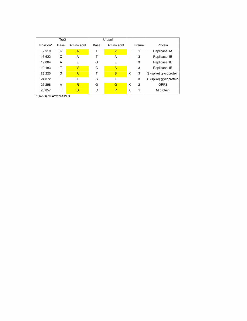
Purification of viral particles and RNA, and DNAsequencing. Virus isolation was performed on abronchoalveolar lavage specimen of a fatal SARS casebelonging to the original case cluster from Toronto, Canada.Viral particles from this Tor2 isolate were purified and thegenetic material (RNA) was extracted (12) from the Tor2isolate (13). The RNA was converted to cDNA using acombined random-priming and oligo-dT priming strategy(12). Size selected cDNA products were cloned and singlesequence reads were generated from each end of the insertfrom randomly picked clones. Sequences were assembled andthe assembly edited to produce a draft sequence of the viralgenome on April 12, 2003 (12). RACE (12) was performed tocapture the 5´ end of the viral genome. The SARSgenomic sequence has been deposited intoGenbank (Accession AY274119.3). The finalsequence we produced (also available as Release 3;www.bcgsc.bc.ca) is essentially identical to that releasedindependently by the CDC (14). We report additional bases inthe Tor2 sequence that correspond to the 3´ (encoded) polyAtail. Eight base differences between the two sequences couldrepresent sequencing errors, PCR artifacts or mutable sites inthe genome. The differences we detect between our sequenceand that of the CDC are summarized in Table 1.
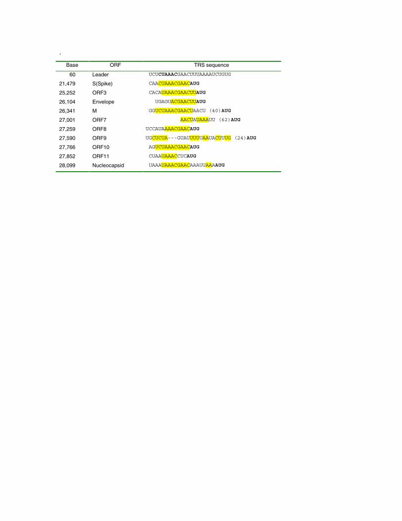
Non-protein coding features of the Tor2 SARS-CoVgenome sequence. At the 5´ end of the genome we detected aputative 5´ leader sequence with similarity to the conservedcoronavirus core leader sequence, 5´-CUAAAC-3 (5, 6).Putative TRS sequences were determined through manualalignment of sequences upstream of potential initiatingmethionine codons (see below) to the region of thecoronavirus genome sequence containing the leader sequence(Table 2). Candidate TRS sequences were scored as strong,weak or absent based on inspection of the alignments.
The 3´ UTR sequence contains a 32 base-pair regioncorresponding to the conserved s2m motif (15). The s2mmotif is believed to be a universal feature of astroviruses thathas also been identified in avian infectious bronchitis virus(avian IBV) and the ERV-2 equine rhinovirus. The highdegree of conservation between the s2m motifs in thesedifferent viruses and their evolutionary distance suggests thatthe avian IBV and ERV-2 have acquired the s2m motifthrough separate horizontal RNA transfer events (15). Theinferred distance of the SARS coronavirus to IBV from our
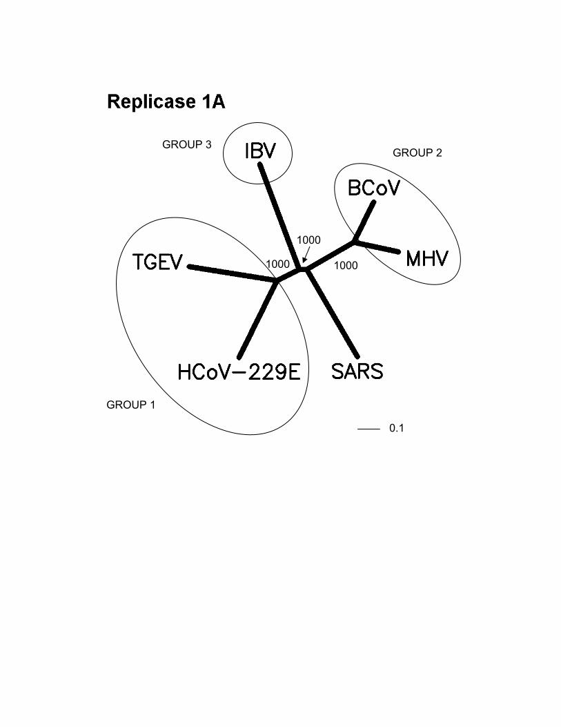
phylogenetic analysis (Fig. 1) would also suggest that theSARS coronavirus has obtained its s2m motif through ahorizontal transfer event.
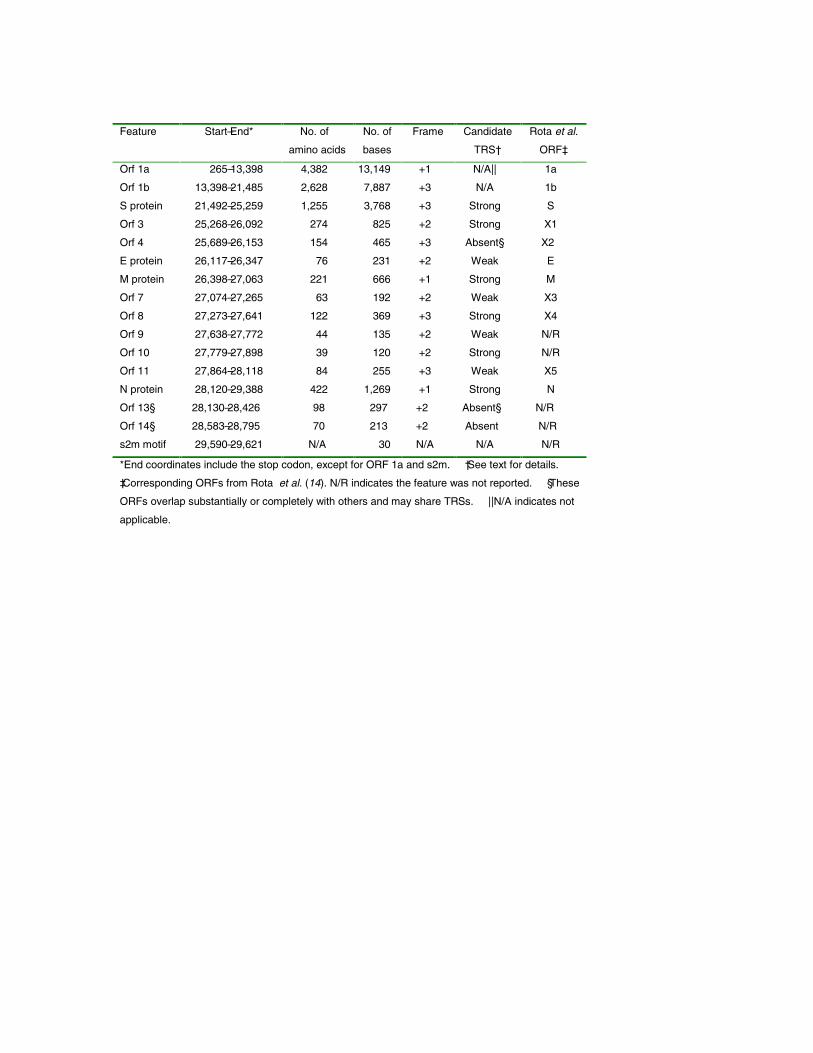
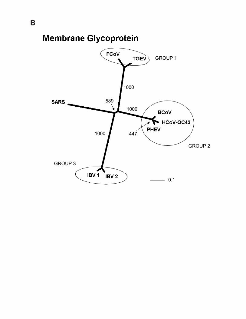
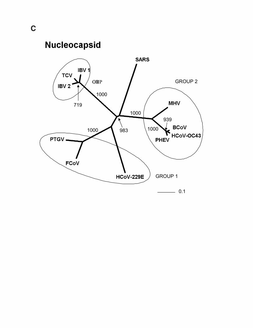
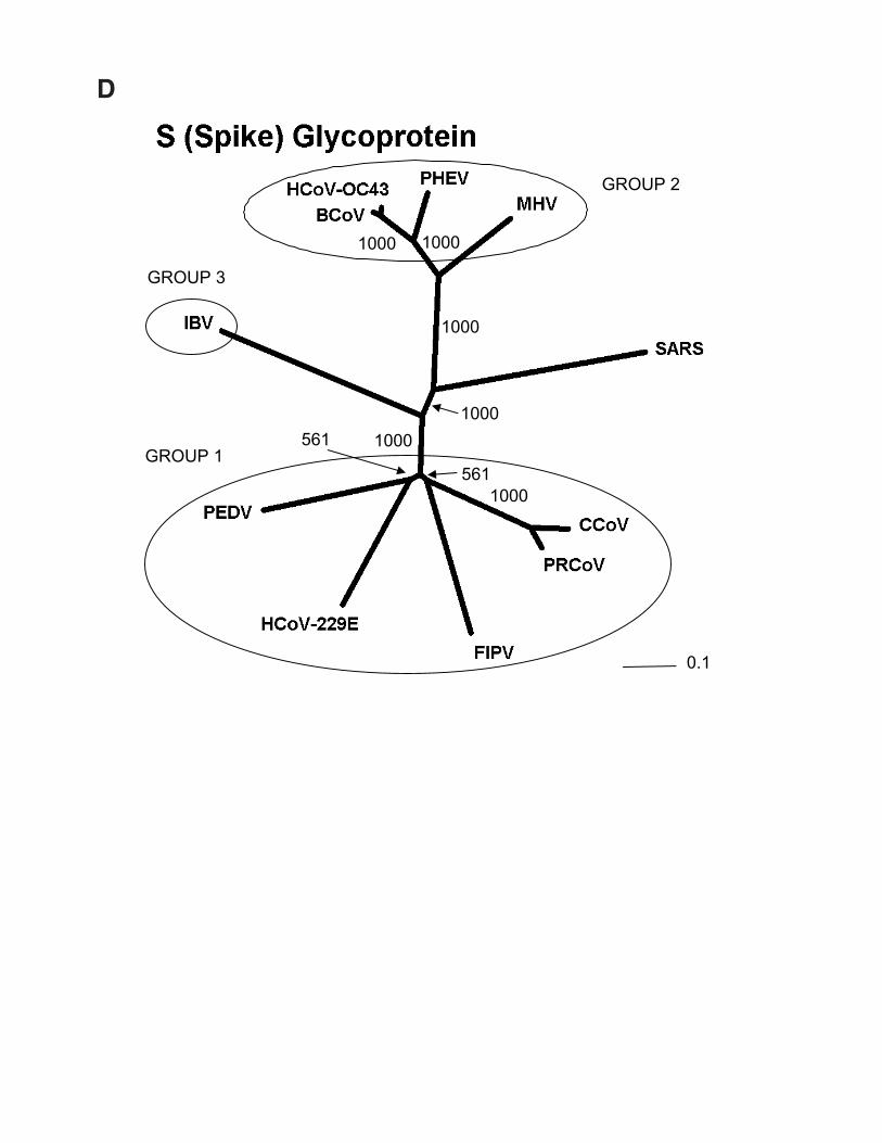
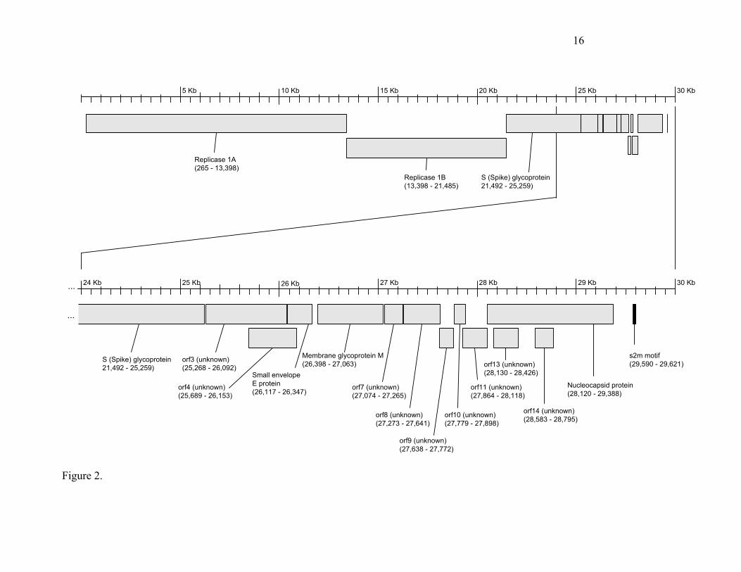
Predicted protein coding features of the Tor2 SARS-CoV genome sequence. Open reading frames weredetermined initially through sequence similarity to knowncoronavirus proteins. This approach identified replicases 1aand 1b, the S protein, the E protein, the M protein and the Nprotein. Orfs that did not match database sequences wereidentified if they were larger than 40 amino acids, unless astrong match to the TRS consensus was found close to andupstream of the potential initiating methionine residue. Wenote that Rota et al. (14) did not identify potential proteins ofless than 50 amino acids. We attempted to identify putativeTRSs upstream of all Orfs, both known and predicted (Tables2 and 3). However, TRSs are not required for transcription ofall coronavirus genes, as internal initiation from larger RNAtranscripts is also able to facilitate translation (16, 17).Certain Orfs overlap (Orf 10 and 11, by 12 amino acids; Fig.2), and some are contained entirely within another Orf or Orfs(Orf 4 and Orfs 13 and 14; Fig. 2). The biological relevanceof these Orf predictions remains to be established, but in thecases of Orfs 10 and 11, we detect strong matches to the TRSconsensus in close proximity to their respective initiatingmethionine codons (Table 2). Construction of unrootedphylogenetic trees using the set of known proteins andrepresentatives of the three known coronaviral groups revealsthat the proteins encoded by the SARS virus do not readilycluster more closely with any one group (Fig. 1). Hence, wepropose that this isolate be considered the first representativeof “Group 4” coronaviruses.
The coding potential of the 29,751-base genome isdepicted in Fig. 2. Recognizable open reading frames includethe replicase 1a and 1b translation products, the Sglycoprotein, the E protein, the M protein and the N protein.We have, in addition, conducted a preliminary analysis of thenine novel Orfs, in an attempt to ascribe to them a possiblefunctional role. These analyses are summarized below.
The replicase 1a (265-13,398 bp) and 1b Orfs (13,398 –21,485 bp) occupy 21.2 kb of the SARS virus genome (Fig.2). Conserved in both length and amino acid sequence toother coronavirus replicase proteins, the genes encode anumber of proteins that are produced by proteolytic cleavageof a large polyprotein (18). As seen in other coronavirusesand as anticipated, a frame shift interrupts the protein-codingregion, separating the 1a and 1b reading frames.
The S (Spike glycoprotein) (Fig. 2; 21,492 to 25,259 bp)encodes a surface projection glycoprotein precursor predictedto be 1,255 amino acids in length. Mutations in this gene havepreviously been correlated with altered pathogenesis andvirulence in other coronaviruses (4). In some coronaviruses,the mature spike protein is inserted in the viral envelope withthe majority of the protein exposed on the surface of the viralparticles. It is believed that three molecules of the Spikeprotein form the characteristic peplomers or corona-likestructures of this virus family. Our analysis of the spikeglycoprotein with SignalP (19) reveals a high probability of asignal peptide (probability 0.996) with cleavage betweenresidues 13 and 14. TMHMM (20) reveals a strongtransmembrane domain near the C-terminal end. Togetherthese data predict a type I membrane protein with the N-terminus and the majority of the protein (residues 14-1195)on the outside of the cell-surface or virus particle, inagreement with other coronavirus spike protein data.Supporting this conclusion, it has recently been shown that
/ www.sciencexpress.org / 1 May 2003 / Page 3/ 10.1126/science.1085953
for HCoV-229E virions, residues 417-546 are required forbinding to the cellular receptor, aminopeptidase N (21).However it is known that various coronaviruses use differentreceptors, hence it is likely that different receptor bindingsites are also used.
Orf 3 (Fig. 2; 25,268 - 26,092) encodes a predicted proteinof 274 amino acids that lacks significant BLAST (22),FASTA (23) or PFAM (24) similarities to any known protein.Analysis of the N-terminal 70 amino acids with SignalPprovides weak evidence for the existence of a signal peptideand a cleavage site (probability 0.540). Both TMpred (25)and TMHMM predict the existence of three trans-membraneregions spanning approximately residues 34-56, 77-99, and103-125. The most likely model from these analyses is thatthe C-terminus and a large 149 amino - terminal domainwould be located inside the viral or cellular membrane. TheC-terminal (interior) region of the protein may encode aprotein domain with ATP-binding properties (PD037277).
Orf 4 (Fig. 2; 25,689 - 26,153) encodes a predicted proteinof 154 amino acids. This Orf overlaps entirely with Orf 3 andthe E protein. Our analysis failed to locate a potential TRSsequence at the 5´ end of this putative Orf. However, it ispossible this protein is expressed from the Orf3 mRNA usingan internal ribosomal entry site. BLAST analyses failed toidentify matching sequences. Analysis with TMPred predictsa single transmembrane helix.
The small envelope protein E (Fig. 2; 26,117 - 26,347)encodes a predicted protein of 76 amino acids. BLAST andFASTA comparisons indicate that the predicted proteinexhibits significant matches to multiple envelope(alternatively known as small membrane) proteins fromseveral coronaviruses. PFAM analysis of the protein revealsthe predicted protein is a member of the well-characterizedNS3_EnvE protein family (24). InterProScan (26, 27)analysis reveals that the protein is a component of the viralenvelope, and conserved sequences are also found in otherviruses, including gastroenteritis virus and murine hepatitisvirus. SignalP analysis predicts the presence of atransmembrane anchor (probability 0.939). TMpred analysisof the predicted protein revealed a similar trans-membranedomain at positions 17-34, consistent with the knownassociation of this protein with the viral envelope. TMHMMpredicts a type II membrane protein with the majority of thehydrophilic domain (46 residues) and C terminus to belocated on the surface of the viral particle. We note that insome coronaviruses such as transmissible gastroenteritiscoronavirus (TGEV) the E protein is essential for virusreplication (28) while in mouse hepatitis virus (MHV) it hasbeen shown that although deletion of gene E reduces virusreplication by more than ten thousand fold, the virus still canreplicate (29).
The Membrane glycoprotein M (Fig. 2; 26,398 - 27,063)encodes a predicted protein of 221 amino acids. BLAST andFASTA analysis of the protein revealed significant matchesto a large number of coronaviral matrix glycoproteins. Theassociation of the spike glycoprotein (S) with the matrixglycoprotein (M) is an essential step in the formation of theviral envelope and in the accumulation of both proteins at thesite of virus assembly (4). Analysis of the amino acidsequence with SignalP predicts a signal sequence (probability0.932) that is not likely cleaved. TMHMM and TMpredanalysis both indicate the presence of three trans-membranehelices, located at approximately residues 15-37, 50-72 and77-99, with the 121 amino acid hydrophilic domain on theinside of the virus particle, where it is believed to interact
with the nucleocapsid. PFAM analysis reveals a match toPFAM domain PF01635, and alignments to 85 othersequences in the PFAM database bearing this domain, whichis indicative of the coronavirus matrix glycoprotein.
Orf7 (Fig. 2; 27,074-27,265) encodes a predicted proteinof 63 amino acids. BLAST and FASTA searches yield nosignificant matches indicative of function. TMHMM andSignalP predict no transmembrane region, however, TMpredanalysis predicts a likely trans-membrane helix locatedbetween residues 3 and 22, with the N-terminus locatedoutside the viral particle. Similarly, the gene encoding Orf8(Fig. 2; 27,273-27,641) encoding a predicted protein of 122amino acids, has no significant BLAST or FASTA matches toknown proteins. Analysis of this sequence with SignalPindicates a cleaved signal sequence (probability 0.995), withthe predicted cleavage site located between residues 15 and16. TMpred and TMHMM analysis also predicts a trans-membrane helix located approximately at residues 99-117.Together these data indicate that Orf7 is likely to be a type Imembrane protein with the major hydrophilic domain of theprotein (residues 16-98) and the amino-terminus are orientedinside the lumen of the ER/Golgi, or on the surface of the cellmembrane or virus particle, depending on the membranelocalization of the protein.
Orf9 (Fig. 2; 27,638-27,772) encodes a predicted proteinof 44 amino acids. FASTA analysis of this sequence revealedsome weak similarities (37% identity over a 35 amino acidoverlap) to Swiss-Prot accession Q9M883, annotated as aputative sterol-C5 desaturase. A similarly weak match to ahypothetical Clostridium perfringens protein (Swiss-Protaccession CPE2366) was also detected. The functionalimplications, if any, of these matches are unknown. TMpredpredicted the existence of a single strong trans-membranehelix, with little preference for alternate models in which theN-terminus was located inside or outside the particle.Similarly Orf10 (Fig. 2; 27,779-27,898) encoding a predictedprotein of 39 amino acids, exhibited no significant matches inBLAST and FASTA searches but was predicted to encode atrans-membrane helix by TMPred, with the N-terminuslocated within the viral particle. The region immediatelyupstream of Orf10 exhibits a strong match to the TRSconsensus (Table 2), providing support for the notion that atranscript initiates from this site. Orf11 (Fig. 2; 27,864-28118) encodes a predicted protein of 84 amino acidsexhibited only very short (9-10 residues) matches to a regionof the human coronavirus E2 glycoprotein precursor (startingat residue 801). Analysis by SignalP and TMHMM predict asoluble protein. As was the case for Orf 10, a detectablealignment to the TRS consensus sequence was found (Table2).
The protein (422 amino acids) encoded by theNucleocapsid gene (Fig. 2; 28,120-29,388) aligns well withnucleocapsid proteins from other representativecoronaviruses, although a short lysine rich region(KTFPPTEPKKDKKKKTDEAQ) (30) appears to be uniqueto SARS. This region is suggestive of a nuclear localizationsignal, and while it contains a hit to InterProDomainIPR001472 (bipartite nuclear localization signal), the functionof this insertion remains unknown. It is possible that theSARS virus nucleocapsid protein has a novel nuclearfunction, which could play a role in pathogenesis. In addition,the basic nature of this peptide suggests it may assist in RNAbinding.
Orf 13 (Fig. 2; 28,130 – 28,426) encodes a predictedprotein of 98 amino acids. BLAST analysis failed to identify
/ www.sciencexpress.org / 1 May 2003 / Page 4/ 10.1126/science.1085953
similar sequences, and no transmembrane helices arepredicted. Orf 14 (Fig. 2; 28,583 – 28,795) encodes apredicted protein of 70 amino acids. BLAST analysis failed toidentify similar sequences. TMPred predicts a singletransmembrane helix.
Conclusions. We have determined through genomesequencing that the virus named by the WHO as causallyassociated with SARS is a novel coronavirus. This has beenconfirmed by the sequence of two independent isolates, theTor2 isolate reported here and the Urbani isolate, reported bythe CDC (14). Although morphologically a coronavirus (2),this SARS virus is not more closely related to any of the threeknown classes of coronavirus, and we propose that it definesa fourth class of coronavirus (Group 4) and that it be referredto as SARS-CoV. Our sequence data do not support a recentinter-viral recombination event between the knowncoronavirus groups in the etiology of this virus, but this maybe due to the limited number of known coronavirus genomesequences. Apart from the s2m motif located in the 3´UTR,there is also no evidence of any exchange of genetic materialbetween the SARS virus and non-Coronaviridae. These dataare consistent with the hypothesis that an animal virus forwhich the normal host is currently unknown recently mutatedand developed the ability to productively infect humans.There also remains the possibility that the SARS virusevolved from a previously harmless human coronavirus.However, preliminary evidence suggests antibodies to thisvirus are absent in those not infected with SARS-CoV (2),implying a benign virus closely related to the Tor2 isolate isnot resident in humans. Identification of the normal host ofthis coronavirus and comparison of the sequences of theancestral and SARS forms will further elucidate the processby which this virus arose.
Availability of the SARS virus genome sequence isimportant from a public health perspective. It will allow therapid development of PCR-based assays for this virus thatcapitalize on novel sequence features allowing thediscrimination between this and other circulatingcoronaviruses. Such assays will allow the diagnosis of SARSvirus infection in humans and, critically, will consolidate theassociation of this virus with SARS. If the association isfurther borne out, SARS virus genome-based PCR assaysmay form an important part of a public health strategy tocontrol the spread of this syndrome. In the longer term, thisinformation will assist in the development of antiviraltreatments including neutralizing antibodies and developmentof a vaccine to treat this emerging and deadly new disease.
References and Notes1. J. S. M. Peiris et al., Lancet, published online 8 April 2003
(http://image.thelancet.com/extras/03art3477web.pdf).2. T. G. Ksiazek et al., N. Engl. J. Med., published online 10
April 2003 (10.1056/NEJMoa030781).3. R. Munch, Microbes Infect. 5, 69 (2003).4. B. N. Fields, D. M. Knipe, P. M. Howley, D. E. Griffin,
Fields Virology (Lippincott Williams & Wilkins,Philadelphia, ed. 4, 2001).
5. M. M. C. Lai, D. Cavanagh, Adv. Virus Res. 48, 1 (1997).6. S. G. Sawicki, D. L. Sawicki, Adv. Exp. Med. Biol. 440,
215 (1998).7. D. L. Sawicki et al., J. Gen. Virol. 82, 386 (2001).8. S. G. Sawicki, D. L. Sawicki, J. Virol. 64, 1050 (1990).9. M. Schaad, R. S. J. Baric, J. Virol. 68, 8169 (1994).10. P. B. Sethna et al., Proc. Natl. Acad. Sci. U.S.A. 86, 5626
(1989).
11. L. Enjuanes et al., in Virus Taxonomy. Classification andNomenclature of Viruses, M. H. V. van Regenmortel et al.,Eds. (Academic Press, New York, 2000), pp. 835–849.
12. Information on materials and methods is available onScience Online.
13. S. M. Poutanen et al., N. Engl. J. Med., published online31 March 2003 (10.1056/NEJMoa030634).
14. P. A. Rota et al., Science, in press; published online 1May 2003 (10.1126/science.1085952).
15. C. M. Jonassen, T. O. Jonassen, B. Grinde, J. Gen. Virol.79, 715 (1998).
16. W. Lapps, B. G. Hogue, D. A. Brian, Virology 157, 47(1987).
17. R. Krishnan, R. Y. Chang, D. A. Brian, Virology 218, 400(1996).
18. J. Ziebuhr, E. J. Snijder, A. E. Gorbalenya, J. Gen. Virol.81, 853 (2000).
19. H. Nielsen, J. Engelbrecht, S. Brunak, G. von Heijne,Protein Eng. 10, 1 (1997).
20. E. L. Sonnhammer, G. von Heijne, A. Krogh, Proc. Int.Conf. Intell. Syst. Mol. Biol. 6, 175 (1998).
21. J. C. Tsai, B. D. Zelus, K. V. Holmes, S. R. Weiss, J.Virol. 77, 841 (2003).
22. S. F. Altschul et al., Nucleic Acids Res. 25, 3389 (1997).23. W. R. Pearson, D. J. Lipman, Proc. Natl. Acad. Sci.
U.S.A. 85, 2444 (1988).24. A. Bateman et al., Nucleic Acids Res. 30, 276 (2002).25. K. Hofman, W. Stoffel, Biol. Chem. Hoppe-Seyler 374,
166 (1993).26. R. Apweiler et al., Nucleic Acids Res. 29, 37 (2001).27. E. M. Zdobnov, R. Apweiler, Bioinformatics 17, 847
(2001).28. J. Ortego et al., J. Virol. 76, 11518 (2002).29. L. Kuo et al., paper presented at the annual meeting of the
American Society for Virology, Lexington, KY, 20 to 24July 2002.
30. Abbreviations for amino acids: A, Ala; C, Cys; D, Asp; E,Glu; F, Phe; G, Gly; H, His; I, Ile; K, Lys; L, Leu; M, Met;N, Asn; P, Pro; Q, Gln; R, Arg; S, Ser; T, Thr; V, Val; W,Trp; and Y, Tyr.
31. J. D. Thompson, D. G. Higgins, T. J. Gibson, NucleicAcids Res. 22, 4673 (1994).
32. J. Felsenstein, PHYLIP (Phylogeny Inference Package)version 3.5c (1993). Distributed by the author, Departmentof Genetics, University of Washington, Seattle.
33. We would like to thank all the staff at the BCCA GenomeSciences Centre for helping to facilitate the rapidsequencing of the SARS-CoV genome. We would like toacknowledge Raymond Tellier, Hospital for Sick Children,for providing us with information on primer sequences thatamplify a 216 base-pair region of the Pol gene. We alsothank Ivan Sadowski, Dept. Biochemistry and MolecularBiology and John Hobbs and his staff at the Nucleic Acidand Protein Services Unit at the University of BritishColumbia for rapid synthesis of PCR primers. We alsoacknowledge the advice and assistance of FrancisOuellette, University of British Columbia BioinformaticsCentre and the staff at the National Center forBiotechnology Information for rapidly processing andmaking available our sequence data. We are indebted toanonymous reviewers who provided useful criticisms. TheBritish Columbia Cancer Agency Genome Sciences Centreis supported by the BC Cancer Foundation, GenomeCanada/Genome British Columbia, Western EconomicDiversification, Canada Foundation for Innovation, BC
/ www.sciencexpress.org / 1 May 2003 / Page 5/ 10.1126/science.1085953
Knowledge Development Fund, Canadian Institutes ofHealth Research, the Michael Smith Foundation for HealthResearch, and the Natural Sciences and EngineeringResearch Council of Canada. Clones derived from theSARS virus are available from the Genome SciencesCentre (www.bcgsc.bc.ca).
Supporting Online Materialwww.sciencemag.org/cgi/content/full/1085953/DC1Materials and MethodsReferences
19 April 2003; accepted 30 April 2003Published online 1 May 2003; 10.1126/science.1085953Include this information when citing this paper.
Fig. 1. Phylogenetic analysis of SARS proteins. Unrootedphylogenetic trees were generated by clustalw 1.74 (31) usingthe BLOSUM comparison matrix and a bootstrap analysis of1000 iterations. Numbers indicate bootstrap replicatessupporting each node. Phylogenetic trees were drawn with thePhylip Drawtree program 3.6a3 (32). Branch lengths indicatethe number of substitutions per residue. Genbank accessionsfor protein sequences: (A) Replicase 1A: BoCoV (BovineCoronavirus): AAL40396, HCoV-229E (HumanCoronavirus): NP_07355, MHV (Mouse Hepatitis Virus):NP_045298, IBV (Avian Infectious bronchitis virus):CAC39113, TGEV (Transmissible Gastroenteritis Virus):NP_058423. (B) Membrane Glycoprotein: PHEV (Porcinehemagglutinating encephalomyelitis virus): AAL80035,BoCoV (Bovine Coronavirus): NP_150082, IBV & IBV2(Avian infectious bronchitis virus): AAF35863 &AAK83027, MHV (Mouse hepatitis virus): AAF36439,TGEV (Transmissible gastroenteritis virus): NP_058427,HCoV-229E & HCoV-OC43 (Human Coronavirus):NP_073555 & AAA45462, FCoV (Feline coronavirus):BAC01160. (C) Nucleocapsid: MHV (Mouse hepatitis virus):P18446, BoCoV (Bovine coronavirus): NP_150083, IBV 1 &2 (Avian infectious bronchitis virus): AAK27162 &NP_040838, FCoV (Feline coronavirus): CAA74230, PTGV(Porcine transmissible gastroenteritis virus): AAM97563,HCoV-229E & HCoV-OC43 (Human coronavirus):NP_073556 & P33469, PHEV (porcine hemagglutinatingencephalomyelitis virus): AAL80036, TCV (Turkeycoronavirus): AAF23873. (D) S (Spike) Protein: BoCoV(Bovine coronavirus): AAL40400, MHV (Mouse hepatitisvirus): P11225, HCoV-OC43 & HCoV-229E (Humancoronavirus): S44241 & AAK32191, PHEV (Porcinehemagglutinating encephalomyelitis virus): AAL80031,PRCoV (Porcine respiratory coronavirus): AAA46905,PEDV (Porcine epidemic diarrhea virus): CAA80971, CCoV(Canine coronavirus): S41453, FIPV (Feline infectiousperitonitis virus): BAA06805, IBV (Avian infectiousbronchitis virus): AAO34396.
Fig. 2. Map of the predicted Orfs and s2m motif in the Tor2SARS virus genome sequence.
Table 1. Nucleotide base differences between the Tor2sequence and the Urbani sequence [(14),www.cdc.gov/ncidod/sars/sequence.htm]. Yellow indicates abase difference resulting in an amino acid change (30); Xindicates a non-conservative amino acid substitution.
Table 2. The nucleotide position, associated open-readingframe and putative transcription regulatory sequences (seetext for details). Numbers in parentheses within the alignmentindicate distance to the putative initiating codon. Theconserved core sequence is indicated in bold in the putativeleader sequence. Contiguous sequences identical to region ofthe leader sequence containing the core sequence are colored.No putative TRSs were detected for Orfs 4, 13 and 14,although Orf 13 could share the TRS associated with the Nprotein.
Table 3. Features of the Tor2 genome sequence.
Tor2 Urbani
Position* Base Amino acid Base Amino acid Frame Protein
7,919 C A T V 1 Replicase 1A
16,622 C A T A 3 Replicase 1B
19,064 A E G E 3 Replicase 1B
19,183 T V C A 3 Replicase 1B
23,220 G A T S X 3 S (spike) glycoprotein
24,872 T L C L 3 S (spike) glycoprotein
25,298 A R G G X 2 ORF3
26,857 T S C P X 1 M protein
*GenBank AY274119.3.
.
Base ORF TRS sequence
60 Leader UCUCUAAACGAACUUUAAAAUCUGUG
21,479 S(Spike) CAACUAAACGAACAUG
25,252 ORF3 CACAUAAACGAACUUAUG
26,104 Envelope UGAGUACGAACUUAUG
26,341 M GGUCUAAACGAACUAACU (40)AUG
27,001 ORF7 AACUAUAAAUU (62)AUG
27,259 ORF8 UCCAUAAAACGAACAUG
27,590 ORF9 UGCUCUA---GUAUUUUUAAUACUUUG (24)AUG
27,766 ORF10 AGUCUAAACGAACAUG
27,852 ORF11 CUAAUAAACCUCAUG
28,099 Nucleocapsid UAAAUAAACGAACAAAUUAAAAUG
Feature Start–End* No. of
amino acids
No. of
bases
Frame Candidate
TRS†
Rota et al.
ORF‡
Orf 1a 265–13,398 4,382 13,149 +1 N/A|| 1a
Orf 1b 13,398–21,485 2,628 7,887 +3 N/A 1b
S protein 21,492–25,259 1,255 3,768 +3 Strong S
Orf 3 25,268–26,092 274 825 +2 Strong X1
Orf 4 25,689–26,153 154 465 +3 Absent§ X2
E protein 26,117–26,347 76 231 +2 Weak E
M protein 26,398–27,063 221 666 +1 Strong M
Orf 7 27,074–27,265 63 192 +2 Weak X3
Orf 8 27,273–27,641 122 369 +3 Strong X4
Orf 9 27,638–27,772 44 135 +2 Weak N/R
Orf 10 27,779–27,898 39 120 +2 Strong N/R
Orf 11 27,864–28,118 84 255 +3 Weak X5
N protein 28,120–29,388 422 1,269 +1 Strong N
Orf 13§ 28,130–28,426 98 297 +2 Absent§ N/R
Orf 14§ 28,583–28,795 70 213 +2 Absent N/R
s2m motif 29,590–29,621 N/A 30 N/A N/A N/R
*End coordinates include the stop codon, except for ORF 1a and s2m. †See text for details.
‡Corresponding ORFs from Rota et al. (14). N/R indicates the feature was not reported. §These
ORFs overlap substantially or completely with others and may share TRSs. ||N/A indicates not
applicable.
GROUP 3
GROUP 1
GROUP 2
0.1
1000
1000 1000
GROUP 3
GROUP 2
GROUP 1
0.1
1000
1000
1000
589
447
B
GROUP 2
GROUP 1
GROUP 3
0.1
1000
1000
1000
1000939
983
719
C
GROUP 1
GROUP 3
GROUP 2
0.1
1000
1000
1000
1000 1000
1000
561
561
D
16
20 Kb15 Kb5 Kb 10 Kb 30 Kb25 Kb
...
28 Kb27 Kb25 Kb 26 Kb 30 Kb29 Kb24 Kb...
Small envelopeE protein(26,117 - 26,347)
orf3 (unknown)(25,268 - 26,092)
S (Spike) glycoprotein21,492 - 25,259)
Membrane glycoprotein M(26,398 - 27,063)
orf7 (unknown)(27,074 - 27,265)
orf8 (unknown)(27,273 - 27,641)
orf9 (unknown)(27,638 - 27,772)
orf10 (unknown)(27,779 - 27,898)
orf11 (unknown)(27,864 - 28,118)
Nucleocapsid protein(28,120 - 29,388)
s2m motif(29,590 - 29,621)
Replicase 1A(265 - 13,398)
Replicase 1B(13,398 - 21,485)
S (Spike) glycoprotein21,492 - 25,259)
orf4 (unknown)(25,689 - 26,153)
orf14 (unknown)(28,583 - 28,795)
orf13 (unknown)(28,130 - 28,426)
Figure 2.