Embed Size (px)
Citation preview
The genome sequence of the corn snake (Pantherophis guttatus), a valuable resource for
EvoDevo studies in squamates ASIER ULLATE-AGOTE1,2,3, MICHEL C. MILINKOVITCH1,2,3 and ATHANASIA C. TZIKA1,2,3,*
1Laboratory of Artificial & Natural Evolution (LANE), Dept. of Genetics & Evolution, University of Geneva, 2SIB Swiss Institute of Bioinformatics and 3Institute of Genetics and Genomics of Geneva (iGE3), University of Geneva, Geneva, Switzerland
ABSTRACT Squamates (snakes and lizards) exhibit a striking variety of phenotypes, with little known on their generative mechanisms. Studies aiming to understand the genetic basis of this wide diversity in morphology, physiology and ecology will greatly benefit from whole genome sequencing initiatives, as they provide the foundation for comparative analyses and improve our understanding of the evolution, development and diversification of traits. Here, we present the first draft genome of the corn snake Pantherophis guttatus, an oviparous snake that we promote as a particularly appropriate model species for evolutionary developmental studies in squamates. We sequenced 100-base paired-end reads from multiple individuals of a single family (parents and offspring) that produced a genome assembly of 1.53 gigabases (Gb), roughly covering 75% of the expected total genome size, and 297,768 scaffolds >1 Kb. We were able to fully retrieve 86, and par-tially another 106, of the 248 CEGMA core genes, indicating that a high genome completeness was achieved, even though the assembly is fragmented. Using MAKER2, we annotated 10,917 genes with high confidence (Annotation Edit Distance (AED)<1) and an additional 5,263 predicted genes matched with the species’ transcriptome. Numerous colour and colour pattern morphs exist in P. guttatus, making it an ideal model to study the genetic determinism, development, and evolution of adaptive colour traits in reptiles. Using our draft genome and a Single-Nucleotide Polymorphism (SNP) calling approach, we confirmed the interval with the causative mutation for the amelanistic phenotype, a result supported by a parallel exome-based study.
KEY WORDS: Pantherophis guttatus, corn snake, reptile, genome, amelanistic, SNP calling
Introduction
An increasing number of non-classical vertebrate genomes have been sequenced during the last decade, facilitating evolutionary and comparative developmental studies (Castoe et al., 2013, Vonk et al., 2013, Wang et al., 2013). However, the order Squamata is still largely underrepresented in these initiatives, despite the wide variety of phenotypes encountered within its 10,000 species (i.e., about twice as many as in mammals). In the last few years, the publications of the Anolis carolinensis genome (Alfoldi et al., 2011), the first draft (Castoe et al., 2011) and the improved version (Cas-toe et al., 2013) of the Burmese python (Python molurus) genome
Int. J. Dev. Biol. 58: 881-888 (2014)doi: 10.1387/ijdb.150060at
www.intjdevbiol.com
*Address correspondence to: Athanasia C. Tzika. Laboratory of Artificial & Natural Evolution (LANE), Dept. of Genetics & Evolution, University of Geneva, Sciences III, 30, Quai Ernest-Ansermet, 1211 Genève 4, Switzerland. Tel: +41(0)22 379 67 85. Fax: +41(0)22 379 67 95. E-mail: [email protected] - Web: www.lanevol.org
Supplementary Material (tables, Figure, XLS files, FASTA files) for this paper is available at: http://dx.doi.org/10.1387/ijdb.150060at - See note at end of paper for a more detailed description of the files.
Accepted: 9 February 2015.
ISSN: Online 1696-3547, Print 0214-6282© 2015 UBC Press (Bilbao, Spain) and Creative Commons CC-BY. This is an open access article distributed under the terms of the Creative Commons Attribution License (http://creativecommons.org/licenses/), which permits you to Share (copy and redistribute the material in any medium or format) and Adapt (remix, transform, and build upon the material for any purpose, even commercia-lly), providing you give appropriate credit, provide a link to the license, and indicate if changes were made. You may do so in any reasonable manner, but not in any way that suggests the licensor endorses you or your use. Printed in Spain
Abbreviations used in this paper: AED, annotation edit distance; BLAST, basic local alignment search tool; CEGMA, core eukaryotic genes mapping approach; CR1, chicken repeat 1; DCT, dopachrome tautomerase; EST, expressed sequence tag; LINE, long interspersed nuclear elements; LTR, long terminal repeat; MIR, mammalian-wide interspersed repeats; Mya, millions years ago; OCA2, oculocu-taneous albinism II; RTE, retro-transposable element; SINE, short interspersed nuclear element; SNP, single-nucleotide polymorphism; amel, amelanistic.
together with draft genomes of the king cobra Ophiophagus hannah (Vonk et al., 2013), of the rattlesnake Crotalus mitchellii (Gilbert et al., 2014) and of the common viper Vipera berus demonstrate an increased interest in Squamata in general, and snakes in particular,
882 A. Ullate-Agote et al.
as they are good models for investigating the mechanisms associ-ated to their modified limb and body plan development (Di-Poi et al., 2010, Woltering, 2012), venom evolution (Vonk et al., 2013), physicochemical sensory perception (Brykczynska et al., 2013), extreme fluctuations in metabolic rates (Castoe et al., 2013), as well as development and patterning of scales and colours.
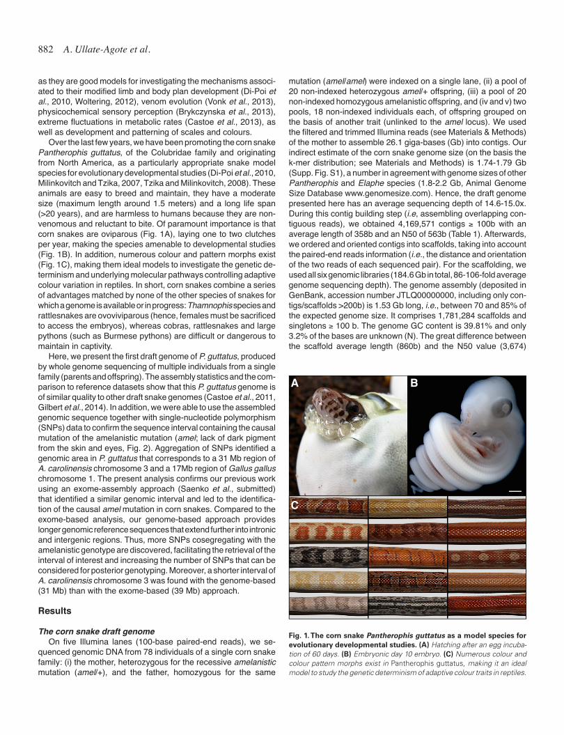
Over the last few years, we have been promoting the corn snake Pantherophis guttatus, of the Colubridae family and originating from North America, as a particularly appropriate snake model species for evolutionary developmental studies (Di-Poi et al., 2010, Milinkovitch and Tzika, 2007, Tzika and Milinkovitch, 2008). These animals are easy to breed and maintain, they have a moderate size (maximum length around 1.5 meters) and a long life span (>20 years), and are harmless to humans because they are non-venomous and reluctant to bite. Of paramount importance is that corn snakes are oviparous (Fig. 1A), laying one to two clutches per year, making the species amenable to developmental studies (Fig. 1B). In addition, numerous colour and pattern morphs exist (Fig. 1C), making them ideal models to investigate the genetic de-terminism and underlying molecular pathways controlling adaptive colour variation in reptiles. In short, corn snakes combine a series of advantages matched by none of the other species of snakes for which a genome is available or in progress: Thamnophis species and rattlesnakes are ovoviviparous (hence, females must be sacrificed to access the embryos), whereas cobras, rattlesnakes and large pythons (such as Burmese pythons) are difficult or dangerous to maintain in captivity.
Here, we present the first draft genome of P. guttatus, produced by whole genome sequencing of multiple individuals from a single family (parents and offspring). The assembly statistics and the com-parison to reference datasets show that this P. guttatus genome is of similar quality to other draft snake genomes (Castoe et al., 2011, Gilbert et al., 2014). In addition, we were able to use the assembled genomic sequence together with single-nucleotide polymorphism (SNPs) data to confirm the sequence interval containing the causal mutation of the amelanistic mutation (amel; lack of dark pigment from the skin and eyes, Fig. 2). Aggregation of SNPs identified a genomic area in P. guttatus that corresponds to a 31 Mb region of A. carolinensis chromosome 3 and a 17Mb region of Gallus gallus chromosome 1. The present analysis confirms our previous work using an exome-assembly approach (Saenko et al., submitted) that identified a similar genomic interval and led to the identifica-tion of the causal amel mutation in corn snakes. Compared to the exome-based analysis, our genome-based approach provides longer genomic reference sequences that extend further into intronic and intergenic regions. Thus, more SNPs cosegregating with the amelanistic genotype are discovered, facilitating the retrieval of the interval of interest and increasing the number of SNPs that can be considered for posterior genotyping. Moreover, a shorter interval of A. carolinensis chromosome 3 was found with the genome-based (31 Mb) than with the exome-based (39 Mb) approach.
Results
The corn snake draft genomeOn five Illumina lanes (100-base paired-end reads), we se-
quenced genomic DNA from 78 individuals of a single corn snake family: (i) the mother, heterozygous for the recessive amelanistic mutation (amel/+), and the father, homozygous for the same
mutation (amel/amel) were indexed on a single lane, (ii) a pool of 20 non-indexed heterozygous amel/+ offspring, (iii) a pool of 20 non-indexed homozygous amelanistic offspring, and (iv and v) two pools, 18 non-indexed individuals each, of offspring grouped on the basis of another trait (unlinked to the amel locus). We used the filtered and trimmed Illumina reads (see Materials & Methods) of the mother to assemble 26.1 giga-bases (Gb) into contigs. Our indirect estimate of the corn snake genome size (on the basis the k-mer distribution; see Materials and Methods) is 1.74-1.79 Gb (Supp. Fig. S1), a number in agreement with genome sizes of other Pantherophis and Elaphe species (1.8-2.2 Gb, Animal Genome Size Database www.genomesize.com). Hence, the draft genome presented here has an average sequencing depth of 14.6-15.0x. During this contig building step (i.e, assembling overlapping con-tiguous reads), we obtained 4,169,571 contigs ≥ 100b with an average length of 358b and an N50 of 563b (Table 1). Afterwards, we ordered and oriented contigs into scaffolds, taking into account the paired-end reads information (i.e., the distance and orientation of the two reads of each sequenced pair). For the scaffolding, we used all six genomic libraries (184.6 Gb in total, 86-106-fold average genome sequencing depth). The genome assembly (deposited in GenBank, accession number JTLQ00000000, including only con-tigs/scaffolds >200b) is 1.53 Gb long, i.e., between 70 and 85% of the expected genome size. It comprises 1,781,284 scaffolds and singletons ≥ 100 b. The genome GC content is 39.81% and only 3.2% of the bases are unknown (N). The great difference between the scaffold average length (860b) and the N50 value (3,674)
Fig. 1. The corn snake Pantherophis guttatus as a model species for evolutionary developmental studies. (A) Hatching after an egg incuba-tion of 60 days. (B) Embryonic day 10 embryo. (C) Numerous colour and colour pattern morphs exist in Pantherophis guttatus, making it an ideal model to study the genetic determinism of adaptive colour traits in reptiles.
B
C
A
Pantherophis draft genome 883
shows that many of the scaffolds are short: 297,768 scaffolds are >1Kb and half of the genome size is covered by 94,091 scaffolds (L50). These statistics are similar to those of the P. molurus and C. mitchellii draft genomes.
The Core Eukaryotic Genes Mapping Approach (CEGMA (Parra et al., 2007)), defines a set of 248 highly-conserved core proteins present in a wide range of eukaryotes (from yeast to human). Our assembly includes the complete gene sequence of 86 out of the 248 CEGMA eukaryotic core genes (34.68%), a lower value com-pared to higher quality genomes (Castoe et al., 2013, Vonk et al., 2013), where more than 200 CEGMA full genes were sequenced. On the other hand, we were able to retrieve partial sequence of 192 core genes (77.42%) from our draft genome, demonstrating that genome coverage is good but fragmented.
We built a library of P. guttatus repeats with RepeatModeler and combined it with all Vertebrate repeats from RepBase (Jurka et al., 2005). The use of this combined library resulted in masking 39.14% of the corn snake genomic sequence with RepeatMas-ker-open 4.0.5 (Smit et al., 1996). We observe a highly diverse landscape of repetitive elements (Fig. 3) as in other non-avian Sauropsida (Alfoldi et al., 2011, Shedlock et al., 2007) and unlike in humans, where most of the non-longterminal-repeats (non-LTR) retrotransposons consist of L1 repeats. The most common long interspersed nuclear elements (LINEs) in the corn snake genome are homologous to the Chicken Repeat 1 (CR1, 2.81%), with L1, L2, RTE (retro-transposable element) and R4 also being abundant, as in A. carolinensis (Novick et al., 2009). The identified
short interspersed nuclear elements (SINEs) mainly belong to the MIR-like family (1.91%) and there are also a few LTR elements that are endogenous retroviruses. The distribution and variety of repetitive elements in the corn snake genome are similar to those of other snakes (Castoe et al., 2013), except that we observe a higher proportion of DNA transposons (“cut and paste DNA”, 6.45%). Finally, 16.58% of the corn snake genome corresponds to repetitive elements from unclassified groups (Fig. 3).
Genome annotation Two iterations of the MAKER2 pipeline (Holt and Yandell, 2011)
yielded 24,258 predicted genes. MAKER2 provides a set of gene predictors, performs similarity searches against cDNA/ESTs or proteins databases, and combines the outputs into a set of high-quality gene predictions. Our corn snake transcriptomic data (Tzika et al., 2015) together with a protein database (including the SwissProt repository and NCBI sequences of A. carolinensis and snakes) were used in the MAKER2 pipeline to complement the gene models computed by three ab-initio/ evidence-based predictors: AUGUSTUS (Stanke and Waack, 2003), SNAP (Korf, 2004) and GeneMark-ES (Ter-Hovhannisyan et al., 2008). As a quality measurement of our annotation, we used the ‘annotation edit distance’ (AED, (Eilbeck et al., 2009)), which is based on the agreement at the nucleotide level (i.e., focusing on overlapping nucleotides rather than substitutions) between the final gene an-notation and the aligned evidence data (cDNA/proteins) supporting that annotation. Values closer to zero correspond to higher quality annotation (i.e, it is more congruent with the cDNA or protein data) and predictions with an AED=1 are interpreted as not supported by experimental data at all. An AED <1 was found for 10,917 of the 24,258 predicted genes (45%) (Supp. Files S2-S4), with the majority of them showing an AED <0.5 (8,597, 35.4%). These rather low numbers are due to the high fragmentation of the draft corn snake genome: many genes span multiple scaffolds, making their annotation impossible. Eighty percent (8,817) of the corn snake proteins with AED < 1 had a blastp hit (e-value <10-5) against A. carolinensis (Ensembl v77 proteins), P. molurus or O. hannah
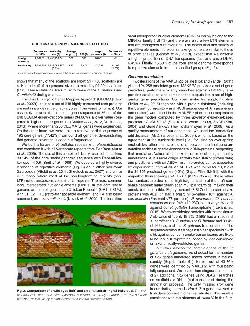
Fig. 2. Comparison of a wild-type (left) and an amelanistic (right) individual. The lack of melanin in the amelanistic individual is obvious in the eyes, around the dorso-lateral blotches, as well as by the absence of the ventral checker pattern.
sequences and 94% (10,237) had a megablast hit against our P. guttatus transcriptome (Tzika et al., 2015). When considering proteins with the maximum AED value of 1, only 19.2% (2,565) had a hit against A. carolinensis, P. molurus or O. hannah and 39.4% (5,263) against the P. guttatus transcriptome. The sequences without a hit against other species but with a hit against our corn-snake transcriptome are likely to be real cDNA/proteins, coded by less-conserved or taxonomically-restricted genes.
To further assess the completeness of the P. guttatus draft genome, we checked for the number of Hox genes annotated and/or present in the as-sembly (Suppl. Table S1). Eleven out of 40 Hox genes were identified by MAKER2, with four being fully sequenced. We located homologous sequences of 27 additional Hox genes using BLAST searches on scaffolds <10Kbp (not considered during the annotation process). The only missing Hox gene in our draft genome is Hoxd12, a gene involved in limb development in other vertebrates. This result is consistent with the absence of Hoxd12 in the fully-
Sequences ≥ 100b
Assembly size (b)
Average length (b) N50 (b)
Longest sequence (b)
Sequences ≥ 10Kb
Contig assembly
4,169,571 1,493,759,741 358 563 16,051 42
Scaffolding 1,781,284 1,532,089,567 (3,2%)
860 3,674 102,731 21,405 (1.2%)
TABLE 1
CORN SNAKE GENOME ASSEMBLY STATISTICS
In parenthesis, the percentage of unknown (N) bases is indicated. b = number of bases

884 A. Ullate-Agote et al.
sequenced clusters of posterior Hox genes in P. guttatus (Di-Poi et al., 2010) and in the P. molurus, O. hannah and other squamata genomes (Vonk et al., 2013). Given the fragmented nature of the assembly, 29 of the 39 corn snake Hox genes were located on distinct scaffolds, besides five pairs of genes found on the same scaffold, preventing the confirmation of their cluster organisation.
Identification of the genomic region with the amelanistic mutation
We performed a Single-Nucleotide Polymorphism (SNP) call-ing approach to find the locus of the amelanistic (amel) mutation. The Mendelian inheritance of this phenotype indicates that it is associated to a single recessive mutation in our captive-bred population. The homozygous amelanistic corn snake individuals can be identified by their lack of dark pigmentation (melanin) in their skin and eyes; hence, they miss both the checker pattern on the ventral skin and the black contour surrounding the red blotches on their dorsal side (Fig. 2).
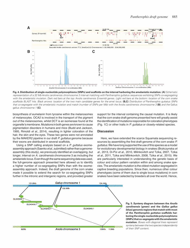
We used the parental genomic DNA libraries (male amel/amel and female amel/+) and the two libraries of their offspring pooled on the basis of their genotype (amel/amel versus amel/+). Each of the four libraries was aligned separately to our newly-assembled P. guttatus draft genome and the SNPs were extracted using Free-Bayes (Garrison and Marth, 2012). We only kept biallelic SNPs, with a defined minimum sequencing depth, that were co-segregating according to the expected genotype for the amelanistic locus in each sample (see Supp. Table S2 and Materials and Methods). This filtering resulted in 19,104 SNPs distributed on 4,740 scaffolds (on average, 4 SNPs per scaffold and 1.16 SNPs/Kb). Of these scaffolds, 751 had a megablast hit (bitscore ≥ 100) against the A. carolinensis masked genome and included 5,273 SNPs (7 SNPs per sequence). Most of the hits (59%) were against A. carolinensis Chromosome 3, and 356 of them densely covered the 82-113Mb interval (Fig. 4A and 4B): 3,735 SNPs were present in this interval and only 131 elsewhere on the chromosome. Note that the SNPs in the 82-113Mb interval are split in two islands with only 50 hits found within the 87-97Mb sub-interval (corresponding, in A. caro-linensis, to a gene desert, i.e., a region of the genome with very few protein-coding genes). As the P. guttatus and A. carolinensis lineages separated ~166.4 million years ago (Mya), it is not sur-
prising that similarity searches (BLAST matches) are inefficient in this, probably fast evolving, intergenic region.
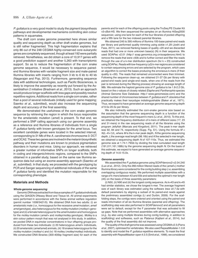
Another 61 P. guttatus scaffolds with informative SNPs aligned to a 2.2 Mb A. carolinensis scaffold (GL343243.1), which includes genes orthologous to those located in the 132.9-134.4 Mb interval of the G. gallus chromosome 1 (Fig. 5). Other parts of the G. gallus chromosome 1 adjacent to this interval are syntenic with A. caro-linensis chromosome 3. Hence, our analysis indicates that the A. carolinensis scaffold GL343243.1 should probably be inserted in A. carolinensis chromosome 3 at about the position 109Mb (more specifically between the TGFBRAP1 and MGAT4A genes), i.e., in the area discussed above where most SNPs informative for the amelanistic locus are localised. Our analysis also indicates that genes present in the 131.8-132.9 Mb interval in G. gallus chromo-some 1 are homologous to A. carolinensis genes located on three short scaffolds: GL343940.1 (0.1 Mb), GL343456.1 (0.66 Mb) and GL343542.1 (0.45 Mb). In total, 435 out of 751 (58%) of the corn snake sequences with SNPs informative for the amelanistic locus had a hit against either one of these four A. carolinensis scaffolds or the 82-113 Mb interval of A. carolinensis chromosome 3. All these hits accounted for 4,674 SNPs (89%) of the 5,273 informa-tive SNPs (10.7 SNPs per scaffold and 1.45 SNPs/Kb), strongly suggesting that the amelanistic locus is in this interval, as SNPs located close to the amelanistic locus tend to present the same co-segregating pattern as the causal mutation.
We also ran a megablast search of the 4,740 filtered scaffolds against the G. gallus masked genome (International Chicken Genome Sequencing, 2004). As snakes are more distantly re-lated to birds (~274 Mya) than to lizards (~166.4 Mya), only 291 P. guttatus scaffolds out of 4,740 (6%) had a hit (bitscore ≥ 100). These matching scaffolds include 2,530 SNPs (13.2%, 8.7 SNPs per scaffold, and 1.09 SNPs/Kb) and 226 (77.5%) of them were against G. gallus chromosome 1, with 178 in the 129.5-146 Mb interval (2,312 SNPs with a density of 1.43 SNPs/Kb; Fig. 4C). These SNPs are distributed more uniformly than in A. carolinensis chromosome 3 (Fig. 4B), probably because this interval in the G. gallus chromosome 1 does not include a gene desert, contrary to the corresponding region of A. carolinensis chromosome 3. The synteny between G. gallus chromosome 1 and the A. carolinensis chromosome 3 and the four short scaffolds (Fig. 5) provides ad-ditional support for the localisation of the amelanistic mutation.
A greater number of P. guttatus scaffolds had a megablast hit against the O. hannah and P. molurus genomes (than against the A. carolinensis and G. gallus genomes), as expected given their closer evolutionary relationship (Supp. Table S2). Although this larger number of P. guttatus scaffolds also corresponds to a greater number of informative SNPs, in total 85 O. hannah and 120 P. molurus scaffolds span the interval of interest on the A. carolinensis chromosome 3, making the comparison with these snake species less informative in terms of synteny.
Candidate genes for the amelanistic mutationAmong the 205 genes in the 82-113 Mb interval of the A.
carolinensis chromosome 3 and the four short scaffolds (Fig. 5), two genes are particularly good candidates for bearing the muta-tion responsible for the amelanistic phenotype in corn snakes: the oculocutaneous albinism II or P protein (OCA2) at one end of the interval and the dopachrome tautomerase (DCT) or Tyrp2 genes at the other end (Fig. 4A). Both proteins participate in the
Fig. 3. Proportions of identified repetitive elements in the Pantherophis guttatus genome.
Pantherophis draft genome 885
biosynthesis of eumelanin from tyrosine within the melanosomes of melanocytes. OCA2 is involved in the transport of the pigment out of the melanosomes, whilst DCT is an isomerase found at the organelle’s membrane. Mutations in both genes are known to cause pigmentation disorders in humans and mice (Budd and Jackson, 1995, Rimoldi et al., 2014), resulting in lighter coloration of the hair, the skin and the eyes. These two genes were not annotated by the MAKER2 pipeline in our draft P. guttatus genome because their exons are distributed in several scaffolds.
Using a SNP calling analysis based on a P. guttatus exome-assembly approach (Saenko et al., submitted) rather than a genome-assembly (this study), we previously identified an overlapping, but longer, interval on A. carolinensis chromosome 3 as including the amelanistic locus. Even though the same sequencing data was used, the full-genome approach presented here allowed us to identify a higher number of co-segregating SNPs than with the exome-assembly approach. Indeed, the draft genome of the corn snake made it possible to extend the search for co-segregating SNPs further in the intronic and intergenic regions, and provided greater
support for the interval containing the causal mutation. It is likely that the corn snake draft genome presented here will greatly assist the identification of mutations responsible for coloration phenotypes (Fig. 1C) or other traits in P. guttatus or closely-related species.
Discussion
Here, we have extended the scarce Squamata sequencing re-sources by assembling the first draft genome of the corn snake P. guttatus. We have long supported the use of this species as a model for evolutionary developmental biology in snakes (Brykczynska et al., 2013, Di-Poi et al., 2010, Milinkovitch and Tzika, 2007, Tzika et al., 2011, Tzika and Milinkovitch, 2008, Tzika et al., 2015). We are particularly interested in understanding the genetic basis of colour and colour pattern variation within and among snake spe-cies. The amelanistic mutation is the oldest mutation segregated in captive breeding populations. Since then, numerous other colour phenotypes (some of them due to single locus mutations) in corn snakes have been selected by breeders all over the world. Hence,
Fig. 4. Distribution of single-nucleotide polymorphisms (SNPs) and scaffolds on the interval harboring the amelanistic mutation. (A) Schematic representation of a 32 Mb Anolis carolinensis chromosome 3 interval matching with Pantherophis guttatus sequences exhibiting SNPs co-segregating with the amelanistic mutation. Dark red bars at the top: Anolis carolinensis Ensembl genes. Light red bars at the bottom: location of the corn snake scaffolds BLAST hits. Black arrows: location of the two main candidate genes for the amel locus. (B,C) Distribution of Pantherophis guttatus SNPs that co-segregate with the amelanistic mutation and match (number of SNPs per Mb) with the Anolis carolinensis chromosome 3 (B) and the Gallus gallus chromosome 1 (C).
B C
A
Fig. 5. Synteny diagram between the Anolis carolinensis (green) and the Gallus gallus (blue) genomic regions that match with most of the Pantherophis guttatus scaffolds har-boring the single-nucleotide polymorphisms (SNPs) that co-segregate with the amelanistic mutation. Boxes with diagonal lines represent synteny between the two species independently of their SNP content.
886 A. Ullate-Agote et al.
P. guttatus is a very good model to study the pigment biosynthesis pathways and developmental mechanisms controlling skin colour patterns in squamates.
The draft corn snake genome presented here shows similar quality and sequencing depth as other draft snake genomes, but is still rather fragmented. This high fragmentation explains that only 86 out of the 248 CEGMA highly-conserved core eukaryotic genes are completely sequenced, though at least 192 were partially retrieved. Nonetheless, we did obtain a core of 10,917 genes with a good prediction support and another 5,263 with transcriptomic support. So as to reduce the fragmentation of the corn snake genome sequence, it would be necessary to sequence paired-end Illumina libraries with variable fragment size and mate-paired Illumina libraries with inserts ranging from 3 kb to 6 kb to 40 kb (Nagarajan and Pop, 2013). Furthermore, generating sequence data with additional technologies, such as Pacific Biosciences, is likely to improve the assembly as recently put forward by the As-semblathon 2 initiative (Bradnam et al., 2013). Such an approach would produce longer scaffolds with less gaps and possibly resolve repetitive regions. Additional reads from a single corn snake, rather than a pool of several individuals (initially used for gene mapping; Saenko et al., submitted), would also increase the sequencing depth and accuracy of the final assembly.
We demonstrated the usefulness of the corn snake genomic sequence by retrieving the region in which the locus responsible for the amelanistic mutation (amel) is present. To that end, we performed a SNP calling approach using our genome assembly as a reference and Illumina libraries of individuals from a single P. guttatus family with known genotypes for the amel locus. Two excellent candidate genes were located in the selected interval, corresponding to 31 Mb of the A. carolinensis chromosome 3: DCT and OCA2. These two genes are involved in the melanin biosynthetic pathway and their mutations are known to produce pigmentation disorders in human and mice. Using our approach, we retrieved a greater number of informative SNPs on longer scaffolds, both in coding and intergenic/intronic regions, compared to the SNPs obtained in a parallel study, based on the same raw Illumina se-quence data but using an exome-assembly approach (Saenko et al., submitted). In that study, we proceeded with the genotyping by PCR and Sanger sequencing of additional individuals of the same P. guttatus family and identified the mutation responsible for the corresponding phenotype.
Materials and Methods
Whole-genome sequencing Genomic DNA was extracted from tissue samples of P. guttatus individuals
using the QIAGEN DNeasy Blood and Tissue kit. All animal experiments were performed in accordance with the Swiss animal welfare regulation (permit number 1008/3421/0). We obtained DNA from two adults: (i) an amelanistic male (i.e., homozygous for the recessive amel mutation; amel/amel genotype), also heterozygous for the motley mutation (motley/+ geno-type) and (ii) a female heterozygous for the amel mutation and homozygous for the motley mutation (amel/+ and motley/motley genotype). Motley is a skin colour pattern morph that was not analysed in this study. In addition, we pooled DNA in equimolar concentrations in four offspring groups pro-duced from these two individuals: (i) 20 heterozygous amel/+ individuals, (ii) 20 amelanistic (amel/amel) animals, (iii) 18 snakes heterozygous for the motley mutation (motley/+) and (iv) 18 motley (motley/motley) individuals. We constructed DNA libraries (300-400bp fragment size) for each of the
parents and for each of the offspring pools using the TruSeq PE Cluster Kit v3-cBot-HS. We then sequenced the samples on an Illumina HiSeq2000 sequencer, using one lane for each of the four libraries of pooled offspring and a fifth lane for the two indexed parental libraries.
We obtained 245 to 395 millions of Illumina 100-base paired-end reads per library and performed quality trimming using sickle v1.29 (Joshi and Fass, 2011); we removed flanking bases of quality <20 and we discarded reads that had one or more unknown base(s) (N) in their sequence. We used SOAPec v2.01 (http:// soap.genomics.org.cn/soapdenovo.html) to remove the reads that were <50b and to further correct and filter the reads through the use of a k-mer distribution spectrum (for k = 23) constructed using SOAPec. Reads with low-frequency (≤3) k-mer regions are considered to contain sequencing errors and are selected by the program, which uses an algorithm to correct the bases producing the erroneous k-mers (if their quality is <30). The reads that remained uncorrected were then trimmed. Following the sequence clean-up, we obtained 21-37 Gb per library with paired-end reads (and single-end reads, when one of the reads from the pair is removed during the filtering process) showing an average length of 98b. We estimate the haploid genome size of P. guttatus to be 1.8-2.2 Gb, based on the c-values of closely related Elaphe and Pantherophis species (Animal Genome Size Database -http:// www.genomesize.com) and the shared number of chromosomes between P. guttatus and P. obsoletus that practically makes their karyotype indistinguishable (Baker et al., 1971). Thus, we expect to have generated an average genome sequencing depth of 9.5x-20.5x per library.
We also indirectly estimated the corn-snake genome size based on the assumption that the genome sequencing depth is a function of the sequencing depth of the most frequent k-mers (Li et al., 2010). To this end, we obtained the frequency distribution of k-mers of different sizes (17, 23 and 31-mers) in the raw sequencing reads of all the libraries using the program Jellyfish (Marcais and Kingsford, 2011). The peak k-mer depth was 92, 84 and 74, respectively (Supp. Fig. S1). Using the formula M = N(L–K+1)/L, where M is the k-mer peak depth, N the genome sequencing depth, L the average read length (98.16b) and K the selected k-mer length, we obtained a sequencing depth N of 107-110x. Thus, we estimated the genome size at 1.74-1.79Gb by dividing the total cumulated read length (191,011,161,168b) by the genome sequencing depth N. On the basis of this estimate, we expect to have generated an average genome sequenc-ing depth of 14.6-15.0x.
Genome assembly We assembled the P. guttatus genome using SOAPdenovo2-v2.04.240
(Luo et al., 2012). Only the 266 million filtered reads of the (amel/+) mother Illumina library were considered for the contig building step (i.e, assembling overlapping contiguous reads). We performed multiple assemblies with a range of k-mers between 43 and 55b and selected the optimal k-mer length (45) on the basis of three assembly parameters:
(i) N50, (ii) N90 and (iii) the longest contig sequence. As k=43 and k=45 had similar statistics, we chose the longest k-mer. The average fragment size of each library was estimated using the software bwa v0.7.5a with default parameters by aligning a subset of its paired-end reads against the preliminary assembled contigs (Li and Durbin, 2009). For the scaf-folding steps, the contigs were ordered and oriented using the paired-end reads information of all six Illumina libraries (parental and offspring). The scaffolding step was also performed in SOAPdenovo2 and all parameters were set to default, except for the F parameter that was activated to fill up gaps. Note that we performed assemblies with alternative approaches (e.g., by also using multiple libraries during contig building, in addition to scaffolding) and softwares, such as Platanus (Kajitani et al., 2014), but the quality of the final assembly did not improve.
The quality of the draft genome was assessed using CEGMA v2.5 (Parra et al., 2007), optimised for vertebrates. We also used RepeatModeler-1.0.8 to identify and model the P. guttatus repetitive elements. To mask the final assembly with RepeatMasker-open 4.0.5 (Smit et al., 1996), we considered
Pantherophis draft genome 887
the newly identified repeats together with all the Vertebrate repeats from RepBase Update 19.07 (Jurka et al., 2005).
Genome annotationThe Core Eukaryotic Genes Mapping Approach (CEGMA (Parra et al.,
2007)) was used for quality control, but it additionally retrieves orthologs of the highly-conserved eukaryotic core genes in a genome, thus determin-ing their exon-intron structure. We performed gene prediction using the automated pipeline MAKER2 (Holt and Yandell, 2011). We considered only those scaffolds that were longer than 10Kb or that were >1Kb and had a predicted annotation using the CEGMA set of highly-conserved genes in eukaryotes. For the gene annotation, we built a protein database including the SwissProt database, as well as all A. carolinensis and snake proteins (including the ones from P. molurus and O. hannah) from NCBI. We also used a P. guttatus transcriptome assembled from Illumina and 454 cDNA libraries obtained from a mix of adult organs (testis, kidneys, brain and vomeronasal organ) and three developmental stages (Brykczynska et al., 2013, Tzika et al., 2015).
We run the first iteration of MAKER2 combining the evidence from known mRNAs and proteins and the ab-initio predictions of SNAP (Korf, 2004) and GeneMark-ES (Ter-Hovhannisyan et al., 2008). For this step, the SNAP hidden Markov models (HMM) were optimised using the CEGMA output and the GeneMark-ES model parameters were obtained from self-training in genome scaffolds greater than 10Kb. We then trained the evidence-based predictor AUGUSTUS (Stanke and Waack, 2003) with the output of the first step and we run it in a second MAKER2 iteration, together with the other two gene predictors. We also modeled new SNAP HMM from the output of the previous iteration. The repetitions library including the P. guttatus repeats identified by RepeatModeler and the Vertebrate repeats from RepBase Update 19.07 was used to mask the genome during the annotation process.
As an additional means to verify the genome completeness, we retrieved the Hox proteins of A. carolinensis from Ensembl version 77, of P. molurus from NCBI, and of the O. hannah scaffolds that include the Hox clusters. We then performed BLAST searches to find their homologous sequences in the P. guttatus draft genome.
SNP callingTo identify the genomic interval where the amel mutation is located, SNP
calling was performed on the genomic libraries of individuals (parents and offspring) with known genotype for the amel locus (excluding the libraries where offspring were segregated on the basis of the motley locus). First, using bwa v0.7.5a and default parameters, we aligned the reads of each library against the corn snake genomic scaffolds >1Kb. Second, we con-verted the output to BAM files and extracted all variants using FreeBayes v0.9.9.2 (Garrison and Marth, 2012). We then used an inhouse Python script to extract from the output VCF file only those variants that (i) were biallelic SNPs (i.e., they had exactly two alleles), (ii) had a FreeBayes-estimated quality greater than 100 and (iii) had a sequencing depth between 8 and 50 for each library. At the next step, we filtered out the SNPs that deviated from the segregation expected for SNPs linked to the causal mutation. More specifically, considering only the two parental libraries, we discarded SNPs that met at least one of the following conditions: (i) presented both alleles in at least one amelanistic individual (i.e., homozygous for the amel mutation) or (ii) showed one of the two alleles in less than 25% of the reads in the heterozygous parental library (amel/+ genotype) because these cannot be informative for mapping. Then, we used the four family libraries to perform the same filtering approach, but also discarding SNPs for which any of the two alleles was sequenced less than twice in the heterozygous offspring library. We compared the two filtered SNPs datasets to identify scaffolds that co-segregate with the amelanistic genotype, as they could hint to the location of the amelanistic mutation.
The identified scaffolds were compared against the A. carolinensis (AnoCar2.0, (Alfoldi et al., 2011)), G. gallus (Galgal4, (International Chicken Genome Sequencing, 2004)), O. hannah and P. molurus masked genomes
using megablast (BLAST+ release 2.2.29). We considered only hits with an e-value < 10-5 and a bitscore ≥ 100, keeping only the best match for each sequence.
Supplementary FilesSupplementary File S1 is a PDF including Supplementary Tables S1-S2 and Supplementary Fig S1; Supplementary File S2 is an XLS file with MAKER2 information on the annotated P. guttatus proteins (AED < 1); Supplementary Files S3 and S4 are FASTA files with sequences of predicted proteins and transcripts (AED < 1); Supplementary File S5 is an XLS file listing the SNPs selected for mapping the amelanistic mutation.
AcknowledgmentsWe are grateful to Adrien Debry and Rosa Afonso for maintenance of
the P. guttatus colony and Suzanne Saenko for setting up the crosses and extracting the DNA. Most computations were performed at the Vital-IT Centre for high-performance computing (www.vital-it.ch) of the SIB Swiss Institute of Bioinformatics. This work was supported by grants from the University of Geneva (Switzerland), the Swiss National Science Foundation (FNSNF, grant 31003A_125060), and the SystemsX.ch initiative (project EpiPhysX).
References
ALFOLDI, J., DI PALMA, F., GRABHERR, M., WILLIAMS, C., KONG, L., MAUCELI, E., RUSSELL, P., LOWE, C.B., GLOR, R.E., JAFFE, J.D. et al., (2011). The genome of the green anole lizard and a comparative analysis with birds and mammals. Nature 477: 587-591.
BAKER, R.J., BULL, J.J. and MENGDEN, G.A. (1971). Chromosomes of Elaphe-Subocularis (Reptilla-Serpentes), with Description of an in-Vivo Technique for Preparation of Snake Chromosomes. Experientia 27: 1228-1229.
BRADNAM, K.R., FASS, J.N., ALEXANDROV, A., BARANAY, P., BECHNER, M., BIROL, I., BOISVERT, S., CHAPMAN, J.A., CHAPUIS, G., CHIKHI, R. et al., (2013). Assemblathon 2: evaluating de novo methods of genome assembly in three vertebrate species. Gigascience 2: 10.
BRYKCZYNSKA, U., TZIKA, A.C., RODRIGUEZ, I. and MILINKOVITCH, M.C. (2013). Contrasted evolution of the vomeronasal receptor repertoires in mammals and squamate reptiles. Genome Biol Evol 5: 389-401.
BUDD, P.S. and JACKSON, I.J. (1995). Structure of the mouse tyrosinase-related protein-2/ dopachrome tautomerase (Tyrp2/Dct) gene and sequence of two novel slaty alleles. Genomics 29: 35-43.
CASTOE, T.A., DE KONING, A.P., HALL, K.T., CARD, D.C., SCHIELD, D.R., FUJITA, M.K., RUGGIERO, R.P., DEGNER, J.F., DAZA, J.M., GU, W. et al., (2013). The Burmese python genome reveals the molecular basis for extreme adaptation in snakes. Proc Natl Acad Sci USA 110: 20645-20650.
CASTOE, T.A., DE KONING, J.A., HALL, K.T., YOKOYAMA, K.D., GU, W., SMITH, E.N., FESCHOTTE, C., UETZ, P., RAY, D.A., DOBRY, J. et al., (2011). Sequencing the genome of the Burmese python (Python molurus bivittatus) as a model for studying extreme adaptations in snakes. Genome Biol 12: 406.
DI-POI, N., MONTOYA-BURGOS, J.I., MILLER, H., POURQUIE, O., MILINKOVITCH, M.C. and DUBOULE, D. (2010). Changes in Hox genes’ structure and function during the evolution of the squamate body plan. Nature 464: 99-103.
EILBECK, K., MOORE, B., HOLT, C. and YANDELL, M. (2009). Quantitative measures for the management and comparison of annotated genomes. Bmc Bioinformat-ics 10: 67.
GARRISON, E. and MARTH, G. (2012). Haplotype-based variant detection from short-read sequencing. arXiv preprint arXiv:1207.3907.
GILBERT, C., MEIK, J.M., DASHEVSKY, D., CARD, D.C., CASTOE, T.A. and SCHAACK, S. (2014). Endogenous hepadnaviruses, bornaviruses and circoviruses in snakes. Proceedings. Biological sciences / The Royal Society 281: 20141122.
HOLT, C. and YANDELL, M. (2011). MAKER2: an annotation pipeline and genome-database management tool for second-generation genome projects. Bmc Bioinformatics 12: 491.
INTERNATIONAL CHICKEN GENOME SEQUENCING, C. (2004). Sequence and
888 A. Ullate-Agote et al.
comparative analysis of the chicken genome provide unique perspectives on vertebrate evolution. Nature 432: 695-716.
JOSHI, N.A. and FASS, J.N. (2011). Sickle: A sliding-window, adaptive, quality-based trimming tool for FastQ files (Version 1.33) [Software]. Available at https://github.com/najoshi/sickle
JURKA, J., KAPITONOV, V.V., PAVLICEK, A., KLONOWSKI, P., KOHANY, O. and WALICHIEWICZ, J. (2005). Repbase Update, a database of eukaryotic repetitive elements. Cytogen. Genome Res. 110: 462-467.
KAJITANI, R., TOSHIMOTO, K., NOGUCHI, H., TOYODA, A., OGURA, Y., OKUNO, M., YABANA, M., HARADA, M., NAGAYASU, E., MARUYAMA, H. et al., (2014). Efficient de novo assembly of highly heterozygous genomes from whole-genome shotgun short reads. Genome Res. 24: 1384-1395.
KORF, I. (2004). Gene finding in novel genomes. Bmc Bioinformatics 5: 59. LI, H. and DURBIN, R. (2009). Fast and accurate short read alignment with Burrows-
Wheeler transform. Bioinformatics 25: 1754-1760. LI, R.FAN, W.TIAN, G.ZHU, H.HE, L.CAI, J.HUANG, Q.CAI, Q.LI, B.BAI, Y. et al.,
(2010). The sequence and de novo assembly of the giant panda genome. Nature 463: 311-317.
LUO, R., LIU, B., XIE, Y., LI, Z., HUANG, W., YUAN, J., HE, G., CHEN, Y., PAN, Q., LIU, Y. et al., (2012). SOAPdenovo2: an empirically improved memory-efficient short-read de novo assembler. GigaScience 1: 18.
MARCAIS, G. and KINGSFORD, C. (2011). A fast, lock-free approach for efficient parallel counting of occurrences of k-mers. Bioinformatics 27: 764-770.
MILINKOVITCH, M.C. and TZIKA, A. (2007). Escaping the mouse trap: The selec-tion of new Evo-Devo model species. In J. Exp. Zool. Part B: Molec. Dev. Evol., vol. 308, pp. 337-346.
NAGARAJAN, N. and POP, M. (2013). Sequence assembly demystified. Nat Rev Genet 14: 157-167.
NOVICK, P.A., BASTA, H., FLOUMANHAFT, M., MCCLURE, M.A. and BOISSINOT, S. (2009). The evolutionary dynamics of autonomous non-LTR retrotransposons in the lizard anolis carolinensis shows more similarity to fish than mammals. Molec. Biol. Evol. 26: 1811-1822.
PARRA, G., BRADNAM, K. and KORF, I. (2007). CEGMA: A pipeline to accurately annotate core genes in eukaryotic genomes. Bioinformatics 23: 1061-1067.
RIMOLDI, V., STRANIERO, L., ASSELTA, R., MAURI, L., MANFREDINI, E., PENCO, S., GESU, G.P., DEL LONGO, A., PIOZZI, E., SOLDÀ, G. et al., (2014). Functional characterization of two novel splicing mutations in the OCA2 gene associated with oculocutaneous albinism type II. Gene 537: 79-84.
SHEDLOCK, A.M., BOTKA, C.W., ZHAO, S., SHETTY, J., ZHANG, T., LIU, J.S., DESCHAVANNE, P.J. and EDWARDS, S.V. (2007). Phylogenomics of nonavian reptiles and the structure of the ancestral amniote genome. Proc Natl Acad Sci USA 104: 2767-2772.
SMIT, A., HUBLEY, R. and GREEN, P. (1996). RepeatMasker Open-3.0. RepeatMasker Open-3.0. www.repeatmasker.org.
STANKE, M. and WAACK, S. (2003). Gene prediction with a hidden Markov model and a new intron submodel. Bioinformatics 19 Suppl 2: ii215-ii225.
TER-HOVHANNISYAN, V., LOMSADZE, A., CHERNOFF, Y.O. and BORODOVSKY, M. (2008). Gene prediction in novel fungal genomes using an ab initio algorithm with unsupervised training. Genome Res. 18: 1979-1990.
TZIKA, A.C., HELAERS, R., SCHRAMM, G. and MILINKOVITCH, M.C. (2011). Reptiliantranscriptome v1.0, a glimpse in the brain transcriptome of five divergent Sauropsida lineages and the phylogenetic position of turtles. Evodevo 2: 19.
TZIKA, A.C. and MILINKOVITCH, M.C. (2008). A Pragmatic Approach for Selecting Evo-Devo Model Species in Amniotes. In Evolving Pathways: Key Themes in Evolutionary Developmental Biology, (ed. A, M. and G, F.). Cambridge University Press, pp.123-143.
TZIKA, A.C., ULLATE-AGOTE, A., GRBIC, D. and MILINKOVITCH, M.C. (2015). Reptilian Transcriptomes v2.0: an extensive resource for Sauropsida genomics and transcriptomics. Genome Biol. Evol. In press (doi:10.1093/gbe/evv106)
VONK, F.J., CASEWELL, N.R., HENKEL, C.V., HEIMBERG, A.M., JANSEN, H.J., MCCLEARY, R.J.R., KERKKAMP, H.M.E., VOS, R.A., GUERREIRO, I., CALVETE, J.J. et al., (2013). The king cobra genome reveals dynamic gene evolution and adaptation in the snake venom system. Proc Natl Acad Sci USA 110: 20651-20656.
WANG, Z., PASCUAL-ANAYA, J., ZADISSA, A., LI, W., NIIMURA, Y., HUANG, Z., LI, C., WHITE, S., XIONG, Z., FANG, D. et al., (2013). The draft genomes of soft-shell turtle and green sea turtle yield insights into the development and evolution of the turtle-specific body plan. Nat Genet 45: 701-706.
WOLTERING, J.M. (2012). From Lizard to Snake; Behind the Evolution of an Extreme Body Plan. Curr. Genomics 13: 289-299.
Further Related Reading, published previously in the Int. J. Dev. Biol.
Sexual dimorphism of AMH, DMRT1 and RSPO1 localization in the developing gonads of six anuran speciesRafal P. Piprek, Anna Pecio, Katarzyna Laskowska-Kaszub,Jacek Z. Kubiak and Jacek M. SzymuraInt. J. Dev. Biol. (2013) 57: 891-895
Dual embryonic origin of the hyobranchial apparatus in the Mexican axolotl (Ambys-toma mexicanum)Asya Davidian and Yegor MalashichevInt. J. Dev. Biol. (2013) 57: 821-828
Clonal analyses in the anterior pre-placodal region: implications for the early lineage bias of placodal progenitorsSujata Bhattacharyya and Marianne E. BronnerInt. J. Dev. Biol. (2013) 57: 753-757
Amphibian interorder nuclear transfer embryos reveal conserved embryonic gene transcription, but deficient DNA replication or chromosome segregationPatrick Narbonne and John B. GurdonInt. J. Dev. Biol. (2012) 56: 975-986
Origins of Cdx1 regulatory elements suggest roles in vertebrate evolutionStephen J. Gaunt and Yu-Lee PaulInt. J. Dev. Biol. (2011) 55: 93-98
Reptile scale paradigm: Evo-Devo, pattern formation and regenerationCheng Chang, Ping Wu, Ruth E. Baker, Philip K. Maini, Lorenzo Alibardi and Cheng-Ming ChuongInt. J. Dev. Biol. (2009) 53: 813-826
Proteomics analysis of regenerating amphibian limbs: changes during the onset of regenerationMichael W. King, Anton W. Neff and Anthony L. MescherInt. J. Dev. Biol. (2009) 53: 955-969
5 yr ISI Impact Factor (2013) = 2.879