Embed Size (px)
Citation preview
Hematol Oncol Clin N Am 22 (2008) 257–269
HEMATOLOGY/ONCOLOGY CLINICSOF NORTH AMERICA
The Genetics of Cancer Survivorship
James M. Allan, DPhilNorthern Institute for Cancer Research, Paul O’Gorman Building, Medical School, FramlingtonPlace, Newcastle University, Newcastle upon Tyne NE2 4HH, UK
The probability of surviving cancer is determined by numerous interactinggenotypic, phenotypic, and treatment-related characteristics. Researchefforts have traditionally focused on phenotypic characteristics, such as
age, comorbidities, and stage of disease [1], and treatment-related characteris-tics, such as dosing regimen [2]. More recently it has become apparent thatgenetics, including constitutional (host-related hereditary) and acquired somatic(disease-specific) genetics, can also have a major impact on cancer outcome andsurvivorship. Importantly, constitutional and somatic genetics can affect canceroutcome at several points during the natural history of disease, including can-cer progression, induction death, chemoresistant disease, relapsing disease, andthe risk for developing comorbidities and other long-term adverse effects.
ACQUIRED SOMATIC GENETICS AND CANCER SURVIVORSHIPThe development of novel therapies for the treatment of malignancy hasfocused on taking advantage of features unique to disease; therapies are tar-geted specifically against acquired phenotypic or genotypic characteristicsunique to the neoplastic clone. For example, fusion of the breakpoint cluster region(BCR) gene on chromosome 22 and the abelson (ABL) tyrosine kinase gene onchromosome 9 gives rise to the Philadelphia chromosome harboring the BCR-ABL fusion gene, with constitutively active tyrosine kinase activity of theexpressed fusion protein [3]. The Philadelphia translocation is characteristicof chronic myeloid leukemia (CML), and is also reported in acute lymphoblas-tic and acute myeloid leukemia (AML) [4]. Targeted therapy using tyrosinekinase inhibitors, such as imatinib mesylate, are effective against leukemiasexpressing the BCR-ABL fusion gene and have significantly improved patientoutcome [5,6]. Similarly, the t(15;17) translocation in AML fuses the retinoicacid receptor alpha (RARalpha) gene on chromosome 15 to the promyelocytic leukemia(PML) gene on chromosome 17, giving rise to acute promyelocytic leukemia(APL), which is responsive to differentiation therapy using all-trans retinoic
JMA gratefully acknowledges the support of Leukaemia Research, Yorkshire Cancer Research, CancerResearch UK, and the Candlelighters Trust.
E-mail address: [email protected]
0889-8588/08/$ – see front matter ª 2008 Elsevier Inc. All rights reserved.doi:10.1016/j.hoc.2008.01.001 hemonc.theclinics.com
258 ALLAN
acid [7]. As a consequence of targeted therapy, APL has emerged as the mostcurable form of AML; up to 80% of adult patients can be cured with treatmentthat includes retinoic acid and anthracyclines [8]. The efficacy of retinoic acid isa direct consequence of an effect on leukemic blasts, which are selectivelydriven toward terminal differentiation by virtue of the expressed APL-RARal-pha fusion gene [8]. Treatment of BCR-ABL–positive leukemias with tyrosinekinase inhibitors and t(15;17)-positive APL with retinoic acid are paradigmsof targeted therapy in hematologic disease in which the diagnosis of anovert cytogenetic alteration indicates treatment and determines outcome andsurvivorship.
Approximately 60% of AML cases are characterized by an overt karyotypicabnormality [9,10], and these remain a marker of patient prognosis [10,11]. Inaddition to t(15;17)-positive leukemia, myeloid disease characterized by inv16or t(8;21) also has a generally favorable outcome, whereas AML characterizedby monosomy of chromosomes 5 or 7, long-arm deletions of chromosome 5,3q alterations, or a complex karyotype with five or more independent alter-ations generally have a poor outcome by comparison [10–13]. The remainingpatients, including those who have a normal karyotype, have an intermediateoutcome [10–13]. Like myeloid disease, good risk and poor risk cytogeneticsubgroups can be identified in B-cell leukemias [14,15], and these can beused to direct therapy in adult patients and children [16,17]. Global geneexpression analysis can be used to cluster myeloid leukemias by karyotypewithout any prior knowledge of abnormality [18–20], demonstrating thatsuch abnormalities define the overt biology of the leukemic clone. Taken to-gether, these data suggest that for many leukemias the presence of an overtkaryotypic abnormality has a major effect on disease biology and ultimatelyon outcome and survivorship.
Despite considerable evidence demonstrating a role for overt karyotype indetermining disease biology, even within these well-defined disease subgroupsthere remains considerable heterogeneity in disease outcome and overall sur-vival. For example, AML with a normal karyotype, which represents approx-imately 40% of cases, is highly heterogeneous with respect to prognosis. Insupport of this, gene expression profiling of leukemic blast cells identifieddistinct subgroups with significantly different outcomes [19,20]. These andother data prompted an intensified search for subcytogenetic somatic alter-ations responsible for the clinical heterogeneity in AML, with considerablesuccess. Several studies have shown that internal tandem duplications or pointmutations in the FLT3 gene, which encodes a tyrosine kinase, are common inAML and that these are independent prognostic markers in some disease sub-types [21–26]. Subcytogenetic alterations affecting other genes also add impor-tant prognostic information to a diagnosis of AML, including WT1 [27],CEBPA [28], KIT [25,29], and NPM1 [30,31].
The molecular genetics underlying the pathogenesis of AML is perhaps betterunderstood than any other human cancer. As a consequence, the developmentof targeted therapies for disease subtypes defined at the subcytogenetic level
259THE GENETICS OF CANCER SURVIVORSHIP
(and the cytogenetic level) is now becoming a reality, including FLT3 tyrosinekinase inhibitors [32–34]. The development of targeted therapies for geneticallycharacterized disease can result in dramatic improvements in long-term cancersurvival, as illustrated by the examples of APL and CML. By extrapolation, wecan predict that elucidating the somatic genetics underlying other hematologicdiseases and also solid malignancies will lead to the development of other tar-geted therapies, improved patient outcome, and increasing numbers of cancersurvivors. Given this, we can predict that acquired somatic genetics will becomeincreasingly important as a determinant of cancer survivorship.
Like somatic genetics at presentation, alterations acquired at disease relapsecan also affect outcome and have significant implications for survivorship. Thepotential impact of acquired somatic genetics on outcome is illustrated by theexample of relapsing CML. Treatment of Philadelphia-positive CML inchronic phase with imatinib mesylate induces a complete cytogenetic responsein more than 80% of cases, but the BCR-ABL fusion is still detectable usingmolecular techniques in most of these cases [35]. Unfortunately, as a conse-quence of acquired point mutations in the kinase domain of BCR-ABL,many patients who have CML relapse with disease that is resistant to imatinibmesylate [36]. This phenomenon is typified by the example of the T315I mu-tation. This and other kinase domain mutations give rise to a chimeric proteinwith altered conformation [37], such that interaction with imatinib mesylate atthe molecular level is physically impaired. The development of clinical resis-tance has stimulated the development of second-generation tyrosine kinaseinhibitors with efficacy in patients who have imatinib mesylate–resistant disease[38], although treatment of T315I-mutated chronic myeloid leukemia remainshighly problematic [39].
As a parallel to targeted therapy that takes advantage of somatically acquireddisease-specific alterations, research efforts have also focused on the develop-ment of therapeutic strategies individualized to the patient. This approachrecognizes that response to cancer therapy and outcome may be independentof disease (as well as being disease dependent), and that constitutional geneticscommon to neoplastic and nonneoplastic cells can significantly affect survival.
CONSTITUTIONAL GENETICS AND CANCER SURVIVORSHIPThe human genome is highly polymorphic and includes an estimated 10 millionsingle nucleotide polymorphisms [40] in addition to numerous structural alter-ations, such as deletions, duplications, and large-scale copy number variants[41]. Attempts to elucidate the contribution of constitutional genetic variationto survival following a diagnosis of cancer have traditionally focused on genes incellular pathways that modify chemotherapy drug metabolism or detoxification.For example, reactive metabolites of several chemotherapeutic alkylating agentsare subject to cellular phase II detoxification by way of conjugation to glutathi-one, mediated by cytosolic glutathione S-transferases, including glutathioneS-transferase P1 (GSTP1) [42]. GSTP1 is encoded by a single locus on chromo-some 11 (GSTP1) that displays allelic variation in humans. A common single
260 ALLAN
nucleotide polymorphism at codon 105 in exon 5 results in an isoleucine to va-line substitution. Codon 105 forms part of the active site for binding of reactiveelectrophiles, and the valine substitution encodes a protein with reduced conju-gation activity and lower thermal stability, relative to the isoleucine-containingvariant at codon 105 [42,43]. There is convincing evidence that the codon 105variant affects outcome following treatment with chemotherapeutic alkylatingagents, in which therapy includes agents conjugated by GSTP1, including coloncancer [44,45], multiple myeloma [46], gastric cancer [47], Hodgkin lymphoma[48], and breast cancer [49]. In addition to genetic variation in pathways thataffect drug metabolism, outcome studies have also focused on candidate genesin pathways that affect drug absorption, renal excretion, and cellular response togenotoxic damage, such as DNA repair [50]. As a consequence of recent devel-opments in single nucleotide polymorphism genotyping technology, however,there is an ongoing shift of emphasis away from candidate gene- and path-way-based studies toward genome-wide studies with the potential to type mil-lions of variants simultaneously. This approach is likely to identify numerouspotentially interacting constitutional genetic variants with an important role indetermining cancer survivorship.
In addition to investigating the constitutional genotype, use of this technol-ogy to interrogate the somatic genome has also identified allelic deletion,uniparental disomy (allelic deletion with reconstitution to the diploid state byway of recombination and duplication of the remaining allele), microdeletions,and gene amplification as common events in cancer. Many of these alterations,particularly uniparental disomy and allelic deletion, frequently affect largeregions of the genome [51–54]. These events result in a polymorphism profilein cancer cells that is markedly different from the constitutional genotype,including heterozygous polymorphisms being rendered homozygous.
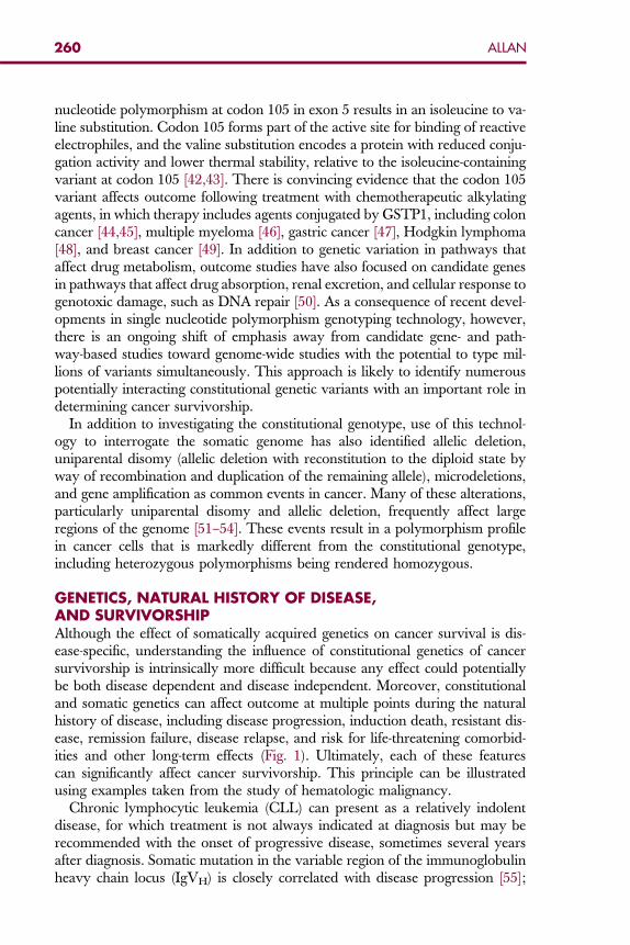
GENETICS, NATURAL HISTORY OF DISEASE,AND SURVIVORSHIPAlthough the effect of somatically acquired genetics on cancer survival is dis-ease-specific, understanding the influence of constitutional genetics of cancersurvivorship is intrinsically more difficult because any effect could potentiallybe both disease dependent and disease independent. Moreover, constitutionaland somatic genetics can affect outcome at multiple points during the naturalhistory of disease, including disease progression, induction death, resistant dis-ease, remission failure, disease relapse, and risk for life-threatening comorbid-ities and other long-term effects (Fig. 1). Ultimately, each of these featurescan significantly affect cancer survivorship. This principle can be illustratedusing examples taken from the study of hematologic malignancy.
Chronic lymphocytic leukemia (CLL) can present as a relatively indolentdisease, for which treatment is not always indicated at diagnosis but may berecommended with the onset of progressive disease, sometimes several yearsafter diagnosis. Somatic mutation in the variable region of the immunoglobulinheavy chain locus (IgVH) is closely correlated with disease progression [55];
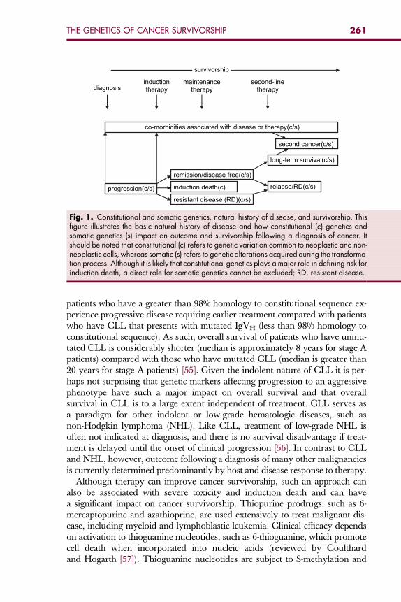
diagnosisinductiontherapy
progression(c/s)
remission/disease free(c/s)
maintenancetherapy
relapse/RD(c/s)
survivorship
long-term survival(c/s)
induction death(c)
resistant disease (RD)(c/s)
second-linetherapy
co-morbidities associated with disease or therapy(c/s)
second cancer(c/s)
Fig. 1. Constitutional and somatic genetics, natural history of disease, and survivorship. Thisfigure illustrates the basic natural history of disease and how constitutional (c) genetics andsomatic genetics (s) impact on outcome and survivorship following a diagnosis of cancer. Itshould be noted that constitutional (c) refers to genetic variation common to neoplastic and non-neoplastic cells, whereas somatic (s) refers to genetic alterations acquired during the transforma-tion process. Although it is likely that constitutional genetics plays a major role in defining risk forinduction death, a direct role for somatic genetics cannot be excluded; RD, resistant disease.
261THE GENETICS OF CANCER SURVIVORSHIP
patients who have a greater than 98% homology to constitutional sequence ex-perience progressive disease requiring earlier treatment compared with patientswho have CLL that presents with mutated IgVH (less than 98% homology toconstitutional sequence). As such, overall survival of patients who have unmu-tated CLL is considerably shorter (median is approximately 8 years for stage Apatients) compared with those who have mutated CLL (median is greater than20 years for stage A patients) [55]. Given the indolent nature of CLL it is per-haps not surprising that genetic markers affecting progression to an aggressivephenotype have such a major impact on overall survival and that overallsurvival in CLL is to a large extent independent of treatment. CLL serves asa paradigm for other indolent or low-grade hematologic diseases, such asnon-Hodgkin lymphoma (NHL). Like CLL, treatment of low-grade NHL isoften not indicated at diagnosis, and there is no survival disadvantage if treat-ment is delayed until the onset of clinical progression [56]. In contrast to CLLand NHL, however, outcome following a diagnosis of many other malignanciesis currently determined predominantly by host and disease response to therapy.
Although therapy can improve cancer survivorship, such an approach canalso be associated with severe toxicity and induction death and can havea significant impact on cancer survivorship. Thiopurine prodrugs, such as 6-mercaptopurine and azathioprine, are used extensively to treat malignant dis-ease, including myeloid and lymphoblastic leukemia. Clinical efficacy dependson activation to thioguanine nucleotides, such as 6-thioguanine, which promotecell death when incorporated into nucleic acids (reviewed by Coulthardand Hogarth [57]). Thioguanine nucleotides are subject to S-methylation and
262 ALLAN
detoxification by thiopurine S-methyltransferase (TPMT) [57,58]. TPMT activ-ity in humans displays considerable heterogeneity, with approximately 90% ofhumans having high activity, 10% having intermediate activity, and 0.3% havingvery low or null activity. This phenotypic heterogeneity is the result of a highdegree of constitutional genetic variation in the TPMT gene [59]. Eight majorTPMT alleles have been identified, with three of these (TPMT2, TPMT3A,and TPMT3C) accounting for approximately 90% of all intermediate-, low-,and null-activity cases. Individuals homozygous or compound heterozygousfor TPMT2, TPMT3A, or TPMT3C are null for TPMT activity, whereas hetero-zygotes (with one wild-type allele) have intermediate TMPT activity [60].TPMT activity is a critical determinant of patient response to thiopurine-basedtherapy. Treatment of low- or null-activity patients with standard-dose thiopur-ine therapy can lead to acute bone marrow toxicity and induction death attribut-able to neutropenia [61,62]. Constitutional TPMT genotype is thus a major riskdeterminant for induction death; routine genetic screening before administrationof thiopurine chemotherapy and appropriate dose modification for low- andnull-activity patients is indicated [63]. Similarly, induction death is a significantcause of treatment failure in patients treated with retinoic acid for APL, in which10% to 15% of patients experience often fatal hemorrhage [64]. Although the un-derlying risk factors remain unidentified it is likely that constitutional geneticvariation plays a major part.
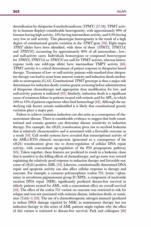
Failure to achieve remission induction can also arise as a consequence of che-moresistant disease. There is considerable evidence to suggest that both consti-tutional and somatic genetics can determine disease sensitivity to inductiontherapy. For example, the t(8;21) translocation gives rise to myeloid leukemiathat is relatively chemosensitive and is associated with a favorable outcome asa result [10]. Cell model systems have revealed that transcriptional activity ofthe AML1-ETO chimeric oncoprotein (generated as a consequence of thet(8;21) translocation) gives rise to down-regulation of cellular DNA repairactivity, with concomitant up-regulation of the P53 proapoptotic pathway[65]. Taken together, these features are predicted to result in a leukemic clonethat is sensitive to the killing effects of chemotherapy, and go some way towardexplaining the relatively good response to induction therapy and favorable out-come of t(8;21)-positive AML [10]. Likewise, constitutionally determined DNArepair and apoptotic activity can also affect cellular response to therapy andoutcome. For example, a common polymorphism (codon 751, lysine/gluta-mine) in xeroderma pigmentosum group D (XPD), a component of nucleotideexcision DNA repair (NER), significantly predicted disease-free survival inelderly patients treated for AML, with a concomitant effect on overall survival[50]. The effect of the codon 751 variant on outcome was restricted to risk forrelapse and was not associated with resistant disease, induction death, or remis-sion (Table 1) [50]. The use of a chemotherapeutic nitrogen mustard (predictedto induce DNA damage repaired by NER) as maintenance therapy but notinduction therapy in this series of AML patients might explain why the effectof this variant is restricted to disease-free survival. Park and colleagues [66]
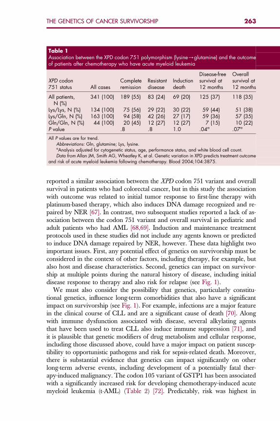
Table 1Association between the XPD codon 751 polymorphism (lysine/glutamine) and the outcomeof patients after chemotherapy who have acute myeloid leukemia
XPD codon751 status All cases
Completeremission
Resistantdisease
Inductiondeath
Disease-freesurvival at12 months
Overallsurvival at12 months
All patients,N (%)
341 (100) 189 (55) 83 (24) 69 (20) 125 (37) 118 (35)
Lys/Lys, N (%) 134 (100) 75 (56) 29 (22) 30 (22) 59 (44) 51 (38)Lys/Gln, N (%) 163 (100) 94 (58) 42 (26) 27 (17) 59 (36) 57 (35)Gln/Gln, N (%) 44 (100) 20 (45) 12 (27) 12 (27) 7 (15) 10 (22)P value .8 .8 1.0 .04a .07a
All P values are for trend.Abbreviations: Gln, glutamine; Lys, lysine.aAnalysis adjusted for cytogenetic status, age, performance status, and white blood cell count.Data from Allan JM, Smith AG, Wheatley K, et al. Genetic variation in XPD predicts treatment outcome
and risk of acute myeloid leukemia following chemotherapy. Blood 2004;104:3875.
263THE GENETICS OF CANCER SURVIVORSHIP
reported a similar association between the XPD codon 751 variant and overallsurvival in patients who had colorectal cancer, but in this study the associationwith outcome was related to initial tumor response to first-line therapy withplatinum-based therapy, which also induces DNA damage recognized and re-paired by NER [67]. In contrast, two subsequent studies reported a lack of as-sociation between the codon 751 variant and overall survival in pediatric andadult patients who had AML [68,69]. Induction and maintenance treatmentprotocols used in these studies did not include any agents known or predictedto induce DNA damage repaired by NER, however. These data highlight twoimportant issues. First, any potential effect of genetics on survivorship must beconsidered in the context of other factors, including therapy, for example, butalso host and disease characteristics. Second, genetics can impact on survivor-ship at multiple points during the natural history of disease, including initialdisease response to therapy and also risk for relapse (see Fig. 1).
We must also consider the possibility that genetics, particularly constitu-tional genetics, influence long-term comorbidities that also have a significantimpact on survivorship (see Fig. 1). For example, infections are a major featurein the clinical course of CLL and are a significant cause of death [70]. Alongwith immune dysfunction associated with disease, several alkylating agentsthat have been used to treat CLL also induce immune suppression [71], andit is plausible that genetic modifiers of drug metabolism and cellular response,including those discussed above, could have a major impact on patient suscep-tibility to opportunistic pathogens and risk for sepsis-related death. Moreover,there is substantial evidence that genetics can impact significantly on otherlong-term adverse events, including development of a potentially fatal ther-apy-induced malignancy. The codon 105 variant of GSTP1 has been associatedwith a significantly increased risk for developing chemotherapy-induced acutemyeloid leukemia (t-AML) (Table 2) [72]. Predictably, risk was highest in
Table 2Association between the glutathione S-transferase P1 codon 105 polymorphism (isoleucine/valine) and risk for therapy-induced acute myeloid leukemia
GSTP1codon 105
de novoAML, N (%)
t-AML, N (%)(all cases)
Odds ratio(95% CI)
t-AML, N (%),(postchemotherapy)
Odds ratio(95% CI)
t-AML, N (%),(post GSTP1substrates)
Odds ratio(95% CI)a
Iso/Iso 202 (49) 33 (37) 1 (�) 14 (27) 1 (�) 4 (19) 1 (�)Iso/Val 151 (36) 40 (45) 1.87 (1.11–3.17) 28 (55) 2.87 (1.45–5.67) 12 (57) 4.43 (1.39–14.12)Val/Val 61 (15) 16 (18) 1.67 (0.84–3.30) 9 (18) 2.17 (0.89–5.29) 5 (24) 4.16 (1.07–16.07)Iso/Val þ
Val/Val212 (51) 56 (63) 1.81 (1.11–2.94) 37 (73) 2.66 (1.39–5.09) 17 (81) 4.34 (1.43–13.20)
Abbreviations: AML, acute myeloid leukemia; CI, confidence interval; Iso, isoleucine; t-AML, therapy-induced acute myeloid leukemia; Val, valine.aOdds ratios and 95% confidence intervals were calculated using unconditional logistic regression and using de novo AML cases as the reference.Data from Allan JM, Wild CP, Rollinson S, et al. Polymorphism in glutathione S-transferase P1 is associated with susceptibility to chemotherapy-induced leukemia. Proc Natl Acad
Sci U S A 2001;98:11596.
26
4A
LLAN
265THE GENETICS OF CANCER SURVIVORSHIP
patients previously treated with chemotherapy agents that are substratesfor glutathione conjugation by GSTP1 (see Table 2) [72]. It is biologically plau-sible that the GSTP1 codon 105 variant has a direct impact on susceptibility tomutation and transformation in non-target bone marrow progenitor cells, con-ferring susceptibility to t-AML. We must also consider the possibility, however,that the population at risk for developing a therapy-induced cancer is geneti-cally biased for this variant because of its role as a prognostic marker after treat-ment of primary cancer, including colon cancer [44,45], multiple myeloma [46],gastric cancer [47], Hodgkin lymphoma [48], and breast cancer [49]. Geneticsusceptibility to iatrogenic malignancies is also complicated by other factors,and these have been reviewed elsewhere [73].
SUMMARYHost and disease phenotype at presentation are critical determinants of out-come and survival following a diagnosis of cancer. Given that phenotype isdetermined to a large extend by genotype it is perhaps not surprising thatgenetics should also play an important role in defining outcome and survivor-ship. A better understanding of constitutional and somatic genetics will allowfor therapeutic regimens to be individualized to the patient and for the devel-opment of therapies targeted against specific disease types. Advances in ourunderstanding of genetics, in conjunction with the development of more sophis-ticated therapies and treatment regimens, can only become more important asa pivotal determinant of survivorship.
References
[1] Lossos IS, Morgensztern D. Prognostic biomarkers in diffuse large B-cell lymphoma. J ClinOncol 2006;24(6):995–1007.[2] Kolitz JE. Current therapeutic strategies for acute myeloid leukaemia. Br J Haematol
2006;134(6):555–72.[3] Mauro MJ, Druker BJ. Chronic myelogenous leukemia. Curr Opin Oncol 2001;13(1):3–7.[4] Cilloni D, Guerrasio A, Giugliano E, et al. From genes to therapy: the case of Philadelphia
chromosome-positive leukemias. Ann N Y Acad Sci 2002;963:306–12.(5) Druker BJ, Talpaz M, Resta DJ, et al. Efficacy and safety of a specific inhibitor of the BCR-ABL
tyrosine kinase in chronic myeloid leukemia. N Engl J Med 2001;344(14):1031–7.[6] Mauro MJ, Druker BJ. STI571: targeting BCR-ABL as therapy for CML. Oncologist
2001;6(3):233–8.[7] Mistry AR, Pedersen EW, Solomon E, et al. The molecular pathogenesis of acute promyelo-
cytic leukaemia: implications for the clinical management of the disease. Blood Rev2003;17(2):71–97.
[8] Tallman MS. Acute promyelocytic leukemia as a paradigm for targeted therapy. SeminHematol 2004;41(2 Suppl 4):27–32.
[9] Bacher U, Kern W, Schnittger S, et al. Population-based age-specific incidences of cytoge-netic subgroups of acute myeloid leukemia. Haematologica 2005;90(11):1502–10.
[10] Grimwade D, Walker H, Oliver F, et al. The importance of diagnostic cytogenetics onoutcome in AML: analysis of 1,612 patients entered into the MRC AML 10 trial. The MedicalResearch Council Adult and Children’s Leukaemia Working Parties. Blood 1998;92(7):2322–33.
[11] Grimwade D, Moorman A, Hills R, et al. Impact of karyotype on treatment outcome in acutemyeloid leukemia. Ann Hematol 2004;83(Suppl 1):S45–8.
266 ALLAN
[12] Grimwade D. The clinical significance of cytogenetic abnormalities in acute myeloid leukae-mia. Best Pract Res Clin Haematol 2001;14(3):497–529.
[13] Grimwade D, Walker H, Harrison G, et al. The predictive value of hierarchical cytogeneticclassification in older adults with acute myeloid leukemia (AML): analysis of 1065 patientsentered into the United Kingdom Medical Research Council AML11 trial. Blood2001;98(5):1312–20.
[14] Moorman AV, Harrison CJ, Buck GA, et al. Karyotype is an independent prognostic factor inadult acute lymphoblastic leukemia (ALL): analysis of cytogenetic data from patients treatedon the Medical Research Council (MRC) UKALLXII/Eastern Cooperative Oncology Group(ECOG) 2993 trial. Blood 2007;109(8):3189–97.
[15] Moorman AV, Richards SM, Robinson HM, et al. Prognosis of children with acute lympho-blastic leukemia (ALL) and intrachromosomal amplification of chromosome 21 (iAMP21).Blood 2007;109(6):2327–30.
[16] Pui CH, Evans WE. Treatment of acute lymphoblastic leukemia. N Engl J Med 2006;354(2):166–78.
[17] Piccaluga PP, Paolini S, Martinelli G. Tyrosine kinase inhibitors for the treatment of Philadel-phia chromosome-positive adult acute lymphoblastic leukemia. Cancer 2007;110(6):1178–86.
[18] Heuser M, Wingen LU, Steinemann D, et al. Gene-expression profiles and their associationwith drug resistance in adult acute myeloid leukemia. Haematologica 2005;90(11):1484–92.
[19] Valk PJ, Verhaak RG, Beijen MA, et al. Prognostically useful gene-expression profiles inacute myeloid leukemia. N Engl J Med 2004;350(16):1617–28.
[20] Bullinger L, Dohner K, Bair E, et al. Use of gene-expression profiling to identify prognosticsubclasses in adult acute myeloid leukemia. N Engl J Med 2004;350(16):1605–16.
[21] Kottaridis PD, Gale RE, Linch DC. Prognostic implications of the presence of FLT3 mutationsin patients with acute myeloid leukemia. Leuk Lymphoma 2003;44(6):905–13.
[22] Kottaridis PD, Gale RE, Frew ME, et al. The presence of a FLT3 internal tandem duplication inpatients with acute myeloid leukemia (AML) adds important prognostic information to cyto-genetic risk group and response to the first cycle of chemotherapy: analysis of 854 patientsfrom the United Kingdom Medical Research Council AML 10 and 12 trials. Blood2001;98(6):1752–9.
[23] Schnittger S, Schoch C, Dugas M, et al. Analysis of FLT3 length mutations in 1003 patientswith acute myeloid leukemia: correlation to cytogenetics, FAB subtype, and prognosis in theAMLCG study and usefulness as a marker for the detection of minimal residual disease.Blood 2002;100(1):59–66.
[24] Boissel N, Cayuela JM, Preudhomme C, et al. Prognostic significance of FLT3 internaltandem repeat in patients with de novo acute myeloid leukemia treated with reinforcedcourses of chemotherapy. Leukemia 2002;16(9):1699–704.
[25] Boissel N, Leroy H, Brethon B, et al. Incidence and prognostic impact of c-Kit, FLT3, and Rasgene mutations in core binding factor acute myeloid leukemia (CBF-AML). Leukemia2006;20(6):965–70.
[26] Stirewalt DL, Kopecky KJ, Meshinchi S, et al. Size of FLT3 internal tandem duplication hasprognostic significance in patients with acute myeloid leukemia. Blood 2006;107(9):3724–6.
[27] Schmid D, Heinze G, Linnerth B, et al. Prognostic significance of WT1 gene expression atdiagnosis in adult de novo acute myeloid leukemia. Leukemia 1997;11(5):639–43.
[28] Barjesteh van Waalwijk van Doorn-Khosrovani S, Erpelinck C, Meijer J, et al. Biallelic mu-tations in the CEBPA gene and low CEBPA expression levels as prognostic markers in inter-mediate-risk AML. Hematol J 2003;4(1):31–40.
[29] Schnittger S, Kohl TM, Haferlach T, et al. KIT-D816 mutations in AML1-ETO-positive AML areassociated with impaired event-free and overall survival. Blood 2006;107(5):1791–9.
267THE GENETICS OF CANCER SURVIVORSHIP
[30] Suzuki T, Kiyoi H, Ozeki K, et al. Clinical characteristics and prognostic implications ofNPM1 mutations in acute myeloid leukemia. Blood 2005;106(8):2854–61.
[31] Thiede C, Koch S, Creutzig E, et al. Prevalence and prognostic impact of NPM1 mutations in1485 adult patients with acute myeloid leukemia (AML). Blood 2006;107(10):4011–20.
[32] Levis M, Allebach J, Tse KF, et al. A FLT3-targeted tyrosine kinase inhibitor is cytotoxic toleukemia cells in vitro and in vivo. Blood 2002;99(11):3885–91.
[33] Knapper S, Burnett AK, Littlewood T, et al. A phase 2 trial of the FLT3 inhibitor lestaurtinib(CEP701) as first-line treatment for older patients with acute myeloid leukemia not consid-ered fit for intensive chemotherapy. Blood 2006;108(10):3262–70.
[34] Stirewalt DL, Radich JP. The role of FLT3 in haematopoietic malignancies. Nat Rev Cancer2003;3(9):650–65.
[35] Druker BJ, Guilhot F, O’brien SG, et al. Five-year follow-up of patients receiving imatinib forchronic myeloid leukemia. N Engl J Med 2006;355(23):2408–17.
[36] Gorre ME, Mohammed M, Ellwood K, et al. Clinical resistance to STI-571 cancer therapycaused by BCR-ABL gene mutation or amplification. Science 2001;293(5531):876–80.
[37] Young MA, Shah NP, Chao LH, et al. Structure of the kinase domain of an imatinib-resistantAbl mutant in complex with the Aurora kinase inhibitor VX-680. Cancer Res 2006;66(2):1007–14.
[38] Kantarjian HM, Giles F, Gattermann N, et al. Nilotinib (formerly AMN107), a highly selec-tive BCR-ABL tyrosine kinase inhibitor, is effective in patients with Philadelphia chromosomepositive chronic myelogenous leukemia in chronic phase following imatinib resistance andintolerance. Blood 2007;110(10):3540–6.
[39] Mughal TI, Goldman JM. Emerging strategies for the treatment of mutant Bcr-Abl T315Imyeloid leukemia. Clin Lymphoma Myeloma 2007;7(Suppl 2):S81–4.
[40] Kruglyak L, Nickerson DA. Variation is the spice of life. Nat Genet 2001;27(3):234–6.[41] Feuk L, Carson AR, Scherer SW. Structural variation in the human genome. Nat Rev Genet
2006;7(2):85–97.[42] Pandya U, Srivastava SK, Singhal SS, et al. Activity of allelic variants of Pi class human
glutathione S-transferase toward chlorambucil. Biochem Biophys Res Commun2000;278(1):258–62.
[43] Johansson AS, Stenberg G, Widersten M, et al. Structure-activity relationships and thermalstability of human glutathione transferase P1-1 governed by the H-site residue 105. J MolBiol 1998;278(3):687–98.
[44] Stoehlmacher J, Park DJ, Zhang W, et al. Association between glutathione S-transferase P1,T1, and M1 genetic polymorphism and survival of patients with metastatic colorectalcancer. J Natl Cancer Inst 2002;94(12):936–42.
[45] Stoehlmacher J, Park DJ, Zhang W, et al. A multivariate analysis of genomic polymorphisms:prediction of clinical outcome to 5-FU/oxaliplatin combination chemotherapy in refractorycolorectal cancer. Br J Cancer 2004;91(2):344–54.
[46] Dasgupta RK, Adamson PJ, Davies FE, et al. Polymorphic variation in GSTP1 modulatesoutcome following therapy for multiple myeloma. Blood 2003;102(7):2345–50.
[47] Goekkurt E, Hoehn S, Wolschke C, et al. Polymorphisms of glutathione S-transferases (GST)and thymidylate synthase (TS)–novel predictors for response and survival in gastric cancerpatients. Br J Cancer 2006;94(2):281–6.
[48] Hohaus S, Di Ruscio A, Di Febo A, et al. Glutathione S-transferase P1 genotype andprognosis in Hodgkin’s lymphoma. Clin Cancer Res 2005;11(6):2175–9.
[49] Sweeney C, McClure GY, Fares MY, et al. Association between survival after treatment forbreast cancer and glutathione S-transferase P1 Ile105Val polymorphism. Cancer Res2000;60(20):5621–4.
[50] Allan JM, Smith AG, Wheatley K, et al. Genetic variation in XPD predicts treatment outcomeand risk of acute myeloid leukemia following chemotherapy. Blood 2004;104(13):3872–7.
268 ALLAN
[51] Fitzgibbon J, Smith LL, Raghavan M, et al. Association between acquired uniparental dis-omy and homozygous gene mutation in acute myeloid leukemias. Cancer Res2005;65(20):9152–4.
[52] Raghavan M, Lillington DM, Skoulakis S, et al. Genome-wide single nucleotide polymor-phism analysis reveals frequent partial uniparental disomy due to somatic recombinationin acute myeloid leukemias. Cancer Res 2005;65(2):375–8.
[53] Strefford JC, van Delft FW, Robinson HM, et al. Complex genomic alterations and geneexpression in acute lymphoblastic leukemia with intrachromosomal amplification ofchromosome 21. Proc Natl Acad Sci U S A 2006;103(21):8167–72.
[54] Irving JA, Bloodworth L, Bown NP, et al. Loss of heterozygosity in childhood acute lympho-blastic leukemia detected by genome-wide microarray single nucleotide polymorphismanalysis. Cancer Res 2005;65(8):3053–8.
[55] Hamblin TJ, Davis Z, Gardiner A, et al. Unmutated Ig V(H) genes are associated with a moreaggressive form of chronic lymphocytic leukemia. Blood 1999;94(6):1848–54.
[56] Ardeshna KM, Smith P, Norton A, et al. Long-term effect of a watch and wait policy versusimmediate systemic treatment for asymptomatic advanced-stage non-Hodgkin lymphoma:a randomised controlled trial. Lancet 2003;362(9383):516–22.
[57] Coulthard S, Hogarth L. The thiopurines: an update. Invest New Drugs 2005;23(6):523–32.
[58] Coulthard SA, Matheson EC, Hall AG, et al. The clinical impact of thiopurine methyltrans-ferase polymorphisms on thiopurine treatment. Nucleosides Nucleotides Nucleic Acids2004;23(8–9):1385–91.
[59] McLeod HL, Siva C. The thiopurine S-methyltransferase gene locus—implications for clinicalpharmacogenomics. Pharmacogenomics 2002;3(1):89–98.
[60] Yates CR, Krynetski EY, Loennechen T, et al. Molecular diagnosis of thiopurine S-methyltrans-ferase deficiency: genetic basis for azathioprine and mercaptopurine intolerance. AnnIntern Med 1997;126(8):608–14.
[61] Evans WE, Rodman J, Relling MV, et al. Individualized dosages of chemotherapy as a strat-egy to improve response for acute lymphocytic leukemia. Semin Hematol 1991;28(3 Suppl4):15–21.
[62] McLeod HL, Miller DR, Evans WE. Azathioprine-induced myelosuppression in thiopurinemethyltransferase deficient heart transplant recipient. Lancet 1993;341(8853):1151.
[63] van den Akker-van Marle ME, Gurwitz D, Detmar SB, et al. Cost-effectiveness of pharmaco-genomics in clinical practice: a case study of thiopurine methyltransferase genotyping inacute lymphoblastic leukemia in Europe. Pharmacogenomics 2006;7(5):783–92.
[64] Tallman MS, Abutalib SA, Altman JK. The double hazard of thrombophilia and bleeding inacute promyelocytic leukemia. Semin Thromb Hemost 2007;33(4):330–8.
[65] Krejci O, Wunderlich M, Geiger H, et al. p53 signaling in response to increased DNAdamage sensitizes AML1-ETO cells to stress-induced death. Blood 2008;111(4):2190–9.
[66] Park DJ, Stoehlmacher J, Zhang W, et al. A Xeroderma pigmentosum group D gene polymor-phism predicts clinical outcome to platinum-based chemotherapy in patients with advancedcolorectal cancer. Cancer Res 2001;61(24):8654–8.
[67] Wang D, Lippard SJ. Cellular processing of platinum anticancer drugs. Nat Rev Drug Discov2005;4(4):307–20.
[68] Mehta PA, Alonzo TA, Gerbing RB, et al. XPD Lys751Gln polymorphism in the etiology andoutcome of childhood acute myeloid leukemia: a Children’s Oncology Group report. Blood2006;107(1):39–45.
[69] Kuptsova N, Kopecky KJ, Godwin J, et al. Polymorphisms in DNA repair genes andtherapeutic outcomes of AML patients from SWOG clinical trials. Blood 2007;109(9):3936–44.
[70] Francis S, Karanth M, Pratt G, et al. The effect of immunoglobulin VH gene mutation statusand other prognostic factors on the incidence of major infections in patients with chroniclymphocytic leukemia. Cancer 2006;107(5):1023–33.

269THE GENETICS OF CANCER SURVIVORSHIP
[71] Mackall CL. T-cell immunodeficiency following cytotoxic antineoplastic therapy: a review.Stem Cells 2000;18(1):10–8.
[72] Allan JM, Wild CP, Rollinson S, et al. Polymorphism in glutathione S-transferase P1 isassociated with susceptibility to chemotherapy-induced leukemia. Proc Natl Acad Sci U S A2001;98(20):11592–7.
[73] Allan JM, Rabkin CS. Genetic susceptibility to iatrogenic malignancy. Pharmacogenomics2005;6(6):615–28.