Embed Size (px)
Citation preview
The genetic code and tRNABiochemistry 302
February 15, 2006
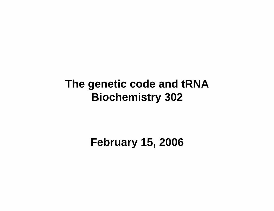
Major advances in defining mechanism of protein biosynthesis• Identification of ribosomes
(Paul Zamecnik, 1950s)– Injected radioactive amino acids
into rats, fractionated liver homogenates at various time points, then analyzed subcellular distribution of labeled proteins.
– At early time points, “hot”proteins only in “small” RNP particles.
• Identification of tRNAs– M. Hoagland & Zamecnik– Amino acids become “activated”
i.e. attached to heat-stable soluble RNA species if incubated with cytosolic extract.
– R. Holley → tRNA• Crick’s adaptor hypothesis
Predicted triplet code 43 = 64 possible combinations
Ribosomes attached to outer face of ER
mRNA-guided protein synthesis
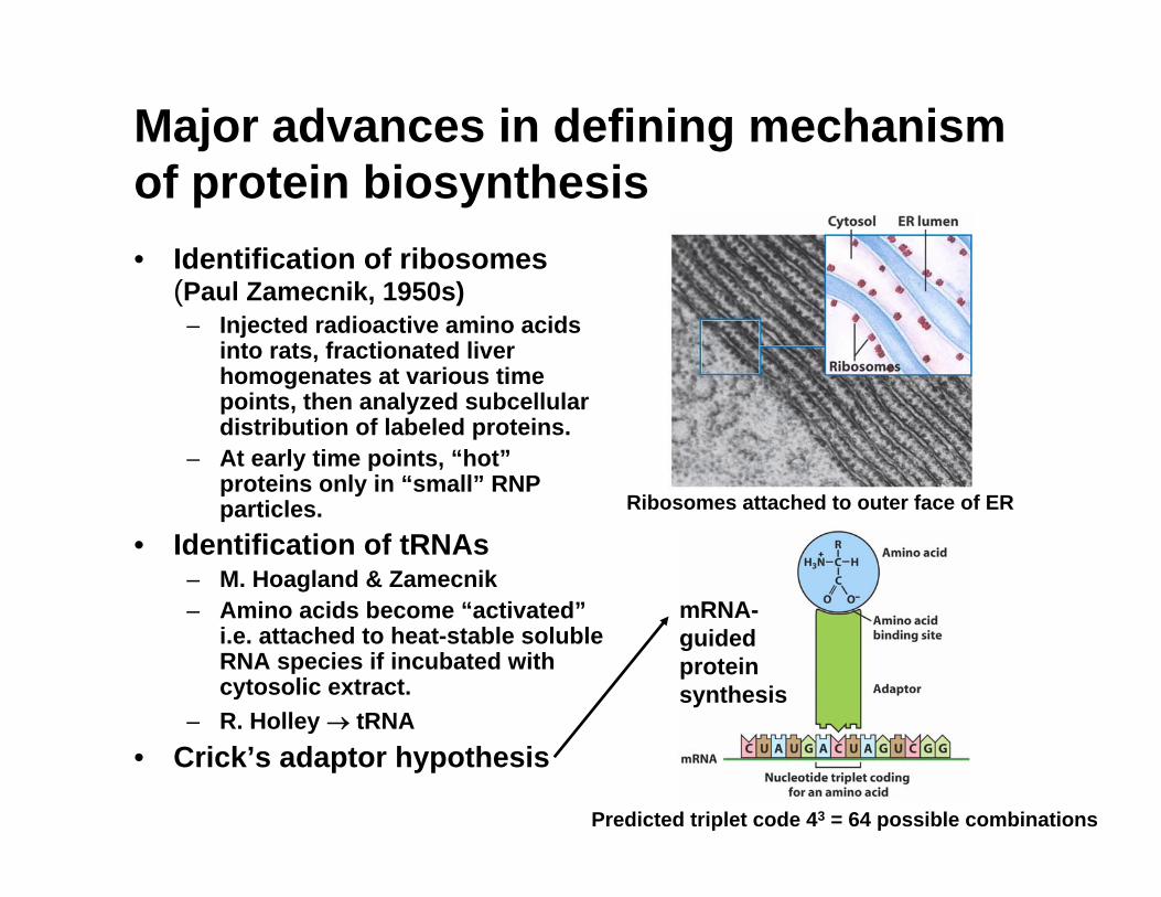
Overall process of mRNA-guided protein biosynthesis: translation
activated amino acid or aminoacyl tRNA
3′
anticodon
Fig. 4-22
5′H2N
COOH
7-meG
A200-250
5′
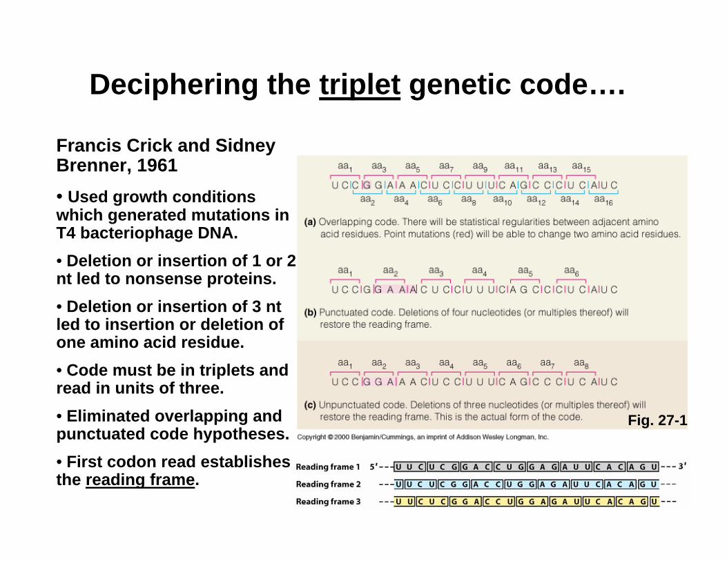
Deciphering the triplet genetic code….
Francis Crick and Sidney Brenner, 1961• Used growth conditions which generated mutations in T4 bacteriophage DNA.• Deletion or insertion of 1 or 2 nt led to nonsense proteins.• Deletion or insertion of 3 nt led to insertion or deletion of one amino acid residue.• Code must be in triplets and read in units of three. • Eliminated overlapping and punctuated code hypotheses.• First codon read establishes the reading frame.
Fig. 27-1
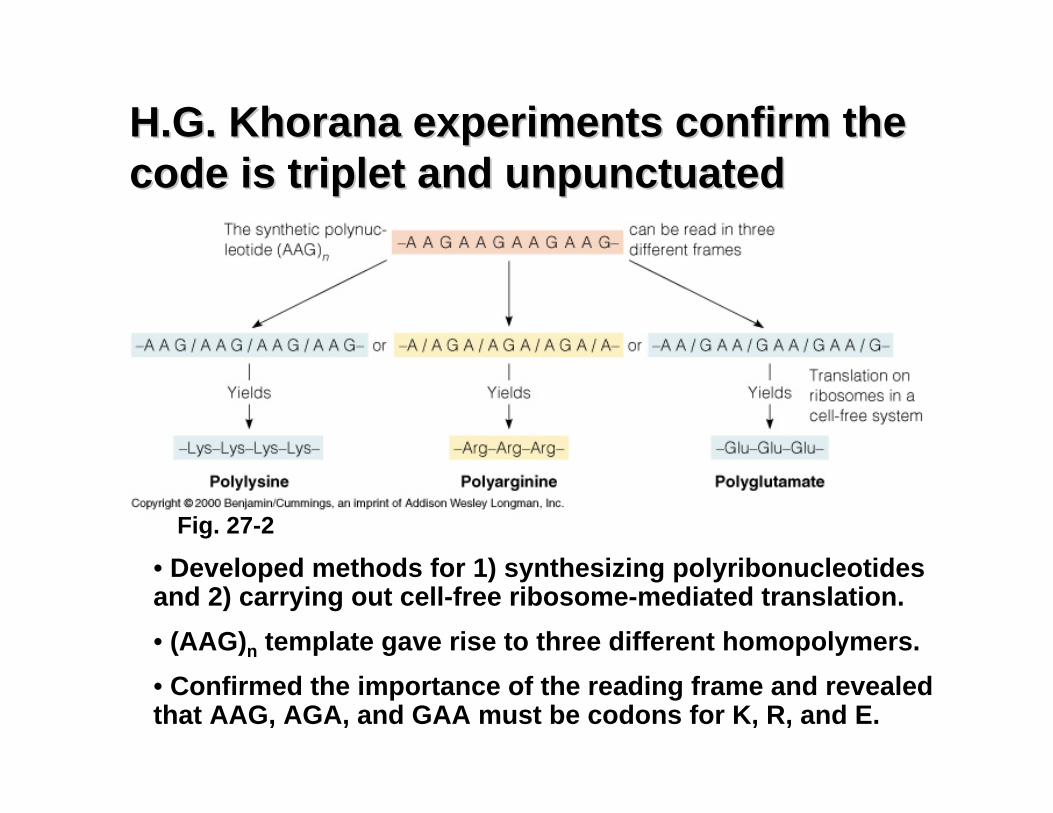
H.G. Khorana experiments confirm the H.G. Khorana experiments confirm the code is triplet and unpunctuatedcode is triplet and unpunctuated
• Developed methods for 1) synthesizing polyribonucleotides and 2) carrying out cell-free ribosome-mediated translation.• (AAG)n template gave rise to three different homopolymers.• Confirmed the importance of the reading frame and revealed that AAG, AGA, and GAA must be codons for K, R, and E.
Fig. 27-2
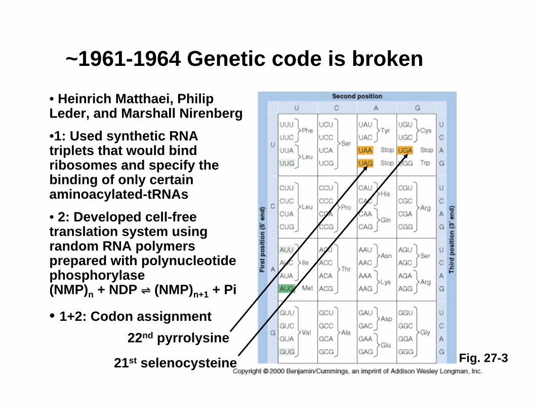
~1961-1964 Genetic code is broken • Heinrich Matthaei, Philip Leder, and Marshall Nirenberg•1: Used synthetic RNA triplets that would bind ribosomes and specify the binding of only certain aminoacylated-tRNAs• 2: Developed cell-free translation system using random RNA polymers prepared with polynucleotide phosphorylase (NMP)n + NDP ⇌ (NMP)n+1 + Pi
• 1+2: Codon assignment
21st selenocysteine Fig. 27-322nd pyrrolysine
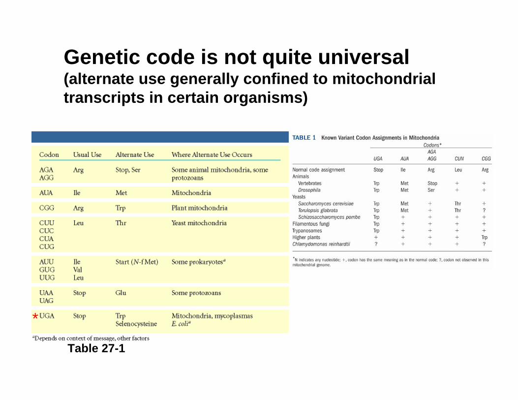
Genetic code is not quite universal (alternate use generally confined to mitochondrial transcripts in certain organisms)
Table 27-1
*
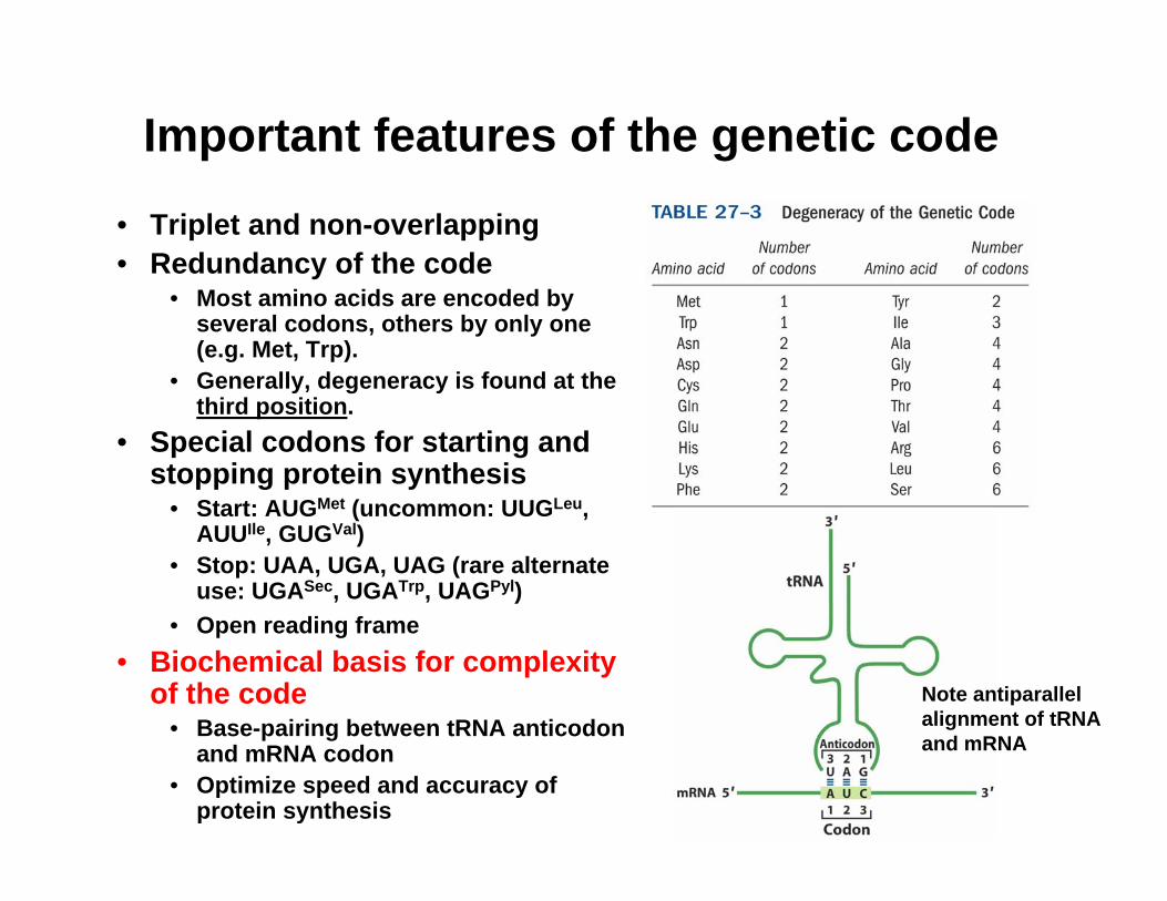
Important features of the genetic code
• Triplet and non-overlapping• Redundancy of the code
• Most amino acids are encoded by several codons, others by only one (e.g. Met, Trp).
• Generally, degeneracy is found at the third position.
• Special codons for starting and stopping protein synthesis
• Start: AUGMet (uncommon: UUGLeu, AUUIle, GUGVal)
• Stop: UAA, UGA, UAG (rare alternate use: UGASec, UGATrp, UAGPyl)
• Open reading frame• Biochemical basis for complexity
of the code• Base-pairing between tRNA anticodon
and mRNA codon• Optimize speed and accuracy of
protein synthesis
Note antiparallel alignment of tRNA and mRNA
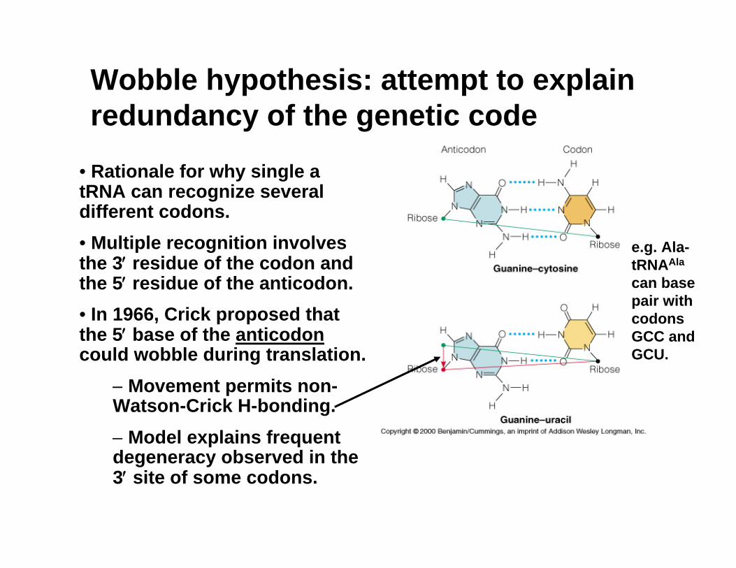
Wobble hypothesis: attempt to explain redundancy of the genetic code
• Rationale for why single a tRNA can recognize several different codons.• Multiple recognition involves the 3′ residue of the codon and the 5′ residue of the anticodon. • In 1966, Crick proposed that the 5′ base of the anticodoncould wobble during translation.
− Movement permits non-Watson-Crick H-bonding.− Model explains frequent degeneracy observed in the 3′ site of some codons.
e.g. Ala-tRNAAla
can base pair with codons GCC and GCU.
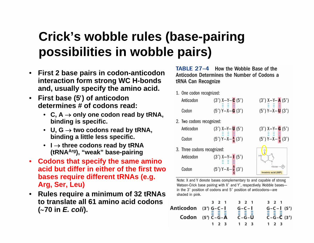
Crick’s wobble rules (base-pairing possibilities in wobble pairs)
• First 2 base pairs in codon-anticodon interaction form strong WC H-bonds and, usually specify the amino acid.
• First base (5′) of anticodon determines # of codons read:
• C, A → only one codon read by tRNA, binding is specific.
• U, G → two codons read by tRNA, binding a little less specific.
• I → three codons read by tRNA (tRNAArg), “weak” base-pairing
• Codons that specify the same amino acid but differ in either of the first two bases require different tRNAs (e.g. Arg, Ser, Leu)
• Rules require a minimum of 32 tRNAsto translate all 61 amino acid codons (∼70 in E. coli).
Table 27-2 Wobble bps
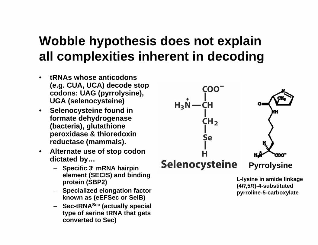
Wobble hypothesis does not explain all complexities inherent in decoding• tRNAs whose anticodons
(e.g. CUA, UCA) decode stop codons: UAG (pyrrolysine), UGA (selenocysteine)
• Selenocysteine found in formate dehydrogenase (bacteria), glutathione peroxidase & thioredoxin reductase (mammals).
• Alternate use of stop codon dictated by…
– Specific 3′ mRNA hairpin element (SECIS) and binding protein (SBP2)
– Specialized elongation factor known as (eEFSec or SelB)
– Sec-tRNASec (actually special type of serine tRNA that gets converted to Sec)
PyrrolysineL-lysine in amide linkage (4R,5R)-4-substituted pyrroline-5-carboxylate
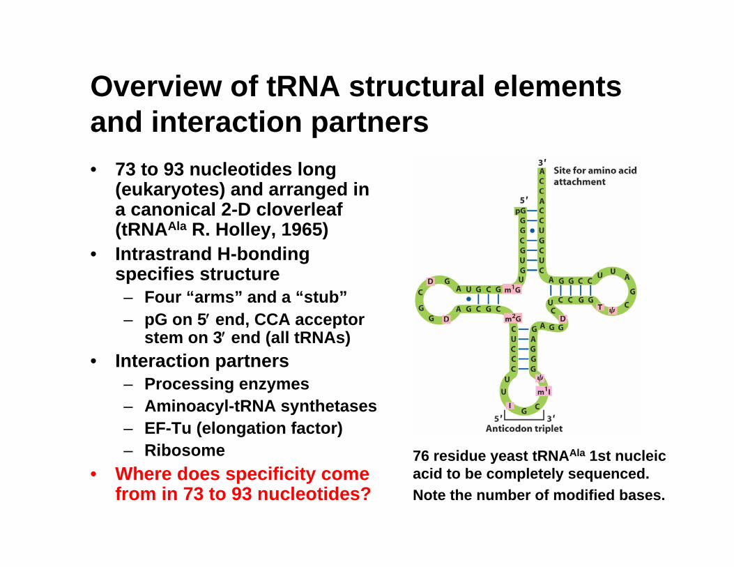
Overview of tRNA structural elements and interaction partners• 73 to 93 nucleotides long
(eukaryotes) and arranged in a canonical 2-D cloverleaf (tRNAAla R. Holley, 1965)
• Intrastrand H-bonding specifies structure– Four “arms” and a “stub”– pG on 5′ end, CCA acceptor
stem on 3′ end (all tRNAs) • Interaction partners
– Processing enzymes– Aminoacyl-tRNA synthetases– EF-Tu (elongation factor)– Ribosome
• Where does specificity come from in 73 to 93 nucleotides?
76 residue yeast tRNAAla 1st nucleic acid to be completely sequenced. Note the number of modified bases.
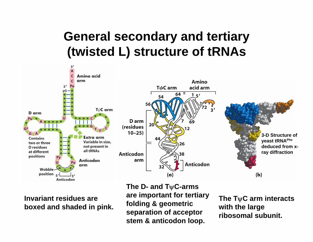
General secondary and tertiary (twisted L) structure of tRNAs
Invariant residues are boxed and shaded in pink.
The D- and TψC-arms are important for tertiary folding & geometric separation of acceptor stem & anticodon loop.
The TψC arm interacts with the large ribosomal subunit.
3-D Structure of yeast tRNAPhe
deduced from x-ray diffraction
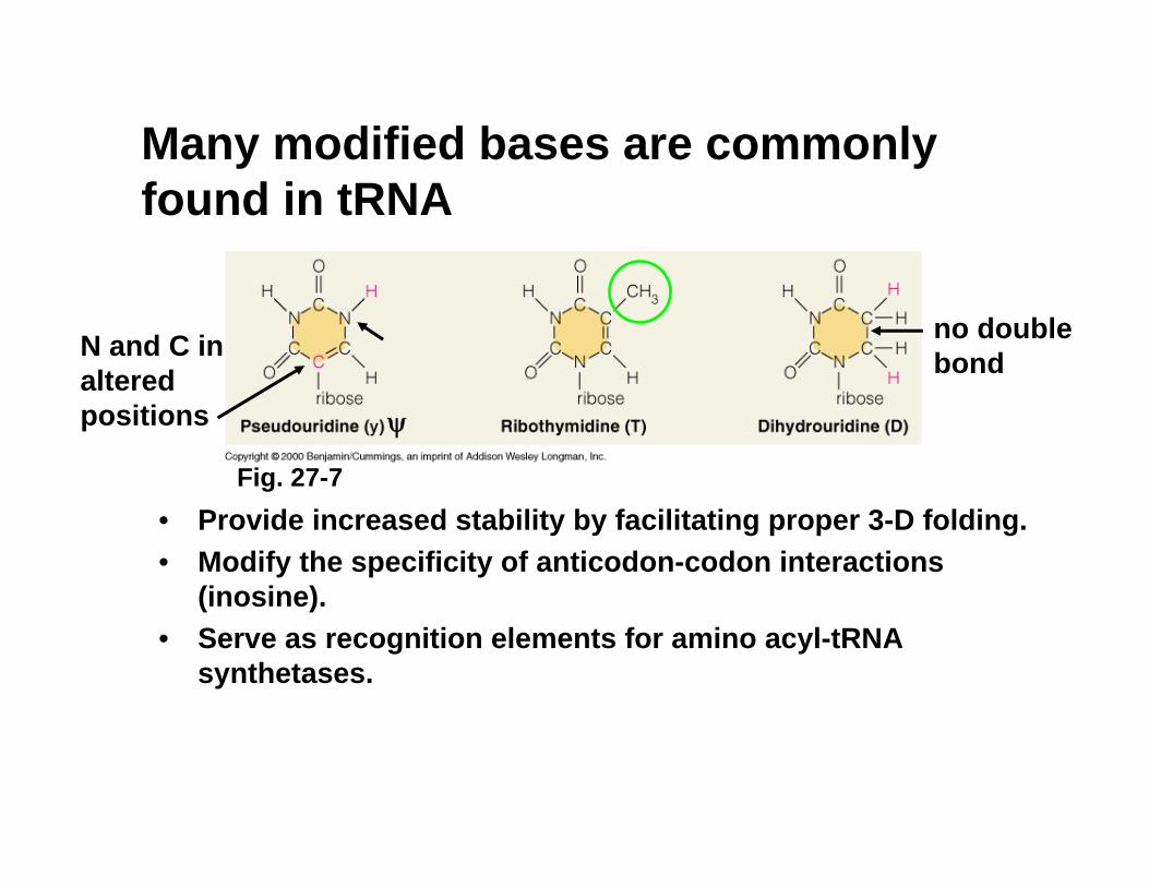
Many modified bases are commonly found in tRNA
• Provide increased stability by facilitating proper 3-D folding.• Modify the specificity of anticodon-codon interactions
(inosine). • Serve as recognition elements for amino acyl-tRNA
synthetases.
no double bondN and C in
altered positions
Fig. 27-7
ψ
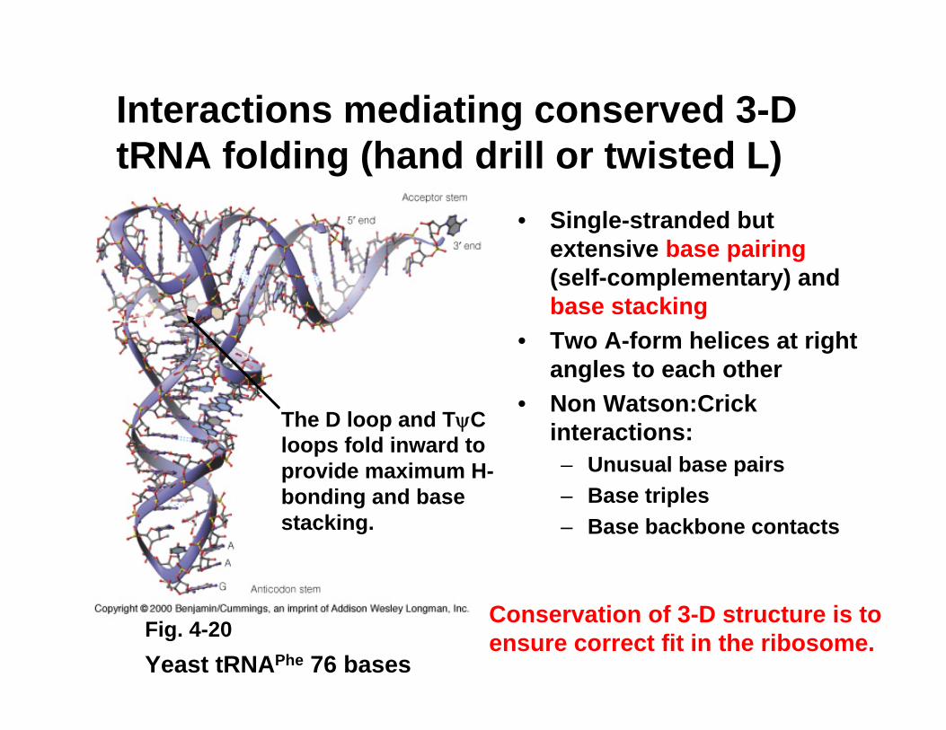
Interactions mediating conserved 3-D tRNA folding (hand drill or twisted L)
• Single-stranded but extensive base pairing(self-complementary) and base stacking
• Two A-form helices at right angles to each other
• Non Watson:Crick interactions:– Unusual base pairs– Base triples– Base backbone contacts
Yeast tRNAPhe 76 bases
The D loop and TψC loops fold inward to provide maximum H-bonding and base stacking.
Conservation of 3-D structure is to ensure correct fit in the ribosome.Fig. 4-20
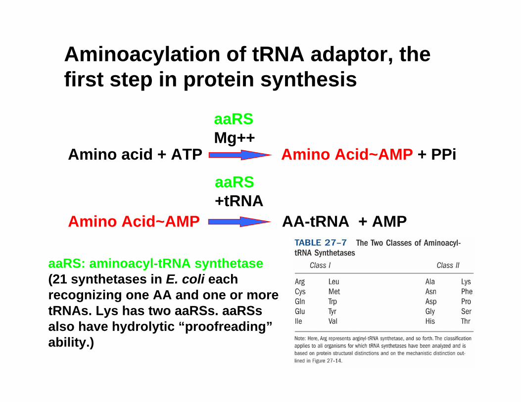
Aminoacylation of tRNA adaptor, the first step in protein synthesis
Amino acid + ATP Amino Acid~AMP + PPi
aaRSMg++
Amino Acid~AMP AA-tRNA + AMP
aaRS+tRNA
aaRS: aminoacyl-tRNA synthetase (21 synthetases in E. coli each recognizing one AA and one or more tRNAs. Lys has two aaRSs. aaRSsalso have hydrolytic “proofreading”ability.)
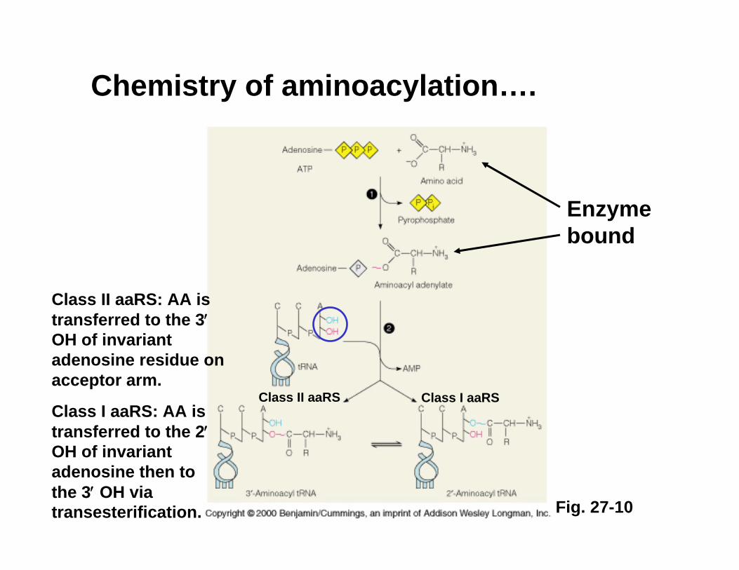
Chemistry of aminoacylation….
Enzyme bound
Class II aaRS: AA is transferred to the 3′OH of invariant adenosine residue on acceptor arm.
Class I aaRS: AA is transferred to the 2′OH of invariant adenosine then to the 3′ OH via transesterification. Fig. 27-10
Class I aaRSClass II aaRS
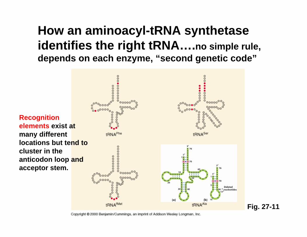
How an aminoacyl-tRNA synthetase identifies the right tRNA….no simple rule, depends on each enzyme, “second genetic code”
Recognition elements exist at many different locations but tend to cluster in the anticodon loop and acceptor stem.
Fig. 27-11
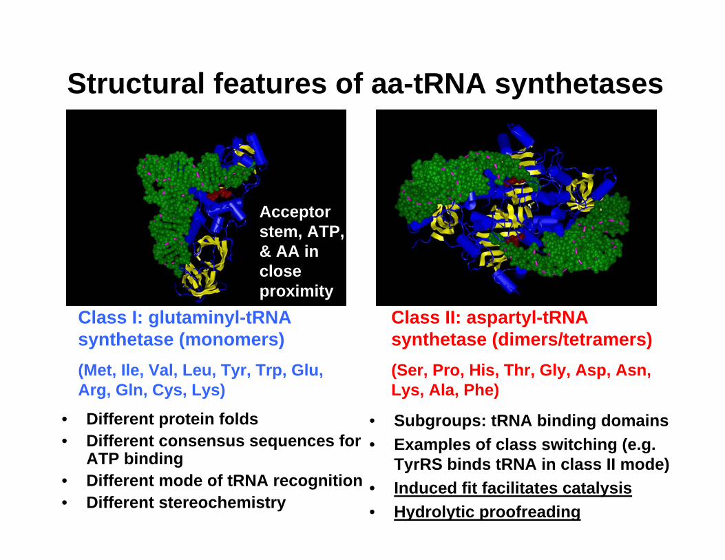
Structural features of aa-tRNA synthetases
• Different protein folds• Different consensus sequences for
ATP binding• Different mode of tRNA recognition• Different stereochemistry
• Subgroups: tRNA binding domains• Examples of class switching (e.g.
TyrRS binds tRNA in class II mode)• Induced fit facilitates catalysis• Hydrolytic proofreading
Class I: glutaminyl-tRNA synthetase (monomers)(Met, Ile, Val, Leu, Tyr, Trp, Glu, Arg, Gln, Cys, Lys)
Class II: aspartyl-tRNA synthetase (dimers/tetramers)(Ser, Pro, His, Thr, Gly, Asp, Asn, Lys, Ala, Phe)
Acceptor stem, ATP, & AA in close proximity


![RESEARCH ARTICLE Open Access Fragmentation of ... - SLU.SE · 18–46 nt pieces derived from mature tRNA or the 3 ′ end of precursor-tRNA (pre-tRNA) [14-16]. tRNA fragmenta-tion](https://img.pdfslide.us/doc/110x75/60474a078cb48655a57c0958/research-article-open-access-fragmentation-of-sluse-18a46-nt-pieces-derived.jpg)