Embed Size (px)
Citation preview
The Gastrointestinal Tract as an Integrator of Mechanicaland Hormonal Response to Nutrient IngestionAdrian Vella1 and Michael Camilleri2
Diabetes 2017;66:2729–2737 | https://doi.org/10.2337/dbi17-0021
Glucose tolerance aftermeal ingestion in vivo is the result ofmultiple processes that occur in parallel. Insulin secretiontogether with reciprocal inhibition of glucagon secretioncontributes to glucose tolerance. However, other factorsbeyond glucose effectiveness and insulin action requireconsideration. The absorption of ingested nutrients andtheir subsequent systemic rate of appearance largelydepend on the rate of delivery of nutrients to the proximalsmall intestine. This is determined by the integrated re-sponse of the upper gastrointestinal tract to a meal. Whilegastric emptying is probably the most significant compo-nent, other factors need to be considered. This reviewwill examine all processes that could potentially alter thefraction and rate of appearance of ingested nutrients in theperipheral circulation. Several of these processes may bepotential therapeutic targets for the prevention and treat-ment of diabetes. Indeed, there is increased interest ingastrointestinal contributions to nutritional homeostasis, asdemonstrated by the advent of antidiabetes therapies thatalter gastrointestinal motility, the effect of bariatric surgeryon diabetes remission, and the potential of the intestinalmicrobiome as a modulator of human metabolism. Theoverall goal of this review is to examine current knowledgeof the gastrointestinal contributions to metabolic control.
Our collective understanding of the physiological responseto meal ingestion has centered around the pancreatic isletresponse to nutrient ingestion. However, while insulinsecretion together with reciprocal inhibition of glucagonsecretion is important for the maintenance of glucosetolerance, other factors require consideration. These includethe ability of glucose and of insulin to stimulate glucoseuptake and suppress glucose production (glucose effective-ness and insulin action, respectively) (1). Given that thegastrointestinal tract is the first organ system to make
contact with ingested nutrients, it is necessary to considerits role in determining the systemic rate of appearance ofingested nutrients and the direct and indirect contributionsof the gastrointestinal tract to postprandial metabolism.The systemic rate of appearance of ingested nutrients islargely determined by the rate of delivery of nutrients tothe proximal small intestine through the rate of gastricemptying. While gastric emptying is arguably the most sig-nificant of the myriad processes occurring within the gas-trointestinal tract, many other factors are either overlookedor misunderstood. This article provides a systematic over-view of the mechanisms that can alter the fraction andrate of appearance of ingested nutrients in the peripheralcirculation.
The role of the upper gastrointestinal tract in the main-tenance of glucose tolerance has been highlighted by theadvent of antidiabetes therapies that can either exclusivelyalter gastrointestinal motility (with secondary effects onsatiation and weight) or have a combined effect on gastro-intestinal motility and b-cell function (2). In addition, thepast decade has witnessed renewed interest in bariatricsurgery given its effects on type 2 diabetes, suggestingthat the gastrointestinal tract produces diabetogenic and/ordiabetogenic mediators whose secretion is respectivelyinhibited or enhanced by surgery (3). The overall goal ofthis review is to examine current knowledge and provide anoverview of the gastrointestinal contributions to metaboliccontrol.
MEAL COMPOSITION AND ITS EFFECTS ONAPPETITE, GLYCEMIA, AND UPPERGASTROINTESTINAL FUNCTION
Dietary fiber is composed predominantly of indigestiblecarbohydrate polymers, and its consumption is thought toconfer benefits such as the prevention of ischemic heart
1Division of Endocrinology, Diabetes, Metabolism, and Nutrition, Mayo Clinic,Rochester, MN2Division of Gastroenterology and Hepatology, Mayo Clinic, Rochester, MN
Corresponding author: Adrian Vella, [email protected].
Received 7 July 2017 and accepted 21 August 2017.
© 2017 by the American Diabetes Association. Readers may use this article aslong as the work is properly cited, the use is educational and not for profit, and thework is not altered. More information is available at http://www.diabetesjournals.org/content/license.
Diabetes Volume 66, November 2017 2729
PERSPECTIVESIN
DIA
BETES
disease, colorectal cancer, and type 2 diabetes. It decreasesenergy density of ingested foods, likely impairs absorption ofsome nutrients through physical interactions, and stimulatessatiety. In epidemiological studies, dietary fiber intake isinversely correlated with adiposity and BMI (4). Theseeffects may not be unique to dietary fiber; indeed, a low-energy density foam has been shown to have some effecton satiety and food intake that is sustained for a fewhours after ingestion. Multiple short-term studies suggestthat increased dietary consumption of fiber has short-term effects that are positive in terms of postprandialglycemia and appetite (5). However, not all interventionalstudies utilizing dietary fiber are associated with beneficialeffects on weight, and their efficacy over the long termremains untested.
Effects of fiber have been reviewed in detail elsewhere(6). In summary, insoluble fibers demonstrate the strongestassociations with decreased risk of diabetes, whereas solubledietary fiber exerts physiological effects on the stomach andsmall intestine that modulate postprandial glycemic responsesthrough delayed gastric emptying, modification of gastrointes-tinal myoelectrical activity and delaying small bowel transit,reduced glucose diffusion through the unstirred water layer,and reduced accessibility of a-amylase to its substrates dueto increased viscosity of gut contents (6). Table 1 shows asummary of dietary fiber effects on gastric emptying, sati-ety, glucose homeostasis, intestinal hormones, and bodyweight regulation (6).
The effect of volume of ingesta on postprandial satietyseems to be greater than that of calories ingested, andindeed a gastric balloon inflated to 400–800 mL rapidlyinduces satiety without altering gastric emptying. Subse-quent studies have suggested that maximal stomach capac-ity affects the volume of (stomach) contents necessary todecrease spontaneous food consumption by half (7). Indeed,Geliebter et al. (7) suggested a significant difference inmaximal stomach capacity between obese and lean subjects.Postprandial gastric volume seems to predict satiationacross a wide range of BMI (8).
GASTRIC EMPTYING AND NUTRIENTCOMPOSITION
The nutrient content of the suspension traversing thepylorus influences the rate of emptying to the extent thatcaloric delivery is nearly constant and ;200 kcal/h are de-livered to the duodenum. This is based on the studies ofHunt et al. (9) who first suggested that the pressure differ-ences between the stomach and the small intestine as wellas the volume of ingested meal govern the emptying half-time. The volume of the meal, its energy density (kcal/mL),and the proportions of fat, carbohydrate, and protein in themeal have minor effects on the rate of gastric emptying ofenergy (10). Regulation is achieved through the osmoticeffect (including calorie content) and calcium binding ofthe products of digestion in the duodenum.
Increased caloric content (increasing sucrose concentra-tions) delayed emptying irrespective of the volume of testmeal ingested (9). Subsequently, the slowing of gastric emp-tying by disaccharides was shown to be consistent with thestimulation of duodenal osmoreceptors after hydrolysisto monosaccharides (11). A later series of studies suggestedsimilar slowing of gastric emptying by isocaloric amounts offat, protein, and carbohydrate. Hunt (10) ultimately sug-gested that the osmotic properties of the stomach contentsreaching the duodenum as well as the saponification ofpartially hydrolyzed triglycerides determine the rate of gas-tric emptying. A similar pattern for small bowel motor ac-tivity (i.e., it is dependent on the caloric value of the liquidmeal ingested) has also been observed (12). The presence offat such as oleate in the duodenum stimulates cholecysto-kinin (CCK) secretion, which in turn inhibits antral motility,stimulates pyloric tone, and therefore delays gastric empty-ing (13). However, there is some adaptation to diet so thata high-fat diet may not always delay gastric emptying inresponse to a test meal.
In addition to the trituration of solid food, the stomachfacilitates nutritional absorption through denaturation byits acidic milieu as well as through secretion of gastric lipaseand pepsins. Pepsins are secreted by the gastric mucosa andare typically activated by the acidic environment within thestomach. Gastric lipase is secreted by the chief cells in thegastric fundus in response to stimuli such as gastrin andacetylcholine that are elicited by food intake. Gastric lipaseinitiates digestion of lipids and triglycerides—free fattyacids liberated by its actions in the duodenum stimulateCCK secretion, which, together with glucagon-like peptide1 (GLP-1), inhibits lipase secretion. In contrast, carbohy-drate digestion commences with salivary amylase (which isinactivated by a pH ,4—conditions typically encounteredwithin the stomach). Further digestion occurs at the surfaceof the intestinal mucosa and in the presence of pancreaticamylase in the duodenum and proximal small intestine (14).
It is controversial whether macronutrient compositionindependently affects upper gastrointestinal function andappetite. The classic studies by Hunt et al. (9,10) suggestedonly minor effects, and Park et al. (15) reported that in-gestion for 2 weeks of four different classes of macronutri-ents (protein, carbohydrate, fat, or a mixture) in excess ofrequired calories by 500 kcal did not significantly changegastric function or aggregate gastric symptoms after inges-tion of a challenge meal. The maximum tolerated volume ofEnsure was higher only in a subset of participants with highbaseline maximum tolerated volume who were randomizedto fat supplementation. Satiety (calories ingested) or foodchoices at an ad libitum buffet meal did not differ betweengroups (15).
A caloric “preload” prior to the ingestion of the “main”meal can alter postprandial glycemic excursion throughmodulation of upper gastrointestinal function. Fat addedto carbohydrate-containing meals stimulates incretin hor-mones and delays gastric emptying (16). Similar resultshave been observed with a whey protein preload (17).
2730 Gastrointestinal Tract and Glucose Tolerance Diabetes Volume 66, November 2017
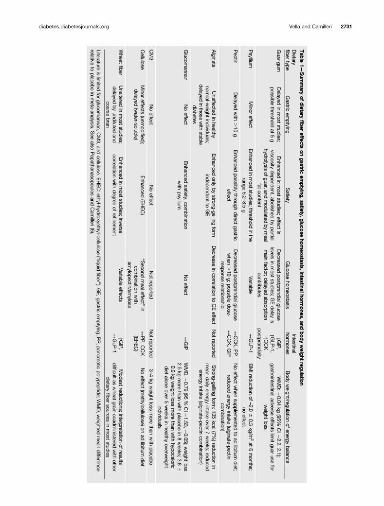
Table
1—Sum
mary
ofdietary
fiber
effectson
gastricem
ptying,satiety,glucosehom
eostasis,intestinalhorm
ones,andbody
weight
regulation
Dietary
fibertype
Gastric
emptying
Satiety
Glucose
homeostasis
Intestinalhorm
onesBody
weight/regulation
ofenergy
balance
Guar
gumDelayed
inmost
studies;possible
thresholdat
5g
Enhanced
inmost
studies;effect
isviscosity
dependent,abolished
bypartial
hydrolysisofguar,and
modulated
bymeal
fatcontent
Decreased
postprandialglucoselevels
inmost
studies;GEdelay
ismain
factor;delayed
absorptioncontributes
↓GIP,
↑GLP
-1,↑C
CK
postprandially
WMD:20.04
kg(95%
CI22.2,
2.1);gastrointestinaladverse
effectslim
itguar
usefor
weight
loss
Psyllium
Minor
effectEnhanced
inmoststudies;threshold
inthe
range5.2
–8.5g
Variable
↔GLP
-1BMIreduction
of–2.0
60.3
kg/m2at
6months;
noeffect
Pectin
Delayed
with
.10
gEnhanced
possiblythrough
directgastric
effectDecreased
postprandialglucosewhen
.10
g;possible
dose-response
relationship
↔CCK,PP
↔CCK,GIP
Noeffect
when
supplemented
toad
libitumdiet;
reducedenergy
intake(alginate-pectin
combination)
Alginate
Unaffected
inhealthy
normal-w
eightindividuals;
delayedinthose
with
stablediabetes
Enhanced
onlyby
strong-gellingform
independentto
GE
Decrease
incorrelation
toGEeffect
Not
reportedStrong-gelling
form:135
kcal(7%)reduction
inmean
dailyenergy
intakeover
7weeks;
reducedenergy
intake(alginate-pectin
combination)
Glucom
annanNoeffect
Enhanced
satiety,com
binationwith
psylliumNoeffect
↔GIP
WMD:2
0.79(95
%CI2
1.53,20.05);w
eightloss2.5
kgmore
thanwith
placeboin
8weeks;3.8
60.9
kgweight
lossmore
thanwith
hypocaloricdiet
aloneover
5weeks
inhealthy
overweight
individuals
CM3
Noeffect
Noeffect
Not
reportedNot
reported3–4
kgweight
lossmore
thanwith
placebo
Cellulose
Minor
effects(unm
odified);delayed
(water-soluble)
Enhanced
(EHEC)
“Second
mealeffect”
incom
binationwith
amylopectin/am
ylose
↔PP,CCK
(EHEC)
Noeffect
(methylcellulose)on
adlibitum
diet
Wheat
fiberUnaltered
inmost
studies;delayed
byundiluted
andcoarse
bran
Enhanced
inmost
studies;inverse
correlationwith
degreeof
refinem
entVariable
effects↑G
IP,
↔GLP
-1Modest
reductions;interpretation
ofresults
difficultas
wheat
graincoadm
inisteredwith
otherdietary
fibersources
inmost
studies
Literatureislim
itedfor
glucomannan,C
M3,and
cellulose.EHEC:ethyl-hydroxyethyl-cellulose
(“liquidfiber”);G
E,gastric
emptying;P
P,pancreatic
polypeptide;WMD,w
eightedmean
differencerelative
toplacebo
inmeta-analysis.S
eealso
Papathanasopoulos
andCam
illeri(6).
diabetes.diabetesjournals.org Vella and Camilleri 2731
GASTRIC ACCOMMODATION AND VAGALFUNCTION
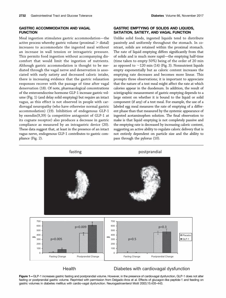
Meal ingestion stimulates gastric accommodation—theactive process whereby gastric volume (proximal . distal)increases to accommodate the ingested meal withoutan increase in wall tension or intragastric pressure.This permits food ingestion without accompanying dis-comfort that would limit the ingestion of nutrients.Although gastric accommodation is thought to be me-diated through the vagal nerve and denervation is asso-ciated with early satiety and decreased caloric intake,there is increasing evidence that the gastric relaxationresponses recover with the passage of time after vagaldenervation (18). Of note, pharmacological concentrationsof the enteroendocrine hormone GLP-1 increase gastric vol-ume (Fig. 1) (and delay solid emptying) but require an intactvagus, as this effect is not observed in people with car-diovagal neuropathy (who have otherwise normal gastricaccommodation) (19). Inhibition of endogenous GLP-1by exendin(9,39) (a competitive antagonist of GLP-1 atits cognate receptor) also produces a decrease in gastriccompliance as measured by an intragastric device (20).These data suggest that, at least in the presence of an intactvagus nerve, endogenous GLP-1 contributes to gastric com-pliance (Fig. 2).
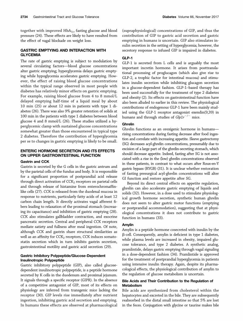
GASTRIC EMPTYING OF SOLIDS AND LIQUIDS,SATIATION, SATIETY, AND VAGAL FUNCTION
Unlike solid foods, ingested liquids tend to distributepassively and uniformly throughout the stomach. In co-ntrast, solids are retained within the proximal stomach.The rate of liquid emptying differs significantly from thatof solids and is much more rapid—the emptying half-time(time taken to empty 50%) being of the order of 20 minas opposed to ;120 min (14) (Fig. 3). Nonnutrient liquidsempty exponentially but as caloric content increases theemptying rate decreases and becomes more linear. Thisprompts three observations; it is important to appreciatethat the nature of a test meal might affect the rate at whichcalories appear in the duodenum. In addition, the result ofscintigraphic measurement of gastric emptying depends to alarge extent on whether it is bound to the liquid or solidcomponent (if any) of a test meal. For example, the use of alabeled egg meal measures the rate of emptying of a differ-ent phase than that measured by the systemic appearance ofingested acetaminophen solution. The final observation tomake is that liquid emptying is not completely passive andthe emptying rate is decreased by increasing caloric content,suggesting an active ability to regulate caloric delivery that isnot entirely dependent on particle size and the ability topass through the pylorus (10).
Figure 1—GLP-1 increases gastric fasting and postprandial volume. However, in the presence of cardiovagal dysfunction, GLP-1 does not alterfasting or postprandial gastric volume. Reprinted with permission from Delgado-Aros et al. Effects of glucagon-like peptide-1 and feeding ongastric volumes in diabetes mellitus with cardio-vagal dysfunction. Neurogastroenterol Motil 2003;15:435–443.
2732 Gastrointestinal Tract and Glucose Tolerance Diabetes Volume 66, November 2017
Solids food particles are subject to trituration by thecircular contractions of the gastric antrum that propel foodtoward the closed pylorus. These forces together with acidicand peptic digestion (which commences in the stomach)breaks food into particles of sufficiently small size (;2 mm)
that they can traverse the pylorus. The corollary of this isthat solid emptying is preceded by a lag phase where noemptying occurs followed by linear, postlag emptying. Sol-ids typically empty over a period of 3–4 h (14). However,the volume, consistency and fat content will affect empty-ing rate so that large, fatty meals may empty over periodslonger than 4 h. Gastric emptying rather than gastric ac-commodation appears to be the major gastric function de-termining postprandial satiation and satiety.
Bilateral truncal vagotomy, previously used to treatpeptic ulcer, results in delayed gastric emptying, earlysatiety and weight loss (18). This is believed to be, atleast in part, due to a decrease in gastric accommodationalthough symptoms decrease in severity over time. Thismay be due to the formation of collateral innervation oradaptation of the intrinsic enteric nervous system overtime. While studies in animals suggest that vagal innerva-tion modulates insulin secretion, insulin action and hepaticglucose metabolism (21), it is uncertain if these effectsmake significant contributions to the regulation of glucosemetabolism in humans (22). The use of chronic, but revers-ible, electrical vagal blockade in humans produced a signif-icant, but temporary, decrease in weight and caloric intake(23). No direct effects on glucose metabolism have beendemonstrated with electrical vagal blockade (22) althoughin 26 subjects with type 2 diabetes electrical vagal block-ade achieved sustained weight loss over a 1-year period,
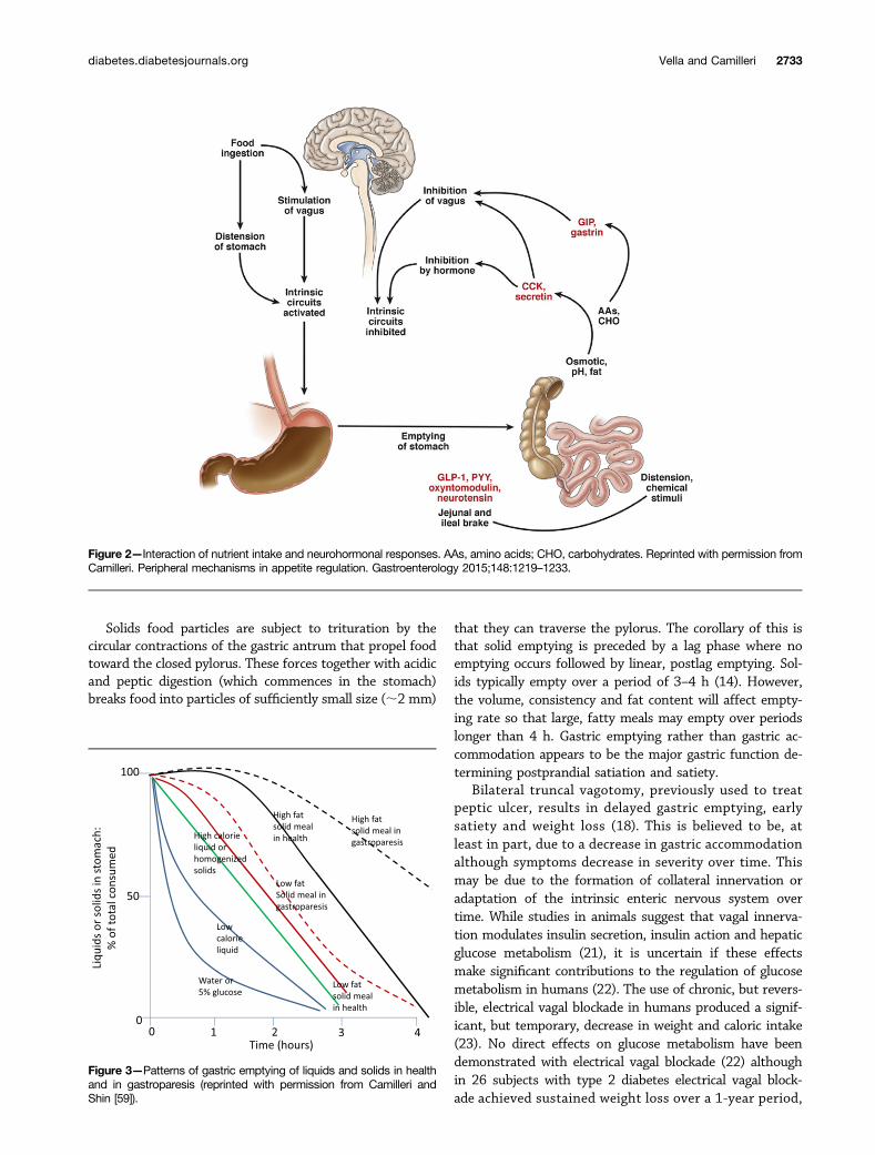
Figure 2—Interaction of nutrient intake and neurohormonal responses. AAs, amino acids; CHO, carbohydrates. Reprinted with permission fromCamilleri. Peripheral mechanisms in appetite regulation. Gastroenterology 2015;148:1219–1233.
Figure 3—Patterns of gastric emptying of liquids and solids in healthand in gastroparesis (reprinted with permission from Camilleri andShin [59]).
diabetes.diabetesjournals.org Vella and Camilleri 2733
together with improved HbA1c, fasting glucose and bloodpressure (24). These effects are likely to have resulted fromthe effect of vagal blockade on weight loss.
GASTRIC EMPTYING AND INTERACTION WITHGLYCEMIA
The rate of gastric emptying is subject to modulation byseveral circulating factors—blood glucose concentrationsalter gastric emptying; hyperglycemia delays gastric empty-ing while hypoglycemia accelerates gastric emptying. How-ever, the effect of raising blood glucose concentrationswithin the typical range observed in most people withdiabetes has relatively minor effects on gastric emptying.For example, raising blood glucose from 4 to 8 mmol/Ldelayed emptying half-time of a liquid meal by about10 min (25) or about 12 min in patients with type 1 di-abetes (26). There was also 9% greater retention of solids at100 min in the patients with type 1 diabetes between bloodglucose 4 and 8 mmol/L (26). These studies utilized a hy-perglycemic clamp with sustained glucose concentrationssomewhat greater than those encountered in typical type2 diabetes. Therefore the contribution of hyperglycemiaper se to changes in gastric emptying is likely to be small.
ENTERIC HORMONE SECRETION AND ITS EFFECTSON UPPER GASTROINTESTINAL FUNCTION
Gastrin and CCKGastrin is secreted by the G cells in the gastric antrum andby the parietal cells of the fundus and body. It is responsiblefor a significant proportion of postprandial acid releasethrough direct activation of CCK2 receptors on parietal cellsand through release of histamine from enterochromaffin-like cells (27). CCK is released from the duodenal mucosa inresponse to nutrients, particularly fatty acids of at least 12carbon chain length. It directly activates vagal afferent fi-bers leading to relaxation of the proximal stomach (increas-ing its capacitance) and inhibition of gastric emptying (28).CCK also stimulates gallbladder contraction, and exocrinepancreatic secretion. Central and peripheral CCK receptorsmediate satiety and fullness after meal ingestion. Of note,although CCK and gastrin share structural similarities aswell as an affinity for CCK2 receptors, CCK induces somato-statin secretion which in turn inhibits gastrin secretion,gastrointestinal motility and gastric acid secretion (29).
Gastric Inhibitory Polypeptide/Glucose-DependentInsulinotropic PolypeptideGastric inhibitory polypeptide (GIP), also called glucose-dependent insulinotropic polypeptide, is a peptide hormonesecreted by K cells in the duodenum and proximal jejunum.It signals through a specific receptor (GIPR). In the absenceof a competitive antagonist of GIP, most of its effects onphysiology are inferred from transgenic mice lacking thereceptor (30). GIP levels rise immediately after nutrientingestion, inhibiting gastric acid secretion and emptying.In humans these effects are observed at pharmacological
(supraphysiological) concentrations of GIP, and thus thecontribution of GIP to gastric acid secretion and gastricemptying in humans is uncertain. GIP also stimulates in-sulin secretion in the setting of hyperglycemia; however, thesecretory response to infused GIP is impaired in diabetes.
GLP-1GLP-1 is secreted from L cells and is arguably the mostimportant incretin hormone. It arises from posttransla-tional processing of proglucagon (which also give rise toGLP-2, a trophic factor for intestinal mucosa) and stimu-lates insulin secretion while inhibiting glucagon secretionin a glucose-dependent fashion. GLP-1–based therapy hasbeen used successfully for the treatment of type 2 diabetesand obesity (2). Its effects on gastrointestinal function havealso been alluded to earlier in this review. The physiologicalcontributions of endogenous GLP-1 have been mainly stud-ied using the GLP-1 receptor antagonist exendin(9,39) inhumans and through studies of Glp1r2/2 mice.
GhrelinGhrelin functions as an orexigenic hormone in humans—rising concentrations during fasting decrease after food inges-tion and correlate with increasing appetite. Sleeve gastrectomy(SG) decreases acyl-ghrelin concentrations, presumably due toexcision of a large part of the ghrelin-secreting stomach, whichshould decrease appetite. Indeed, fasting after SG is not asso-ciated with a rise in the (low) ghrelin concentrations observedin these patients, in contrast to what occurs after Roux-en-Ygastric bypass (RYGB) (31). It is unclear whether restorationof fasting presurgical acyl-ghrelin concentrations will alterGI function and restore appetite after SG.
Beyond its direct central effects on appetite regulation,ghrelin can also accelerate gastric emptying of liquids andsolids (32). However, in a dose used to stimulate physiolog-ical growth hormone secretion, synthetic human ghrelindoes not seem to alter gastric motor functions (emptyingor postprandial accommodation), suggesting that at physi-ological concentrations it does not contribute to gastricfunction in humans (33).
AmylinAmylin is a peptide hormone cosecreted with insulin by theb-cell. Consequently, amylin is deficient in type 1 diabetes,while plasma levels are increased in obesity, impaired glu-cose tolerance, and type 2 diabetes. A synthetic analog,pramlintide, delays gastric emptying through vagal signalingin a dose-dependent fashion (34). Pramlintide is approvedfor the treatment of postprandial hyperglycemia in patientsusing intensive insulin therapy. Again, despite its pharma-cological effects, the physiological contribution of amylin tothe regulation of glucose metabolism is uncertain.
Bile Acids and Their Contribution to the Regulation ofMetabolismBile acids are synthesized from cholesterol within thehepatocytes and excreted in the bile. They are subsequentlyreabsorbed in the distal small intestine so that 5% are lostin the feces. Conjugation with glycine or taurine makes bile
2734 Gastrointestinal Tract and Glucose Tolerance Diabetes Volume 66, November 2017
acids impermeable to cell membranes and confers detergent-like properties that help solubilize lipid micelles andfacilitate the absorption of fat in the small intestine (35).Bile acids are stored in the gallbladder during indigestiveperiods, but activation of CCK by fat emptying into duode-num stimulates gallbladder contraction and relaxation ofthe sphincter of Oddi, delivering bile to the small bowel.Bile acids act as natural ligands for the transcription factorfarnesoid X receptor (FXR). Activation of this nuclear receptorstimulates the expression of genes encoding proteins involvedin bile acid synthesis, transport, and metabolism. Activationby ligand binding causes heterodimerization with the retinoicX receptor a and subsequent binding to the promoter re-gions of target DNA (36). Bile acids differ in their ability toactivate FXR, suggesting that changes in bile acid composi-tion may alter the metabolic effects of FXR activation (37).
In addition, bile acids can also influence metabolismthrough the membrane-bound, G-protein–coupled bile acidreceptor 1 (also known as TGR5). TGR5 is expressed inadipose tissue, the enteric nervous system, and the enter-oendocrine L cells that produce GLP-1. Activation of thereceptor leads to activation of protein kinase A and phos-phorylation of target proteins. Increased GLP-1 secretionhas been attributed to increased bile acid delivery to thesmall intestine after bariatric surgery (35).
FGF19 production is almost exclusively restricted to theterminal ileum, which corresponds to the site where bileacids are actively taken up by the ileal sodium/bile acidcotransporter (38). Its expression is increased by FXR sig-naling, and it is secreted into the portal circulation where itsuppresses the rate-controlling enzyme of bile acid synthe-sis. It also stimulates hepatic glucose uptake and glycogensynthesis in an insulin-independent manner (39).
Changes in bile acid composition (and in the enterohepaticcirculation) may contribute to the metabolic benefits ofbariatric surgery. Certainly, differences among the variousbariatric procedures might shed some light on the contributionof bile acids to metabolic regulation in humans. Plasma bileacid concentrations are higher after RYGB compared withthose of weight-matched control subjects. However, thesechanges are not immediately apparent after surgery (at thetime when most of the metabolic changes are occurring) andare only evident ;1 year after the procedure (40).
In contrast, bile acid sequestration using bile acid bindingresins decreases bile acid reabsorption by the terminal ileum,leading to increased fecal loss of bile acids. This is accompa-nied by decreased FGF19 concentrations and increased bileacid synthesis (41). However, in humans the decrease inFGF19 is accompanied by decreased fasting glucose concen-trations and decreased meal ce of increased splanchnic ex-traction of the meal (42). Changes in GLP-1 concentrationshave been variable (increase or no change) (41,42).
Bariatric Surgery and Mechanistic Insights Into UpperGastrointestinal FunctionBariatric surgery is the most effective intervention forweight loss. Nonrandomized retrospective studies suggest
that the remission rate is associated with length of by-passed intestine (;85% for standard RYGB and ;98%with duodenal switch [43]). However, prospective, random-ized controlled trials have reported lower remission ratesfor diabetes with RYGB (44). Of note, caloric restrictionacutely improves b-cell function, suggesting that alteredcaloric intake in addition to altered intestinal anatomyand/or function is a key contributor to improved glucosetolerance (45).
On the other hand, remission of diabetes after bariatricsurgery seems to depend on the duration and severity ofdiabetes (as quantified by the number of oral medicationsand/or use of insulin) prior to the procedure (46). Thiswould suggest that the effects of bariatric surgery on theability to synthesize and secrete insulin are limited. Indeed,insulin secretion in response to hyperglycemia with GLP-1or GIP—a test of supramaximal b-cell function—did notchange after surgical invention (47). This effect is less ap-parent after oral challenges where quantification of isletfunction as a disposition index demonstrates improvementsin b-cell function that are mainly due to improved insulinaction (48). Acute caloric restriction improves insulin actionprior to any discernable weight loss (45), and indeed directcomparison of caloric restriction to the early changes afterbariatric surgery might suggest equivalence.
Procedures that result in more rapid appearance ofcalories in the proximal intestine result in increased GLP-1secretion. The extent to which this contributes to theremission of diabetes has been debated with some divergencein the literature. Acute blockade of the GLP-1 receptordecreases b-cell function after RYGB (49). However, in sub-jects with intact glucose effectiveness, the effect on glucosetolerance is marginal. GLP-1 also delays gastrointestinalmotility after RYGB (49).
GLP-1 also has direct effects on hypothalamic nucleioutside of the blood-brain barrier, and GLP-1 or GLP-1receptor agonists decrease food intake and cause weightloss (2). More recently, activation of the GLP-1 receptordecreased food intake and food-related brain responsesin patients with type 2 diabetes and in obese subjects asmeasured by functional MRI—an action blocked byexendin(9,39) (50). However, the effect of postprandialGLP-1 concentrations on satiety after RYGB or SG is atpresent uncertain (51).
There is some evidence that intestinal absorption ofnutrients is more rapid and efficient in obese individualsthan it is in lean humans, predisposing them to furtherweight gain (52). Active glucose transport measured using3-O-methylglucose (3OMG)—which is taken up by SGLT-1and GLUT2 but is not metabolized, being excreted un-changed in the urine—is increased in subjects post-RYGBcompared with lean subjects. In addition, intestinal glucoseabsorption is increased in obese subjects (53). While intrigu-ing, the methods used in those studies were qualitativebecause they measured differences in area under the curveof plasma 3OMG concentrations (reflecting the sum of in-testinal absorption and renal clearance) after a 30 min bolus
diabetes.diabetesjournals.org Vella and Camilleri 2735
of 3OMG. Moreover, they provided no information as tohow glucose absorption changes after RYGB.
The Intestinal Barrier and Nutrient AbsorptionThe gastrointestinal epithelium provides a selective barrierlimiting permeation of toxins while allowing passage ofnutrients and water. Disruption of the barrier may play arole in the pathogenesis of multiple gastrointestinal tractdisorders. Selectivity is achieved by the tight junctionsthat respond to extracellular stimuli and alter para-cellular permeability through changes in the multipleproteins that comprise the complex (54). Factors thatinfluence permeability include fatty acids in the intestinallumen (whether ingested directly or as products of bacterialfermentation). In addition, bile acids such as deoxycholicacid and chenodeoxycholic acid can increase paracellu-lar permeability (55). Intriguingly, bile acids alter bothpermeability and GLP-1 secretion (56) by signaling throughthe same receptor—TGR5. The intestinal barrier has beenassessed in vivo using oral administration of inert, water-soluble probe molecules such as sugars and radiolabeledEDTA that passively traverse the intestinal mucosa intothe bloodstream and are recovered unchanged in the urine.Such techniques suggest that intestinal permeability is in-creased in diabetes, contributing to postprandial hypergly-cemia and systemic inflammation (57) through mediatorssuch as lipopolysaccharide (58).
CONCLUSIONS
The upper gastrointestinal tract is not a passive conduitof nutrients that subsequently traverse the gut wall and stim-ulate an endocrine response. Instead, it serves to integrateintraluminal nutrients and neural, mechanical, and hormonalmechanisms to modulate the response to caloric ingestion.We hope that this brief overview helps crystallize currentknowledge and suggest future areas of research that mightlead to novel therapies for the prevention and treat-ment of obesity and type 2 diabetes in humans.
Acknowledgments. The authors thank Monica M. Davis from the EndocrineResearch Unit, Mayo Clinic, Rochester, MN, for secretarial assistance.Funding. A.V. and M.C. are supported by the National Institutes of Health NationalInstitute of Diabetes and Digestive and Kidney Diseases (DK78646 and DK92179,respectively).Duality of Interest. A.V. has received research grants from Novo Nordisk andhas served as an advisory board member for Sanofi. No other potential conflicts ofinterest relevant to this article were reported.
References1. Vella A, Shah P, Basu R, Basu A, Holst JJ, Rizza RA. Effect of glucagon-likepeptide 1(7-36) amide on glucose effectiveness and insulin action in people withtype 2 diabetes. Diabetes 2000;49:611–6172. Drucker DJ, Nauck MA. The incretin system: glucagon-like peptide-1 receptoragonists and dipeptidyl peptidase-4 inhibitors in type 2 diabetes. Lancet 2006;368:1696–17053. Rubino F, Forgione A, Cummings DE, et al. The mechanism of diabetes controlafter gastrointestinal bypass surgery reveals a role of the proximal small intestine inthe pathophysiology of type 2 diabetes. Ann Surg 2006;244:741–749
4. McKeown NM, Meigs JB, Liu S, Wilson PW, Jacques PF. Whole-grain intakeis favorably associated with metabolic risk factors for type 2 diabetes andcardiovascular disease in the Framingham Offspring Study. Am J Clin Nutr 2002;76:390–3985. Silva FM, Kramer CK, Crispim D, Azevedo MJ. A high-glycemic index, low-fiberbreakfast affects the postprandial plasma glucose, insulin, and ghrelin responses ofpatients with type 2 diabetes in a randomized clinical trial. J Nutr 2015;145:736–7416. Papathanasopoulos A, Camilleri M. Dietary fiber supplements: effects in obesityand metabolic syndrome and relationship to gastrointestinal functions. Gastroen-terology 2010;138:65–72.e1–e27. Geliebter A. Gastric distension and gastric capacity in relation to food intake inhumans. Physiol Behav 1988;44:665–6688. Vazquez Roque MI, Camilleri M, Stephens DA, et al. Gastric sensorimotorfunctions and hormone profile in normal weight, overweight, and obese people.Gastroenterology 2006;131:1717–17249. Hunt JN, MacDonald I. The influence of volume on gastric emptying. J Physiol1954;126:459–47410. Hunt JN. Mechanisms and disorders of gastric emptying. Annu Rev Med 1983;34:219–22911. Elias E, Gibson GJ, Greenwood LF, Hunt JN, Tripp JH. The slowing of gastricemptying by monosaccharides and disaccharides in test meals. J Physiol 1968;194:317–32612. von Schönfeld J, Evans DF, Renzing K, Castillo FD, Wingate DL. Human smallbowel motor activity in response to liquid meals of different caloric value and dif-ferent chemical composition. Dig Dis Sci 1998;43:265–26913. Heddle R, Dent J, Read NW, et al. Antropyloroduodenal motor responses tointraduodenal lipid infusion in healthy volunteers. Am J Physiol 1988;254:G671–G67914. Camilleri M. Integrated upper gastrointestinal response to food intake. Gas-troenterology 2006;131:640–65815. Park MI, Camilleri M, O’Connor H, et al. Effect of different macronutrients inexcess on gastric sensory and motor functions and appetite in normal-weight,overweight, and obese humans. Am J Clin Nutr 2007;85:411–41816. Feinle C, O’Donovan D, Doran S, et al. Effects of fat digestion on appetite, APDmotility, and gut hormones in response to duodenal fat infusion in humans. Am JPhysiol Gastrointest Liver Physiol 2003;284:G798–G80717. Ma J, Stevens JE, Cukier K, et al. Effects of a protein preload on gastricemptying, glycemia, and gut hormones after a carbohydrate meal in diet-controlledtype 2 diabetes. Diabetes Care 2009;32:1600–160218. Smith DK, Sarfeh J, Howard L. Truncal vagotomy in hypothalamic obesity.Lancet 1983;1:1330–133119. Delgado-Aros S, Kim DY, Burton DD, et al. Effect of GLP-1 on gastric volume,emptying, maximum volume ingested, and postprandial symptoms in humans. Am JPhysiol Gastrointest Liver Physiol 2002;282:G424–G43120. Schirra J, Nicolaus M, Woerle HJ, Struckmeier C, Katschinski M, Goke B. GLP-1regulates gastroduodenal motility involving cholinergic pathways. NeurogastroenterolMotil 2009;21:609–618, e21–e2221. Nishi S, Seino Y, Ishida H, et al. Vagal regulation of insulin, glucagon, andsomatostatin secretion in vitro in the rat. J Clin Invest 1987;79:1191–119622. Sathananthan M, Ikramuddin S, Swain JM, et al. The effect of vagal nerveblockade using electrical impulses on glucose metabolism in nondiabetic subjects.Diabetes Metab Syndr Obes 2014;7:305–31223. Sarr MG, Billington CJ, Brancatisano R, et al.; EMPOWER Study Group. TheEMPOWER study: randomized, prospective, double-blind, multicenter trial ofvagal blockade to induce weight loss in morbid obesity. Obes Surg 2012;22:1771–178224. Shikora S, Toouli J, Herrera MF, et al. Vagal blocking improves glycemic controland elevated blood pressure in obese subjects with type 2 diabetes mellitus. J Obes2013;2013:24568325. Hebbard GS, Samsom M, Sun WM, Dent J, Horowitz M. Hyperglycemia affectsproximal gastric motor and sensory function during small intestinal triglyceride in-fusion. Am J Physiol 1996;271:G814–G819
2736 Gastrointestinal Tract and Glucose Tolerance Diabetes Volume 66, November 2017
26. Schvarcz E, Palmér M, Aman J, Horowitz M, Stridsberg M, Berne C. Physio-logical hyperglycemia slows gastric emptying in normal subjects and patients withinsulin-dependent diabetes mellitus. Gastroenterology 1997;113:60–6627. Schmidt WE, Schmitz F. Genetic dissection of the secretory machinery in thestomach. Gastroenterology 2004;126:606–60928. Lal S, McLaughlin J, Barlow J, et al. Cholecystokinin pathways modulatesensations induced by gastric distension in humans. Am J Physiol Gastrointest LiverPhysiol 2004;287:G72–G7929. Beglinger C, Degen L. Fat in the intestine as a regulator of appetite–role of CCK.Physiol Behav 2004;83:617–62130. Hansotia T, Maida A, Flock G, et al. Extrapancreatic incretin receptors modulateglucose homeostasis, body weight, and energy expenditure. J Clin Invest 2007;117:143–15231. Lee WJ, Chen CY, Chong K, Lee YC, Chen SC, Lee SD. Changes in postprandialgut hormones after metabolic surgery: a comparison of gastric bypass and sleevegastrectomy. Surg Obes Relat Dis 2011;7:683–69032. Murray CD, Martin NM, Patterson M, et al. Ghrelin enhances gastric emptying indiabetic gastroparesis: a double blind, placebo controlled, crossover study. Gut 2005;54:1693–169833. Cremonini F, Camilleri M, Vazquez Roque M, et al. Obesity does not increaseeffects of synthetic ghrelin on human gastric motor functions. Gastroenterology2006;131:1431–143934. Samsom M, Szarka LA, Camilleri M, Vella A, Zinsmeister AR, Rizza RA.Pramlintide, an amylin analog, selectively delays gastric emptying: potential role ofvagal inhibition. Am J Physiol Gastrointest Liver Physiol 2000;278:G946–G95135. Kuipers F, Bloks VW, Groen AK. Beyond intestinal soap–bile acids in metaboliccontrol. Nat Rev Endocrinol 2014;10:488-49836. Staels B, Fonseca VA. Bile acids and metabolic regulation: mechanisms andclinical responses to bile acid sequestration. Diabetes Care 2009;32(Suppl. 2):S237–S24537. Albaugh VL, Flynn CR, Cai S, Xiao Y, Tamboli RA, Abumrad NN. Early increasesin bile acids post Roux-en-Y gastric bypass are driven by insulin-sensitizing, sec-ondary bile acids. J Clin Endocrinol Metab 2015;100:E1225–E123338. Inagaki T, Choi M, Moschetta A, et al. Fibroblast growth factor 15 functions asan enterohepatic signal to regulate bile acid homeostasis. Cell Metab 2005;2:217–22539. Dong X, Park S, Lin X, Copps K, Yi X, White MF. Irs1 and Irs2 signaling isessential for hepatic glucose homeostasis and systemic growth. J Clin Invest 2006;116:101–11440. Patti ME, Houten SM, Bianco AC, et al. Serum bile acids are higher in humanswith prior gastric bypass: potential contribution to improved glucose and lipid me-tabolism. Obesity (Silver Spring) 2009;17:1671–167741. Beysen C, Murphy EJ, Deines K, et al. Effect of bile acid sequestrants onglucose metabolism, hepatic de novo lipogenesis, and cholesterol and bile acid ki-netics in type 2 diabetes: a randomised controlled study. Diabetologia 2012;55:432–44242. Smushkin G, Sathananthan M, Piccinini F, et al. The effect of a bile acid se-questrant on glucose metabolism in subjects with type 2 diabetes. Diabetes 2013;62:1094–1101
43. Buchwald H, Avidor Y, Braunwald E, et al. Bariatric surgery: a systematic reviewand meta-analysis. JAMA 2004;292:1724–173744. Ikramuddin S, Korner J, Lee WJ, et al. Roux-en-Y gastric bypass vs intensivemedical management for the control of type 2 diabetes, hypertension, and hyper-lipidemia: the Diabetes Surgery Study randomized clinical trial. JAMA 2013;309:2240–224945. Kelley DE, Wing R, Buonocore C, Sturis J, Polonsky K, Fitzsimmons M. Relativeeffects of calorie restriction and weight loss in noninsulin-dependent diabetesmellitus. J Clin Endocrinol Metab 1993;77:1287–129346. Still CD, Wood GC, Benotti P, et al. Preoperative prediction of type 2 diabetesremission after Roux-en-Y gastric bypass surgery: a retrospective cohort study.Lancet Diabetes Endocrinol 2014;2:38–4547. Dirksen C, Bojsen-Møller KN, Jørgensen NB, et al. Exaggerated release andpreserved insulinotropic action of glucagon-like peptide-1 underlie insulin hyperse-cretion in glucose-tolerant individuals after Roux-en-Y gastric bypass. Diabetologia2013;56:2679–268748. Nguyen KT, Billington CJ, Vella A, et al. preserved insulin secretory capacity andweight loss are the predominant predictors of glycemic control in patients withtype 2 diabetes randomized to Roux-en-Y gastric bypass. Diabetes 2015;64:3104–311049. Shah M, Law JH, Micheletto F, et al. Contribution of endogenous glucagon-likepeptide 1 to glucose metabolism after Roux-en-Y gastric bypass. Diabetes 2014;63:483–49350. van Bloemendaal L, IJzerman RG, ten Kulve JS., et al. GLP-1 receptor activationmodulates appetite- and reward-related brain areas in humans. Diabetes 2014;63:4186–419651. Lee W-J, Chong K, Ser K-H, et al. Gastric bypass vs sleeve gastrectomy fortype 2 diabetes mellitus: a randomized controlled trial. Arch Surg 2011;146:143–14852. Seimon RV, Brennan IM, Russo A, et al. Gastric emptying, mouth-to-cecumtransit, and glycemic, insulin, incretin, and energy intake responses to a mixed-nutrient liquid in lean, overweight, and obese males. Am J Physiol Endocrinol Metab2013;304:E294–E30053. Nguyen NQ, Debreceni TL, Bambrick JE, et al. Upregulation of intestinal glucosetransporters after Roux-en-Y gastric bypass to prevent carbohydrate malabsorption.Obesity (Silver Spring) 2014;22:2164–217154. Suzuki T. Regulation of intestinal epithelial permeability by tight junctions. CellMol Life Sci 2013;70:631–65955. Camilleri M. Peripheral mechanisms in irritable bowel syndrome. N Engl J Med2012;367:1626–163556. Katsuma S, Hirasawa A, Tsujimoto G. Bile acids promote glucagon-like peptide-1secretion through TGR5 in a murine enteroendocrine cell line STC-1. Biochem BiophysRes Commun 2005;329:386–39057. Damci T, Nuhoglu I, Devranoglu G, Osar Z, Demir M, Ilkova H. Increased in-testinal permeability as a cause of fluctuating postprandial blood glucose levels intype 1 diabetic patients. Eur J Clin Invest 2003;33:397–40158. Koeth RA, Wang Z, Levison BS, et al. Intestinal microbiota metabolism ofL-carnitine, a nutrient in red meat, promotes atherosclerosis. Nat Med 2013;19:576–58559. Camilleri M, Shin A. Novel and validated approaches for gastric emptyingscintigraphy in patients with suspected gastroparesis. Dig Dis Sci 2013;58:1813–1815
diabetes.diabetesjournals.org Vella and Camilleri 2737