Embed Size (px)
Citation preview

SC I ENCE S I GNAL ING | R E S EARCH ART I C L E
CANCER
1Cancer Science Institute of Singapore, National University of Singapore, Singapore117599, Singapore. 2NUS Graduate School for Integrative Sciences and Engineering,National University of Singapore, Singapore 117456, Singapore. 3Department of Sur-gery and Cancer, Imperial College London, LondonW120NN, U.K. 4Department of Ob-stetrics and Gynecology, National University Health System, Singapore 119228,Singapore. 5Department of Biochemistry, Yong Loo Lin School of Medicine, NationalUniversity of Singapore, Singapore 117596, Singapore. 6Institute of Molecular and CellBiology, A*STAR (Agency for Science, Technology and Research), Singapore 138673,Singapore. 7Department of Anatomy, Yong Loo Lin School of Medicine, National Uni-versity of Singapore, Singapore 117597, Singapore.*Corresponding author. Email: [email protected]
Antony et al., Sci. Signal. 9, ra97 (2016) 4 October 2016
2016 © The Authors,
some rights reserved;
exclusive licensee
American Association
for the Advancement
of Science.
10.1126/scisignal.aaf8175
http:/D
ownloaded from
The GAS6-AXL signaling network is a mesenchymal(Mes) molecular subtype–specific therapeutic targetfor ovarian cancerJane Antony,1,2,3 Tuan Zea Tan,1 Zoe Kelly,3 Jeffrey Low,4 Mahesh Choolani,4 Chiara Recchi,3
Hani Gabra,3 Jean Paul Thiery,1,5,6 Ruby Yun-Ju Huang1,4,7*
Ovarian cancer is a complex disease with heterogeneity among the gene expression molecular subtypes (GEMS)between patients. Patients with tumors of a mesenchymal (“Mes”) subtype have a poorer prognosis than patientswith tumors of an epithelial (“Epi”) subtype. We evaluated GEMS of ovarian cancer patients for molecularsignaling profiles and assessed how the differences in these profiles could be leveraged to improve patient clin-ical outcome. Kinome enrichment analysis identified AXL as a particularly abundant kinase in Mes-subtype tumortissue and cell lines. In Mes cells, upon activation by its ligand GAS6, AXL coclustered with and transactivated thereceptor tyrosine kinases (RTKs) cMET, EGFR, and HER2, producing sustained extracellular signal–regulated kinase(ERK) activation. In Epi-A cells, AXL was less abundant and induced a transient activation of ERK without evidenceof RTK transactivation. AXL-RTK cross-talk also stimulated sustained activation of the transcription factor FRA1,which correlated with the induction of the epithelial-mesenchymal transition (EMT)–associated transcriptionfactor SLUG and stimulation of motility exclusively in Mes-subtype cells. The AXL inhibitor R428 attenuatedRTK and ERK activation and reduced cell motility in Mes cells in culture and reduced tumor growth in a chickchorioallantoic membrane model. A higher concentration of R428 was needed to inhibit ERK activation and cellmotility in Epi-A cells. Silencing AXL in Mes-subtype cells reversed the mesenchymal phenotype in culture andabolished tumor formation in an orthotopic xenograft mouse model. Thus, AXL-targeted therapy may improveclinical outcome for patients with Mes-subtype ovarian cancer.
/stkon October 5, 2016
e.sciencemag.org/
INTRODUCTIONEpithelial ovarian cancer is a lethal malignancy with nonspecific symp-toms in early stages that enable silent progression of the disease. Mostcases are diagnosed in the advanced stages, and despite treatment, prog-nosis is poor with 5-year survival lower than 30%. Locoregional dissem-ination of the tumor and metastases contribute to immense diseaseburden. Furthermore, epithelial ovarian cancer is hallmarked by a highdegree of genetic and epigenetic aberrations, resulting in a diverserepertoire of modified cellular networks that can be delineated into dis-tinct molecular subtypes based on gene expression profiling (1, 2).These gene expression molecular subtypes (GEMS) are robust and re-producible as reported in the Australian Ovarian Cancer Study (1), TheCancer Genome Atlas (TCGA) (2), and by a large-scale meta-analysisstudy (3). At least five distinct GEMS of epithelial ovarian cancer havebeen identified: epithelial-A (Epi-A), epithelial-B (Epi-B),mesenchymal(Mes), Stem-A, and Stem-B (3). Elucidation of key signaling networkshas further identified distinct players that promote tumorigenesis ineach subtype.
The Stem-A subtype, which corresponds to the C5 subtype fromthe Tothill data set and the proliferative subtype from the TCGA dataset, is driven by MYCN amplification (4) and FZD7 overexpression(5), is enriched in microtubule-related gene sets, and is dependent
on microtubule-associated genes for growth (3). The Mes subtype,which corresponds to the C1 subtype from the Tothill data set andthe mesenchymal subtype from the TCGA data set, shows high cor-relation with transforming growth factor–b (TGF-b) pathway in geneset enrichment analysis (GSEA) with poor survival outcomes (1–3).The TGF-b pathway promotes epithelial-mesenchymal transition(EMT) (6). Mes-subtype tumors display extensive desmoplastic stromalreactions (1) that are associated with EMT and a microRNA networksuppressing the expression of genes encoding E-cadherin (CDH1), vi-mentin (VIM), SLUG (SNAI2), and the epithelial transcriptional gate-keeper GRHL2 (7).
The RTK AXL belongs to the Tyro3-Axl-Mer (TAM) family ofRTKs (8) along with MER and Tyro-3 and is activated by its ligandgrowth arrest–specific 6 (GAS6) (9). Its expression has been shown tobe of prognostic value in breast (10) and lung (11) cancers. AXL has alsobeen suggested as a therapeutic target for breast (12) and pancreaticcancer (13), and cancers demonstrating resistance to epidermal growthfactor receptor (EGFR) and phosphatidylinositol 3-kinase (PI3K) inhi-bition (14–16). In epithelial ovarian cancer, AXL is highly expressed inadvanced-stage diseases, predominantly in peritoneal deposits andmetastases (17). Silencing AXL or blocking the GAS6-AXL pathwayprevents regional dissemination of ovarian cancer cells in vivo (18).However, the role of AXL in the heterogeneous molecular backgroundsof epithelial ovarian cancer and the potential functional and therapeuticimplication are yet to be defined.
The concept of GEMS-specific therapeutics has been well devel-oped in breast cancer (19). However, data supporting the GEMS-specific management in epithelial ovarian cancer are newly emerging.The GEMS-specific pathways in epithelial ovarian cancer further pointto the direction for developing new therapeutic strategies. The Stem-Asubtype exhibits preferential sensitivity to microtubule destabilizers,such as vinorelbine (3). Inhibiting the TGF-b pathway might be a
1 of 15

SC I ENCE S I GNAL ING | R E S EARCH ART I C L E
D
potential option for patients with the Mes subtype (20). The feasibilityto stratify epithelial ovarian cancer patients on the basis of GEMSusing a laboratory-based assay on formalin-fixed, paraffin-embedded(FFPE) diagnostic blocks has been demonstrated recently (21). There-fore, there is an eminent need in the field to quest for GEMS-specifictherapeutic targets for epithelial ovarian cancer patients to achievepersonalized precision management.
Here, we investigated the molecular differences in kinase signalingpatterns among the various ovarian cancer GEMS and whether theseprofiles are predictive of therapeutic efficacy. Our data suggest that, incontrast to the Epi-A subtype, the Mes subtype is hallmarked by a re-ceptor tyrosine kinase (RTK) cross-talk network sustained by theGAS6-AXL signaling node. The findings show how particularly theMes and Epi-A subtypes differ, in molecular and cell behavior pheno-types as well as in drug response, thereby supporting the future devel-opment of GEMS-stratified clinical trials for epithelial ovarian cancerpatients.
on October 5, 2016
http://stke.sciencemag.org/
ownloaded from
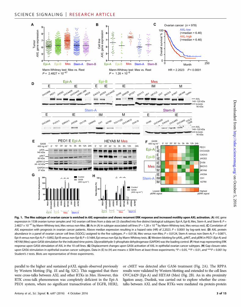
RESULTSInterrogation of kinome profiles identifies AXL as highlyexpressed in Mes ovarian tumors and cell linesUpon interrogating the kinome gene expression patterns in a pub-lished data set of ovarian cancer tumors and cell lines (3), we foundthat AXL was among the most highly expressed kinases in the Messubtype (fig. S1, A and B). Tumors and cell lines of the Mes subtypein this data set (3) had significantly greater AXL gene expression thandid the other subtypes (Fig. 1, A and B). Survival analysis showed thatAXL overexpression correlated with poor prognosis in ovarian cancerpatients (Fig. 1C) in both univariate and multivariate analyses (tableS1).AXLwas a prognostic factor for poor overall survival independentof known prognostic factors: patient age, tumor stage, tumor grade,and metastasis status. After stratification by the five GEMS, multi-variate Cox’s regression showed that AXL was still an independentprognostic factor for poor overall survival (table S2). The prognosticrelevance of AXL has been validated in an independent cohort (fig. S1,C and D).
Mes, but not Stem-A, was largely positive for AXL when exam-ining the total AXL protein expression in the SGOCL panel of ovar-ian cancer cell lines (22) matched with the GEMS (Fig. 1D) (3). Cellsof the Epi-A and Epi-B subtypes collectively were moderately posi-tive for AXL protein expression (Fig. 1D and fig. S1E). Because AXLhas been associated with EMT, we further examined, within eachGEMS, whether AXL expression would correlate with different phe-notypes along the previously described EMT spectrum: epithelial (E),intermediate epithelial (IE), intermediate mesenchymal (IM), andmesenchymal (M) (22). Whereas the GEMS are principally deli-neated on the basis of molecular profiles, the EMT phenotypes aresegregated on the basis of immunostaining and visualization of clas-sic EMT markers such as E-cadherin, vimentin, and cytokeratins.From a phenotypic standpoint, cell lines of the IM phenotype, whichhas previously been shown to be an aggressive, anoikis-resistant state(22), were all positive for AXL abundance (Fig. 1D and fig. S1F) andalso had the highest AXL abundance among the classifications (Fig.1D and fig. S1G). AXL protein abundance was high in both Epi-Aand Mes subtypes, whereas it was low in both Stem-A and Stem-Bsubtypes (Fig. 1D). These data suggest that although AXL is highlyexpressed in cells of the Mes and the IM phenotypes, it is alsoexpressed abundantly in cells of the Epi-A and the IE phenotypes.
Antony et al., Sci. Signal. 9, ra97 (2016) 4 October 2016
GEMS-specific AXL activation has divergent consequences indownstream ERK activation, cell proliferation, and motilityWe next asked whether AXL played different roles in Mes and Epi-A.Cells representing the different ovarian cancer GEMS (fig. S2A) werestimulated with GAS6 over time to study the kinetics of downstreamsignaling by Western blotting for phosphorylated extracellular signal–regulated kinase (pERK) and pAKT responses. Upon GAS6-AXL acti-vation, Mes showed two inductions of ERK phosphorylation at early(10, 20, and 30 min) and late (6 and 12 hours) time points (Fig. 1Eand fig. S2C). The other subtypes, such as Epi-A and Stem-A, showedsingle induction of ERK activation only at the early time points (10, 20,and 30min) (Fig. 1E and fig. S2C). Additional time course experimentsshowed that the pERK response in Epi-A subtype PEO1 cells is trulytransient with no recurrence, whereas Mes-subtype SKOV3 cellsshowed multiple waves of ERK activation (fig. S2D). Within eachGEMS, the EMT phenotypic classification further dictated the patternof AKT activation (Fig. 1E and fig. S2, B and C). In GEMS with singleinductions of pERK, such as Epi-A, Epi-B, and Stem-A subtype cells,AKT activation was induced once in cells of the epithelial orintermediate epithelial phenotype. However, in those of amesenchymalor intermediate mesenchymal phenotype, AKT activation occurred indual peaks (Fig. 1E and fig. S2, B and C). For Mes, AKT activation per-sisted from the early to the late time points regardless of the EMT phe-notype (Fig. 1E and fig. S2, B andC).Densitometric quantitation furthervalidated high abundance of pERK at both early and late time points inMes, whereas Epi-A, Epi-B, and Stem-A showed pERK only at initialstages (Fig. 1F). Because changes in the temporal duration of ERKsignaling elicit diverse biological outcomes (23, 24), the recurrence ofthe pERK signal upon GAS6-AXL activation suggests that the temporalduration of the signal is sustained in Mes and that this might affect rel-evant downstream biological functions.
AXL signaling promotes cancer cell survival (13, 25). A moderate in-crement in cell proliferation was observed upon GAS6 stimulation in allovarian cancer GEMS without specific differences (fig. S2E). AXLsignaling also enhances cancer cell invasion and metastasis (13, 26).We utilized single-cell tracking to monitor cell motility by measuringseveral kinetic parameters such as displacement, velocity, speed, and di-rectional persistence. Upon GAS6-AXL stimulation, Mes cells showedsignificant increase in all four kinetic parameters (fig. S2F). UponGAS6stimulation, Mes cell lines showed a significant enhancement indisplacement (Fig. 1G and fig. S2G) and in gap closuremigration assays(Fig. 1H and fig. S2H), whereas the other GEMS did not.
Together, GAS6-AXL activation has divergent control indownstream signaling and functions that are GEMS-specific in ovar-ian cancer. In Mes, it results in a recurrent sustained ERK response,which causes an increase in both proliferation and cell motility. How-ever, in Epi-A, GAS6-AXL activation stimulates a single and transientERK response that only results in an increase in proliferation.
GEMS-specific AXL activation reveals an extensive RTKcross-talk network in MesTo understand the signaling circuitry that is connected to the recur-rent ERK activation response specific to Mes-subtype cells, a reverse-phase protein array (RPPA) experiment was carried out using a Mescell line (SKOV3) and an Epi-A cell line (PEO1) treated with GAS6 at30 min and at 12 hours to evaluate the early and late responses, re-spectively. SKOV3 cells displayed increased abundance of pEGFR,pHER2, and pMET at both 30 min and 12 hours after GAS6 stimu-lation compared to the 0-min control (Fig. 2A). This pattern was
2 of 15
SC I ENCE S I GNAL ING | R E S EARCH ART I C L E
on October 5, 2016
http://stke.sciencemag.org/
Dow
nloaded from
parallel to the higher and sustained pAXL signals observed previouslyby Western blotting (Fig. 1E and fig. S2C). This suggested that therewere cross-talks between AXL and other RTKs in Mes. However, thisRTK cross-talk phenomenon was completely deficient in the Epi-APEO1 system, where no significant transactivation of EGFR, HER2,
Antony et al., Sci. Signal. 9, ra97 (2016) 4 October 2016
or cMET was detected after GAS6 treatment (Fig. 2A). The RPPAresults were validated by Western blotting and extended to the cell linesOVCA429 (Epi-A) and HEYA8 (Mes) (Fig. 2B). An in situ proximityligation assay, Duolink, was carried out to explore whether the cross-talks between AXL and these RTKs were mediated via protein-protein
**
PE
O1
OV
CA
429
CA
OV
3
JHO
S3
OV
56
SK
OV
3
HE
YA
8
CH
1
JHO
S4
A27
80
0
50
100
Gap
clo
sure
(%)
ControlGAS6 (400 ng ml-1)
*
Epi-A Epi-B Stem-A Mes
H
AX
L ge
ne e
xpre
ssio
n
A
Tum
or
4
5
6
7
8
9
p
Mann-Whitney test: Mes vs. Rest P = 2.4827 × 10−47
Stem-B Epi-A Epi-B Stem-A Mes 4
5
6
7
8
9
Mann-Whitney test: Mes vs. Rest P = 1.26 × 10⁻⁸
Cel
l lin
e
B
AX
L ge
ne e
xpre
ssio
n
Stem-B Epi-A Epi-B Stem-A Mes
HR = 2.2023 P < 0.0001
AXL-low (<median = 6.46) AXL-high (>median = 6.46)
Ove
rall
surv
ival
% 100
0 0 250Month
Ovarian cancer (n = 978) C
IE
Epi-A Epi-B Mes
Stem-A Stem-B
E IE IE E IE IM M
E IE IM M E
AXL 140−120 kDa
-Actin 40 kDa
AXL 140−120 kDa
-Actin 40 kDa
D
24h
pERK1/2 44−42 kDa
PEO1 E Epi-A HEYA8 M Mes 0’
GAS6 400 ng ml-1 10’ 20’ 30’ 1h 3h 6h 12h 24h 0’ 10’ 20’ 30’ 1h 3h 6h 12h
pAXL140 kDa
pAKT 60 kDa
GAPDH 37 kDa
E 12h 1h 3h
Lowest pERK signal
GAS6 400 ng ml-1
F
Highest pERK signal
PEO1
CAOV3
JHOS3
OV56
SKOV3
HEYA8
JHOS4
CH1
A2780
OVCA429
0’ 10’ 20’ 30’ 6h
Epi-A
Epi-B
Mes
E IE E IE IE IM M IE IM M
Stem-A
PE
O1
OV
CA
429
CA
OV
3
JHO
S3
OV
56
SK
OV
3
HE
YA
8
CH
1
JHO
S4
A27
80
0
50
100
150
200
No
rmal
ized
dis
pla
cem
ent Control
GAS6 (400 ng.ml-1)
* ** **
Epi-A Epi-B Stem-A Mes
G
24h
Fig. 1. The Mes subtype of ovarian cancer is enriched in AXL expression and shows recurrent ERK response and increasedmotility upon AXL activation. (A) AXL geneexpression in 1538 ovarian tumor samples and 142 ovarian cell lines from a data set (3) classified into five distinct biological subtypes: Epi-A, Epi-B, Mes, Stem-A, and Stem-B. P =2.4827 × 10−47 byMann-Whitney test, Mes versus non-Mes. (B) As in (A) in subtype-associated cell lines (P = 1.28 × 10⁻⁸ byMann-Whitney test, Mes versus rest). (C) Correlation ofAXL expression with prognosis in ovarian cancer patients. Above median expression resulting in a hazard ratio (HR) of 2.2023. P < 0.0001 by log-rank test. (D) AXL proteinabundance in a panel of ovarian cancer cell lines (SGOCL) assigned to the five subtypes. P = 0.0138, Mes versus non-Mes; P = 0.0124, Stem-A versus non-Stem-A; P = 0.0871,Epi-A versus non-Epi-A;P=0.892, Epi-B versus non-Epi-B; P= 0.1684, Epi versus non-Epi; byMann-Whitney tests. (E)Western blotting for pAXL, pAKT, and pERK in PEO1 (Epi-A) andHEYA8 (Mes) uponGAS6 stimulation for the indicated time points. Glyceraldehyde-3-phosphate dehydrogenase (GAPDH)was the loading control. (F) Heatmap representing ERKresponse upon GAS6 stimulation of AXL in the 10 cell lines. (G) Displacement changes upon GAS6 activation of AXL in epithelial ovarian cancer subtypes. (H) Gap closure ratesupon GAS6 stimulation in epithelial ovarian cancer subtypes. Data in (E) to (H) are means ± SEM from at least three experiments; *P < 0.05, **P < 0.01, and ***P < 0.001 byStudent’s t tests. Blots are representative of three experiments.
3 of 15

SC I ENCE S I GNAL ING | R E S EARCH ART I C L E
on October 5, 2016
http://stke.sciencemag.org/
Dow
nloaded from
interactions. Upon GAS6 stimulation in Mes cell lines SKOV3 andHEYA8, Duolink showed signals for interactions between AXL andthe phosphorylated forms of the RTKs Met, HER2 and EGFR (Fig. 2,C and D). This transactivation was not seen at the resting state whenGAS6 is absent (fig. S3A). This indicated that ligand-activated AXLmight trigger the phosphorylation of other RTKs through receptorclustering. This phenomenon of AXL-RTK clustering was not ob-
Antony et al., Sci. Signal. 9, ra97 (2016) 4 October 2016
served in Epi-A lines PEO1 and OVCA429 (Fig. 2, C and D). Thedifference in transactivation was not due to the expression status ofthe RTKs since all chosen cell lines were positive for cMET, HER2,and EGFR expression (fig. S3B).
Downstream of the RTK network, adapter proteins such as SHC,SRC, and SHP2 and the mitogen-activated protein kinase (MAPK)pathway kinases MEK1, ERK, and JNK had increased phosphorylation
PEO1 E Epi-A
OVCA429 IE Epi-A
SKOV3 IM Mes
HEYA8 M Mes
AX
L-A
XL P
ositive control
AX
L-pM
ET
A
XL-
pEG
FR
A
XL-
pHE
R2
30 min
(+GA
S6)
30 min
(+GA
S6)
30 min
(+GA
S6)
PEO1 E Epi-A
OVCA429 IE Epi-A
SKOV3 IM Mes
HEYA8 M Mes
C
0’ 12h pEGFR (pY1068) pEGFR (pY1173)
pcMET (Y1234 Y1235)Shc (pY317) Src (pY416)
SHP-2 (pY542) PKC- (pS660)
MEK1 (pS217 S221) pERK (pT202 Y204)
JNK (pT183 Y185) Elk1 (pS383)
FRA1 p90RSK (pT573)
pHER2 (pY1248)
elF4G
SKOV3 Mes
PEO1 Epi-A
Lowest median centred fold change
Highest median centred fold change
A
GAS6 400 ng ml-1 0’ 30’ 12h 30’
pEGFR 175 kDa
SLUG 30 kDa
pERK1/2 44 42 kDa
GAPDH 37 kDa
OVCA429 IE Epi-A
SKOV3 IM Mes
pHER2 185 kDa
PEO1 E Epi-A
HEYA8 M Mes
B
pMET 145 kDa
pAXL 140 kDa
pFRA1 40 kDa
12h 0’ 30’ 12h 0’ 30’ 12h 0’ 30’ 12h 0’ 30’ GAS6 400 ng ml-1
AX
L-p
ME
T
AX
L-p
EG
FR
AX
L-p
HE
R2
AX
L-p
ME
T
AX
L-p
EG
FR
AX
L-p
HE
R2
AX
L-p
ME
T
AX
L-p
EG
FR
AX
L-p
HE
R2
AX
L-p
ME
T
AX
L-p
EG
FR
AX
L-p
HE
R2
0
50
100
150
200 0 min30 min + GAS6
DU
Olin
k si
gn
al/c
ell
D
GAS6 24h
GRHL2
pAXL
pHER2 pEGFR
pERK
GAPDH
GAS6 0’ 10’ 20’ 30’ 1h 3h 6h 12h
OVCA429 shLuciferase control
10’ 20’ 30’ 1h 3h 6h 12h 24h 0’
OVCA429 shGRHL2_12
TWIST1
pAXL
pHER2 pEGFR
pERK
GAPDH
0’ 10’ 20’ 30’ 1h 3h 6h 12h 24h
OVCA429 control
10’ 20’ 30’ 1h 3h 6h 12h 24h 0’
OVCA429 TWIST1 E
Fig. 2. AXL is enmeshed in an RTK network in the Mes subtype. (A) RPPA depicting global proteomic and phosphoproteomic changes upon AXL stimulation by GAS6.(B) Western blotting or phosphorylated RTKs and downstream signaling moieties in epithelial ovarian cancer cell lines PEO1 and OVCA429 (Epi-A) and SKOV3 and HEYA8 (Mes)uponAXL activation. (C) Proximity ligation assay showing the extent of interactions of pMET, pHER2, and pEGFRwithAXL upon activationwith GAS6 in SKOV3 andHEYA8 (Mes) orPEO1 andOVCA429 (Epi-A) cells. As a positive control, AXL-AXL interactionwas assessedwith two different antibodies targeting the C terminus and N terminus of AXL. Scale bars,50 mm. (D) Quantification of Duolink signals in (C) using ImageJ. Data aremeans ± SEM from at least three experiments; *P < 0.05, **P < 0.01, and ***P< 0.001 by Student’s t tests.(E) Western blotting for pAXL, pEGFR, pHER2, and pERK in GAS6-stimulated OVCA429 cells, transfected with GRHL2 short hairpin–mediated RNA (shRNA), TWIST1, or thecorresponding control. Blots and microscopy images are representative of three experiments.
4 of 15

SC I ENCE S I GNAL ING | R E S EARCH ART I C L E
on October 5, 2016
http://stke.sciencemag.org/
Dow
nloaded from
upon GAS6 stimulation in SKOV3 but not in PEO1 cells (Fig. 2A).Their activation correlated with the phosphorylation of sustained re-sponse proteins such as ELK1, p90RSK, and FRA1 at 12 hours inSKOV3 cells, whichwas absent in PEO1 cells (Fig. 2A). FRA1 inductionalongside the sustained ERK activation was confirmed byWestern blotanalysis (Fig. 2B). Sustained activation of ERK stabilizes FRA1 (27), theactivity of which has been linked to the induction of EMT transcriptionfactors ZEB1 and SLUG and, consequently, increased motility (28).SLUG induction in Mes was confirmed by Western blotting (Fig. 2B).The MEK inhibitor PD0325901 (29) prevented phosphorylation of theMEK-ERK axis in Epi-A and Mes and consequently prevented induc-tion of FRA1 and SLUG in Mes (fig. S3C). Furthermore, MEK inhibi-tion preventedGAS6-AXL–mediatedmotility induction inMes (fig. S3,D to G), suggesting that AXL signaling relies on theMEK-ERK cascadeto induce motility. Furthermore, the ERK inhibitor SCH772984 (30)prevented the induction of pERK in a time course–dependent manner(fig. S3, H and I).
Together, our data indicate that the AXL-RTK network and thesignaling cascade responsible for the increased cell motility in Mesare deficient in other GEMS, such as Epi-A. The Mes subtype is wiredwith a unique signaling cross-talk network that contributes to itsbiological functions. Indeed, knocking down epithelial status main-taining transcription factor GRHL2 (7) or overexpressing EMTinducing TWIST1 (31) in epithelial OVCA429 promoted a mesenchy-mal phenotype (fig. S3J) and induced AXL-RTK cross-talk and recur-rent pERK response (Fig. 2E).
AXL kinase domain is crucial to initiate phosphorylationof RTKsTo study the functional relevance of the AXL kinase domian, we trans-fected AXL-null Epi-A cell line PEO4, and Mes cell line TYKnu with aplasmid encoding either functional AXL or a kinase-deficient K567Rmutant AXL (Fig. 3A) (32). Stimulation of AXL-transfected PEO4cells with GAS6 for 30 min induced the phosphorylation of AXLbut not EGFR, as expected of epithelial systems (Fig. 3B). Stimulationof transfected TYKnu cells with GAS6 induces the phosphorylation ofAXL as well as EGFR. However, in AXLK567R (kinase-deficient)–transfectedTYKnu cells, neither AXL nor EGFR was activated (Fig. 3B). Duolinkassays were carried out to corroborate these Western blot results andinvestigate the interaction between AXL and EGFR. AXL-EGFRclustering occurred in both AXL- and AXLK567R-transfected Mes-subtypeTYKnu cells; however, no pEGFR was detected in AXLK567R-transfectedcells (Fig. 3C). This suggests that GAS6-AXL signaling–inducedRTK cross-talk relies on the AXL kinase activity to phosphorylatethe other RTKs.
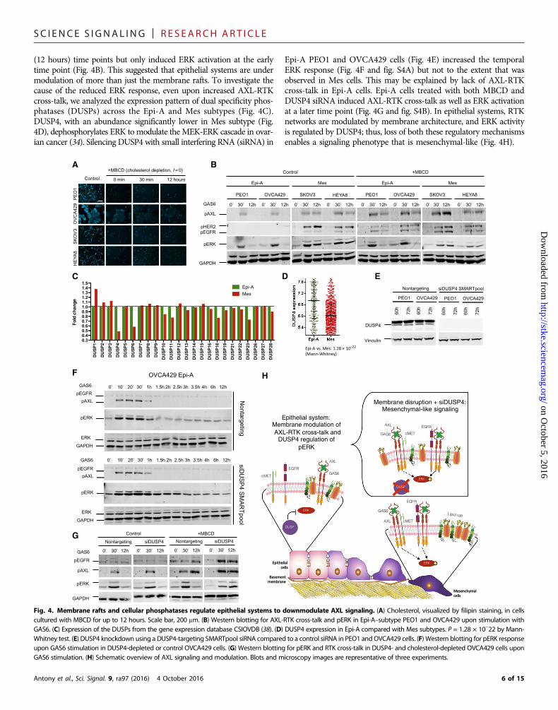
Epithelial systems are under dual regulation ofmembrane rafts and cellular phosphatases tocurtail AXL signalingTo decipher the differential modulation of AXL signaling in Epi andMes systems, we investigated the role of membrane-organized lipidrafts, which regulate signal transduction (33). We depleted cholesterol,an integral component of the lipid rafts, using methyl-b-cyclodextrin(MBCD) in Epi-A cell lines PEO1 and OVCA429 and Mes cell linesSKOV3 and HEYA8. Cholesterol depletion, as measured by the inten-sity of cholesterol-bound filipin staining, was observed up to 12 hoursafter MBCD treatment (Fig. 4A). Subsequent to loss of membranearchitecture integrity, GAS6 stimulation resulted in AXL-RTK cross-talk in Epi-A subtype PEO1 and OVCA429 at early (30 min) and late
Antony et al., Sci. Signal. 9, ra97 (2016) 4 October 2016
pAXL
pERK
GAPDH
AXL
ERK
pEGFR
EGFR
0’ 30’ 0’ 30’ 0’ 30’ 0’ 30’ 0’ 30’ 0’ 30’
Non transfected
AXLWT AXL K567R Non transfected
AXLWT AXLK567R
GAS6
PEO4 TYKnu
PE
O4
Epi
-A
TY
Knu
Mes
A
C
B
PE
O4
Epi
-A
TY
Knu
Mes
Nontransfected AXLWT AXLK567R
Nontransfected AXLWT AXLK567R
DAPI/pEGFR/AXL-EGFR complex
Fig. 3. AXL kinase domain is crucial for phosphorylation of other RTKs. (A) Im-munostaining of AXL-deficient Epi-A subtype PEO4 cells andMes-subtype TYKnu cells,transfected with wild-type AXL (AXLWT) or AXLK567R. (B) Western blotting for pAXL,pEGFR, and pERK in Mes TYKnu and in Epi-A PEO1 cells transfected with AXLWT orAXLK567R and stimulated with GAS6. (C) Proximity ligation assay to assess AXL-EGFRcross-talk (red dots) upon stimulation with GAS6 in Mes TYKnu and in Epi-A PEO4 cellstransfected with AXLWT or AXLK567R, alongside immunostaining for pEGFR (green).Scale bars, 50 mm. Blots and microscopy images are representative of threeexperiments.
5 of 15
SC I ENCE S I GNAL ING | R E S EARCH ART I C L E
(12 hours) time points but only induced ERK activation at the earlytime point (Fig. 4B). This suggested that epithelial systems are undermodulation of more than just the membrane rafts. To investigate thecause of the reduced ERK response, even upon increased AXL-RTKcross-talk, we analyzed the expression pattern of dual specificity phos-phatases (DUSPs) across the Epi-A and Mes subtypes (Fig. 4C).DUSP4, with an abundance significantly lower in Mes subtype (Fig.4D), dephosphorylates ERK to modulate the MEK-ERK cascade in ovar-ian cancer (34). Silencing DUSP4 with small interfering RNA (siRNA) in
Antony et al., Sci. Signal. 9, ra97 (2016) 4 October 2016
Epi-A PEO1 and OVCA429 cells (Fig. 4E) increased the temporalERK response (Fig. 4F and fig. S4A) but not to the extent that wasobserved in Mes cells. This may be explained by lack of AXL-RTKcross-talk in Epi-A cells. Epi-A cells treated with both MBCD andDUSP4 siRNA induced AXL-RTK cross-talk as well as ERK activationat a later time point (Fig. 4G and fig. S4B). In epithelial systems, RTKnetworks are modulated by membrane architecture, and ERK activityis regulated by DUSP4; thus, loss of both these regulatory mechanismsenables a signaling phenotype that is mesenchymal-like (Fig. 4H).
on October 5, 2016
http://stke.sciencemag.org/
Dow
nloaded from
DU
SP
1
DU
SP
2
DU
SP
3
DU
SP
4
DU
SP
5
DU
SP
6
DU
SP
7
DU
SP
8
DU
SP
9
DU
SP
10
DU
SP
11
DU
SP
12
DU
SP
13
DU
SP
14
DU
SP
15
DU
SP
16
DU
SP
18
DU
SP
19
DU
SP
21
DU
SP
22
DU
SP
23
DU
SP
26
DU
SP
27
DU
SP
28
0.30.40.50.60.70.80.91.01.11.21.31.41.5
Fold
cha
nge
Epi-AMes
ERK
AXL
GAS6cMET
EGFR
DUSP
AXL
GAS6
cMET
EGFR
Lipid raft
ERK
AXL
GAS6 cMET
EGFR
ERK
DUSP
12h 12h 12h 12h 12h 12h 12h 12h
PEO1 OVCA429 SKOV3 HEYA8 PEO1 OVCA429 SKOV3 HEYA8
Epi-A Mes Epi-A Mes
Control +MBCD
pAXL
pHER2 pEGFR
pERK
GAPDH
GAS6
3h 2h 2.5h 1h 1.5h 3.5h 4h 6h 12h
OVCA429 Epi-A
pAXL
pERK
GAPDH
ERK
pEGFR 3h 2h 2.5h 1h 1.5h 3.5h 4h 6h 12h GAS6
Nontargeting
siD
US
P4 S
MA
RT
pool
pAXL
pERK
GAPDH
ERK
pEGFR
GAS6
G
pAXL
pERK
GAPDH
Nontargeting siDUSP4 Nontargeting siDUSP4
Control +MBCD
GAS6
pEGFR
Epithelial system: Membrane modulation of AXL-RTK cross-talk and
DUSP4 regulation of pERK
60h
72h
60h
72h
60h
72h
60h
72h
DUSP4
Vinculin
PEO1 OVCA429 PEO1 OVCA429
Nontargeting siDUSP4 SMARTpool
0’ 10’ 20’ 30’
0’ 10’ 20’ 30’
12h 12h 12h 12h 0’ 30’ 0’ 30’ 0’ 30’ 0’ 30’
-
Fig. 4. Membrane rafts and cellular phosphatases regulate epithelial systems to downmodulate AXL signaling. (A) Cholesterol, visualized by filipin staining, in cellscultured with MBCD for up to 12 hours. Scale bar, 200 mm. (B) Western blotting for AXL-RTK cross-talk and pERK in Epi-A–subtype PEO1 and OVCA429 upon stimulation withGAS6. (C) Expression of the DUSPs from the gene expression database CSIOVDB (38). (D) DUSP4 expression in Epi-A compared with Mes subtypes. P = 1.28 × 10⁻²² by Mann-Whitney test. (E) DUSP4 knockdown using a DUSP4-targeting SMARTpool siRNA compared to a control siRNA in PEO1 and OVCA429 cells. (F) Western blotting for pERK responseupon GAS6 stimulation in DUSP4-depleted or control OVCA429 cells. (G) Western blotting for pERK and RTK cross-talk in DUSP4- and cholesterol-depleted OVCA429 cells uponGAS6 stimulation. (H) Schematic overview of AXL signaling and modulation. Blots and microscopy images are representative of three experiments.
6 of 15

SC I ENCE S I GNAL ING | R E S EARCH ART I C L E
The Mes subtype is more sensitive to the AXLinhibitor R428R428, an AXL-selective inhibitor, blocks tumor dissemination and pro-longs survival in mouse models of metastatic breast cancer (12). Tosupport specificity of the inhibitor, we first confirmed that R428 didnot disrupt EGF-EGFR signaling (fig. S5A). To test whether R428causes different effects among ovarian cancer GEMS, we treated 10selected cell lines representing the different GEMS (fig. S2A) withR428 for 48 hours to determine the 50% growth inhibition (GI50) con-
Antony et al., Sci. Signal. 9, ra97 (2016) 4 October 2016
centrations. Mes cell lines had a significantly lower mean GI50 of 2.91mM compared to the other subtypes with a mean GI50 of 9.38 mM (P =0.016) (Fig. 5A). Furthermore, AXL-deficient Epi-A PEO4 cells, andMes TYKnu cells, were transfected with wild-type or kinase-deficient(K567R) AXL and treated with R428. The R428-mediated GI50 inAXL-null PEO4 and TYKnu cells was high at 195.9 and 262.86 mM,respectively, enforcing the specificity of the AXL inhibitor (fig. S5B).High GI50 was also observed in kinase-deficient AXLK567R-transfectedPEO4 cells (276.6 mM) and TYKnu cells (245.5 mM), confirming the
on October 5, 2016
http://stke.sciencemag.org/
Dow
nloaded from
Non-Mes Mes0
5
10
15
20
25
GI 50
R42
8 (
m)
*
DMSO 2.91 M 9.38 M DMSO 2.91 M 9.38 M
0
5
10
0
20
40
60
80
SKOV3 IM Mes
0
5
10
15
10
20
30
40
HEYA8 M Mes
0’ 30’ 3h 12h 0’ 30’ 3h 12h 0’ 30’ 3h 12h 0’ 30’ 3h 12h 0’ 30’ 3h 12h 0’ 30’ 3h 12h
OVCA429 IE Epi-A
0
5
10
0
20
40
60
80
100
0
2
4
6
8
0
50
100
150
PEO1 E Epi-A
pER
K in
tens
ity
pAX
L in
tens
ity
0’ 30’ 3h 12h GAS6 0’ 30’ 3h 12h 0’ 30’ 3h 12h 0’ 30’ 3h 12h 0’ 30’ 3h 12h 0’ 30’ 3h 12h
DMSO 2.91 M 9.38 M DMSO 2.91 M 9.38 M R428
*****
* * ** * *
0.0
0.1
0.2
0.3
Vel
ocity
(µm
/s)
DM
SO
con
trol
R42
8 2.
91 µ
MR
428
9.38
µM
DM
SO
con
trol
R42
8 2.
91 µ
MR
428
9.38
µM
DM
SO
con
trol
R42
8 2.
91 µ
MR
428
9.38
µM
DM
SO
con
trol
R42
8 2.
91 µ
MR
428
9.38
µM
PEO1 OVCA429 SKOV3 HEYA8
A
D
DM
SO
Ctr
l
R42
8 3
µM
R42
8 10
µM
DM
SO
Ctr
l
R42
8 3
µM
R42
8 10
µM
DM
SO
Ctr
l
R42
8 3
µM
R42
8 10
µM
0
100
200
300
400
Tum
or v
olum
e (m
m3 )
HEYA8SKOV3PEO1
**
**
*E F
12h 3h 0’ 30’ 0’ 3h 12h 30’ 3h 12h 0’ 30’ 0’ 3h 12h 30’ 3h 12h 0’ 30’ 0’ 12h 30’ 3h
DMSO Ctrl 2.91 M 9.38 M R428 DMSO Ctrl 2.91 M 9.38 M B PEO1 E Epi-A OVCA429 IE Epi-A
SKOV3 IM Mes HEYA8 M Mes
pERK 1/2 44−42 kDa
pAXL 140 kDa
GAPDH 37 kDa
GAS6 400 ng ml-1
pERK 1/2 44 − 42 kDa
pAXL 140 kDa
GAPDH 37 kDa
HE
YA
8
M M
es
PE
O1
E
Epi
-A
SK
OV
3
IM M
es
DMSO Ctrl 2.91 M 9.38 M R428
C
Fig. 5. TheMes subtype ismore sensitive to theAXL inhibitorR428. (A) GI50 values for AXL inhibitor R428 in epithelial ovarian cancer cell lines.P=0.0167,Mes versus non-Mesby Mann-Whitney test. (B) Western blotting for pAXL, pAKT, and pERK in epithelial ovarian cancer cell lines PEO1 and OVCA429 (Epi-A) or SKOV3 and HEYA8 (Mes), preincubatedwith DMSO [control (Ctrl)], 2.91 mM R428 (GI50
Mes), or 9.38 mM R428 (GI50non-Mes) for an hour, then stimulated with GAS6 for 0.5, 3, or 12 hours. (C) Quantitation of immunofluore-
sence staining for pAXL, pAKT, and pERK in epithelial ovarian cancer cell lines PEO1 and OVCA429 (Epi-A) or SKOV3 and HEYA8 (Mes) upon treatment with the indicated dose ofR428. (D) Velocity of Epi-A– and Mes-subtype cell lines exposed to 2.91 mM (GI50
Mes) or 9.38 mM R428 (GI50non-Mes). (E) Chick CAMmodel of Mes or Epi-A cell line tumor growth in
response to R428. Scale bars, 2000 mm. (F) Tumor volume as the mean radius (r) of the fluorescent tumor was calculated relative to background. Tumor was assumed to be aspheroid with volume 4pr3/3. Data in (A), (C), (D), and (F) are means ± SEM from at least three experiments; *P < 0.05, **P < 0.01, and ***P < 0.001 by Student’s t tests. Blots andmicroscopy images are representative of three experiments.
7 of 15

SC I ENCE S I GNAL ING | R E S EARCH ART I C L E
on October 5, 2016
http://stke.sciencemag.org/
Dow
nloaded from
relevance of the AXL kinase domain (fig. S5B). Transfections withfunctional wild-type AXL showed a GI50 of 30.48 mM in Epi-APEO4 cells and a significantly lower GI50 of 3.45 mM in Mes TYKnucells (fig. S5B).
Because Mes has a unique GAS6-AXL signaling network, we ex-plored the effect of R428 on this signaling network in comparisonto Epi-A. Two Epi-A lines (PEO1 and OVCA429) and two Meslines (SKOV3 and HEYA8) were chosen and preincubated with di-methyl sulfoxide (DMSO; control), 2.91 mM (GI50
Mes), and 9.38 mM(GI50
non-Mes) of R428. From both Western blotting and immuno-staining, the early-phase ERK activation in Epi-A was only inhibited atthe higher concentration of R428 at 9.38 mM, whereas the early- andlate-phase ERK activation in Mes were effectively inhibited at the low-er concentration of 2.91 mM (Fig. 5, B and C, and fig. S5C). In addi-tion, these four cell lines also represent different phenotypes along theEMT spectrum (E, PEO1; IE, OVCA429; IM, SKOV3; M, HEYA8).From the phenotypic perspective, lower R428 concentration of 2.91mM had no effect on the pERK and pAXL activations in PEO1 (E),whereas OVCA429 (IE) showed a moderate reduction (Fig. 5C andfig. S5C). The SKOV3 (IM) line showed a drastic decrease, whereasthe HEYA8 (M) line showed complete inhibition at 2.91 mM (Fig.5C and fig. S5C). Mes shows dependence on the GAS6-AXL axisand is therefore very sensitive to AXL inhibition. In addition, complexRTK networks connected by AXL might govern the phenotypictransition along the EMT gradient.
To analyze the functional consequences following AXL signaling in-hibition, we preincubated PEO1, OVCA429, SKOV3, andHEYA8withR428 and tracked for motility. Similar to the pERK and pAXL signalinginhibition, Epi-A cell lines showedmotility inhibition only at the higherconcentration of R428, whereas Mes cell lines showed effective inhibi-tion at the lower concentration (Fig. 5D and fig. S5, D to F).
To translate the AXL signaling inhibition and functional repressionto a biologically relevant system, we embedded the four cell lines inMa-trigel and deposited on top of the chick chorioallantoic membrane(CAM) model to study the relevance of AXL inhibition in ovo. Thein ovo findings substantiated the in vitro results, with HEYA8 andSKOV3 forming large tumors in the CAM, which were significantlysuppressed at 2.91 mM as well as at 9.38 mM of R428 (Fig. 5, E andF). Unlike its Mes counterparts, tumor formation in PEO1 was notinhibited at 2.91 mM but only at 9.38 mM (Fig. 5, E and F). TheOVCA429 line proved to be nontumorigenic in the CAM system.Together, these results indicated the preferential application of in-hibiting AXL in the Mes subtype of epithelial ovarian cancer, in whichsignificant sensitization to R428 can be achieved.
AXL governs the sequential phenotypic and functionaltransition of EMTApart from pharmacological inhibition of AXL, shRNA-mediated si-lencing of AXL was also utilized to better understand its functional rolein ovarian cancer. Two shRNAs (shAXL#40 and shAXL#41) were usedto knock down AXL, and a nontargeting shRNA (shLuciferase) wasused as a control, in Epi-A OVCA429 andMes SKOV3 cell lines. Morethan 90% reduction in total AXL protein expression was achieved byboth shRNAs (Fig. 6A). Phase-contrast images revealed that in shAXLclones,OVCA429 formedmore compact colonies, and SKOV3 revertedfrom a fibroblastic to a loosely defined colony structure (Fig. 6B). Im-munofluorescence staining revealed that shAXL OVCA429 cellsincreased the intensity of E-cadherin staining at the cell-cell junctionstogether with a complete restoration of cytoplasmic b-catenin to the
Antony et al., Sci. Signal. 9, ra97 (2016) 4 October 2016
junctions (Fig. 6C). The shAXL SKOV3 cells showed reexpression ofE-cadherin, which was mostly cytoplasmic with partial restoration tothe cell-cell junctions (Fig. 6C). These data suggested that AXLknockdown causes a partial reversal along the EMT spectrum. The re-versal inOVCA429 is froman IE state to amore epithelial state, whereasthe SKOV3 reverts from an IM state to an IE state. This implies that theextent of EMT reversal upon AXL knockdown is sequential and de-pends on the cell line’s original EMT state (Fig. 6D). Note that althoughtheMes subtype is indeed more sensitive to pharmacological inhibitionof AXL at lower doses of R428, shRNA-mediated AXL silencing did notinduce differential survival benefits but induced EMT reversal in bothEpi-A and Mes.
To substantiate the functional effect of partial EMT reversal inshAXL cells, we carried out motility assays. Single-cell motility analysisand gap closure assays in shAXL#40 and shAXL#41 clones showed asignificant reduction (fig. S6, A to C). These results were consistent withthe functional changes in cell motility after R428 inhibition.
AXL knockdown reduces anchorage-independent growth,invasion, and tumor formationIn addition to the effect on EMT reversal, proliferation, soft agar, andinvasion assays were carried out in the shAXL cells. Both Mes SKOV3and Epi-A OVCA429 shAXL cell lines showed reduced proliferation(Fig. 6, E and F). This is concordant with the proliferative effects ob-served after GAS6 stimulation that both Epi-A and Mes require AXLsignaling for cell growth. For soft agar assays, OVCA429 cells showedminimal colony formation, which was completely abrogated in shAXL(Fig. 6G and fig. S6D). In SKOV3, the control cells showed significantcolony formation in soft agar, and shAXLattenuated its clonogenic abil-ity (Fig. 6G and fig. S6D). Spheroid invasion assays showed a similartrend with OVCA429, forming minimally invasive spheroids, an effectthat was completely prevented in shAXL. SKOV3 showed numerousinvasive protrusions from the spheroids, which was abolished in shAXL(Fig. 6H and fig. S6E).
When the spheroid invasion assay was performed in reduced serumconditions with GAS6 treatment, only theMes SKOV3 cell line showedinvasion, but not the Epi-A OVCA429 cell line (fig. S6, F and G). Ourdata suggested that AXL played an important role in both the anchorage-dependent and anchorage-independent growth. It is particularly essentialfor the mesenchymal phenotype such as Mes that AXL is required foranchorage independency.
In an orthotopic xenograft tumor model in mice, shAXL#40 andshAXL#41 SKOV3 cells showed complete inhibition of tumor forma-tion and growth compared to shLuciferase control. Tumor inhibitionwas 100%, with SKOV3 shLuciferase control forming tumors [averageweight of 0.3658 g (SEM, 0.1563); average number of 5.8 (SEM, 0.74)],and the SKOV3 shAXL#40 and shAXL#41 clones forming no tumors(P = 0.0474; P = 0.0001) (Fig. 6, I and J). This is consistent with theR428-mediated tumor reduction observed in the in ovo CAM model,suggesting that AXL is indeed a key signaling node for Mes.
The AXL signature is clinically relevant forpatient stratificationA gene signature of the top 30 genes correlating with AXL expression(Fig. 7A) was derived from the gene expression microarray analysis(3). This “AXL gene signature” was significantly overexpressed inMes-type tumors (Fig. 7B). EMT scoring is a quantitative measureof the effects of EMT on cancer progression (35); GEMS classificationand EMT status in ovarian tumors robustly correlate with a poor
8 of 15

SC I ENCE S I GNAL ING | R E S EARCH ART I C L E
on October 5, 2016
http://stke.sciencemag.org/
Dow
nloaded from
prognosis in patients (35). By analyzing the gene expression profilingmeta-analysis cohort (3), the AXL gene signature positively correlatedwith the ovarian-specific EMT score (Rho = +0.4148, P = 5.23 × 10−65)(Fig. 7C) and negatively correlated with overall patient survival (Fig.7D). These findings were validated in an independent cohort (fig. S7,
Antony et al., Sci. Signal. 9, ra97 (2016) 4 October 2016
A to C). Multivariate analysis also shows a significant prognosticrelevance of the AXL signature (table S3). In addition, the AXL genesignature was significantly overexpressed in omental metastases (36)(P = 0.0078) and platinum-resistant relapsed tumors (37) (P =0.0059) compared to their paired primary tumors (Fig. 7E). This is
Epithelial mesenchymal Intermediate epithelial
Intermediate mesenchymal
SKOV3 OVCA429 shAXL shAXL
D
shLu
cife
rase
sh
AX
L#41
s
hAX
L#40
OVCA429 IE Epi-A SKOV3 IM Mes Bsh
Luci
fera
se
shA
XL#
40
shA
XL#
41
AXL 140 kDa
-Actin 40 kDa
shLu
cife
rase
shA
XL#
40
shA
XL#
41
OVCA429 IE Epi-A SKOV3 IM Mes A
shLuciferase shAXL#41 shAXL#40 shLuciferase shAXL#41 shAXL#40 OVCA429 IE Epi-A SKOV3 IM Mes G
shLuciferase shAXL#41 shAXL#40 shLuciferase shAXL#41 shAXL#40 OVCA429 IE Epi-A SKOV3 IM Mes H
shControl shAXL#40 shAXL#410.0
0.5
1.0
1.5
Tu
mo
r w
eig
ht
(g)
**
shControl shAXL#40 shAXL#410
2
4
6
8
Tu
mo
r n
um
ber
********
0 2 4 6 80.0
2.5 106
5.0 106
7.5 106
1.0 107
1.3 107
Day
Num
ber o
f cel
ls
shLuciferaseshAXL40shAXL41
*** ***
0 2 4 6 80
2 106
4 106
6 106
Day
Num
ber o
f cel
ls
shLuciferaseshAXL40shAXL41
*** ***
E F I JOVCA429 IE Epi-A SKOV3 IM Mes SKOV3 IM Mes SKOV3 IM Mes
shLu
cife
rase
sh
AX
L#41
s
hAX
L#40
E-cadherin E-cadherin Merge Merge
OVCA429 IE Epi-A SKOV3 IM Mes C
Fig. 6. AXL governs the sequential phenotypic and functional transition of EMT. (A) Western blotting of AXL in OVCA429 and SKOV3 cells upon transfectionwith one of thetwo AXL shRNAs or a control (shLuciferase). (B) Phase-contrast images showing colony formation phenotype upon AXL knockdown inOVCA429 and SKOV3 cells. Scale bars, 200 mm.(C) Immunostaining for E-cadherin and b-catenin abundance and localization in OVCA429 and SKOV3 cells upon AXL knockdown. Scale bars, 50 mm. (D) Schematic of partial EMTreversal facilitated by AXL knockdown, from an “intermediatemesenchymal” to an “intermediate epithelial” state in SKOV3 cells, and from an “intermediate epithelial” state to amore“epithelial” state inOVCA429 cells. (E and F) Proliferation in cultured SKOV3 (E) andOVCA429 (F) cells uponAXL knockdown. (G) Colony formation in soft agar in SKOV3 andOVCA429cell lines upon AXL knockdown. Scale bars, 200 mm. (H) Spheroid invasion in completemedium in SKOV3 and OVCA429 cell lines upon AXL knockdown. Scale bars, 200 mm. (I and J)Tumor number (I) and mass (J) in severe combined immunodeficient (SCID)–Beige mice 4 weeks after intraperitoneal injection with SKOV3 cells (5 × 10⁶ cells, 200 ml of suspension)transfectedwith either shLuciferase (control) or one of two AXL shRNAs. Data aremeans ± SEM from at least three experiments (E and F) or fivemice per condition (I and J); *P< 0.05,**P < 0.01, ***P < 0.001, and ****P < 0.0001 by Student’s t tests. Blots and microscopy images are representative of 3 experiments.
9 of 15

SC I ENCE S I GNAL ING | R E S EARCH ART I C L E
on October 5, 2016
http://stke.sciencemag.org/
Dow
nloaded from
consistent with the notion that AXL overexpression is associated withpoor prognosis (26). The strong correlation between AXL and EMTfurther highlighted its relevance in EMT modulation and subsequenteffects in promoting tumor dissemination and drug resistance, whichtranslates to patient survival. Analyzing RPPA data in the TCGA re-vealed greater abundance of pMAPK in Mes-subtype cells (P =0.0282) (Fig. 7F); we propose that this could at least partially be theresult of increased GAS6-AXL signaling and RTK cross-talk. Anincreased phosphorylation of ERK in Mes-type cells was validatedby Western blotting tumor samples that had been previously stratifiedby GEMS (Fig. 7G) (38).
Antony et al., Sci. Signal. 9, ra97 (2016) 4 October 2016
Together, our data indicate that cells of different GEMS in ovariancancer show varied responses to the same kinase signaling axis that di-rect functional and therapeutic consequences. In particular, our datasuggest that the Mes subtype is addicted to the GAS6-AXL-RTKcross-talk. The recurrent temporal activation of ERK downstream toGAS6-AXL results functionally in a motile and invasive phenotype thatis synonymous with EMT. Therapeutically, the amplified signaling sen-sitizes the Mes subtype to AXL inhibition, thus providing evidence toutilize AXL as a GEMS-specific target. Moreover, because our data in-dicate that AXL mediates EMT, stratifying patients for AXL inhibitionon the basis of EMT status may also be a rational clinical strategy.
Mes vs. rest, Mann-Whitney P = 1.39 ×10–120
P = 5.23 ×10–65
Ovarian cancer (n = 1142) B
Mes
Ste
m-B
Epi
-A
Epi
-B
Ste
m-A
Tum
our
Ov.
aX
L si
gnat
ure
Ovarian-specific EMT
Rho = +0.4148
AEBP1 AXL CDH11 COL10A1 COL1A1 COL3A1 COL5A1 COL5A2 CRISPLD2 CTSK
PTGIS SERPINF1 SFRP4 SNAI2 SPARC THBS1 THBS2 TIMP3 VCAM1 VCAN ZEB1
CXCL12 DCN EFEMP1 FAP FBN1 FN1 INHBA LUM MMP2 OLFML3
AXL gene signature A C
P = 0.0059
E-MTAB-611 E
P = 0.0078
GSE30587
HR = 1.263 P = 0.0096
AXLsig-high AXLsig-low
D
HR = 1.58 P = 0.0003
Sur
viva
l % 100
0 0 150 Month
Sur
viva
l %
100
0 0 150 Month
Ovarian cancer overall survival (n = 978)
AXLsig-Q4 (highest 25%) AXLsig-Q1 (lowest 25%)
G
pERK1/2 44−42 kDa
1-11
40
1-04
24
1-04
44
1-12
50
Stem-B
1-02
86
1-10
21
1-12
81
1-06
68
1-06
82
1-06
83
1-01
56
Epi-A Epi-B
1-03
78
1-05
85
Stem-A 1-
0237
1-03
72
1-06
26
Mes
-Actin 40 kDa
GSE69207 subtyped ovarian tumors
Mes vs. rest, Mann-Whitney P = 0.0282
pMA
PK
(pT
202)
TCGA ovarian tumor RPPA
Ste
m-B
Epi
-A
Epi
-B
Mes
Ste
m-A
F
Fig. 7. AXL signature positively correlateswith EMT and confersworse prognosis. (A) AXL gene signature, defined by the cluster of genes that positively correlatedwithAXLexpression in a publishedmicroarray (3). (B) Expression pattern of the AXL gene signature in ovarian tumor samples [n = 1142 (3)] classified by ovarian GEMS. P = 1.39 × 10−120 byMann-Whitney test, Mes versus the rest. Ov., ovarian. (C) Correlation of AXL gene signature with ovarian-specific EMT score in tumor samples (3). Rho = +0.4148; P = 5.23 × 10−65.(D) Survival in ovarian cancer patients (3) classified by expression of the AXL gene signature (AXLsig) and analyzed either by median expression (left) or by the lowest and highestquadrants (right). (E) AXL signature inplatinum-resistant relapsed tumors from the E-MTAB-611data set (left; P=0.0059) and in omentalmetastases from theGSE30587data set (right;P= 0.0078), each compared to the paired primary tumor. (F) RPPA-based abundance of pMAPK (pThr202) inMes subtype ovarian tumors from the TCGA, P=0.0282 byMann-Whitneytest, Mes versus the rest. (G) Western blotting for pERK1 (Thr202/Tyr204) and phosphorylated (Thr185/Tyr187) ERK2 in epithelial ovarian cancer tumor samples classified by subtype.
10 of 15
SC I ENCE S I GNAL ING | R E S EARCH ART I C L E
on October 5, 2016
http://stke.sciencemag.org/
Dow
nloaded from
DISCUSSIONAXLwas originally identified as a transforming gene in humanmyeloidleukemia cells (39). There is ectopic expression or overexpression in sev-eral cancers including glioblastoma, lung, breast, colorectal, gastric, pan-creatic, bladder, renal, endometrial, and ovarian cancer (17). In ovariancancer, AXL is crucial to initiate and sustain metastasis and tumor dis-semination in mouse models (18). However, given the vast molecularheterogeneity of the disease, the biology of AXL in the ovarian context isunclear. Here, we illustrated the divergent roles of AXL among theGEMS in ovarian cancer. AXL is significantly enriched in Mes, andits activation potentiates cross-talk with EGFR and cMET, amplifyingdownstream oncogenic signaling such as recurrent pERK peaks. Thedifferential pERK responses between Epi-A and Mes could potentiallybe linked to an increased complex network of RTK interactions alongthe EMT spectrum, resulting in the amplification of RTK signaling. Ourresults indicate a reliance of theMes subtype on theGAS6-AXLmoduleto propagate RTK signaling interactions. AXL has been previouslyshown to interact with cMET (40), EGFR (14), and HER2 (41) in lungand breast cancers. In head and neck and esophageal squamous cellcarcinomas, it has been reported that AXLdimerizes with EGFR to con-fer resistance to PI3K inhibition (16). This AXL-RTK cross-talk mightexist in reciprocal control since activation of EGFRwas shown to trans-activate AXL in triple-negative breast cancer, thereby promoting addi-tional signaling cascades to effect motility responses (42). Notably,inhibiting AXL in MCF7 and T47D cell lines had no effect, whereasthe MDA-MB-231 andMDA-MB-157 showed loss of EGFR-mediatedfunctionswhenAXLwas inhibited (42). The former two cell lines have anegative EMT score and are classified as epithelial, whereas the latterhave a positive EMT score and are classified as mesenchymal (35).Therefore, this AXL-RTK reciprocal cross-talk might be tightlygoverned by the intrinsic EMT status that only exists in the mesenchy-mal phenotype. This alludes to the conserved ability of one oncogenicRTK to recruit and activate other receptors through clustering, leadingto the diversification of signaling pathways in cells that have undergonean EMT, conferring the mesenchymal state with a more aggressivephenotype. Additional evidence comes from targeting AXL with ahigh-affinity RNA-based inhibitory aptamer GL21.T that abrogatesAXL-mediated invasion and tumor growth inmice (43). This aptamerselectively accumulates in the AXL-overexpressing, mesenchymal-likeA549 xenograft (43) but not in the AXL-low, epithelial MCF7. In mes-enchymal systems such as A459 and MDA-MB-231, AXL inhibitionimproves responses to erlotinib and reducesmetastasis in vivo (44). Col-lectively, it is clear that anti-AXL therapy will principally be efficaciousin systems that have undergone EMT.However, it is worthwhile to notethat in pancreatic cancermodels inmice, anti-AXLmonoclonal antibo-dies (mAbs) have inhibited the growth of xenografts derived from ep-ithelial cell lines (such as BxPC-3) and mesenchymal cell lines (such asMiaPaCa-2) alike (45).
Downstream the AXL-RTK network, several adapter proteins suchas SHC, SRC, and SHP-2 are activated in Mes but not in Epi-A. This isparalleled to the recurrent ERK activation followed by FRA1 induction.Consequently, the sustained ERK and FRA1 activation contributes toan EMT-like phenotype in terms of enhanced motility. ERK activationhas been previously linked to motility (46) via the EMT-inducingtranscription factor Slug (47). The ERK/FRA1 axis is involved in mod-ulating the EMT transcription factors Slug and ZEB1 and cell motility(27, 48). Our data not only corroborate with these findings but also pro-vide additional significance in the specific activation of this signalingcascade in a clinically relevant subset of ovarian cancer, the Mes sub-
Antony et al., Sci. Signal. 9, ra97 (2016) 4 October 2016
type. Thismight lead to a targetable conduit for theAXL-RTK-ERKaxisby precise patient stratification in ovarian cancer. The tendency of Mesto amplify AXL signaling results into an increase in the sensitivity topharmacological inhibition of AXL compared to other ovarian cancerGEMS. In lung cancer, mesenchymal cells are more sensitive to AXLinhibition by the small-molecule inhibitor SGI-7079, and this is primar-ily because of increased AXL abundance (15). However, our findingsfurther show that in addition to AXL abundance, the AXL-RTK-ERKcross-talk pattern is another important feature to determine the sensi-tivity to AXL inhibition.
AXL-associated EMT has been shown to mediate resistance to tar-geted therapy (12, 14, 15, 49, 50). Pharmacologic inhibition of AXL ac-tivity has been demonstrated to reverse the resistance. In non–small celllung cancer,mesenchymal cells show greater resistance to EGFR-targetedtherapy due to increased abundance of AXL. This resistance can bereversed by inhibiting AXL (15), which suggests oncogenic dependenceon AXL in systems that have undergone EMT. Increased activation ofAXL in the absence of acquired EGFR T790M mutation or cMET ac-tivation confers erlotinib resistance in multiple lung cancer modelsin vitro and in vivo, with genetic or pharmacologic inhibition of AXL re-storing sensitivity (14). These findings highlight the therapeuticpotential of AXL inhibition in drug-resistant tumors. Currently, thereare several AXL-targeted inhibitors, such asMGCD265,NPS-1034 (51),S49076 (52), andCEP-40783, that cross-inhibit otherRTKs, particularlycMet. Most of these inhibitors are positioned to be used as the second-line treatment after acquiring EGFR resistance (51, 53). However, an-other potential approach would be to target the mesenchymal systemwith AXL inhibition as the first-line treatment. This rationale is basedon our finding that the RTK activation network exhibit increasinglyrobust cross-talk along the EMT gradient, thereby drastically sensitizingmesenchymal-like cancer cells to inhibition of the AXL signaling node.Our data also imply that tumors of an epithelial phenotype will also showsome sensitivity at higher doses. We speculate that the epithelial cells arelikely to retain linear signaling axis, which shows a dose-dependent inhi-bition. The linear signaling nature of the AXL axis in Epi-A–type tumorshas decreased dependency on AXL signaling and suggests that resistanceto selective AXL inhibitors would be more likely. It will be worth explor-ing whether those inhibitors targeting both AXL and other RTKs wouldbe a better choice for the epithelial Epi-A subtype.
There are several strategies to target AXL. Several small–molecularweight compounds that inhibit the kinase pocket of AXL are in theclinical pipelines (54). Amonoclonal antibody YW327.6S2 has provento be effective in blocking the GAS6-AXL axis by binding and inter-nalizing AXL (44). Apart from targeting AXL directly, a “decoy recep-tor” has been engineered to bindGAS6 and inhibit its function and hasshown to markedly reduce metastatic spread in SKOV3 intra-peritoneal mouse model (55). Apart from pharmacological inhibition,genetic inhibition of AXL using shRNA showed partial reversal ofEMT in ovarian cancer cell lines and reduction in motility and inva-sion and in tumorigenesis in vivo.
In conclusion, molecular stratification of ovarian cancer by GEMSenables the identification of key players in molecular networks thatmodulate the functions of a specific molecular subtype, with AXL beinga crucial player in theMes subtype. The GAS6-AXL pathway initiates arecurrent and sustained ERK response exclusively for the Mes subtype,which contributes to an increase inmotility and signal amplification viaextensive cross-talks with other RTKs. This inherent amplification ofoncogenic signaling makes the Mes subtype more sensitive to AXLinhibition. Thus, identifying the Mes-subtype ovarian cancer patients
11 of 15

SC I ENCE S I GNAL ING | R E S EARCH ART I C L E
followed by targeted AXL inhibition would be promising in a prospec-tive clinical setting, such as in platinum-resistant diseases.
on October 5, 2016
http://stke.sciencemag.org/
Dow
nloaded from
MATERIALS AND METHODSCell cultureThirty eight of the 43 SGOCL panel of ovarian cancer cell lines usedhave been documented previously (22). Theywere cultured in their spe-cified media with requisite amounts of fetal bovine serum and 1% pen-icillin and streptomycin (Sigma-Aldrich) and grown at 37°C in a 95%air and 5% CO2–regulated incubator. For kinetic stimulation studies,the 10 epithelial ovarian cancer cell lines were seeded to attain 70% con-fluence, serum-starved overnight, and activated with GAS6 (400 ngml−1;#885-GS, R&D Systems) for 0, 10, 20, and 30 min and 1, 3, 6, 12, and24 hours, and lysates were collected for analysis. For AXL inhibitor stu-dies, the 10 epithelial ovarian cancer cell lineswere seeded to obtain 80%confluence at the end of study, incubated with R428 (#BGB324, Selleck-chem) at varying doses between 0 and 80 mM for 48 hours to calculateGI50 doses. CellTiter MTS cell proliferation assay (#G3580, Promega)was used to evaluate 50% inhibition. For GAS6 kinetics upon AXL in-hibition, cells were serum-starved overnight and preincubated withGI50
non-Mes or GI50Mes concentrations of R428 for an hour, whereas con-
trol cells were incubated with DMSO and then stimulated with GAS6(400 ng ml−1) for requisite time points. For the MEK inhibitor studies,cellswerepreincubatedwith eithernodrug (DMSO-only control) or 2mMPD0325901 for 4 hours and then stimulated with GAS6 to visualizesignaling kinetics and motility. For ERK inhibitor studies, cells were pre-incubated with either DMSO (control) or 5 mM SCH772984 for the re-quired time to study the time course–dependent effects. For transfectionexperiments inPEO4andTYKnucells, IRES-GFP-AXL-KDandpIRESpuro2AXLwere a gift fromA.Meyer (plasmids #65498 and #65627, Addgene).
AntibodiesAXL C89E7 rabbit mAb (#8661), phospho-AXL Tyr702 D12B2 rabbitmAb (#5724), phospho-AKT Ser473 D9E XP rabbit mAb (#4060),phospho-AKT Thr308 D25E6 XP rabbit mAb (#13038), AKT pan11E7 rabbit mAb (#4685), GAPDH D16H11 XP rabbit mAb (#5174),Slug C19G7 rabbit mAb (#9585), phospho-FRA1 Ser265 D22B1 rabbitmAb (#5841), phospho-HER2/Erb2 Tyr1248 rabbit mAb (#2247),HER2/Erb2 29D8 rabbit mAb (#2165), phospho-EGFR Tyr1068 D7A5XP rabbit mAb (#3777), phospho-Met Tyr1234/1235 3D7 rabbit mAb(#3129), and cMET 25H2 mouse mAb (#3127) were purchased fromCell Signaling Technology (CST). Phospho-ERK1 pT202/pY204 +phospho-ERK2 pT185/pY187 mouse mAb (#ab50011) and ERK1/2rabbit polyclonal antibody (pAb) (#ab17942)werepurchased fromAbcam.EGFR/ErbB1 goat pAb (#E1157) was purchased from Sigma-Aldrich. Theabove listed primary antibodies were used in immunoblotting at 1:1000dilution. b-Catenin D10A8 XP rabbit mAb (#8480) from CST andE-CadherinClone 36/EmousemAb (#610182) fromBDBiosciences wereused 1:100 and 1:2000 for immunostaining, respectively. The phospho-HER2, phospho-EGFR, phospho-Met, and AXL antibodies were usedat 1:100 for Duolink and immunostaining. The phospho-AXL andphospho-ERK antibodies were used at 1:100 for immunostaining.
Western blottingWhole-cell lysates were generated by lysis with radioimmunoprecipita-tion assay (RIPA) buffer (Sigma-Aldrich), along with protease andphosphatase inhibitor cocktails (Calbiochem). Tumor samples fromthe National University Hospital cohort (GSE69207) that have been
Antony et al., Sci. Signal. 9, ra97 (2016) 4 October 2016
previously subtyped (38) were lysed in 200 ml of RIPA buffer with pro-tease and phosphatase inhibitor cocktails using a tissue homogenizer.Bicinchoninic acid assay was used to evaluate protein concentrations(#23225, Thermo Scientific). The Bio-Rad Mini Protean II apparatuswas used to perform gel electrophoresis on the lysates, and the Bio-RadMini Trans-Blot apparatus was used to transfer the gel onto poly-vinylidene difluoride membranes (#IPFL00010, Millipore). Membraneswere blockedwith LI-CORblocking buffer (#927-40000) and subsequent-ly probed with primary antibodies. Incubation with secondary antibodiescontaining fluorophores at 1:20,000 dilution [IRDye 800CW–conjugatedgoat anti-rabbit (#926-32211) and IRDye 680–conjugated goat anti-mouse antibodies (#926-32220), LI-COR Biosciences] enabled visualiza-tion on the Odyssey Infrared Imaging System from LI-COR Biosciences.Full-length blots for the various experiments are shown in fig. S8.
Single-cell motility trackingThe 10 epithelial ovarian cancer cell lineswere seeded at 50%confluencein ibidi 24-well microplate and serum-starved overnight. They werestained for an hour with Hoechst nuclear dye (1:500,000; #62249, LifeTechnologies) and then stimulated with GAS6 (400 ng ml−1), whereascontrol cells were left untreated and visualized under the SP5 LeicaCon-focal system overnight at intervals of 10 min. The results were analyzedin ImageJ software using TrackMate to calculate individual cellular dis-placements, velocity of locomotion, speed, distance travelled, and direc-tional persistence. In the R428 studies, the cells were serum-starvedovernight and preincubated with GI50
non-Mes or GI50Mes concentrations
of R428 for an hour, whereas control cells were incubated with DMSOand then stimulated with GAS6 (400 ng ml−1).
Gap closure assaysThe 10 epithelial ovarian cancer cell lines were seeded at 100% confluenceinto culture inserts (#80209, ibidi) in a 24-well microplate and serum-starved overnight. CellMask membrane dye (1:4000; #C10049, Life Tech-nologies) was added for an hour, the insert was removed to generate a500-mm gap and stimulated with GAS6 (400 ng ml−1). Control cells werenot stimulated. The cells were visualized under the SP5 Leica Confocal sys-tem overnight at intervals of 10 min. The results were analyzed in ImageJsoftware to generate the gap closure at 12 hours after GAS6 stimulation.
Reverse-phase protein arrayMes-subtype SKOV3 cell line and Epi-A subtype PEO1 cell line wereserum-starved overnight and treatedwithGAS6 (400 ngml−1) for 30minor 12 hours, whereas control cells were untreated. RPPA procedureinvolved serial dilution of samples and subsequent colorimetric detectionby antibodies upon tyramid amplification. Spot intensity assessment wasdoneusing theRpackagedeveloped in-house at theMDAndersonCancerCenter with protein levels quantified by SuperCurve method (http://bioinformatics.mdanderson.org/OOMPA). Data were log-transformed(base 2) and median-control–normalized across all proteins within asample. The RPPA antibody list can be obtained at www.mdanderson.org/education-and-research/resources-for-professionals/scientific-resources/core-facilities-and-services/functional-proteomics-rppa-core/antibody-lists-protocols/functional-proteomics-reverse-phase-protein-array-core-facility-antibody-lists-and-protocols.html.
Also, for the abundance of pMAPK (pThr202) in ovarian tumors,level 3 RPPA data of ovarian cancer clinical samples were downloadedfrom TCGA data portal (https://tcga-data.nci.nih.gov/tcga/). The sub-type information was extracted from our previous gene expressionanalysis of the corresponding samples (3).
12 of 15

SC I ENCE S I GNAL ING | R E S EARCH ART I C L E
on October 5, 2016
http://stke.sciencemag.org/
Dow
nloaded from
Duolink PLA assayFor this proximity ligation amplification (PLA) assay (#DUO92102and #DUO92104, Duolink, OLink Biosceinces, Sigma-Aldrich), cellswere seeded on glass coverslips, serum-starved overnight, and treatedwith GAS6 (400 ng ml−1) for 30 min; control cells were untreated. Themanufacturer’s instructions were followed.
Immunofluorescence stainingCells were seeded on coverslips in full medium and left to adhere for24 hours and to reach 50% confluence. After necessary serum deple-tion and/or treatments, cells were fixed with ice-cold 100% methanolfor 5 min at −20°C and then rehydrated thrice in phosphate-bufferedsaline (PBS) for 5min each. Coverslips were blocked for 30minwith 3%bovine serum albumin (BSA)/PBS and then incubated with primaryantibodies in 1% BSA/PBS for 1 hour in a moist environment. Afterthree washes with PBS, cells were incubated with fluorescently labeledsecondary antibodies (1:400; Alexa Fluor Life Technologies, donkey anti-mouse, donkey anti-goat or donkey anti-mouse) in 1% BSA/PBS for1 hour. After three washes with PBS, the coverslips weremounted usingProLong Gold antifade with 4′,6-diamidino-2-phenylindole (DAPI)with mounting medium.
Cholesterol depletion protocolMBCD (#C4555, Sigma-Aldrich) was used to deplete cholesterol. PEO1,OVCA429, SKOV3, and HEYA8 cells were grown on 13-mm coverslipsand treated with 5 mM MBCD for 30 min at 37°C; control cells weremaintained without cholesterol depletion. After MBCD treatment, cellswere allowed to recover for 0 and 30 min, and 12 hours in serum-freemedium and then fixed with ice-coldmethanol for 5min and rehydratedthrice with PBS. The cells were stained with Filipin (0.05 mg ml−1;#C4767, Sigma-Aldrich), diluted in PBS for 1 hour at room temperature,and rinsed thrice with PBS. The coverslips were mounted in non-DAPImounting medium, and the filipin staining was observed under the405-nm wavelength of a fluorescent microscope.
Chick CAM modelFertilized chicken eggs were purchased fromHenry Stewart & Co. Ltd.,washed, and incubated at 37°C with 60% relative humidity on embry-onic day 1 (ED1), for implantation on ED4. The method for the prep-aration of the CAM was a modification of a previously describedtechnique (56). Under sterile conditions, a small hole was piercedthrough the pointed pole of the shell using a 19-gauge needle, and a2-cm diameter window of shell was removed to expose the CAM.The inner shell membrane was carefully removed with sterile forcepsto expose the CAM. This window was covered with a sterile 10-mmtissue culture dish and sealed with Suprasorb F sterile wound healingfilm (Lohmann & Rauscher). Between ED7 and ED10 of incubation,the membranes are ready for the grafting of cancer cells. PEO1,OVCA429, SKOV3, and HEYA8 cells were preincubated withDMSO only (control cells) or with GI50
non-Mes or GI50Mes concentra-
tions of R428 for 48 hours. Grafts were prepared by suspending 106
cells in 100 ml ofMatrigel (BD Biosciences). Amedium to large bloodvessel was then bruised using a sterile cotton bud, and 25 ml of theprepared graft, containing 1 × 10⁶ cells was then inoculated onto thisarea. After inoculation, the window was covered, sealed, and placedback in the incubator. Tumor growth and viability of the embryowere checked daily. Tumor dimensions were measured usingSteREO Discovery.V8 microscope (Zeiss) with the Zen 2.0 blue edi-tion software (Zeiss).
Antony et al., Sci. Signal. 9, ra97 (2016) 4 October 2016
AXL knockdownFive validatedAXLMISSIONshRNAand control shRNA (shLuciferasetargeting) bacterial glycerol stocks were purchased from Sigma-Aldrich.DNA was purified and extracted using PureYield Plasmid MiniprepSystem (Promega) and used to transfect 293T cells according to themanufacturer’s protocol. The virus generated was subsequently filteredto remove 293T cell debris and used to infect OVCA429 and SKOV3with polybrene (4 mg ml−1). The sequences that gave the maximum de-crease in AXL protein abundance after infection and selection with pu-romycin (5 mg ml−1) were the following: TRCN0000001040 (clone ID,NM_021913.x-2909s1c1; sequence, CCGGCGAAATCCTCTATGT-CAACATCTCGAGATGTTGACATAGAGGATTTCGTTTTT) andTRCN0000001041 (clone ID, NM_021913.x-2151s1c1; sequence,CCGGGCTGTGAAGACGATGAAGATTCTCGAGAATCTT-CATCGTCTTCACAGCTTTTT). The nonspecific shLuciferase had thesequence CCGGCGCTGAGTACTTCGAAATGTCCTCGAGGA-CATTTCGAAGTACTCAGCGTTTTT (#SHC007). For the single-cellmotility and gap closure assays, cells were seeded in complete mediumand the experiments were carried out as described.
Soft agar assayCells (106) from shAXL clones and control of SKOV3 and OVCA429were seeded in a six-well plate using a previously established protocol.The anoikis-resistant cells formed colonies on the soft agar and werevisualized on day 14.
Spheroid invasion assayCells (5000) from shAXL clones and control of SKOV3 and OVCA429were seeded in a Cultrex 96-well kit (3-D Spheroid BME Cell InvasionAssay; #3500-096-K, Trevigen), and the assay was performed followingthe manufacturer’s protocol. Cell invasion was monitored over time,and optimal results were documents on day 3.
SKOV3 orthotopic xenograft mouse modelThe SKOV3 orthotopic xenograft model was established by GenScript.The procedures involving the care and use of animals were reviewedand approved by the Institutional Animal Care and Use Committeeto ensure compliance with the regulations of the Association for Assess-ment and Accreditation of Laboratory Animal Care. Five 6-week-oldfemale SCID-Beige mice were used per condition and inoculated by in-traperitoneal injection with a single volume of 200 ml of cell suspensionwith 5 × 10⁶ cells to establish SKOV3 shLuciferase control, shAXL#40,and shAXL#41 ovarian cancermodels. The studywas concluded 4weeksafter injection of cells, and the number of tumors and tumor mass wasassessed in each mouse using calibrated calipers. Tumor volume wascalculated using the formula width2 × length × 0.5.
Gene expression analysis and AXL signatureGene expression data were extracted from previously processed meta-analysis of 1538 ovarian cancer (3). To derive the AXL signature, genesthat are most strongly and positively correlated with AXL gene expres-sion (Rho > +0.45) were selected.
Statistical analysisStatistical analyses were conducted using MATLAB R2012a version7.14.0.739 and statistics toolbox version 8.0 (MathWorks). Statisticalsignificance of differential expressionwas evaluated using either Student’st test or Mann-Whitney U test. A Spearman correlation coefficient testwas applied to assess significance of correlation. Kaplan-Meier analyses
13 of 15

SC I ENCE S I GNAL ING | R E S EARCH ART I C L E
were conducted using GraphPad Prism version 5.04 (GraphPad Soft-ware). Statistical significance of the Kaplan-Meier analysis was cal-culated by log-rank test. To perform Kaplan-Meier analysis of overalland disease-free survival for AXL signature in various cancers, stratifi-cation of patients was based on AXL signature score, which segregatespatients into AXL-high and AXL-low groups referring tomedian score,or AXL-Q1 and AXL-Q4 referring to lowest 25% and highest 25% sig-nature scores, respectively.
httpD
ownloaded from
SUPPLEMENTARY MATERIALSwww.sciencesignaling.org/cgi/content/full/9/448/ra97/DC1Fig. S1. AXL rank and expression in GEMS and EMT phenotype.Fig. S2. AXL signaling in GEMS has divergent biological consequences.Fig. S3. GAS6-AXL signaling relies on MEK-ERK pathway to promote motility in Mes-subtypetumor cells.Fig. S4. Epi-A cells show membrane modulation of AXL-RTK networks and DUSP4 regulation ofthe pERK response.Fig. S5. Mes- subtype cells are more sensitive to AXL-specific inhibition.Fig. S6. AXL mediates the phenotypic transitions of EMT.Fig. S7. The AXL gene signature is enriched in Mes-subtype tumors and correlates with worseprognosis in patients.Fig. S8. Full-length blots.Table S1. High AXL expression predicts poor prognosis in ovarian cancer in multivariateanalysis.Table S2. High AXL expression predicts poor prognosis in ovarian cancer even whenaccounting for the effect arising from the GEMS.Table S3. The AXL gene signature is prognostically relevant in ovarian cancer.
on October 5, 2016
://stke.sciencemag.org/
REFERENCES AND NOTES1. R. W. Tothill, A. V. Tinker, J. George, R. Brown, S. B. Fox, S. Lade, D. S. Johnson, M. K. Trivett,
D. Etemadmoghadam, B. Locandro, N. Traficante, S. Fereday, J. A. Hung, Y.-E. Chiew,I. Haviv; Australian Ovarian Cancer Study Group, D. Gertig, A. deFazio, D. D. L. Bowtell,Novel molecular subtypes of serous and endometrioid ovarian cancer linked to clinicaloutcome. Clin. Cancer Res. 14, 5198–5208 (2008).
2. Cancer Genome Atlas Research Network, Integrated genomic analyses of ovariancarcinoma. Nature 474, 609–615 (2011).
3. T. Z. Tan, Q. H. Miow, R. Y.-J. Huang, M. K. Wong, J. Ye, J. A. Lau, M. C. Wu,L. H. Bin Abdul Hadi, R. Soong, M. Choolani, B. Davidson, J. M. Nesland, L.-Z. Wang,N. Matsumura, M. Mandai, I. Konishi, B.-C. Goh, J. T. Chang, J. P. Thiery, S. Mori,Functional genomics identifies five distinct molecular subtypes with clinical relevanceand pathways for growth control in epithelial ovarian cancer. EMBO Mol. Med. 5, 983–998(2013).
4. A. Helland, M. S. Anglesio, J. George, P. A. Cowin, C. N. Johnstone, C. M. House,K. E. Sheppard, D. Etemadmoghadam, N. Melnyk, A. K. Rustgi, W. A. Phillips, H. Johnsen,R. Holm, G. B. Kristensen, M. J. Birrer; Australian Ovarian Cancer Study Group,R. B. Pearson, A.-L. Børresen-Dale, D. G. Huntsman, A. deFazio, C. J. Creighton, G. K. Smyth,D. D. L. Bowtell, Deregulation of MYCN, LIN28B and LET7 in a molecular subtype ofaggressive high-grade serous ovarian cancers. PLOS ONE 6, e18064 (2011).
5. M. Asad, M. K. Wong, T. Z. Tan, M. Choolani, J. Low, S. Mori, D. Virshup, J. P. Thiery,R. Y.-J. Huang, FZD7 drives in vitro aggressiveness in Stem-A subtype ofovarian cancer via regulation of non-canonical Wnt/PCP pathway. Cell Death Dis.5, e1346 (2014).
6. J. P. Thiery, H. Acloque, R. Y. J. Huang, M. A. Nieto, Epithelial-mesenchymal transitions indevelopment and disease. Cell 139, 871–890 (2009).
7. V. Y. Chung, T. Z. Tan, M. Tan, M. K. Wong, K. T. Kuay, Z. Yang, J. Ye, J. Muller,C. M. Koh, E. Guccione, J. P. Thiery, R. Y.-J. Huang, GRHL2-miR-200-ZEB1 maintainsthe epithelial status of ovarian cancer through transcriptional regulation and histonemodification. Sci. Rep. 6, 19943 (2016).
8. R. M. A. Linger, A. K. Keating, H. S. Earp, D. K. Graham, TAM receptor tyrosine kinases:Biologic functions, signaling, and potential therapeutic targeting in human cancer.Adv. Cancer Res. 100, 35–83 (2008).
9. S. Goruppi, E. Ruaro, C. Schneider, Gas6, the ligand of Axl tyrosine kinase receptor, hasmitogenic and survival activities for serum starved NIH3T3 fibroblasts. Oncogene 12,471–480 (1996).
10. C. Gjerdrum, C. Tiron, T. Høiby, I. Stefansson, H. Haugen, T. Sandal, K. Collett,S. Li, E. McCormack, B. T. Gjertsen, D. R. Micklem, L. A. Akslen, C. Glackin, J. B. Lorens,Axl is an essential epithelial-to-mesenchymal transition-induced regulator of breast
Antony et al., Sci. Signal. 9, ra97 (2016) 4 October 2016
cancer metastasis and patient survival. Proc. Natl. Acad. Sci. U.S.A. 107, 1124–1129(2010).
11. M. Ishikawa, M. Sonobe, E. Nakayama, M. Kobayashi, R. Kikuchi, J. Kitamura, N. Imamura,H. Date, Higher expression of receptor tyrosine kinase Axl, and differential expression ofits ligand, Gas6, predict poor survival in lung adenocarcinoma patients. Ann. Surg. Oncol.20 (Suppl. 3), S467–S476 (2013).
12. S. J. Holland, A. Pan, C. Franci, Y. Hu, B. Chang, W. Li, M. Duan, A. Torneros, J. Yu,T. J. Heckrodt, J. Zhang, P. Ding, A. Apatira, J. Chua, R. Brandt, P. Pine, D. Goff, R. Singh,D. G. Payan, Y. Hitoshi, R428, a selective small molecule inhibitor of Axl kinase, blockstumor spread and prolongs survival in models of metastatic breast cancer. Cancer Res. 70,1544–1554 (2010).
13. X. Song, H. Wang, C. D. Logsdon, A. Rashid, J. B. Fleming, J. L. Abbruzzese, H. F. Gomez,D. B. Evans, H. Wang, Overexpression of receptor tyrosine kinase Axl promotestumor cell invasion and survival in pancreatic ductal adenocarcinoma. Cancer 117,734–743 (2011).
14. Z. Zhang, J. C. Lee, L. Lin, V. Olivas, V. Au, T. LaFramboise, M. Abdel-Rahman,X. Wang, A. D. Levine, J. K. Rho, Y. J. Choi, C.-M. Choi, S.-W. Kim, S. J. Jang, Y. S. Park,W. S. Kim, D. H. Lee, J.-S. Lee, V. A. Miller, M. Arcila, M. Ladanyi, P. Moonsamy,C. Sawyers, T. J. Boggon, P. C. Ma, C. Costa, M. Taron, R. Rosell, B. Halmos, T. G. Bivona,Activation of the AXL kinase causes resistance to EGFR-targeted therapy in lungcancer. Nat. Genet. 44, 852–860 (2012).
15. L. A. Byers, L. Diao, J. Wang, P. Saintigny, L. Girard, M. Peyton, L. Shen, Y. Fan, U. Giri,P. K. Tumula, M. B. Nilsson, J. Gudikote, H. Tran, R. J. G. Cardnell, D. J. Bearss, S. L. Warner,J. M. Foulks, S. B. Kanner, V. Gandhi, N. Krett, S. T. Rosen, E. S. Kim, R. S. Herbst,G. R. Blumenschein, J. J. Lee, S. M. Lippman, K. K. Ang, G. B. Mills, W. K. Hong,J. N. Weinstein, I. I. Wistuba, K. R. Coombes, J. D. Minna, J. V. Heymach, Anepithelial–mesenchymal transition gene signature predicts resistance to EGFRand PI3K inhibitors and identifies Axl as a therapeutic target for overcoming EGFRinhibitor resistance. Clin. Cancer Res. 19, 279–290 (2013).
16. M. Elkabets, E. Pazarentzos, D. Juric, Q. Sheng, R. A. Pelossof, S. Brook,A. O. Benzaken, J. Rodon, N. Morse, J. J. Yan, M. Liu, R. Das, Y. Chen, A. Tam, H. Wang,J. Liang, J. M. Gurski, D. A. Kerr, R. Rosell, C. Teixidó, A. Huang, R. A. Ghossein,N. Rosen, T. G. Bivona, M. Scaltriti, J. Baselga, AXL mediates resistance to PI3Ka inhibitionby activating the EGFR/PKC/mTOR axis in head and neck and esophageal squamouscell carcinomas. Cancer Cell 27, 533–546 (2015).
17. D. K. Graham, D. DeRyckere, K. D. Davies, H. S. Earp, The TAM family: Phosphatidylserine-sensing receptor tyrosine kinases gone awry in cancer. Nat. Rev. Cancer 14, 769–785(2014).
18. E. B. Rankin, K. C. Fuh, T. E. Taylor, A. J. Krieg, M. Musser, J. Yuan, K. Wei, C. J. Kuo,T. A. Longacre, A. J. Giaccia, AXL is an essential factor and therapeutic target formetastatic ovarian cancer. Cancer Res. 70, 7570–7579 (2010).
19. C. Sotiriou, L. Pusztai, Gene-expression signatures in breast cancer. N. Engl. J. Med. 360,790–800 (2009).
20. D. Yang, Y. Sun, L. Hu, H. Zheng, P. Ji, C. V. Pecot, Y. Zhao, S. Reynolds, H. Cheng,R. Rupaimoole, D. Cogdell, M. Nykter, R. Broaddus, C. Rodriguez-Aguayo,G. Lopez-Berestein, J. Liu, I. Shmulevich, A. K. Sood, K. Chen, W. Zhang, Integratedanalyses identify a master microRNA regulatory network for the mesenchymal subtype inserous ovarian cancer. Cancer Cell 23, 186–199 (2013).
21. H. S. Leong, L. Galletta, D. Etemadmoghadam, J. George; Australian Ovarian CancerStudy, M. Köbel, S. J. Ramus, D. Bowtell, Efficient molecular subtype classification ofhigh-grade serous ovarian cancer. J. Pathol. 236, 272–277 (2015).
22. R. Y.-J. Huang, M. K. Wong, T. Z. Tan, K. T. Kuay, A. H. C. Ng, V. Y. Chung, Y.-S. Chu,N. Matsumura, H.-C. Lai, Y. F. Lee, W.-J. Sim, C. Chai, E. Pietschmann, S. Mori, J. J. H. Low,M. Choolani, J. P. Thiery, An EMT spectrum defines an anoikis-resistant andspheroidogenic intermediate mesenchymal state that is sensitive to e-cadherinrestoration by a src-kinase inhibitor, saracatinib (AZD0530). Cell Death Dis. 4, e915(2013).
23. C. J. Marshall, Specificity of receptor tyrosine kinase signaling: Transient versus sustainedextracellular signal-regulated kinase activation. Cell 80, 179–185 (1995).
24. B. N. Kholodenko, J. F. Hancock, W. Kolch, Signalling ballet in space and time. Nat. Rev.Mol. Cell Biol. 11, 414–426 (2010).
25. T. Sawabu, H. Seno, T. Kawashima, A. Fukuda, Y. Uenoyama, M. Kawada, N. Kanda,A. Sekikawa, H. Fukui, M. Yanagita, H. Yoshibayashi, S. Satoh, Y. Sakai, T. Nakano, T. Chiba,Growth arrest-specific gene 6 and Axl signaling enhances gastric cancer cell survival viaAkt pathway. Mol. Carcinog. 46, 155–164 (2007).
26. K. Rea, P. Pinciroli, M. Sensi, F. Alciato, B. Bisaro, L. Lozneanu, F. Raspagliesi, F. Centritto,S. Cabodi, P. Defilippi, G. C. Avanzi, S. Canevari, A. Tomassetti, Novel Axl-drivensignaling pathway and molecular signature characterize high-grade ovarian cancerpatients with poor clinical outcome. Oncotarget 6, 30859–30875 (2015).
27. E. Vial, C. J. Marshall, Elevated ERK-MAP kinase activity protects the FOS family memberFRA-1 against proteasomal degradation in colon carcinoma cells. J. Cell Sci. 116,4957–4963 (2003).
14 of 15

SC I ENCE S I GNAL ING | R E S EARCH ART I C L E
on October 5, 2016
http://stke.sciencemag.org/
Dow
nloaded from
28. L. Bakiri, S. Macho-Maschler, I. Custic, J. Niemiec, A. Guío-Carrión, S. C. Hasenfuss,A. Eger, M. Müller, H. Beug, E. F. Wagner, Fra-1/AP-1 induces EMT in mammaryepithelial cells by modulating Zeb1/2 and TGFb expression. Cell Death Differ. 22,336–350 (2015).
29. K.-N. Chua, W.-J. Sim, V. Racine, S.-Y. Lee, B. C. Goh, J. P. Thiery, A cell-based smallmolecule screening method for identifying inhibitors of epithelial-mesenchymaltransition in carcinoma. PLOS ONE 7, e33183 (2012).
30. E. J. Morris, S. Jha, C. R. Restaino, P. Dayananth, H. Zhu, A. Cooper, D. Carr, Y. Deng, W. Jin,S. Black, B. Long, J. Liu, E. Dinunzio, W. Windsor, R. Zhang, S. Zhao, M. H. Angagaw,E. M. Pinheiro, J. Desai, L. Xiao, G. Shipps, A. Hruza, J. Wang, J. Kelly, S. Paliwal, X. Gao,B. S. Babu, L. Zhu, P. Daublain, L. Zhang, B. A. Lutterbach, M. R. Pelletier, U. Philippar,P. Siliphaivanh, D. Witter, P. Kirschmeier, W. R. Bishop, D. Hicklin, D. G. Gilliland,L. Jayaraman, L. Zawel, S. Fawell, A. A. Samatar, Discovery of a novel ERK inhibitor withactivity in models of acquired resistance to BRAF and MEK inhibitors. Cancer Discov. 3,742–750 (2013).
31. J. Yang, S. A. Mani, J. L. Donaher, S. Ramaswamy, R. A. Itzykson, C. Come, P. Savagner,I. Gitelman, A. Richardson, R. A. Weinberg, Twist, a master regulator of morphogenesis,plays an essential role in tumor metastasis. Cell 117, 927–939 (2004).
32. A. S. Meyer, A. J. M. Zweemer, D. A. Lauffenburger, The AXL receptor is a sensor of ligandspatial heterogeneity. Cell Syst. 1, 25–36 (2015).
33. K. Simons, E. Ikonen, Functional rafts in cell membranes. Nature 387, 569–572(1997).
34. N. L. G. Sieben, J. Oosting, A. M. Flanagan, J. Prat, G. M. J. M. Roemen, S. M. Kolkman-Uljee,R. van Eijk, C. J. Cornelisse, G. J. Fleuren, M. van Engeland, Differential gene expression inovarian tumors reveals Dusp 4 and Serpina 5 as key regulators for benign behavior ofserous borderline tumors. J. Clin. Oncol. 23, 7257–7264 (2005).
35. T. Z. Tan, Q. H. Miow, Y. Miki, T. Noda, S. Mori, R. Y.-J. Huang, J. P. Thiery, Epithelial-mesenchymal transition spectrum quantification and its efficacy in decipheringsurvival and drug responses of cancer patients. EMBO Mol. Med. 6, 1279–1293(2014).
36. A. S. Brodsky, A. Fischer, D. H. Miller, S. Vang, S. MacLaughlan, H.-T. Wu, J. Yu, M. Steinhoff,C. Collins, P. J. S. Smith, B. J. Raphael, L. Brard, Expression profiling of primary andmetastatic ovarian tumors reveals differences indicative of aggressive disease. PLOS ONE9, e94476 (2014).
37. S. Marchini, R. Fruscio, L. Clivio, L. Beltrame, L. Porcu, I. Fuso Nerini, D. Cavalieri,G. Chiorino, G. Cattoretti, C. Mangioni, R. Milani, V. Torri, C. Romualdi, A. Zambelli,M. Romano, M. Signorelli, S. di Giandomenico, M. D’Incalci, Resistance to platinum-basedchemotherapy is associated with epithelial to mesenchymal transition in epithelialovarian cancer. Eur. J. Cancer 49, 520–530 (2013).
38. T. Z. Tan, H. Yang, J. Ye, J. Low, M. Choolani, D. S. P. Tan, J.-P. Thiery, R. Y.-J. Huang,CSIOVDB: A microarray gene expression database of epithelial ovarian cancer subtype.Oncotarget 6, 43843–43852 (2015).
39. J. P. O’Bryan, R. A. Frye, P. C. Cogswell, A. Neubauer, B. Kitch, C. Prokop, R. Espinosa III,M. M. Le Beau, H. S. Earp, E. T. Liu, axl, a transforming gene isolated from primary humanmyeloid leukemia cells, encodes a novel receptor tyrosine kinase. Mol. Cell. Biol. 11,5016–5031 (1991).
40. T. S. Gujral, R. L. Karp, A. Finski, M. Chan, P. E. Schwartz, G. MacBeath, P. Sorger, Profilingphospho-signaling networks in breast cancer using reverse-phase protein arrays.Oncogene 32, 3470–3476 (2013).
41. L. Liu, J. Greger, H. Shi, Y. Liu, J. Greshock, R. Annan, W. Halsey, G. M. Sathe, A.-M. Martin,T. M. Gilmer, Novel mechanism of lapatinib resistance in HER2-positive breast tumor cells:Activation of AXL. Cancer Res. 69, 6871–6878 (2009).
42. A. S. Meyer, M. A. Miller, F. B. Gertler, D. A. Lauffenburger, The receptor AXL diversifiesEGFR signaling and limits the response to EGFR-targeted inhibitors in triple-negativebreast cancer cells. Sci. Signal. 6, ra66 (2013).
43. L. Cerchia, C. L. Esposito, S. Camorani, A. Rienzo, L. Stasio, L. Insabato, A. Affuso,V. de Franciscis, Targeting Axl with an high-affinity inhibitory aptamer. Mol. Ther. 20,2291–2303 (2012).
Antony et al., Sci. Signal. 9, ra97 (2016) 4 October 2016
44. X. Ye, Y. Li, S. Stawicki, S. Couto, J. Eastham-Anderson, D. Kallop, R. Weimer, Y. Wu, L. Pei,An anti-Axl monoclonal antibody attenuates xenograft tumor growth and enhances theeffect of multiple anticancer therapies. Oncogene 29, 5254–5264 (2010).
45. W. Leconet, C. Larbouret, T. Chardès, G. Thomas, M. Neiveyans, M. Busson, M. Jarlier,N. Radosevic-Robin, M. Pugnière, F. Bernex, F. Penault-Llorca, J.-M. Pasquet, A. Pèlegrin,B. Robert, Preclinical validation of AXL receptor as a target for antibody-based pancreaticcancer immunotherapy. Oncogene 33, 5405–5414 (2014).
46. E. Vial, E. Sahai, C. J. Marshall, ERK-MAPK signaling coordinately regulates activity of Rac1and RhoA for tumor cell motility. Cancer Cell 4, 67–79 (2003).
47. H.-J. Lee, Y.-M. Jeng, Y.-L. Chen, L. Chung, R.-H. Yuan, Gas6/Axl pathway promotes tumorinvasion through the transcriptional activation of Slug in hepatocellular carcinoma.Carcinogenesis 35, 769–775 (2014).
48. S. Shin, J. Blenis, ERK2/Fra1/ZEB pathway induces epithelial-to-mesenchymal transition.Cell Cycle 9, 2483–2484 (2010).
49. C. Wilson, X. Ye, T. Pham, E. Lin, S. Chan, E. McNamara, R. M. Neve, L. Belmont,H. Koeppen, R. L. Yauch, A. Ashkenazi, J. Settleman, AXL inhibition sensitizesmesenchymal cancer cells to antimitotic drugs. Cancer Res. 74, 5878–5890 (2014).
50. F. Wu, J. Li, C. Jang, J. Wang, J. Xiong, The role of Axl in drug resistance and epithelial-to-mesenchymal transitionofnon-small cell lungcarcinoma. Int. J. Clin. Exp.Pathol.7, 6653–6661 (2014).
51. J. K. Rho, Y. J. Choi, S. Y. Kim, T. W. Kim, E. K. Choi, S.-J. Yoon, B. M. Park, E. Park, J. H. Bae,C.-M. Choi, J. C. Lee, MET and AXL inhibitor NPS-1034 exerts efficacy against lung cancercells resistant to EGFR kinase inhibitors because of MET or AXL activation. Cancer Res. 74,253–262 (2014).
52. M. F. Burbridge, C. J. Bossard, C. Saunier, I. Fejes, A. Bruno, S. Léonce, G. Ferry,G. Da Violante, F. Bouzom, V. Cattan, A. Jacquet-Bescond, P. M. Comoglio, B. P. Lockhart,J. A. Boutin, A. Cordi, J.-C. Ortuno, A. Pierré, J. A. Hickman, F. H. Cruzalegui, S. Depil,S49076 is a novel kinase inhibitor of MET, AXL, and FGFR with strong preclinical activityalone and in association with bevacizumab. Mol. Cancer Ther. 12, 1749–1762 (2013).
53. X. Wu, X. Liu, S. Koul, C. Y. Lee, Z. Zhang, B. Halmos, AXL kinase as a novel target forcancer therapy. Oncotarget 5, 9546–9563 (2014).
54. R. A. Okimoto, T. G. Bivona, AXL receptor tyrosine kinase as a therapeutic target in NSCLC.Lung Cancer Targets and Ther. 2015, 27–34 (2015).
55. M. S. Kariolis, Y. R. Miao, D. S. Jones II, S. Kapur, I. I. Mathews, A. J. Giaccia, J. R. Cochran, Anengineered Axl ‘decoy receptor’ effectively silences the Gas6-Axl signaling axis. Nat.Chem. Biol. 10, 977–983 (2014).
56. D. H. Ausprunk, D. R. Knighton, J. Folkman, Vascularization of normal and neoplastictissues grafted to the chick chorioallantois. Role of host and preexisting graft bloodvessels. Am. J. Pathol. 79, 597–618 (1975).
Funding: This work was supported by the National Research Foundation Singapore andthe Singapore Ministry of Education under its Research Centres of Excellence initiative to J.P.T.,and the National Medical Research Council under its Centre Grant scheme to the NationalUniversity Cancer Institute under the EMT Theme to J.P.T. and R.Y.-J.H. Author contributions:H.G., C.R., J.P.T., and R.Y.-J.H. devised the project, and H.G., J.P.T., and R.Y.-J.H. obtainedfunding; J.A., T.Z.T., C.R., and R.Y.-J.H. wrote the paper; J.A. performed all the experiments;T.Z.T. performed bioinformatics analyses; Z.K. performed the CAM assays; and J.L. andM.C. provided clinical samples. Competing interests: The authors declare that theyhave no competing interests.
Submitted 4 April 2016Accepted 13 September 2016Published 4 October 201610.1126/scisignal.aaf8175
Citation: J. Antony, T. Z. Tan, Z. Kelly, J. Low, M. Choolani, C. Recchi, H. Gabra, J. P. Thiery,R. Y.-J. Huang, The GAS6-AXL signaling network is a mesenchymal (Mes) molecularsubtype–specific therapeutic target for ovarian cancer. Sci. Signal. 9, ra97 (2016).
15 of 15

(448), ra97. [doi: 10.1126/scisignal.aaf8175]9Science Signaling Yun-Ju Huang (October 4, 2016) Choolani, Chiara Recchi, Hani Gabra, Jean Paul Thiery and Ruby Jane Antony, Tuan Zea Tan, Zoe Kelly, Jeffrey Low, Maheshcancer
specific therapeutic target for ovarian−molecular subtype The GAS6-AXL signaling network is a mesenchymal (Mes)
This information is current as of October 5, 2016. The following resources related to this article are available online at http://stke.sciencemag.org.
Article Tools
http://stke.sciencemag.org/content/9/448/ra97article tools: Visit the online version of this article to access the personalization and
MaterialsSupplemental
http://stke.sciencemag.org/content/suppl/2016/09/30/9.448.ra97.DC1"Supplementary Materials"
Related Content
http://stke.sciencemag.org/content/sigtrans/6/287/ra66.fullhttp://stke.sciencemag.org/content/sigtrans/9/448/fs14.full
's sites:ScienceThe editors suggest related resources on
Referenceshttp://stke.sciencemag.org/content/9/448/ra97#BIBLThis article cites 56 articles, 16 of which you can access for free at:
Permissionshttp://www.sciencemag.org/about/permissions.dtlObtain information about reproducing this article:
reserved. DC 20005. Copyright 2016 by the American Association for the Advancement of Science; all rightsAmerican Association for the Advancement of Science, 1200 New York Avenue, NW, Washington,
(ISSN 1937-9145) is published weekly, except the last December, by theScience Signaling
on October 5, 2016
http://stke.sciencemag.org/
Dow
nloaded from

![Configuring VG224 Using AXL SQL Direct Queries [AXL THIN ... · VERSION: 03-01-2008 Configuring VG224 Using AXL SQL Direct Queries [AXL THIN API], Thick API [CM7]](https://img.pdfslide.us/doc/110x75/5e48329b43b7a701dd344f4b/configuring-vg224-using-axl-sql-direct-queries-axl-thin-version-03-01-2008.jpg)