Embed Size (px)
Citation preview
The Future of Immuno-PET in Drug Development, September 2012, www.cyclotron.nl
The Future of Immuno-PET in Drug Development
Zirconium-89 and Iodine-124 as Key Factors in Molecular Imaging
2
The Future of Immuno-PET in Drug Development, September 2012, www.cyclotron.nl
Abstract
Monoclonal antibodies (mAbs) have been approved for therapeutic use in a broad range of medical indications, especially in oncology. Cur-rently, hundreds of new mAbs are under clinical development, directed against validated or novel targets. Although engineered mAb frag-ments and nontraditional antibody-like scaffolds have been receiving increasing attention, most of such candidates evaluated in clinical trials are full-length mAbs.
Immuno-PET, the tracking and quantification of mAbs using positron emission tomography (PET) cameras, is an exciting innovation to in-crease the comprehension of in vivo behavior and efficacy of mAbs. Immuno-PET has recently reached maturity in terms of technical devel-opment and is now entering the phase of broad-scale clinical appli-cation.
This white paper will focus on immuno-PET using full-length mAbs and the associated application of the long-lived positron emitters zirco-nium-89 and iodine-124.
Table of Contents
Introduction: Immuno-PET applications 3
Immuno-PET: Principles and technical developments 4
Immuno-PET applications in research 6
From bench to bedside 7
Clinical results 9
A research challenge: reducing the radiation dose 11
Outlook 12
Research partners 13
References 15
3
The Future of Immuno-PET in Drug Development, September 2012, www.cyclotron.nl
Introduction: Immuno-PET applications
Monoclonal antibodies (mAbs) are the most rapidly expanding cat-egory of therapeutics. At present, the US Food and Drug Administration (FDA) has approved 30 mAbs for therapy, most of which for systemic treatment of cancer. Thus far, the FDA has approved 12 mAbs: ritux-imab, 90Y-ibritumomab tiuxetan, 131I-tositumomab, trastuzumab, cetux-imab, panitumumab, bevacizumab, brentuximab vedotin, alemtuzumab, ipilimumab, ofatumumab and gemtuzumab ozogamicin. Annual sales for these mAbs were estimated at $30 billion in 2010 [1,2,3]. In fact, despite the economic turndown, the mAb market grew to a massive $48 billion as of 2010 [4,5]. Each of the top five mAbs had sales worth over $5 billion in 2010. The average clinical development phase for FDA-approved mAbs is about 80 months [3].
More than 200 new mAb candidates are currently under clinical development by biotechnology and pharmaceutical companies world-wide. Most of these are humanized or human mAbs for treatment of cancer or immunological diseases. They are full-size immunoglobulin G (IgG) antibodies, rather than antibody fragments or third-generation, nontraditional antibody-like scaffolds [6,7]. At present, the pharmaceu-tical industry and academic centers are evaluating a large variety of mAbs in preclinical studies.
Treatment with mAbs is very expensive. For example, the cost of bevacizumab, also known as Avastin, for the treatment of advanced breast cancer can reach up to $100,000 per patient per year [8]. Costs skyrocket if mAb treatment is used for managing chronic diseases, such as rheumatoid arthritis; orphan diseases, e.g. Pompe disease; cancer; or when combined mAb therapies are necessary. In view of the high costs for the development of mAbs and their low efficacy, a method should be in place to identify patients with the highest chance of benefiting from antibody-based therapy.
What can we do about this? New therapeutic mAbs already pose a heavy economic burden on national healthcare systems today. Recent national reports show that hospitals will not always treat all patients with the optimal mAb therapy due to excessive costs and limited likeli-hood of success [9]. How can we improve the situation?
Immuno-PET, the tracking and quantification of mAbs using positron emission tomography (PET), may offer a solution. It is a powerful inno-vation to improve the understanding of in vivo behavior and efficacy of mAbs. It combines the high sensitivity and resolution of a PET camera with the precision of a mAb [10]. Immuno-PET is like performing “com-prehensive immunohistochemical staining in vivo”: the mAb is labeled with a positron emitter to enable visualization with a PET camera. In the past decade, crucial breakthroughs have been made that allow for broad-scale application of immuno-PET, both in clinical and research settings. Clinically, immuno-PET makes it possible to monitor individual patients before administration of expensive medicine, providing an op-portunity to individualize mAb therapy. With immuno-PET, oncologists have a fast, efficient, reliable method at hand for matching patients with mAbs, for the greatest likelihood of success.
4
The Future of Immuno-PET in Drug Development, September 2012, www.cyclotron.nl
In research, immuno-PET is also being increasingly recognized as a powerful tool: R&D departments of pharmaceutical companies, for in-stance, now have an instrument at hand for improved selection of novel high-potential mAbs.
Immuno-PET enables the detailed characterization of drugs even at early stages of development (phase 0, I and II trials). This can help to select the most promising mAbs for large-scale phase III trials.Overall, the stakeholders in the use of immuno-PET are:
Æ Individual patients and patient groups, who hope for the highest probability of cure with minimum morbidity rates.
Æ Physicians, who want their patients to have the best possible treat-ment.
Æ Pharmaceutical companies, which aim for rapid and inexpensive drug development and for application of mAbs in the appropriate patient group.
Æ Insurance companies and health care authorities, which demand optimal efficacy of medicaments at minimum cost [10].
In this white paper we will provide an overview of the main principles and technical developments around immuno-PET. This will be followed by a discussion of the benefits of immuno-PET in research settings. Finally, we will consider labeling mAbs according to good manufactur-ing practice (GMP) guidelines. Here, the commercial availability of a new coordinating ligand (chelate) for labeling mAbs with zirconium-89 (89Zr) achieves a dramatic simplification in favor of 89Zr-immuno-PET. Finally, we will review some impressive recent clinical results.
Immuno-PET: Principles and technical developments
Selection of a positron emitter
Immuno-PET is based on detection of a positron-emitting radionuclide that is labeled to a specific mAb. For a positron-emitting nuclide to be suitable for immuno-PET, it has to meet several requirements:
Æ Having appropriate decay characteristics for optimal resolution and quantitative accuracy, while its physical half-life should match the time needed for a mAb to provide optimal targeting (typically 2-4 days for intact mAbs).
Æ Simple and low-cost production, while enabling easy, efficient, sta-ble labeling of mAbs.
Æ Offering labeling properties that do not influence the pharmacoki-netics and biodistribution of the mAb.
In our view, two positron emitters are especially well suited for labeling intact mAbs: zirconium-89 (89Zr) and iodine-124 (124I). With their rela-tively long half-lives, these nuclides are ideal for obtaining maximum information when imaging is carried out several days after injection.
The tracking and quantification of
monoclonal antibodies (mAbs) with positron
emission tomography leads to improved
understanding of the in vivo behavior and
efficacy of mAbs.
By applying molecular imaging methods,
pilot or phase 1 studies can much more effectively
assess the potential of new drugs.
5
The Future of Immuno-PET in Drug Development, September 2012, www.cyclotron.nl
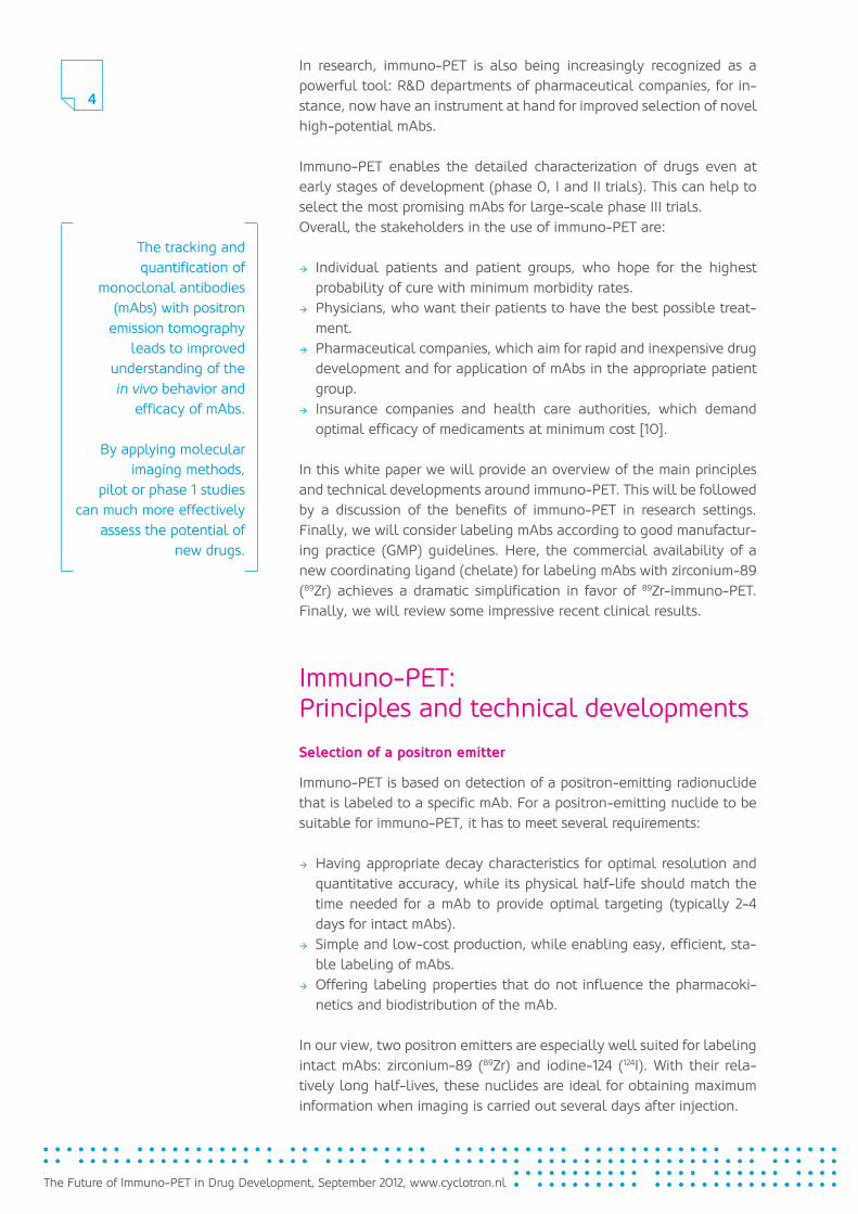
The advantages of 89Zr are manifold. It can be obtained at high yields with high radionuclidic purity. It has no prompt gamma emission that interferes with the overall image quality and accurate quantification. Recent studies revealed that 89Zr residualizes outstandingly after in-ternalization of the antibody, a phenomenon also observed with other radiometals, such as yttrium-90, indium-111 and lutetium-177 [11]. In addition, the half-life values of 89Zr and 124I show evident advantages for logistics relating to labeling and transportation. A major advantage of 124I is that the radioiodination of proteins is a common practice within radiochemistry labs. Contrarily, alternative positron emitters like cop-per-64, yttrium-86 and bromine-76 are less suitable for imaging intact mAbs, especially due to their relatively short half-lives.
Table 1 below shows the main characteristics of positron emitters used in preclinical and clinical studies of immuno-PET.
Table 1Main characteristics of positron emitters used in preclinical and clinical immuno-PET studies
Positronemitter
Commonproductionroutes
Half-life(h)
Main ß+-energies(keV)
(%) Intrinsic spatialresolution loss (mm)
64 Cu 64 Ni(d,2n)64 Ni(p,n)
12.7 653 17.4 0.7
86 Y 86 Sr(p,n) 14.7 12211545
11.95.6
1.8
76 Br 76 Se(p,n) 16.2 87199033823941
6.35.225.86.0
5.3
89 Zr 89Y(p,n) 78.4 897 22.7 1.0
124 I 124 Te(p,n)124 Te(d,2n)125 Te(p,2n)
100.3 15352138
11.810.9
2.3
Immuno-PET can deliver fast, reliable proof of principle. The scientific community and industry alike can now instantly obtain quantifiable results of in vivo research.
6
The Future of Immuno-PET in Drug Development, September 2012, www.cyclotron.nl
Immuno-PET applications in research
In research settings, quantitative PET imaging of therapeutic mAbs is an invaluable tool at several stages of mAb development and applica-tion. For instance, preclinical immuno-PET studies in xenograft-bearing nude mice can help measure the efficiency of tumor targeting with a particular mAb. In toxicology studies of non-human primates, the regulation of target expression can be useful when assessing cross-reactivity with normal tissues, in relation to mAb protein doses.
First-in-man clinical trials with new mAbs particularly benefit from immuno-PET. In these studies, it is important to establish the ideal mAb dose required for optimal targeting (e.g. saturation of receptors), the uptake in normal critical organs to anticipate toxicity, as well as inter-patient variations in pharmacokinetics. Imaging mAbs with immuno-PET provides this information efficiently and safely. As a result, fewer patients are treated at suboptimal levels. This approach is particularly favorable when the mAb of interest is directed against a novel target that has not been validated in clinical trials before.
Quantitative mAb imaging may also be of value as a guide to the optimal use of FDA-approved mAbs.In all these settings, immuno-PET offers the following advantages:
Æ assessment of target status (“comprehensive immunohistochemistry in vivo”);
Æ assessment of pharmacokinetics and biodistribution; Æ confirmation of selective targeting and prediction of toxicity; Æ optimization of mAb dose scheduling; Æ identification of indications and patient groups; Æ therapy planning and individualization.
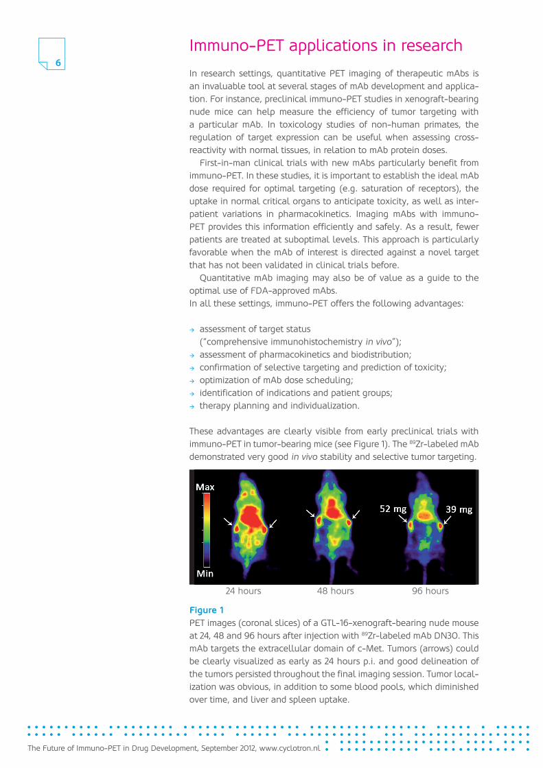
These advantages are clearly visible from early preclinical trials with immuno-PET in tumor-bearing mice (see Figure 1). The 89Zr-labeled mAb demonstrated very good in vivo stability and selective tumor targeting.
Figure 1PET images (coronal slices) of a GTL-16-xenograft-bearing nude mouse at 24, 48 and 96 hours after injection with 89Zr-labeled mAb DN30. This mAb targets the extracellular domain of c-Met. Tumors (arrows) could be clearly visualized as early as 24 hours p.i. and good delineation of the tumors persisted throughout the final imaging session. Tumor local-ization was obvious, in addition to some blood pools, which diminished over time, and liver and spleen uptake.
24 hours 48 hours 96 hours
7
The Future of Immuno-PET in Drug Development, September 2012, www.cyclotron.nl
From bench to bedside
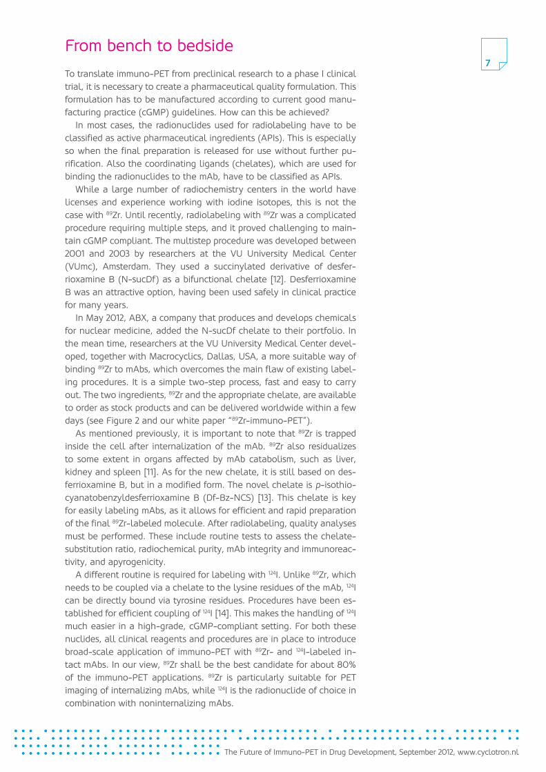
To translate immuno-PET from preclinical research to a phase I clinical trial, it is necessary to create a pharmaceutical quality formulation. This formulation has to be manufactured according to current good manu-facturing practice (cGMP) guidelines. How can this be achieved?
In most cases, the radionuclides used for radiolabeling have to be classified as active pharmaceutical ingredients (APIs). This is especially so when the final preparation is released for use without further pu-rification. Also the coordinating ligands (chelates), which are used for binding the radionuclides to the mAb, have to be classified as APIs.
While a large number of radiochemistry centers in the world have licenses and experience working with iodine isotopes, this is not the case with 89Zr. Until recently, radiolabeling with 89Zr was a complicated procedure requiring multiple steps, and it proved challenging to main-tain cGMP compliant. The multistep procedure was developed between 2001 and 2003 by researchers at the VU University Medical Center (VUmc), Amsterdam. They used a succinylated derivative of desfer-rioxamine B (N-sucDf) as a bifunctional chelate [12]. Desferrioxamine B was an attractive option, having been used safely in clinical practice for many years.
In May 2012, ABX, a company that produces and develops chemicals for nuclear medicine, added the N-sucDf chelate to their portfolio. In the mean time, researchers at the VU University Medical Center devel-oped, together with Macrocyclics, Dallas, USA, a more suitable way of binding 89Zr to mAbs, which overcomes the main flaw of existing label-ing procedures. It is a simple two-step process, fast and easy to carry out. The two ingredients, 89Zr and the appropriate chelate, are available to order as stock products and can be delivered worldwide within a few days (see Figure 2 and our white paper “89Zr-immuno-PET”).
As mentioned previously, it is important to note that 89Zr is trapped inside the cell after internalization of the mAb. 89Zr also residualizes to some extent in organs affected by mAb catabolism, such as liver, kidney and spleen [11]. As for the new chelate, it is still based on des-ferrioxamine B, but in a modified form. The novel chelate is p-isothio-cyanatobenzyldesferrioxamine B (Df-Bz-NCS) [13]. This chelate is key for easily labeling mAbs, as it allows for efficient and rapid preparation of the final 89Zr-labeled molecule. After radiolabeling, quality analyses must be performed. These include routine tests to assess the chelate-substitution ratio, radiochemical purity, mAb integrity and immunoreac-tivity, and apyrogenicity.
A different routine is required for labeling with 124I. Unlike 89Zr, which needs to be coupled via a chelate to the lysine residues of the mAb, 124I can be directly bound via tyrosine residues. Procedures have been es-tablished for efficient coupling of 124I [14]. This makes the handling of 124I much easier in a high-grade, cGMP-compliant setting. For both these nuclides, all clinical reagents and procedures are in place to introduce broad-scale application of immuno-PET with 89Zr- and 124I-labeled in-tact mAbs. In our view, 89Zr shall be the best candidate for about 80% of the immuno-PET applications. 89Zr is particularly suitable for PET imaging of internalizing mAbs, while 124I is the radionuclide of choice in combination with noninternalizing mAbs.
The Future of Immuno-PET in Drug Development, September 2012, www.cyclotron.nl
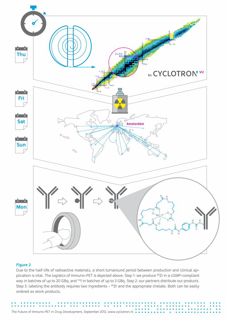
Figure 2Due to the half-life of radioactive materials, a short turnaround period between production and clinical ap-plication is vital. The logistics of immuno-PET is depicted above. Step 1: we produce 89Zr in a cGMP-compliant way in batches of up to 20 GBq, and 124I in batches of up to 3 GBq. Step 2: our partners distribute our products. Step 3: labeling the antibody requires two ingredients - 89Zr and the appropriate chelate. Both can be easily ordered as stock products.
Thu
Fri
Sat
Sun
Mon
Amsterdam
89Zr
89 4+

9
The Future of Immuno-PET in Drug Development, September 2012, www.cyclotron.nl
The technology for labeling antibodies with 89Zr and 124I has become very accessible. A simple two-step procedure makes it ideal for preclinical and clinical settings.
Clinical results89Zr-labeled mAbs
In recent years, highly successful clinical trials have been carried out using immuno-PET.
The first-in-human 89Zr-immuno-PET trial was conducted between 2003 and 2005, at the VUmc in Amsterdam, with 89Zr-labeled-cmAb U36 [15,16]. The objective of this study was to verify whether the imag-ing probe was safe and capable of clearly delineating CD44v6-positive tumors. The study aimed to determine the diagnostic value of immuno-PET with 89Zr-mAb U36 in patients with head and neck squamous cell carcinoma (HNSCC) who were at high risk of having neck lymph node metastases. Twenty HNSCC patients scheduled to undergo resection of the primary tumor and unior bilateral neck dissection underwent com-puted tomography (CT) or magnetic resonance imaging (MRI), or both, and 89Zr-cmAb U36 immuno-PET prior to surgery.
Compared to CT and MRI scans, immuno-PET results were remark-ably accurate. The 89Zr-cmAb U36 immuno-PET scans detected all pri-mary tumors (n=17), in addition to 18 lymph node metastases, out of 25 positive neck nodes. The missed lymph nodes were relatively small in size, and they contained only a small proportion of tumor tissue.
After this first clinical trial with 89Zr, other Dutch sites also began clinical trials using 89Zr. Several trial programs were started at academic hospitals in Groningen, Nijmegen and Maastricht. Shortly thereafter, sites in Brussels and London also initiated their first clinical trials with 89Zr-immuno-PET.
One of the first examples of clinical immuno-PET trials is currently being conducted at the Jules Bordet Institute in Brussels, Belgium. Researchers performed immuno-PET imaging with 89Zr-labeled ritux-imab in patients with CD20+ B-cell lymphoma. Excellent images were obtained (see Figure 3). One of the objectives of this research is to compare the diagnostic accuracy of 89Zr-rituximab-PET/CT with stan-dard [18F]FDG-PET/CT. A preliminary evaluation in 14 patients with follicular lymphoma showed significant uptake of [89Zr]rituximab in all FDG-positive lesions. In some patients, 89Zr-rituximab-PET/CT revealed additional CD20+ lesions that were strictly negative on [18F]FDG-PET/CT (unpublished data). Those lesions corresponded to particularly small (≤ 1 cm) lymph nodes and mesenteric nodules on CT images.
It was established that 89Zr-rituximab-PET/CT is an outstanding diag-nostic tool for precise quantification of CD20 antigen expression. This is of particular interest for dosimetry as a prelude to radioimmunotherapy with 90Y-rituximab.
The European results also led to the first clinical trials for 89Zr-immu-no-PET in the United States. The trials currently carried out in clinical research centers in Frederick (MD), New York (NY) and Sacramento (CA) are part of programs being conducted at the National Cancer In-stitute, Memorial Sloan-Kettering Cancer Center and the University of California (see Figure 4).
Furthermore, pharmaceutical companies such as Genentech, con-sidered the forerunner of the biotechnology industry, have recently started to incorporate 89Zr-immuno-PET into their antibody develop-
10
The Future of Immuno-PET in Drug Development, September 2012, www.cyclotron.nl
ment research programs [17]. In this type of research, immuno-PET technology can be especially beneficial when the mAb of interest is directed against a novel target that has not previously been validated in clinical trials. This includes, for instance, mAbs directed against new tumor cell and tumor stroma targets, such as c-Met, vascular endothe-lial growth factor receptors (VEGFRs), insulin-like growth factor recep-tors and death receptors.
The overall effect of these applications is that the number of papers published about preclinical and clinical results with 89Zr-immuno-PET is steadily growing [10].
124I-labeled mAbs
124I-labeled mAbs were already used for clinical immuno-PET about 15 years ago, but the number of patients included in these studies was small [18,19]. At that time, diagnostic results were suboptimal, partly because of poor quality of the murine mAbs used, which lacked the precision of today’s mAbs [20]. Now, however, interest in 124I-labeled mAbs has returned.
This is, to a certain degree, due to the improved methods of 124I production and its coupling to mAbs. Recently, two clinical applica-tions have particularly attracted much attention. Jayson et al. [21] used various doses of 124I-HuMV833, a mAb binding to VEGF121 and VEGF165, to perform PET-imaging studies in 12 patients with various progressive solid tumors. The results were very promising: by using 124I-immuno-PET imaging, it was demonstrated that antibody distribution and clearance were markedly heterogeneous between and within patients, varying substantially even between and within individual tumors. These dif-ferences may represent the variation in available targets for the mAb.
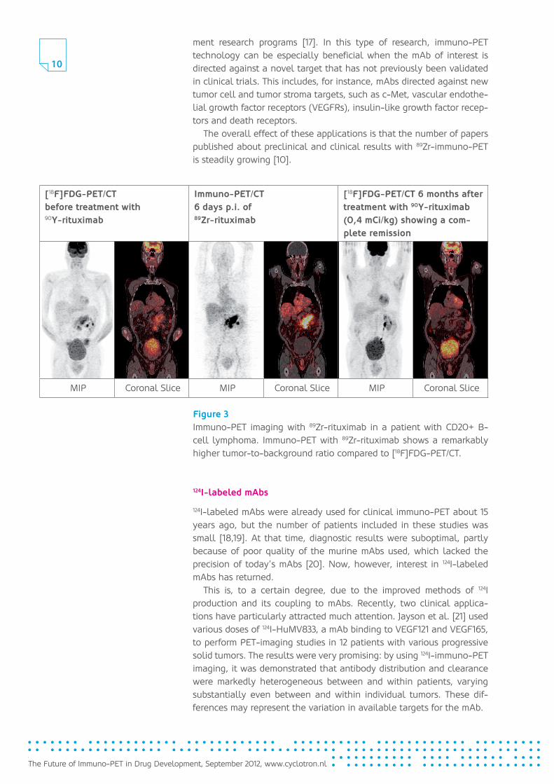
[ 18F]FDG-PET/CTbefore treatment with 90 Y-rituximab
Immuno-PET/CT6 days p.i. of 89 Zr-rituximab
[ 18F]FDG-PET/CT 6 months after treat ment with 90 Y-rituximab (0,4 mCi/kg) showing a com-plete remission
MIP Coronal Slice MIP Coronal Slice MIP Coronal Slice
Figure 3 Immuno-PET imaging with 89Zr-rituximab in a patient with CD20+ B-cell lymphoma. Immuno-PET with 89Zr-rituximab shows a remarkably higher tumor-to-background ratio compared to [18F]FDG-PET/CT.
11
The Future of Immuno-PET in Drug Development, September 2012, www.cyclotron.nl
Both 89Zr and 124I are produced commercially on a weekly basis. Worldwide distribution channels make high-grade material available within two days.
This is highly valuable knowledge, and may have implications for fu-ture anti-VEGF therapies. Similar studies have begun using 89Zr-bevaci-zumab as imaging probe [22,23].
In another clinical setting, 124I-immuno-PET was used for in vivo pro-filing of renal cancer. Divgi et al. [24] used 124I-cmAb G250 to predict the presence of clear-cell renal carcinomas in 25 patients scheduled for surgical tumor resection. G250 is directed against carbonic anhy-drase-IX and overexpressed in clear-cell renal carcinoma. It might be worthwhile to know which renal cancer patients have this aggressive tumor type, an information that can be used to guide future treatment decisions, although opinions on this point vary [25]. With immuno-PET, it was possible to accurately identify 15 out of 16 clear-cell carcinomas, as well as all nine non-clear-cell renal masses. In addition, 124I-immu-no-PET with G250 is currently in a phase III clinical trial in the United States, aiming to assess the potential of the G250 antibody as adjuvant therapy, i.e., a therapy given after surgical removal of the tumor, in patients with non-metastatic RCC.
These studies illustrate how molecular imaging with specific probes can contribute to personalized medicine.
The overall results of clinical trials with 89Zr-immuno-PET and 124I-immuno-PET show that both methods have already been successful in clinical settings. Immuno-PET provides a new, clinically safe precision tool for enhancing knowledge on the efficacy of mAb therapy. In turn, this may lead to more efficient mAb development and to more patient-tailored therapy. These crucial achievements have prepared the way for broad-scale clinical application of 89Zr- and 124I-immuno-PET.
A research challenge: reducing the radiation dose
At present, one of the most challenging research questions in the field of immuno-PET is how to reduce the radiation dose of an immuno-PET imaging procedure. The mean radiation dose for patients receiving 74 MBq 89Zr is about 40 mSv [17], which is high and limits repeated appli-cations of 89Zr-immuno-PET.
However, solutions have already been found: higher-sensitivity PET scanners are currently being developed and entering the market. These scanners require less radioactivity to be injected, without any loss of resolution. Better scanning softwares will further improve sensitivity and resolution, which directly translates into lower doses of injected radioactivity. Indeed, recent PET/CT studies using 37 MBq of 89Zr-trastu-zumab produced images of excellent quality at an effective dose of about 20 mSv [26].
Immuno-PET makes itpossible to identifyindividual patients orpatient groups. Even thedifferences in antibodyhalf-time in individualpatients can be madevisible this way.Mechanisms of antibodiesare now much easier tounderstand. This can leadto highly effective drugoptimization strategies.
12
The Future of Immuno-PET in Drug Development, September 2012, www.cyclotron.nl
Outlook
Molecular imaging with the innovative PET tracers 89Zr and 124I can con-siderably accelerate drug development and facilitate improved patient selection. The ability to understand the added value of new antibodies during early stages of preclinical or clinical trials amounts to nothing less than a paradigm shift. The pharmaceutical industry realizes that molecular imaging may help transform its research and development operations and find more efficient ways to be innovative. Academic biomedical research institutions are also able to initiate potential part-nerships with investors more easily if the results of new research can be quantified with greater accuracy.
Overall, this may support researchers in both academia and industry to more effectively translate their ideas and research into improved drug therapies. A review of recent literature on clinical trials shows that immuno-PET with 89Zr and 124I may be an effective instrument for im-proving knowledge on the efficacy of mAb therapy. This comprehension will ultimately contribute to the development of more efficient mAbs, as well as therapies more tailored towards patient needs.
We strongly believe that the overall positive clinical results of recent years will develop into broad-scale preclinical and clinical application of 89Zr- and 124I-immuno-PET. This is a major breakthrough as it is a cru-cial step forward on the path towards personalized medicine.
Immuno-PET supports academic research
institutes to more easily initiate potential industry and investor partnerships,
as new research results are quantified with greater
accuracy.
Immuno-PET is an important step forward on
the path towards personalized medicine.
13
The Future of Immuno-PET in Drug Development, September 2012, www.cyclotron.nl
Research partners
About BV Cyclotron VU
BV Cyclotron VU is a private company located in the campus of the VU University Medical Center in Amsterdam, the Netherlands. Since 1987, BV Cyclotron VU has been producing SPECT and PET radiopharmaceu-ticals and PET radiochemicals for medical diagnostics and research.
BV Cyclotron VU produces [18F]FDG, [18F]NaF, 124I, 89Zr and other 18F-labeled compounds for the PET community, as well as 123I and 81Rb/81mKr generators for the SPECT community.
In Amsterdam, BV Cyclotron VU runs the only commercial production site of 89Zr in the world. Academic sites such as the Memorial Sloan Kettering in New York, University of Wisconsin, Madison, and Paul Scherrer Institute in Zurich, Switzerland, produce 89Zr on request and for research purposes only [27].
BV Cyclotron VU’s mission is to make a wide range of PET com-pounds available. The goal is to make products of the highest pharma-ceutical quality, and to be a highly reliable supplier of those products. To achieve this, BV Cyclotron VU continuously invests in the best equipment available on the market.
The products are distributed by our worldwide distributor partners see our website: www.cyclotron.nl/distributors). The department of Nuclear Medicine and PET Research of the VU University Medical Center is our partner in research and development of new PET tracers.www.cyclotron.nl
About VUmc
The core activities of the VU University Medical Center (VUmc) are patient care, education and scientific research. The scientific research carried out at VUmc is closely linked to issues that are relevant both for the hospital itself and for society in general. Its main focal areas are oncology, neurology, immunology, vital functions and extramural re-search. The research output at VUmc translates to approximately 2,000 publications and reports per year, of which around 100 are doctoral theses. The aim of the research is to expand knowledge (fundamental research) and to apply this knowledge to health services (strategic and applied research). A staff of around 7,000 professionals and trainees ensure the smooth and efficient running of the medical center.www.vumc.nl
VU University Medical Center
The Future of Immuno-PET in Drug Development, September 2012, www.cyclotron.nl
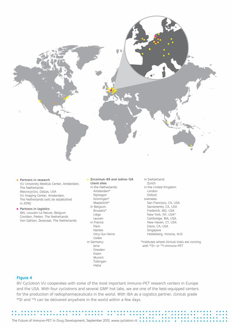
Figure 4BV Cyclotron VU cooperates with some of the most important immuno-PET research centers in Europe and the USA. With four cyclotrons and several GMP hot labs, we are one of the best-equipped centers for the production of radiopharmaceuticals in the world. With IBA as a logistics partner, clinical grade 89Zr and 124I can be delivered anywhere in the world within a few days.
Partners in research VU University Medical Center, Amsterdam, The Netherlands Macrocyclics, Dallas, USA VU Imaging Center, Amsterdam, The Netherlands (will be established in 2016)
Partners in logistics IBA, Louvain-La Neuve, Belgium Covidien, Petten, The Netherlands Von Gahlen, Zevenaar, The Netherlands
Zirconium-89 and iodine-124 client sites
in the Netherlands: Amsterdam* Nijmegen Groningen* Maastricht* in Belgium: Brussels* Liège Leuven in France: Paris Nantes Vitry-Sur-Seine Cedexin Germany: Jena Dresden Essen Munich Tübingen Halle
in Switzerland: Zurich in the United Kingdom: London Oxford overseas: San Francisco, CA, USA Sacramento, CA, USA Frederick, MD, USA New York, NY, USA* Cambridge, MA, USA New Haven, CT, USA Davis, CA, USA Singapore Heidelberg, Victoria, AUS
* Institutes where clinical trials are running with 89Zr- or 124I-immuno-PET

15
The Future of Immuno-PET in Drug Development, September 2012, www.cyclotron.nl
References
1 Maggon K. Monoclonal antibody “gold rush”. Curr Med Chem. 2007;14:1978.
2 Lawrence S. Billion dollar babies-biotech drugs as blockbusters. Nat Biotechnol. 2007;25:380.
3 Reichert JM. Monoclonal antibodies as innovative therapeutics. Curr Pharm Biotechnol. 2008;9:423.
4 Reichert JM. Antibody-based therapeutics to watch in 2011. MAbs. 2011; 3: 76-99.
5 Maggon K. Top Ten Monoclonal Antibodies. Knol 2012.
6 Nelson AL, Reichert JM. Development trends for therapeutic antibody fragments. Nat Biotechnol. 2009;27:331.
7 Reichert JM. Monoclonal antibody. MAbs. 2009;1:189.
8 Kolata G, Pollack A. The evidence gap--Costly cancer drug offers hope, but also a dilemma. New York Times. July 6, 2008.
9 Neyt M, Albrecht J, Cocquyt V. An economic evaluation of Herceptin in adjuvant setting: The breast Cancer International Research Group 006 trial. Ann Oncol. 2006;17:381.
10 van Dongen GAMS, Vosjan MJWD. Immuno-positron emission tomography: Shedding light on clinical antibody therapy. Cancer Biother Radiopharm. 2010;25;375.
11 Perk LR, Visser GWM, Vosjan MJWD, et al. (89)Zr as a PET surrogate radioisotope for scouting biodistribution of the therapeutic radiometals (90)Y and (177)Lu in tumor-bearing nude mice after coupling to the internalizing antibody cetuximab. J Nucl Med. 2005;46:1898.
12 Verel I, Visser GWM, Boellaard R, et al. 89Zr immuno-PET: Comprehensive procedures for the production of 89Zr-labeled monoclonal antibodies. J Nucl Med. 2003;44:1271.
13 Perk LR, Vosjan MJWD, Visser GWM, et al. p-Isothiocyanatobenzyl- desferrioxamine: A new bifunctional chelate for facile radiolabeling of monoclonal antibodies with zirconium-89 for immuno-PET imaging. Eur J Nucl Med Mol Imaging. 2010;37:250.
14 Tijink BM, Perk LR, Budde M, et al. (124)I-L19-SIP for immuno-PET imaging of tumour vasculature and guidance of (131)I-L19-SIP radioimmunotherapy. Eur J Nucl Med Mol Imaging. 2009;36:1235.
15 Börjesson PKE, Jauw YWS, Boellaard R, et al. Performance of immunopositron emission tomography with zirconium-89-labeled chimeric monoclonal antibody U36 in the de-tection of lymph node metastases in head and neck cancer patients. Clin Cancer Res. 2006;12:2133.
16 Börjesson PKE, Jauw YWS, De Bree R, et al. Radiation dosimetry of zirconium-89- labeled chimeric monoclonal antibody U36 as used for immuno-PET in head and neck cancer patients. J Nucl Med. 2009;50:1828.
17 Tinianow JN, Gill HS, Ogasawara A, et al. Site-specifically 89Zr-labeled monoclonal antibodies for immunoPET. Nucl Med Biol. 2010;37:289.
16
The Future of Immuno-PET in Drug Development, September 2012, www.cyclotron.nl
18 Wilson CB, Snook DE, Dhokia B, et al. Quantitative measurement of monoclonal antibody distribution and blood flow using positron emission tomography and 124I in patients with breast cancer. Int J Cancer. 1991;47:344
19 Larson SM, Pentlow KS, Volkow ND, et al. PET scanning of iodine-124-3F9 as an approach to tumor dosimetry during treatment planning for radioimmunotherapy in a child with neuroblastoma. J Nucl Med. 1992;33:2020.
20 Zukier LS, DeNardo GL. Trials and tribulations: oncological antibody imaging comes to the fore. Semin Nucl Med. 1997;27:10.
21 Jayson GC, Zweit J, Jackson A, et al. Molecular imaging and biological evaluation of huMV833 anti-VEGF antibody: implications for trial design of antiangiogenic anti-bodies. J Natl Cancer Inst. 2002;94:1484.
22 Nagengast WB, De Vries EG, Hospers GA, et al. In vivo VEGF imaging with radio-labeled bevacizumab in a human ovarian tumor xenograft. J Nucl Med. 2007;48:1313.
23 Nagengast WB, De Korte A, Oude Munnink TH, et al. 89Zr-bevacizumab PET of early anti-angiogenic tumor response to treatment with HSP90 inhibitor NVP-AUY922. J Nucl Med. 2010;51:761.
24 Divgi CR, Pandit-Taskar N, Jungbluth AA, et al. Preoperative characterization of clear-cell renal carcinoma using iodine-124-labeled antibody chimeric G250 (124I-cG250) and PET in patients with renal masses: a phase I trial. Lancet Oncol. 2007;8:304.
25 Powles T, Ell PJ. Does PET imaging have a role in renal cancers after all? Lancet Oncol. 2007;8:279.
26 Dijkers EC, Oude Munnink TH, Kosterink JG, et al. Biodistribution of 89Zr-trastuzumab and PET imaging of HER2-positive lesions in patients with metastatic breast cancer. Clin Pharmacol Ther. 2010;87:586.
27 Holland JP, Sheh Y, Lewis JS. Standardized methods for the production of high specific-activity zirconium-89. Nucl Med Biol. 2009;36:729.
Imprint
Text
Dr. Lars Perk, Dr. Sybe I. Rispens
Editing and Production
www.knowledgeatwork.eu
Infographics and Layout
WERNERWERKE GbR


![FDA-AACR Immuno-Oncology Drug Development … Immuno-Oncology Drug Development Workshop Transcript from October 13, 2016 Section 1 of 46 [00:00:00 - 00:10:04] (NOTE: speaker names](https://img.pdfslide.us/doc/110x75/5ad128de7f8b9a86158bbbbc/fda-aacr-immuno-oncology-drug-development-immuno-oncology-drug-development-workshop.jpg)