Embed Size (px)
Citation preview
The Future of Critical Care MedicineDavid E. Tannehill, DO
CCM Physician, St. John’s Mercy Medical Center
Assistant Clinical Professor of Medicine, Saint Louis U.

The Future…
• Targeted Temperature Therapy
• Understanding & manipulating the microcirculation
• Endotoxin adsorption
• Point-of-care ultrasound
• Telemedicine

Therapeutic Hypothermia
Hypothermia

Hippocrates (460 BC - 377 BC)
•“Extreme Remedies are very appropriate for Extreme diseases.”
Hypothermia

The Facts• 250,000 Americans die every year from SCA occurring outside of the
hospital
• Out-of-hospital cardiac arrest only has an average survival rate of 6% worldwide
• Approximately 20% of patients who survive and are comatose during the post resuscitation period will awaken with a good neurological outcome
•
• Circulation 2005. IV-206
Hypothermia

Reperfusion Injury
• Global ischemia occurs during periods of circulatory collapse
• During reperfusion, free radicals and mediators are released causing cerebral ischemia, which may persist for several hours after return of spontaneous circulation (ROSC)
Hypothermia

Post–Cardiac Arrest Brain Injury:
• Patients who survived to ICU admission but subsequently died in the hospital, brain injury was the cause of death in 68% after out-of-hospital cardiacarrest and in 23% after in-hospital cardiac arrest.
Hypothermia
Hypothermia

Post–cardiac arrest syndrome ILCOR
• 1-Post–cardiac arrest brain injury.
• 2-Post—cardiac arrest myocardial dysfunction.
• 3-Systemic ischemia/Reperfusionresponse.
• 4-Persistent precipitating pathology
Hypothermia

Hypothermia
Hypothermia

Hypothermia
• Definitions of hypothermia:– Mild: 33-36 degrees Celsius
– Moderate: 26-32 degrees Celsius
– Deep: 20-25 degrees Celsius
– Profound: < 20 degrees Celsius
Hypothermia

Physiologic Effects on CNS:
•Hypothermia decreases Intracranial Pressure.
•Clifton et al.Lack of Effect of induction of Hypothermia after Acute Brain Injury. N Engl J Med 2001

Physiologic Effects on CNS:
• Hypothermia may act as an anticonvulsant.(1)• Hypothermia decreases excitatory AA and lactate during ischemia and reperfusion.(2,3)• In a rat model, Hypothermia reduced the microvessel expression od ICAM-1 protein and the
number of neutrophils migrating into ischemic tissue.(4)• In head injury patients, IH decreased IL-10 concentrations in CSF.(5)
• 1.Orlowski et al. Hypothermia and Barbiturate coma for refractory status epilepticus. Crit Care Med 1984.• 2.Illievich et al. Effects of Hypothermic metabolic suppression on hippocampal glutamate concentrations after transient global cerebral ischemia. Anaesth Analg
1994.• 3.berger et al. Effects of Hypothermia on excitatory amino acids and metabolism in stroke patients.A Microdialysis Study. Stroke 2002.• 4.Inamasu et al. Intraischemic Hypothermia attenuates intracerebral ICAM-1 and migration of neutrophils. Neurol Res 2001.• 5.Clifton et al. Lack of effect of induction of hypothermia after acute brain injury. N Engl J Med 2001.
Hypothermia
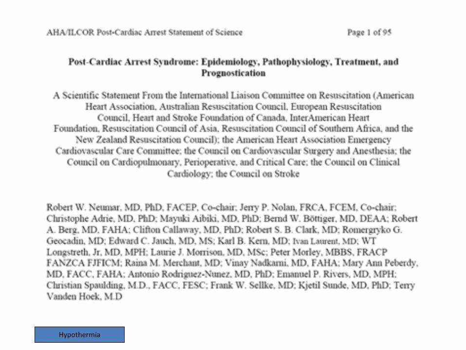
Physiologic Effects on CVS:
• Accidental Hypothermia34C < intense shivering < 36C.
• Shivering increased metabolic rate and oxygen demand.
• Increase incidence of MI in patients with Ischemic heart Disease.
Frank et al. Perioperative maintenance of normothermia reduces the incidenceof morbid cardiac events. A Randomized Clinical Trial. JAMA 1997.

Physiologic Effects on CVS:
• During IHthe patient is sedated/paralyzed to avoid shivering.
Mild Hypothermia(32C-34C)
SVR HR SV MAP
Bernard et al. Treatment of Comatose survivors of out-of-hospital cardiac arrest with induced hypothermia.N Engl J Med 2002; 346:557-63.

Physiologic Effects on CVS:
• Cardiac arrhythmias are rarely seen at 33C, even in patients with myocardial ischemia.
• In accidental hypothermia, there is a risk of V-fib if core temperature decreases below 28C.
• Danzl et al. Accidental Hypothermia. N Engl J Med 1994; 331: 1756-1760.
Hypothermia

Physiologic Effects of Therapeutic Hypothermia on CVS:
• Sinus bradycardia ensues as temperature drops below 35.5°C, with a progressive decrease in heart rate as temperature decreases further.
• At core temperatures of 32°C the heart rate typically decreases to around 40–45 beats/min or even lower(wide inter and intraindividual variability)
• Electrocardiographic changes include:
- prolonged PR interval
- widening of the QRS complex
- increased QT interval
- Osborne waves.
Hypothermia
Physiologic Effects of Therapeutic Hypothermia on Respiratory system:
• At 33C, metabolic rate decreases by 25-33%, Minute volume is decreased to maintain normal pH/pCO2.
• Pneumonia is a risk of IH uncommon during brief periods of hypothermia(12-24H).(1,2)
• In a series of patients who underwent 7 days of IH (for severe head injury)Nosocomial pneumonia in 45%.(3).
• 1.Bernard et al. Treatment of Comatose Survivors of out-of-hospital cardiac arrest with induced hypothermia. N Engl J Med 2002.
• 2.The Hypothermia after Cardiac Arrest Study Group. N Engl J Med 2002.
• 3.Bernard et al. Experience with prolonged induced hypothermia in severe head injury. Crit Care 1999.
Hypothermia

Physiologic Effects of Therapeutic Hypothermia on Renal System:
• Diuresis decreased reabsorption of solute in the ascending limb of the loop of henle.(1).
• Induction of hypothermia shifts K into the cells.(Hyperkalemia during rewarming).(2,3).
• Hypothermia decreased phosphate concentrations.(4).
• Volume status,Potassium and phosphate concentrations require careful monitoring.
1.Wong. Physiology and Pharmacology of Hypothermia. West J Med 1983. 2.Machida et al. Changes in tissue Distribution of potassium during simple hypothermia.Jpn J Thorac Surg 1977.3.Koht et al. Serum Potassium levels during prolonged hypothermia. Int Care Med 1983. 4.Aibiki et al. Reversible hypophosphatemia during moderate hypothermia therapy for brain injured patients.
Crit Care Med 2001.

Physiologic Effects of hypothermia on ABG:
•As temperature decreases ,The solubility of gases in blood increases.
•When ABG of hypothermic patients are corrected for temperature,patients appear to have a respiratory alkalosis.

Physiologic Effects of hypothermia on ABG:
• If it is not possible to obtain blood gas results measured at the patient’s true core temperature, values can be estimated using the following rule of thumb:
- -For PO2, subtract 5 mm Hg for every 1°C below 37°C.
- - For PCO2, subtract 2 mm Hg for every 1°C below 37°C.
• -For pH, add 0.012 points for every 1°C below 37°C.
Hypothermia

Physiologic effects of Therapeutic Hypothermia on GI system:
• Decreased gut motilitydelay enteral feeding.(1)
• Increases blood glucose concentration(? Decrease insulin release)(2) keep tight glucose control by administering Insulin.
• 1.Bernard et al. Experience with prolonged induced hypothermia in severe head injury. Crit Care 1999.
• 2.Curry. Hypothermia and insulin secretion. Endocrinology 1970.
Hypothermia

Physiologic effects of Therapeutic Hypothermia on hematologic system:
• The numbers and function of WBCs decreaseincrease incidence of sepsis(pneumonia during >24H IH).(1)
• Prolonged hypothermia decreases the number and function of platelets.(<35 degrees)(1)
• Hypothermia prolongs clotting times(<33 degrees)increase in risk of bleeding in the setting of major trauma.(2)
• 1.Bernard et al. Experience with prolonged induced hypothermia in severe head injury. Crit Care 1999.• 2.Valerie et al. Effect of temperature on bleeding time and clotting time in normal male and female volunteers.Crit Care Med 1995.
Hypothermia
Effects of Therapeutic Hypothermia on the skin:
• Increases the risk of wound infections :
• -diminished leukocyte function
• -hypothermia-induced vasoconstriction in the skin.
• Thus extra care should be taken in cooled patients to prevent bedsores.
Hypothermia
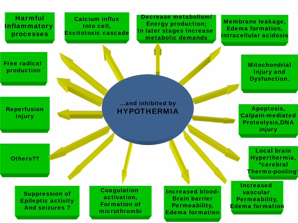
HarmfulInflammatory
processes
Calcium influxInto cell,
Excitotoxic cascade
Decrease metabolism/Energy production;
In later stages Increase metabolic demands
Membrane leakage,Edema formation,
Intracellular acidosis
Free radical production
ReperfusionInjury
Others??
Suppression ofEpileptic activity
And seizures ?
Coagulation activation,
Formation ofmicrothrombi
Increased blood-Brain barrierPermeability,
Edema formation
Increased vascular
Permeability,Edema formation
Local brainHyperthermia,
“cerebralThermo-pooling”
Apoptosis,Calpain-mediatedProteolysis,DNA
injury
MitochondrialInjury and
Dysfunction.
are all stimulated by
FEVER
…and inhibited byHYPOTHERMIA

Research on Hypothermia
• 2 studies published in New England Journal of Medicine in February, 2002
– HACA
– Bernard
Hypothermia

HACA Study: Hypothermia After Cardiac Arrest Group
• 9 centers, 5 countries in Europe
• CPR within 5 to 15 minutes of arrest
• Presumed ventricular tachycardia or fibrillation
• ROSC within 60 minutes from collapse
• 275 patients randomized to hypothermia or normothermia
• Cooled to 32º - 34ºC for 24 hours
Hypothermia

HACA Study: 6 to 8 hours to reach target
Hypothermia

Outcomes
• Primary end point was favorable neurologic outcome (live independently and work at least part-time) within 6 months after cardiac arrest
• Secondary end points were mortality within 6 months and rate of complications within 7 days
Hypothermia

Results• 75/136 patients (55%) in the hypothermia group had a favorable neurologic
outcome within six months after arrest as compared with 54/137 patients (39%) in the normothermia group
• Six month mortality was 41% in the hypothermia group (56/137) as compared with 55% (76/138) in the normothermia group
• No significant difference in complications between the groups
Hypothermia

Australian Study, Bernard et al
• 4 hospitals in Melbourne
• Initial cardiac rhythm of ventricular fibrillation
• Successful ROSC with persistent coma
• 77 patients randomized to hypothermia or normothermia
• Cooling initiated in ambulance
• Cooled to 33ºC for 12 hours
Hypothermia
Outcomes
• Survival to hospital discharge either to home or to a rehabilitation facility
Hypothermia
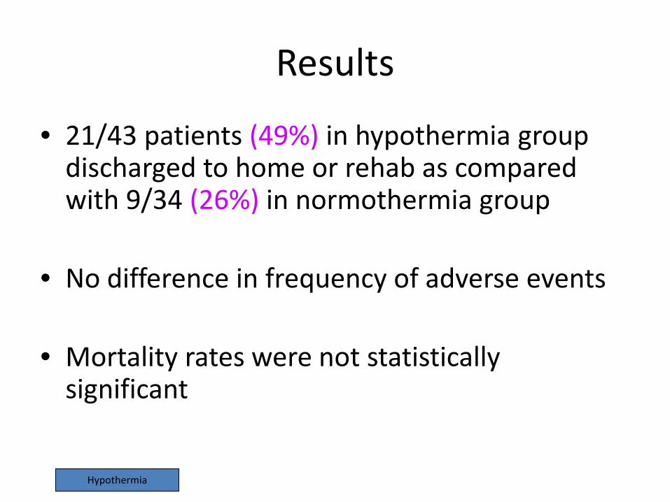
Results
• 21/43 patients (49%) in hypothermia group discharged to home or rehab as compared with 9/34 (26%) in normothermia group
• No difference in frequency of adverse events
• Mortality rates were not statistically significant
Hypothermia

Metaanalysis:
• -Therapeutic Hypothermia is associated with a risk ratio of 1.68 (95% CI,1.29-2.07) favoring a good neurologic outcome when compared with Normothermia.
• -NNT to generate one favorable neurological recovery = 6.
• -Improve neurological recovery in > 10,000 patients/Year.
Hypothermia

Updated 2005 AHA Guidelines for CPR
Circulation 2005; 112:IV-206 – IV-211Hypothermia

ACC/AHA Guidelines
Class II: Conditions for which there is conflicting evidence and/or a divergence of opinion about the usefulness/efficacy of a procedure or treatment.
IIa. Weight of evidence/opinion is in favor of usefulness/efficacyIIb. Usefulness/efficacy is less well established by evidence/opinion.
Hypothermia

Cooling Techniques: Early CoolingDoes it matter?
• Wolff et al. Early achievement of mild therapeutic hypothermia and the neurologic outcome after cardiac arrest. International J of Cardiology 133 (2009): 223-228– 49 pt good (28) vs poor (21) outcome
• Predictors of good outcome:
– Time to Coldest Temperature– Time to Target Temperature

Cooling Techniques: Early CoolingCan it be done?
• Bernard et al reported the results of a clinical trial of the rapid infusion of large-volume (30 mL/kg), ice-cold (4°C) lactated Ringer’s solution in comatose survivors of out-of-hospital cardiac arrest. (1)
– Decreased core temperature by 1.6°C over 25 mins – Associated with improvements in MAP, renal f(x), and acid-base.– No pulmonary edema.– Inexpensive, convenient, and applicable to the prehospital setting. – Higher BP may be of additional benefit after cardiac arrest.
• Minneapolis Heart Institute (2)– Each 1hr delay in time to target temperature results in 25% higher risk of
death or poor neurologic outcome
Bernard et al: Induced hypothermia using large volume, ice-cold intravenous fluid in comatose survivors ofout-of-hospital cardiac arrest: A preliminary report. Resuscitation 2003 The Miracle on Ice: Therapeutic Hypothermia for Cardiac Arrest Patients. December 2009. Unpublished data

PreHospital Cooling: Randomized trials
• Kim et al.Circulation. 2007;115:3064-3070– EMS infused up to 2L of 4°C normal saline
– 125pt randomized to receive standard care with or without intravenous cooling
– 49/63 randomized to cooling received an infusion of 500 to 2000 mL of 4°C normal saline before hospital arrival.
– Cooling group had mean T decrease of 1.241°C to 34.7°C
– Control group had mean T increase of 0.1°C (P0.0001) with a hospital arrival temperature of 35.7°C.
– Secondary end points of awakening and discharged alive from hospital trended toward improvement in ventricular fibrillation patients randomized to in-field cooling.
PreHospital Cooling: Randomized trials
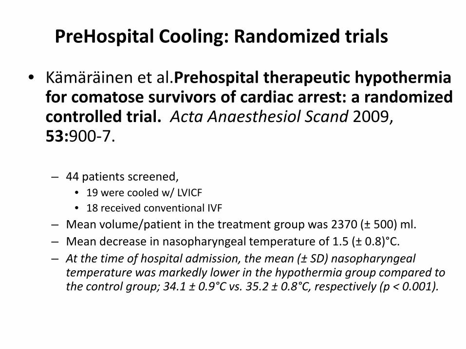
• Kämäräinen et al.Prehospital therapeutic hypothermia for comatose survivors of cardiac arrest: a randomized controlled trial. Acta Anaesthesiol Scand 2009, 53:900-7.
– 44 patients screened, • 19 were cooled w/ LVICF • 18 received conventional IVF
– Mean volume/patient in the treatment group was 2370 (± 500) ml.– Mean decrease in nasopharyngeal temperature of 1.5 (± 0.8)°C.– At the time of hospital admission, the mean (± SD) nasopharyngeal
temperature was markedly lower in the hypothermia group compared to the control group; 34.1 ± 0.9°C vs. 35.2 ± 0.8°C, respectively (p < 0.001).

PreHospital Cooling: During CPR?
• Kämäräinen et al.Induction of therapeutic hypothermia during prehospital CPR using ice-cold intravenous fluid. Resuscitation 2008, 79:205-11.
– 17 patients, paramedics initiated cooling using infusion of cold fluid during CPR and after ROSC.
– Infusion rate= 57 ± 21 ml/min with a target temperature of 33°C.
– The mean infused volume of cold fluid per patient was 1571 ml.
– Mean admission temperature = 33.83 ± 0.77°C (n = 11, -1.34°C decrease compared to initial nasopharyngeal temperature)
– No apparent increase in the rate of re-arrest or hemodynamic instability.
– The treatment was easily carried out by paramedics.

PreHospital Cooling: Improve Outcome?
Bruel et al. Mild hypothermia during advanced lifesupport: a preliminary study in out-of-hospital cardiac arrest. Crit Care 2008, 12:R31.
– 33 patients, 20 w/ ROSC
– A mean esophageal temperature decrease of 2.1°C
– Mean rate of infusion: 67 ml/min
– Volume of cold saline per patient was 2L
– Cooling was continued in hospital
– 4 patients out of 11 surviving to ICU admission were alive after 6 months.
– 3 with a CPC score </= 2

PreHospital Cooling
“Prehospital TH appears to be safe and remains a promising approach that not only decreases the time to therapeutic temperature, but if applied by emergency management service protocols, may increase the overall utilization of TH, resulting in important epidemiological gains.”
Majersik JJ, Silbergleit R, Meurera WJ, et al: Public health impact of full implementation of therapeutic hypothermia after cardiac arrest. Resuscitation 2008; 77:189–194

Temperature Control and Brain Injury
Hypothermia

Current Strategies to Manage Fever
Carhuapoma et al (2003) found pharmacological therapy (acetaminophen and nonsteroidal anti-inflammatory drugs) to be ineffective, with only 36% success rate
Mayer et al (2004) have found water-circulating cooling blankets ineffective to moderate temperature, with success rates <40% (Mayer, 2004)
Hypothermia

Endovascular Cooling Devices:
Hypothermia
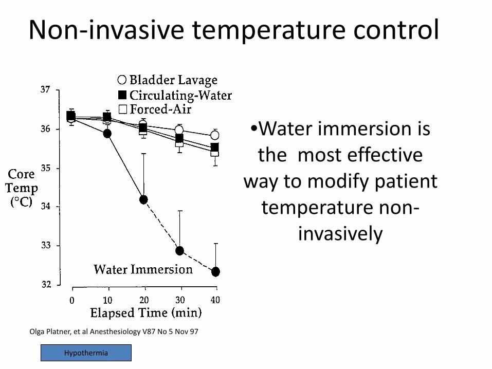
Non-invasive temperature control
•Water immersion is the most effective
way to modify patient temperature non-
invasively
Olga Platner, et al Anesthesiology V87 No 5 Nov 97
Hypothermia

Arctic Sun Energy Transfer Pads
Biocompatible hydrogel material: 50% water based matrix with temperature controlled water flowing beneath the thin film.
Simulates water immersion
Hypothermia

Arctic Sun Energy Transfer Pad ™Placement
Hypothermia

Contraindication…?
- Hypotension ? NO
- Active Bleeding? Maybe
- Arrhythmias ? NO (provided Temp stays>30)
- Older Age ? NOIf the patient is worth admitting to the ICU,
He/she should receive therapeutic HypothermiaHypothermia

Clinical experiences* Neurogenic fever control Heat stroke Malignant Hyperthermia Sudden cardiac arrest Refractory ICP. SAH/ICH Ischemic stroke(COOL AID) Traumatic Brain Injury(EUROTHERM) Spinal Cord Injury. Hepatic Encephalopathy – bridge to transplant Myocardial Infarction(COOL MI, ICE-T..) Contrast-Induced Nephropathy(COOL-RCN…) Refractory Status Epilepticus(case reports/series).
Hypothermia


Sepsis: A Disease of the Microcirculation.
David Tannehill, DO
Farid Sadaka,MD

“Five to fifteen minutes after its [endotoxin] intravenous administration, there were strong waves of contraction along the small arteries, arterioles and metarterioles.These could arrest flow and last for several minutes. There would afterwards be a phase of dilatation, followed again by a strong contraction. As time went on,the phases of relaxation became more prominent until preagonally there was a general and permanent vasodilation. The circulation would slow progressively until death.”
From Delauney and coworkers (1955),

Number of publications regarding microcirculation in humans

EGDT
• Individuals with severe sepsis who initially presented with a MAP >100 mmHg and a serum lactate level > 4 mM (n = 23) had a significantly increased mortality rate (60.9%) compared with patients who had originally presented with hypotension (MAP < 70 mmHg) (n = 68, 42% mortality).

• The microcirculation consists of the smallest blood vessels (<100 μm diameter)-Arterioles, capillaries, and venules.
• The main cell types comprising the microcirculation are the endothelial cells lining the inside of the microvessels, smooth muscle cells (mostly in arterioles), RBCs, leukocytes, and plasma.
• The microcirculation, with its huge endothelial surface, is in fact the largest ‘organ’ in the human body.
• The Microcirculation is the primary site of oxygen and nutrient exchange.
• The Microcirculation delivers therapeutic drugs to target cells.

What exactly comprises the Microcirculation?
-It is where oxygen exchange takes place.
-Every parameter in the microcirculation is different than in the systemic circulation.
-It plays a central role in the immune system.
-During sepsis and shock it the first to go and last to recover.
-Rescue of the microcirculation = resuscitation end-point.

Spronk P, Zandstra D, Ince C (2004) Critical Care 8:462-468
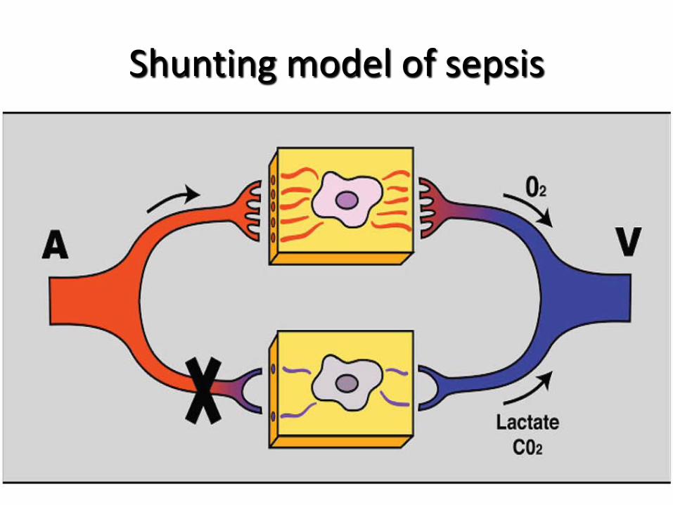
Shunting model of sepsis

How perfused vessel density plays a critical role in oxygen transport.

MIROCIRCULATION:
-Inflammatory activation-Coagulatory/RBC dysfunction
-Endothelial barrier dysfunction-Capillary fall out
-Weak microcirculatory units are shunted-Hypoxia, apoptosis, organ dysfunction
Not detected by systemic variables

Mitochondrial Dysfunction in Sepsis.
Lancet 2002;360:219-23

Co-morbidityGenes
Initial Hit
Circulatory Shock + InflammationResuscitation based on
correction of systemic hemodynamics+ inflammation
Microcirculatory Dysfunction
TimeTherapy
RBC
Deformability,AggregationO2 transport
Coagulation
↓Natural AnticoagulantsMicrovascular Thrombosis
Leukocytes
Adhesion, Cytokines, ROS
Endothelial Dysfunction
Barrier, CommunicationCoagulation, Regulation
Dysfunction Autoregulation
Microcirculatory shunting supply-demand mismatch
Hypoxia
Cellular Distress
MitochondriaHibernationApoptosis
Organ Failure
TimeTherapy
Microcirculatory Mitochondrial Distress SyndromeInce C (2005) Critical Care 9:S13-S19

SDF: Sidestream Dark Field

-Vessel density = the number of vessels crossing the lines divided by the total length of the lines. ---Perfusion :categorized by eye as present (continuous flow for at least
20 s), absent (no flow for at least 20 s), or intermittent (at least
50% of the time with no flow).
-The proportion of perfused vessels
(PPV [%]) =100 × (total
number of vessels - [no flow + intermittent flow])/total number
of vessels.
-Perfused vessel density (PVD),:calculated by multiplying
vessel density by the proportion of perfused vessels

The image is divided into four quadrants and the predominant type of flow (absent = 0, intermittent = 1, sluggish = 2, and normal = 3) is assessed in each quadrant.
-The MFI : represents the averaged values of the four.
A 20 μm cut-off is used to separate small vessels (mostly capillaries) from large vessels (mostly venules)
-Heterogeneity index:
Evaluate three to five sites and measure the MFI in the quadrants. Take the difference between highest MFI minus the lowest site MFI divided by the mean flow velocity of all sublingual sites at a single time point.

NORMAL

SEVERE SEPSIS

SEPTIC SHOCK

De Backer, Creteur, Preiser, Dubois, Vincent
Am J Respir Crit Care Med (2002) 166:98-104.

Sakr et al. Crit Care Med (2004) 32:1825-1831
There was no difference in sytemic hemodynamic and oxygenation variables or the amount or type of drugs used between survivors and non-survivors.
Microcirculatory dysfunction was the single most sensitive and specific predictor of outcome.

Sublingual OPS imaging in a patient with septic shock after pressure guided
volume resuscitation.
the same patient after subsequent nitroglycerin 0.5 mg ivbolus
Nitroglycerin promotes microvascularrecruitment in septic and cardiogenic shock
patients
Spronk, Ince, Gardien, Mathura, Oudemans-van Straaten, Zandstra DF. (2002) The Lancet 360:1395-1396.

.
• Capillary flow but to a much lesser degree venular flow, is impaired during pressure guided resuscitation from septic shock.
• NO donor can recruit the microcirculation by promoting flow.
Nitroglycerin promotes microvascular recruitment in pressure resuscitated septic shock patients sub-lingual OPS imaging
Spronk, Ince, Gardien, Mathura, Oudemans-van Straaten, Zandstra DF. The Lancet 2002; 360(9343):1395-1396.

The effects of Dobutamine on microcirculatory alterations in patients with septic shock are independent of its systemic effects.
De Backer D et al. (2006) Crit Care Med 2006; 34:403–408)

Hydrocortisone in septic shock resulted in a consistentimprovement in capillary perfusion, independent of the response
to the ACTH test. DeBacker et al. Crit Care Med 2009 Vol. 37, No. 4

-Activated protein C(DAA) rapidly improves sepsis-inducedmicrovascular alterations.
-Its cessation is associated with a transient deterioration.
De Backer et al. Crit Care Med 2006 Vol. 34, No. 7

There was no relation between the changes in cardiac index or in arterial pressure and changes in capillary perfusion.
De Backer et al. Crit Care Med 2006 Vol. 34, No. 7
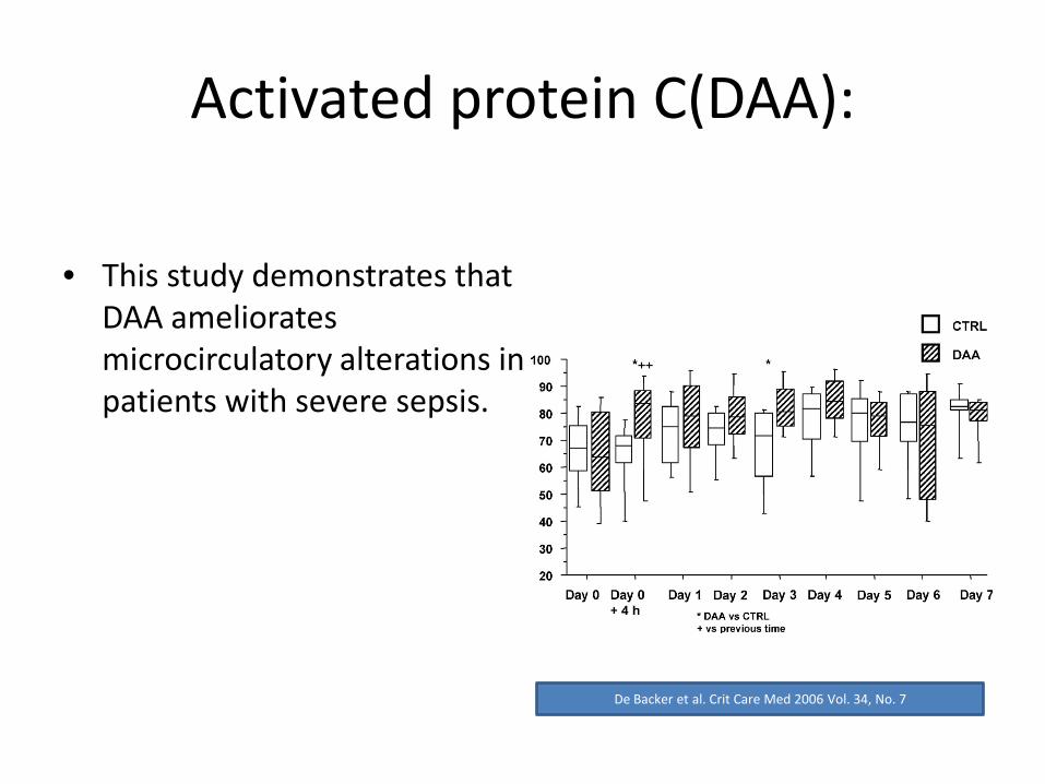
Activated protein C(DAA):
• This study demonstrates that DAA ameliorates microcirculatory alterations in patients with severe sepsis.
De Backer et al. Crit Care Med 2006 Vol. 34, No. 7

Nitroglycerin dose-dependently increases microcirculatory perfusion in patients with severe heart failure as observed by an increase in
sublingual perfused capillary density.Corstiaan et al. Intensive Care Medicine: 35, Nov 2009 1893 - 1899

Near InfraRed Spectroscopy:

Near InfraRed Spectroscopy:

Near-Infrared Spectroscopy:
• A non-invasive technique that uses the differential absorption properties of oxygenated and deoxygenated hemoglobin to evaluate skeletal muscle oxygenation.
• Near-infrared light (680–800 nm) easily crosses biological tissues, which have a low absorption power and is absorbed only by hemoglobin, myoglobin, and oxidized cytochrome ( the contribution of the latter two to the light attenuation signal is very small.)
• Hence, the NIRS signal is derived predominantly from hemoglobin present within the volume of tissue crossed by the near-infrared light.
• NIRS monitors only vessels with a diameter < 1 mm (arterioles, capillaries, and venules)

Near InfraRed Spectroscopy:
Thenar site minimally affected by
– age– gender– edema– adipose
StO2 not confounded by hypothermia

Near-Infrared Spectroscopy:
Total Hemoglobin Index(THI):an indicator of blood volume in the region of microvasculature sensed by the probe expressed in arbitrary units (AU)

Both the current value and trend of StO2 are important.
75%
100%
0%
StO2 low;assess patient; resuscitate if indicated
StO2rising toward normal; assess continued resuscitation
StO2 falling; assess patient; resume resuscitation if indicated
StO2 high; consider late sepsis/spinal cord damage
StO2 adequate; assess need for further resuscitation; stop if indicated
StO
2

StO2-ScvO2

Early Goal-Directed therapy:
• Patients who failed to normalize StO2 during early treatment in the ICU had more severe organ dysfunction and disease severity.
• Low StO2 levels does not reflec global hemodynamic effects(HR,CVP,MAP, ScvO2) or vasoconstriction from pharmacologic intervention.
• Patients who consistently showed low StO2 values within the first 8 hours of ICU treatment had a significantly higher rate of unfavorable outcome.
• The absence of low StO2 levels identified patients with a more favorable outcome.
Lima et al.Critical Care 2009, 13(Suppl 5):S13
Reactive Hyperemia:
• Reactive hyperemia may be considered as a test of microcirculatory reactivity
• It evaluates the tissue’s ability to adjust oxygen extraction capabilities to oxygen delivery after a hypoxic stimulus induced by a transient interruption of blood flow.
• This process is complex, involving capillaries, arterioles, and small arteries, increasing flow in previously patent capillaries and recruiting additional capillaries.
• Altered reactive hyperemia has been reported in septic patients using various techniques.
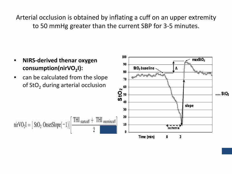
Arterial occlusion is obtained by inflating a cuff on an upper extremity to 50 mmHg greater than the current SBP for 3-5 minutes.
• NIRS-derived thenar oxygen consumption(nirVO2I):
• can be calculated from the slope of StO2 during arterial occlusion

Reactive Hyperemia:
• On release of the arterial cuff, StO2 rises rapidly and eventually peaks at a level of tissue oxygen saturation greater than before occlusion.
• This peak is part of the hyperemic response after arterial occlusion.
• The area under the hyperemic response curve but above the baseline StO2 value is the hyperemic area .
• The time required to reach 63% of the hyperemic peak from the release of the cuff is the StO2 recovery time.

Correlation between SOFA score and the thenar muscleStO2 decrease rate during stagnant ischemia.

StO2 decrease rate :
• NIRS-derived Thenar oxygen consumption(nirVO2I):
• is reduced significantly in severe sepsis subjects.

The rate of increase of StO2 in the hyperemic phase (slope)was lower in the septic patients than in the ICU controls and the healthy
volunteers.

The hyperemic phase (slope):
• Altered recovery in StO2 after an ischemic challenge is frequent in septic patients and more pronounced in the presence of shock.
• The presence and persistence of these alterations in the first 24 h of sepsis are associated with worse outcome.

Reoxygenation Rate(RR)=The hyperemic phase (slope)
• Reoxygenation rate (RR) is related to organ injury in sepsis.
• RR was impaired significantly in sepsis subjects with severe organ failure (SOFA 10, dark gray bar) compared with septic subjects with modest organ dysfunction (light gray bar) and control subjects(white bar).
• RR tended to be slower in those that did not survive hospitalization than in those who survived.

• a linear relationship between nirVO2I and RR in severe sepsis subjects.

Activated Protein C:
• rh-aPC treatment significantly lowered the SOFA score, increased the mean arterial pressure, and reduced the blood lactate concentration
Donati et al.Critical Care 2009, 13(Suppl 5):S12

-rh-aPC had positive effect on StO2 downslope indicating raised oxygen consumption/metabolism.
- rh-aPC had positive effect on StO2 upslope indicating improved microvascular reperfusion following ischemia.
Microvascular function is therefore improved by rh-aPC treatment.
Donati et al.Critical Care 2009, 13(Suppl 5):S12

Signs of regional dysoxia in the presence of apparent adequate oxygen delivery.
• Cytopathic hypoxia: mitochondrial dysfunction in the presence of adequate tissue oxygenation. Fink MP (1997) Acta Anaesth. Scan.110:87-95.
• Shunting theory of sepsis:microcirculatory shut down of weak microcirculatory units creating hypoxic pockets. Ince C & Sinaasappel M (1999) Crit Care Med. 27:1369-1377.


Conclusion:
• The microcirculation consists of the smallest blood vessels (<100 μm diameter)-Arterioles, capillaries, and venules.
• The microcirculation, with its huge endothelial surface, is the largest ‘organ’ in the human body.
• During sepsis and shock it the first to go and last to recover.
• Microcirculatory dysfunction is the single most sensitive and specific predictor of outcome.(irrespective of sytemic hemodynamic and oxygenation variables).
• Noninvasive means to evaluate Microcirculation( SDF, NIRS).(Therapeutic Interventions...).
• MMDS.
• Resuscitation end-point: Microcirculation =Mitochondria


Endotoxin adsorption
• Lipopolysaccharide (LPS) endotoxin– Important in sepsis
• Especially gram negative
• Polymixins– Bind LPS– Hemadsorption of endotoxin
• Japan, Europe• EUPHUS EUPHRATES
Endotoxin adsorption
• 1970’s – benchwork• 1980’s – animal studies
– Plasmapheresis in rat model cleared LPS
• 1990’s – Human studies in Japan– UF or hemadsorption through a special cartridge
w/ polymixin coated fibers.– Clinical use since 1994– Subsequent small, poor quality studies
• 2005 European study suggested benefit

Endotoxin adsorption
• Mechanism of action– Removes endotoxin– Decreases cytokines
• By way of decreasing LPS
– Decreased PAI-1 activity– Decreased neutrophil respiratory burst– Removal of monocytes– Removal of endogenous cannabinoids
• (effects microcirculation…)
Davies, Cohen. Lancet Infect Dis 2011;11:65-71

• Multicenter, RCT in Italy
• 64pt w/ severe sepsis/shock– Intraabdominal sepsis
• Primary endpoint– Change in MAP/pressors
• Secondary endpoint– p/F
– Change in SOFA
– 28day mortality


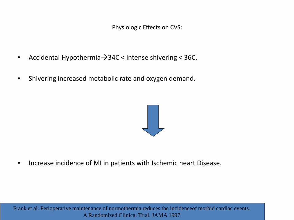
Ultrasound Guidance for Vascular Access and Other Procedures Involving Needles.
Moore CL, Copel JA. N Engl J Med 2011;364:749-757
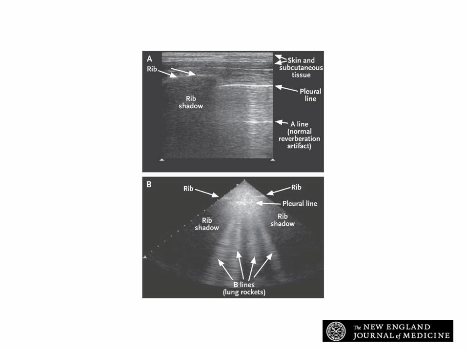
Ultrasound Images of the Pleural Line in a Healthy Patient and in a Patient with Alveolar Interstitial Syndrome.
Moore CL, Copel JA. N Engl J Med 2011;364:749-757

Telemedicine