Embed Size (px)
Citation preview

The fungus Mucor racemosus as a model of phenotypic multidrug resistance in lower eukaryotes
K. A. McGee1 and G. Shearer*2 1Department of Biological Sciences, Alcorn State University, Lorman MS, USA 2Department of Biological Sciences, The University of Southern Mississippi, Hattiesburg, MS, USA.
Keywords: Fungi, antibiotic adaptation, P-glycoprotein, trichodermin, amphotericin B, efflux pump, esterase
1. Introduction
On Monday the 3rd of September 1928 Alexander Fleming, a bacteriologist at St. Mary’s Hospital in London, returned from vacation to his nondescript, cramped and dusty laboratory to change the world. More precisely, he returned to fulfill his role as one of the essential links in the chain of individuals who’s serendipity, scientific curiosity and dogged determination eventually led to the dawn of the antibiotic era. The chain of discovery actually began, as far as we are aware, with observations of 19th century scientists who noted that mold contamination sometimes was associated with the lack of bacterial growth. The English surgeon Joseph Lister (1827-1912), who advocated a ‘carbolic acid’ spray as one of the first antiseptics also noted that urine samples contaminated with mold frequently failed to grow bacteria. Louis Pasteur (1822-1899), who developed the process of pasteurization, noted that the growth of anthrax bacteria was inhibited by the presence of mold. Perhaps most poignant however is the discovery by a French medical student, Ernest Duchesne, who described in his dissertation in 1897 the partial purification and animal testing of a substance from the mold Penicillium which inhibited bacterial growth [1]. His work, preceding Fleming’s discovery by over 30 years, went largely unrecognized until long after his death at the early age of 37 - apparently from tuberculosis. Although the chain of discovery was temporarily severed, Alexander Fleming re-discovered that molds can produce substances that inhibit bacteria and published his discovery in 1929 in the British Journal of Experimental Pathology [2]. Fleming, however, failed to appreciate the potential of this ‘penicillin’ as a therapeutic agent. Ten years later in 1939 three scientists at Oxford University, Howard Florey, Ernst Chain and Edward Abraham expanded Fleming’s work to develop a purified stable form of penicillin and conducted the first human trial in 1941. The development and testing of penicillin was greeted with great enthusiasm and the hope that infectious diseases and death from wound sepsis would soon be a thing of the past. In October 1941 a note in the Monthly Science News [3] published in London stated:
“One of the most important advances in medicine has been the discovery of substances which are more poisonous to certain kinds of bacteria than they are to mice and men, so that they can be used against bacteria when they are circulating in the blood or lurking in odd corners of the body causing disease.”
Even during this early period of the new antibiotic era, worrisome observations were made that microbes also had defenses against these ‘magic bullets’. In 1946 Fleming edited a book reviewing the clinical applications of penicillin [4] in which he made a prescient warning against indiscriminate use of the
* Corresponding author: e-mail: [email protected], Phone 601-266-4722
Communicating Current Research and Educational Topics and Trends in Applied Microbiology A. Méndez-Vilas (Ed.)
201©FORMATEX 2007
_____________________________________________________________________

antibiotic. In his review of Fleming’s book one year later, JF Mahoney made the following observation [5]:
“...the warning against the indiscriminate use of the antibiotic is timely. Possibly Fleming might have further emphasized the dangers which may accrue from the promiscuous use of the product in conditions in which the causative organisms are not identified or in which other forms of treatment are known to be effective. Not only is this practice unsound but it may have the effect of reducing the efficacy of penicillin in patients confronted by a lethal emergency caused by pathogens which would have been susceptible under ordinary circumstances"
In fact, microbes resistant to penicillin as well as other recently discovered antibiotics were being isolated from clinical samples with greater and greater frequency. Now as we fast forward over 60 years to the current time, penicillin resistance is so widespread that this antibiotic is nearly useless. Microbes are very skilled in the art of survival and well equipped by nature to do so. Given that the vast majority of antibiotics we use are natural products of microbes, it should be no surprise that other microbes must have developed defenses over the eons. Our current concerns are not so much that a pathogen is now resistant to penicillin but that these microbes are now frequently multi-drug resistant to first-, second- and third-, etc. generation antibiotics. For example, the common pathogen Staphylococcus aureus is now typically resistant to most common antibiotics. In the 1990s, a dramatic increase in methicillin resistant S. aureus (MRSA) was noted in clinical isolates in the United States [6]. These multi-drug resistant staphlococci were still sensitive to vancomycin which began to be referred to as “the antibiotic of last resort”. In 2002, the first vancomycin-resistant S. aureus was isolated from a clinical sample [7] and concerns in the lay press about a ‘super bug’ and untreatable ‘flesh-eating bacteria’ became widespread. This point/counterpoint battle must continue as scientists develop new agents to combat pathogens and the pathogens continue to demonstrate their amazing survival skills and develop resistance. Nowadays, our main task appears to be to just stay one step ahead in this struggle.
2. Mechanisms and examples of antibiotic resistance
Microbes may become resistant to antibiotics via intrinsic as well as extrinsic means. Intrinsic mechanisms involve existing abilities of the organism itself and we will divide this category into mutation and phenotypic resistance, the latter being the primary concern of this chapter. Extrinsic mechanisms involve the acquisition of genetic information from other microbes via transformation, transduction, etc. These new genes give the organism the ability to resist one or more antibiotics to which it was once sensitive. Before discussing the fungus Mucor as a model of phenotypic multi-drug resistance, it is useful to briefly review resistance via mutation and acquisition of exogenous genetic sequences as they fit into the three major categories of resistance mechanisms. We can divide resistance mechanisms into three fundamental categories: (i) Modification of the antibiotic to render it less toxic, (ii) Modification of the target to render it less sensitive and (iii) Reduced access of the drug to the target. Subcategories can easily be envisioned, but these will still fit within one of these three major categories.
2.1 Resistance via degradation/modification of drugs
Microbes may degrade or modify an antibiotic to render it less toxic. The classic example of this type of resistance is penicillin resistance via production of a β-lactamase enzyme which cleaves the lactam ring to render the drug ineffective. As early as 1941 E. coli strains were reported which produce ‘penicillinase’ [8]. The plasmid borne gene for β-lactamase is easily passed among bacteria and is now common in clinical isolates. In a similar manner, numerous enzymes carried on transferable genetic elements are known which modify (e.g., via acetylation or phosphorylation) aminoglycosides, chloramphenicol and other antibiotics [9, 10].
Communicating Current Research and Educational Topics and Trends in Applied Microbiology A. Méndez-Vilas (Ed.)
202 ©FORMATEX 2007
_____________________________________________________________________

2.2 Resistance via modification of target
Alterations in the target of the antibiotic is an effective means of resistance. For example, mutations in DNA gyrase, the target of fluoroquinolones such as ciprofloxacin [11] render the cells resistant. Mutation in the ribosomal genes, which make cells resistant to ribosome binding agents such as tetracycline, amikacin, kanamycin, etc. [12, 13], is a well known resistance mechanism in clinical isolates.
2.3 Resistance via altered permeability and efflux
The third major mechanism of resistance, denying access to the target, is demonstrated by the commonly found tetracycline efflux system. Numerous efflux proteins capable of pumping tetracycline have been identified [14]. This active efflux of drug helps prevent inhibitory levels from reaching the ribosome. In a similar manner efflux pumps with broad substrate range may render the cells resistant to multiple drugs at once [15]. Although efflux pumps are arguably the most common example of this major mechanism of antibiotic resistance, other changes that alter permeability are also effective. For example, recent data indicate that one mechanism of vancomycin resistance in S. aureus is the production of a thickened cell wall which impedes entry of the drug into the cell. [16].
3. Phenotypic multi-drug resistance in Mucor racemosus
In nearly all the examples discussed above, resistance is ultimately the result of a mutation in some gene(s) in the pathogen or acquisition of new genetic material which enables the recipient cell to effect resistance. Very little is known about phenotypic drug resistance, in particular phenotypic multi-drug resistance. In this case the cell does not acquire any new genetic information, nor is any gene mutated, rather the microbe possesses a set of genes that can be up regulated to enable the cell to resist a number of antibiotics. The cell has some sensory mechanism to rapidly activate and express these genes while the antibiotic is present and then to de-activate the genes when the danger is past. In this manner, the entire population is protected de novo rather than relying on a fortuitous mutation or lucky happenstance that a fragment of DNA with an appropriate resistance gene might be available. The common fungus Mucor racemosus serves as an excellent example of this ability to phenotypically adapt to multiple antibiotics after exposure to a single agent.
3.1 Introduction to the system
The fungus Mucor racemosus (syn. M. circinelloides) is a common member of the Mucorales, the largest and most studied order of the class Zygomycetes. The Mucorales contains over 300 species and exhibits wide, aseptate, fast-growing hyphae typical of this group of fungi. Abundant asexual spores are contained within sporangia borne on elongated sporangiophores. Germination of these spores produces new haploid hyphae of the same mating type. Sexual reproduction occurs when hyphae of opposite mating types touch and, under proper conditions, fuse to form a thick-walled zygospore. Germination of the zygospore leads to the growth of hyphae which then give rise to asexual spores of both the + and – mating type. The Mucorales, including M. racemosus, are typically saprophytes and are problematic as spoilage organisms of fruits, vegetables and other foodstuffs. Although infrequent, human infections with a few species of the Mucorales has been reported. These zygomycoses can be quite severe and life-threatening [17, 18]. M. racemosus exhibits the particularly intriguing ability to grow in two radically different forms: mold and yeast. Generally the organism is known as a mold. The mold form is differentiated and consists of non-septate hyphae with specialized cells for asexual and sexual reproduction. In contrast, M. racemosus can grow in an undifferentiated yeast form under anaerobic (or perhaps microaerophilic) conditions when sparged with CO2 or N2. Either the extensive mold form or the multi-budded yeast form can be
Communicating Current Research and Educational Topics and Trends in Applied Microbiology A. Méndez-Vilas (Ed.)
203©FORMATEX 2007
_____________________________________________________________________
grown in the laboratory simply by maintaining the appropriate growth conditions. The mold form may be ‘converted’ to the yeast form by sparging cultures with CO2 or N2. The yeast form may be ‘converted’ to the mold form simply by increasing the oxygen tension of the growth medium, e.g., by sparging with air. This yeast-mold dimorphism, which represents an easily reversible differentiation process in a lower eukaryote, has been the subject of numerous studies. Bartnicki-Garcia demonstrated in 1962 that yeast growth could be obtained by sparging CO2 through the liquid growth medium [19], while growth in air or with N2 sparging resulted in the mold growth form. Later Mooney and Sypherd demonstrated that the yeast form would also grow in N2 sparged medium provided that the flow rate was greater than 1ml/min per ml of culture medium [20]. They interpreted this result to indicate that a volatile factor is needed to maintain mold growth and removal of this factor by increased flow rate of gas resulted in growth in the yeast morphotype. This apparent relationship of dimorphism to respiratory flux was further studied by Paznokas and Sypherd, who noted that the change in 3’-5’-cyclic adenosine monophosphate (cAMP) levels as a result of growth conditions influenced dimorphism [21]. In fact, addition of exogenous cAMP prevents growth of the mold morphotype [22]. cAMP phosphodiesterase activities were found to be relatively constant in both the mold and yeast morphotypes leading to the conclusion that adenylate cyclase activity may be responsible for changes in internal cAMP levels [22]. Rose, et al. demonstrated in 1999 that Ras genes in M. racemosus exhibit differential expression in mold versus yeast and that exogenous addition of the cAMP analog dibutyryl-cAMP caused a strong repression of RAS1 (but not RAS3) concurrent with the inhibition of growth of the mold morphotype [23]. It is likely that cAMP plays a role in the signal cascade leading to the dimorphic shift from one morphotype to the other. More recent work in this area has implicated pkaR, a cAMP-dependent protein kinase regulatory subunit, which when overexpressed favors growth in the mold morphotype [24, 25].
3.2 Phenotypic Multi-drug Resistance in Mucor
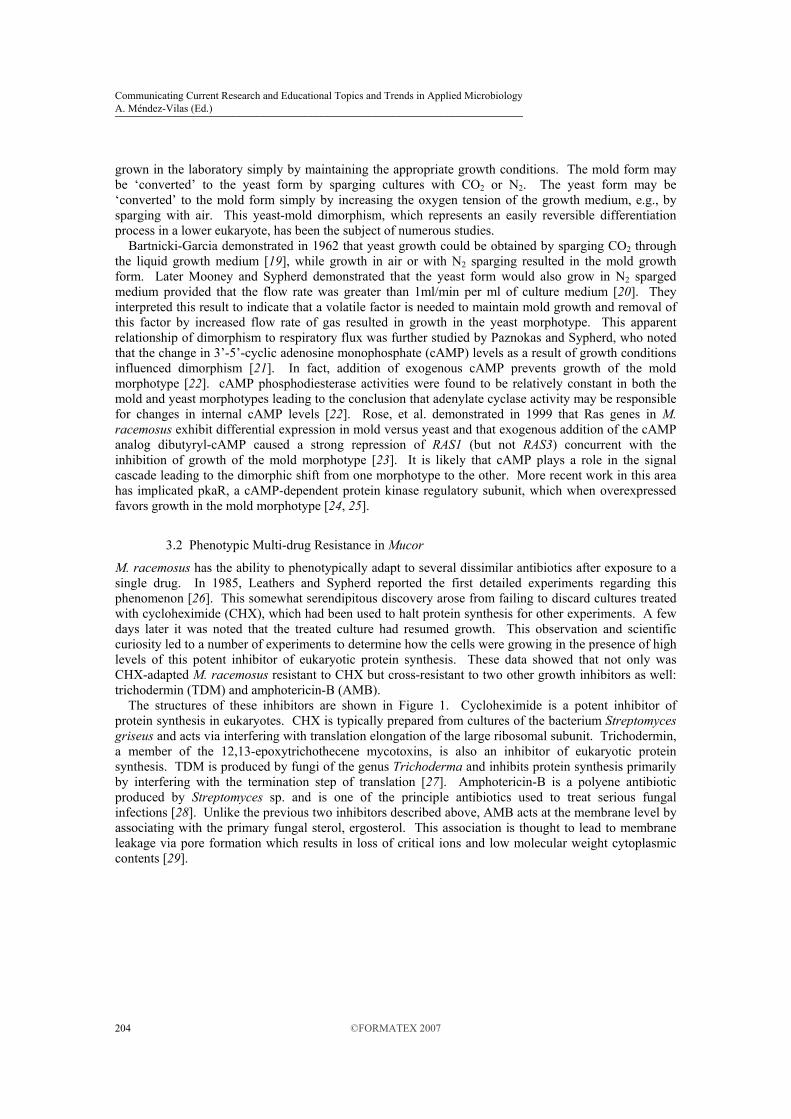
M. racemosus has the ability to phenotypically adapt to several dissimilar antibiotics after exposure to a single drug. In 1985, Leathers and Sypherd reported the first detailed experiments regarding this phenomenon [26]. This somewhat serendipitous discovery arose from failing to discard cultures treated with cycloheximide (CHX), which had been used to halt protein synthesis for other experiments. A few days later it was noted that the treated culture had resumed growth. This observation and scientific curiosity led to a number of experiments to determine how the cells were growing in the presence of high levels of this potent inhibitor of eukaryotic protein synthesis. These data showed that not only was CHX-adapted M. racemosus resistant to CHX but cross-resistant to two other growth inhibitors as well: trichodermin (TDM) and amphotericin-B (AMB). The structures of these inhibitors are shown in Figure 1. Cycloheximide is a potent inhibitor of protein synthesis in eukaryotes. CHX is typically prepared from cultures of the bacterium Streptomyces griseus and acts via interfering with translation elongation of the large ribosomal subunit. Trichodermin, a member of the 12,13-epoxytrichothecene mycotoxins, is also an inhibitor of eukaryotic protein synthesis. TDM is produced by fungi of the genus Trichoderma and inhibits protein synthesis primarily by interfering with the termination step of translation [27]. Amphotericin-B is a polyene antibiotic produced by Streptomyces sp. and is one of the principle antibiotics used to treat serious fungal infections [28]. Unlike the previous two inhibitors described above, AMB acts at the membrane level by associating with the primary fungal sterol, ergosterol. This association is thought to lead to membrane leakage via pore formation which results in loss of critical ions and low molecular weight cytoplasmic contents [29].
Communicating Current Research and Educational Topics and Trends in Applied Microbiology A. Méndez-Vilas (Ed.)
204 ©FORMATEX 2007
_____________________________________________________________________
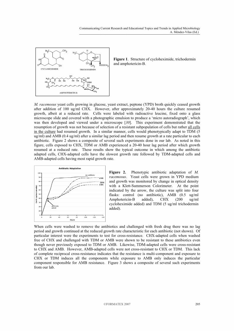
Figure 1. Structure of cycloheximide, trichodermin and amphotericin-B.
M. racemosus yeast cells growing in glucose, yeast extract, peptone (YPD) broth quickly ceased growth after addition of 100 ug/ml CHX. However, after approximately 20-40 hours the culture resumed growth, albeit at a reduced rate. Cells were labeled with radioactive leucine, fixed onto a glass microscope slide and covered with a photographic emulsion to produce a ‘micro autoradiograph’, which was then developed and viewed under a microscope [30]. This experiment demonstrated that the resumption of growth was not because of selection of a resistant subpopulation of cells but rather all cells in the culture had resumed growth. In a similar manner, cells would phenotypically adapt to TDM (5 ug/ml) and AMB (0.4 ug/ml) after a similar lag period and then resume growth at a rate particular to each antibiotic. Figure 2 shows a composite of several such experiments done in our lab. As noted in this figure, cells exposed to CHX, TDM or AMB experienced a 20-40 hour lag period after which growth resumed at a reduced rate. These results show the typical outcome in which among the antibiotic adapted cells, CHX-adapted cells have the slowest growth rate followed by TDM-adapted cells and AMB-adapted cells having most rapid growth rate.
Figure 2. Phenotypic antibiotic adaptation of M. racemosus. Yeast cells were grown in YPD medium and growth was monitored by change in optical density with a Klett-Summerson Colorimeter. At the point indicated by the arrow, the culture was split into four flasks: control (no antibiotic), AMB (0.5 ug/ml Amphotericin-B added), CHX (200 ug/ml cycloheximide added) and TDM (5 ug/ml trichodermin added).
When cells were washed to remove the antibiotics and challenged with fresh drug there was no lag period and growth continued at the reduced growth rate characteristic for each antibiotic (not shown). Of particular interest were the experiments to test for cross-resistance. CHX-adapted cells when washed free of CHX and challenged with TDM or AMB were shown to be resistant to these antibiotics even though never previously exposed to TDM or AMB. Likewise, TDM-adapted cells were cross-resistant to CHX and AMB. However, AMB-adapted cells were not cross-resistant to CHX or TDM. This lack of complete reciprocal cross-resistance indicates that the resistance is multi-component and exposure to CHX or TDM induces all the components while exposure to AMB only induces the particular component responsible for AMB resistance. Figure 3 shows a composite of several such experiments from our lab.
Communicating Current Research and Educational Topics and Trends in Applied Microbiology A. Méndez-Vilas (Ed.)
205©FORMATEX 2007
_____________________________________________________________________

Figure 3. Phenotypic antibiotic adaptation cross-resistance of M. racemosus. Yeast cells were grown in YPD medium and growth was monitored by change in optical density with a Klett-Summerson Colorimeter. A control culture (no antibiotic) and a culture adapted to 0.5 ug/ml AMB were grown in YPD medium. At the point indicated by the arrow, the AMB-adapted culture was washed to remove the AMB and split into three flasks: AMB (0.5 ug/ml Amphotericin-B added), CHX (200 ug/ml cycloheximide added) and TDM (5 ug/ml trichodermin added).
Leathers and Sypherd [26] also demonstrated that the entire population of cells would deadapt when grown in the absence of the antibiotic. These cells then showed full sensitivity to the three antibiotics and required a period of re-adaptation as shown in Figure 2. The deadaptation process was quite rapid for TDM- and AMB-adapted cells, requiring only 1.5 to 2 cell doublings in antibiotic-free medium. CHX-adapted cells, however, required more than 15 generations in drug-free medium to become fully susceptible again.
3.3 Mechanism of trichodermin resistance.
The experiments described above demonstrated that a multi-component resistance system was induced upon exposure to one or more drugs and was then shut down when the inhibitors were removed. Although the three antibiotics used were different in structure and mode of action (CHX and TDM are both protein synthesis inhibitors but they act at different points of the translation process), the mode(s) of resistance could fall within any of the three major resistance mechanisms: (i) Modification of the drug, (ii) Modification of the target or (iii) Reduced access of the drug to the target. Experiments were then designed to address the particular mechanism(s) of this phenotypic resistance by examining each of these major mechanisms.
Figure 4. TDM-esterase activity of cell extracts. M. racemosus yeast were adapted to CHX (200 ug/ml), TDM (5 ug/ml) or AMB (0.5 ug/ml). After growth had resumed, cells were broken by ballistic disruption with glass beads and a cell-free supernatant prepared. Identical amounts of cellular protein were incubated with 14C-trichodermin (label on acetate moiety) and the amount of released 14C acetate was measured. Enzyme assays were optimized to ensure linearity.
Fonzi and Sypherd prepared radiolabeled TDM (14C label on the acetate moiety) and determined that adapted cells did not show decreased uptake of the drug. In fact, adapted cells had far greater uptake of the radioisotope than non-adapted cells [31]. This puzzling result was explained after analysis of cell extracts which showed that adapted cells rapidly deacylate TDM to its alcohol derivative, trichodermol. The increased uptake of radioisotope in adapted cells was due to the rapid uptake of the released 14C-acetate. Assays of TDM esterase activity showed that CHX- and TDM-adapted cells had dramatically increased TDM-esterase as compared to control cells. AMB-adapted cells, however, had essentially the same low level of TDM-esterase activity as control cells. These data provided a working hypothesis for
Communicating Current Research and Educational Topics and Trends in Applied Microbiology A. Méndez-Vilas (Ed.)
206 ©FORMATEX 2007
_____________________________________________________________________

TDM adaptation: cells exposed to CHX or TDM up regulate production of one or more enzymes which can deacylate TDM to the much less potent trichodermol. This modification of drug is almost certainly the mechanism of TDM resistance. In contrast, AMB-adapted cells are not cross-resistant to TDM because AMB does not induce the TDM-esterase(s). As shown in Figure 4 of data from our lab, CHX-adapted cells had approximately 6 times and TDM-adapted cells over 5 times more TDM-esterase activity as compared to control cells. AMB-adapted cells, however, had essentially the same amount of TDM-esterase activity as control cells. We have further investigated the induction of the TDM-esterase by attempting to detect a new or upregulated esterase via native gel electrophoresis followed by staining for esterase activity with the non-specific substrate alpha-napthyl acetate. As shown in Figure 5, cell-free supernatants are rich in esterase activity, as noted by the numerous dark bands where the napthyl acetate was deacetylated. We noted that a new esterase band was apparent only in CHX-adapted and TDM-adapted cells which correlates well with the data of Fonzi and Sypherd and with our data in Figure 4. Since this polyacrylamide gel was non-denaturing, we cannot determine a molecular weight for this band. It is also possible, albeit unlikely, that this result is coincidental and this esterase band is not related to TDM resistance. Future work will need to utilize proteomic and genomic tools to identify this esterase and its gene(s) to determine if it is in fact responsible for TDM resistance.
Figure 5. Esterase activity in control and antibiotic-adapted cells. Cells were grown and adapted as described for Figure 4. Labels above each lane indicate the antibiotic to which the cells were adapted. Identical amounts of total cellular protein were separated via non-denaturing polyacrylamide gel electrophoresis and the gel was stained for esterase activity with the non-specific substrate alpha-napthyl acetate. Dark bands indicate areas of esterase activity. As shown by the arrow, CHX-adapted and TDM-adapted (but not control or AMB-adapted) cells had a new band of esterase activity near the bottom of the gel. The molecular weight cannot be determined from this non-denaturing gel.
3.4 Mechanism of Cycloheximide resistance.
As shown in Figure 1, it is unlikely that enhanced esterase activity could play a direct role in CHX resistance since there are no ester links in this antibiotic. At the time of these early studies (mid 1980’s), a review of the literature revealed that numerous CHX resistant strains of microorganisms were known; for example, Neurospora crassa [32], Podospora anserina [33], Tetrahymena [34], Acanthamoeba castellanii [35], Physarum polycephalum [36], Saccharomyces cerevisiae [37], and Coprinus cinereus [38]. In each of these cases, CHX resistance was shown to be due to a mutation in the ribosome. Based on these data, Shearer and Sypherd [39] conducted experiments to determine if adapted M. racemosus ribosomes showed decreased sensitivity to CHX. Ribosomes were isolated from control cells as well as CHX-adapted and TDM-adapted cells. An in vitro translation assay with poly(U) template revealed that ribosomes from adapted cells as compared to control cells were fully sensitive to the inhibitory effects of CHX. Ribosomes from control cells, CHX-adapted and TDM-adapted cells all required 0.4 ug/ml CHX to inhibit 50% of the poly(Phe) synthesis. Therefore, CHX resistance in M. racemosus was clearly not due to a modification of the ribosomal target. Several organisms were known to modify CHX to yield a compound with markedly decreased toxicity [40, 41]. To determine if adapted cells were modifying CHX, 3H-radiolabeled CHX was used to adapt cells after which thin layer chromatography was used to detect modification and/or degradation of the
Communicating Current Research and Educational Topics and Trends in Applied Microbiology A. Méndez-Vilas (Ed.)
207©FORMATEX 2007
_____________________________________________________________________

compound. No changes in CHX amount or mobility were detected indicating that adapted cells do not degrade CHX or modify it to a less inhibitory compound [39]. Apparently adapted cells did not modify or degrade CHX and their ribosomes were fully sensitive to the inhibitory effects on translation. The remaining major mechanism to consider was a decrease in access of the drug for the target. To test this hypothesis, control and adapted cells were pre-incubated with radiolabeled CHX for 30 min. after which they were rapidly washed and resuspended in drug-free medium. Samples were taken at various times to measure the amount remaining within the cell. As shown in Figure 6, which represents a composite of several experiments, adapted cells (both CHX-adapted and TDM-adapted) had essentially no detectable CHX remaining after approximately 10 minutes while non-adapted control cells had nearly 60% CHX remaining at this time. AMB-adapted cells were similar to non-adapted control cells (not shown). This rapid loss of CHX in adapted cells is most simply explained by an active efflux of the drug. To test this hypothesis, efflux experiments were repeated with sodium azide to collapse the energy charge of the cell. These experiments showed that efflux in adapted cells treated with azide were identical to non-adapted control cells, indicating the presence of an energy-dependent efflux system (not shown).
Figure 6. Efflux of cycloheximide in un-adapted (solid line) and adapted (dashed line) cells. M. racemosus yeast cells were pre-loaded with 3H-CHX for 30 min., washed free of drug and resuspended in drug-free medium. The amount of CHX remaining in the cells was measured over a period of 45 min.
The increased energy dependent efflux of CHX was reminiscent of increased drug efflux of chemotherapy agents in many cases of human cancer via over expression of the P-glycoprotein pump [42-44]. Based on the hypothesis that M. racemosus might use a similar P-glycoprotein efflux pump for CHX, we isolated total cellular protein from non-adapted control cells as well as CHX-adapted cells and performed a western blot with anti-P-glycoprotein monoclonal antibody C219 [45]. As shown in Figure 7, CHX adapted cells had a single band of approximately 170 kd, consistent with the size of other known P-glycoproteins (Figure 7 lane B). CHX-adapted cells allowed to partially de-adapt for 5 generations had reduced amount of this reactive band (Figure 7, lane A) and non-adapted control cells had no detectable signal with the C219 antibody (Figure 7, ctrl lane). This result is consistent with the hypothesis that the Mucor P-glycoprotein homolog is up regulated during CHX adaptation and is a likely candidate for the energy-dependent efflux of cycloheximide.
Figure 7. Western blot of M. racemosus against C219 antibody. Total cellular protein was extracted from non-adapted control cells, CHX-adapted cells allowed to deadapt for 5 generations in drug-free medium (lane A) and CHX-adapted cells (lane B). Equal amounts of protein in each lane were separated by SDS polyacrylamide gel electrophoresis and probed with the anti P-glycoprotein monoclonal antibody C219. Prior staining of the blot with Ponceau-S confirmed equivalent amounts of protein were transferred in each lane.
Communicating Current Research and Educational Topics and Trends in Applied Microbiology A. Méndez-Vilas (Ed.)
208 ©FORMATEX 2007
_____________________________________________________________________

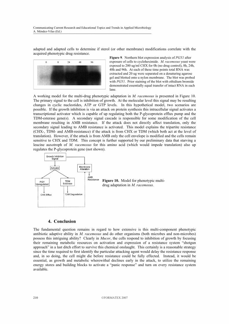
We isolated the putative M. racemosus P-glycoprotein homolog by probing genomic and cDNA libraries with the Drosophila MDR49 gene [46], which we theorized would be likely to cross hybridize given that P-glycoprotein genes share significant base homology. We identified numerous clones from this approach and isolated the complete Mucor P-glycoprotein, PGY1 (GenBank AY029279). Analysis of the predicted protein sequence demonstrated extensive similarity to other characterized P-glycoproteins known to function as efflux pumps with broad substrate range. As shown in Figure 8, the predicted amino acid sequence of the PGY1 gene product had transmembrane regions and ATP binding domains consistent with ABC transporters. The Mucor P-glycoprotein gene was strongly up regulated after exposure to CHX, as shown in the northern blot analysis of Figure 9. Expression of the PGY1 transcript was present, albeit weak, in control cultures and after 2 hours exposure to CHX the gene was dramatically up regulated. These data are consistent with the detection of the putative P-glycoprotein on western blots as shown above. In a similar manner, quantitative real-time PCR analysis of RNA extracted from CHX-adapted and TDM-adapted cells (but not AMB-adapted) showed up to a 7-fold increase in PGY1 transcript level (not shown).
3.5 Mechanism of Amphotericin-B resistance.
The method by which M. racemosus phenotypically adapts (and de-adapts in the absence of AMB) is unknown. It is unlikely that the TDM-esterase is capable of modifying amphotericin since no ester links are available for attack. Also it is not probable that the Mucor P-glycoprotein is able to make the cells AMB resistant, since this drug affects the cell membrane by binding to sterols and thus needs only access the cell surface rather than have to enter the cell to attack an intracellular target. We think it most likely that the cell is altering the membrane in some manner.
Figure 8. Predicted transmembrane regions and homology of the PGY1-encoded P–glycoprotein. of M. racemosus. A, predicted transmembrane domains; B, conserved regions within M. racemosus P-glycoprotein; C, conserved regions within Rattus norvegicus P-glycoprotein.
AMB resistance is known in several other organisms although the mechanism is not fully understood. In most cases, a mutation in some part of the ergosterol (the primary fungal sterol and target of AMB) biosynthetic pathway is implicated. For example, AMB resistance has been correlated with mutation in the Erg2 gene in Candida lusitaniae [47, 48], Candida albicans [49] and Cryptococcus neoformans [50]. In a similar manner, deletion of the Erg6 gene (S-adenosylmethionine methyltransferase) in C. albicans [51], C. lusitaniae [52] and S. cerevisiae [53] has been shown to result in increased polyene resistance. In C. albicans and S. cerevisiae, however, this deletion results in increased CHX sensitivity. Although mutations in the ergosterol biosynthetic genes sometimes yield AMB resistant strains, this cannot explain the phenotypic AMB resistance in Mucor. The same mutation would not be expected to arise simultaneously in the entire cell population in 20-40 hours nor would it be expected to revert after 1-2 generations in drug-free medium. If we consider the possibilities of how a genomic change could explain the phenotypic adaptation, it is possible that a cassette switching mechanism similar to the mating type switch in Saccharomyces cerevisiae [54] could alter sterol biosynthesis in a rapidly reversible manner. However, even in this case, it is not clear how the pre-existing ergosterol in the membrane could be replaced so rapidly to effect the AMB resistance phenotype. Future studies will need to examine the membrane composition of non-
Communicating Current Research and Educational Topics and Trends in Applied Microbiology A. Méndez-Vilas (Ed.)
209©FORMATEX 2007
_____________________________________________________________________
adapted and adapted cells to determine if sterol (or other membrane) modifications correlate with the acquired phenotypic drug resistance.
Figure 9. Northern blot expression analysis of PGY1 after exposure of cells to cycloheximide. M. racemosus yeast were exposed to 200 ug/ml CHX for 0h (no drug control), 8h, 24h, 48h and 96h. At each of these time points total RNA was extracted and 20 ug were separated on a denaturing agarose gel and blotted onto a nylon membrane. The blot was probed with PGY1. Prior staining of the blot with ethidium bromide demonstrated essentially equal transfer of intact RNA in each lane.
A working model for the multi-drug phenotypic adaptation in M. racemosus is presented in Figure 10. The primary signal to the cell is inhibition of growth. At the molecular level this signal may be resulting changes in cyclic nucleotides, ATP or GTP levels. In this hypothetical model, two scenarios are possible. If the growth inhibition is via an attack on protein synthesis this intracellular signal activates a transcriptional activator which is capable of up regulating both the P-glycoprotein efflux pump and the TDM-esterase gene(s). A secondary signal cascade is responsible for some modification of the cell membrane resulting in AMB resistance. If the attack does not directly affect translation, only the secondary signal leading to AMB resistance is activated. This model explains the tripartite resistance (CHX-, TDM- and AMB-resistance) if the attack is from CHX or TDM (which both act at the level of translation). However, if the attack is from AMB only the cell envelope is modified and the cells remain sensitive to CHX and TDM. This concept is further supported by our preliminary data that starving a leucine auxotroph of M. racemosus for this amino acid (which would impede translation) also up regulates the P-glycoprotein gene (not shown).
Figure 10. Model for phenotypic multi-drug adaptation in M. racemosus.
4. Conclusion
The fundamental question remains in regard to how extensive is this multi-component phenotypic antibiotic adaptive ability in M. racemosus and do other organisms (both microbes and non-microbes) possess this intriguing ability? Clearly in Mucor, the cells respond to inhibition of growth by focusing their remaining metabolic resources on activation and expression of a resistance system “shotgun approach” in a last ditch effort to survive this chemical onslaught. This certainly is a reasonable strategy since the time required to first identify the particular attacking agent would delay the resistance response and, in so doing, the cell might die before resistance could be fully effected. Instead, it would be essential, as growth and metabolic wherewithal declines early in the attack, to utilize the remaining energy stores and building blocks to activate a “panic response” and turn on every resistance system available.
Communicating Current Research and Educational Topics and Trends in Applied Microbiology A. Méndez-Vilas (Ed.)
210 ©FORMATEX 2007
_____________________________________________________________________

Microbes in complex ecological communities battling for space, nutrients, etc. produce and secrete a staggering array of antibiotics designed to kill or inhibit their competitors. In particular, soil bacteria of the genus Streptomyces (which synthesize both CHX and AMB) are notorious for this chemical warfare. Fungi, like Mucor, have the difficult task of surviving in this environment. We think it likely that many microbes in such environments may possess a tactical rapid response system to defend against a complex offensive armamentarium of antibiotics. The example described here for M. racemosus which responds to such attack by simultaneously activating an energy-dependent efflux pump (probably capable of pumping a wide variety of compounds), at least one esterase (probably capable of degrading multiple targets which possess ester links) and an as-yet-unknown AMB resistance system (probably via a modification of the cell envelope) may only be the ‘tip of the iceberg’ in regard to microbe-microbe interactions in complex environments. Certainly we anticipate that there are many antibiotics awaiting discovery, and many resistance systems designed by nature to protect against them. It is possible that such phenotypic drug resistance may play an important role in pathogens during antibiotic therapy. If so, this would be difficult to ascertain from conventional laboratory procedures which typically involve isolation and growth of the microbe on standard laboratory media (i.e., lacking antibiotic) during which time the pathogen would de-adapt (i..e, the phenotypic resistance system would be turned off). If these cells are then checked for antibiotic sensitivity, they would appear fully sensitive. Only if the protocols were altered to allow extended incubation time for re-adaptation to occur would the possibility of phenotypic drug resistance be noted.
Acknowledgements. KM and GS are supported in part by NIH Grant Number RR016476 from the National Center for Research Resources.
References [1] J. Pouillard, Hist Sci Med 36, 11 (2002). [2] A. Fleming, Brit. J. Exp. Pathol 10, 226 (1929). [3] unknown, Can J Comp Med Vet Sci 6, 27 (1942). [4] A. Fleming, Penicillin: Its Practical Application (Blakiston, Philadelphia, 1946), [5] J. F. Mahoney, Am J Public Health Nations Health 37, 1189 (1947). [6] A. L. Panlilio et al., Infect Control Hosp Epidemiol 13, 582 (1992). [7] L. M. Weigel et al., Science 302, 1569 (2003). [8] E. P. Abraham et al., Lancet 2, 177 (1941). [9] J. Davies, G. D. Wright, Trends Microbiol 5, 234 (1997). [10] S. Schwarz, C. Kehrenberg, B. Doublet, A. Cloeckaert, FEMS Microbiol Rev 28, 519 (2004). [11] K. Drlica, M. Malik, Curr Top Med Chem 3, 249 (2003). [12] M. M. Gerrits, M. R. de Zoete, N. L. Arents, E. J. Kuipers, J. G. Kusters, Antimicrob Agents Chemother 46,
2996 (2002). [13] G. J. Alangaden et al., Antimicrob Agents Chemother 42, 1295 (1998). [14] F. M. Sapunaric, M. Alderna-Ramos, L. M. McMurry, Tetracycline resistance: efflux, mutation and other
mechanisms. (ASM Press, Washingon, DC, 2005), pp. 3-18. [15] L. J. Piddock, Clin Microbiol Rev 19, 382 (2006). [16] L. Cui et al., Antimicrob Agents Chemother 50, 428 (2006). [17] L. Pagano, L. Fianchi, G. Leone, J Chemother 18, 339 (2006). [18] D. Jain et al., Mod Pathol 19, 1221 (2006). [19] S. Bartnicki-Garcia, W. J. Nickerson, J Bacteriol 84, 829 (1962). [20] D. T. Mooney, P. S. Sypherd, J Bacteriol 126, 1266 (1976). [21] J. L. Paznokas, P. S. Sypherd, J Bacteriol 124, 134 (1975). [22] M. Orlowski, Arch Microbiol 126, 133 (1980). [23] L. V. Roze, N. Mahanti, R. Mehigh, D. G. McConnell, J. E. Linz, Fungal Genet Biol 28, 171 (1999). [24] A. M. Wolff, K. F. Appel, J. B. Petersen, U. Poulsen, J. Arnau, FEMS Yeast Res 2, 203 (2002). [25] T. Lubbehusen et al., Microbiology 150, 143 (2004). [26] T. D. Leathers, P. S. Sypherd, Antimicrob Agents Chemother 27, 892 (1985).
Communicating Current Research and Educational Topics and Trends in Applied Microbiology A. Méndez-Vilas (Ed.)
211©FORMATEX 2007
_____________________________________________________________________

[27] C. M. Wei, B. S. Hansen, M. H. J. Vaughan, C. S. McLaughlin, Proc Natl Acad Sci U S A 71, 713 (1974). [28] H. A. Gallis, R. H. Drew, W. W. Pickard, Rev Infect Dis 12, 308 (1990). [29] B. Venegas, J. Gonzalez-Damian, H. Celis, I. Ortega-Blake, Biophys J 85, 2323 (2003). [30] M. Orlowski, P. S. Sypherd, J Bacteriol 133, 399 (1978). [31] W. A. Fonzi, P. S. Sypherd, Antimicrob Agents Chemother 29, 570 (1986). [32] H. Rothschild, J. Germershausen, S. R. Suskind, Biochem Genet 13, 283 (1975). [33] M. Crouzet, M. Perrot, M. Nogueira, J. Begueret, Biochem Genet 16, 271 (1978). [34] C. A. Sutton, M. J. Ares, R. L. Hallberg, Proc Natl Acad Sci U S A 75, 3158 (1978). [35] G. E. t. Chisholm, M. H. Vaughan, J Bacteriol 138, 280 (1979). [36] T. E. Evans, H. H. Evans, J Bacteriol 143, 897 (1980). [37] N. F. Kaufer, H. M. Fried, W. F. Schwindinger, M. Jasin, J. R. Warner, Nucleic Acids Res 11, 3123 (1983). [38] J. D. Traynor, I. Sardharwalla, J. North, J Gen Microbiol 132, 757 (1986). [39] G. Shearer, P. S. Sypherd, Antimicrob Agents Chemother 32, 341 (1988). [40] R. Howe, R. H. Moore, Experientia 24, 904 (1968). [41] S. B. Sullia, R. Maria, Proc. Indian Acad. Sci. Plant Sci. 95, 417 (1985). [42] I. B. Roninson et al., Proc Natl Acad Sci U S A 83, 4538 (1986). [43] K. Ueda, C. Cardarelli, M. M. Gottesman, I. Pastan, Proc Natl Acad Sci U S A 84, 3004 (1987). [44] C. R. Fairchild et al., Proc Natl Acad Sci U S A 84, 7701 (1987). [45] N. Kartner, D. Evernden-Porelle, G. Bradley, V. Ling, Nature 316, 820 (1985). [46] C. T. Wu, M. Budding, M. S. Griffin, J. M. Croop, Mol Cell Biol 11, 3940 (1991). [47] F. Peyron et al., J Clin Microbiol 39, 339 (2001). [48] F. Peyron et al., Antimicrob Agents Chemother 46, 531 (2002). [49] M. C. Broughton, M. Bard, N. D. Lees, Mycoses 34, 75 (1991). [50] S. L. Kelly et al., FEMS Microbiol Lett 122, 39 (1994). [51] K. L. Jensen-Pergakes et al., Antimicrob Agents Chemother 42, 1160 (1998). [52] L. Y. Young, C. M. Hull, J. Heitman, Antimicrob Agents Chemother 47, 2717 (2003). [53] R. F. Gaber, D. M. Copple, B. K. Kennedy, M. Vidal, M. Bard, Mol Cell Biol 9, 3447 (1989). [54] J. N. Strathern, E. Spatola, C. McGill, J. B. Hicks, Proc Natl Acad Sci U S A 77, 2839 (1980).
Communicating Current Research and Educational Topics and Trends in Applied Microbiology A. Méndez-Vilas (Ed.)
212 ©FORMATEX 2007
_____________________________________________________________________