Embed Size (px)
Citation preview
Copyright 197J. All rights reserved
THE FUNGAL HOST-PARASITE
RELATIONSHIp!
H. L. Barnett and F. L. Binder Division of Plant Sciences, West Virginia University, Morgantown, West Virginia, and Department of Biology, Marshall University, Huntington, West Virginia
Introduction
Knowledge of the host-parasite relationship between economic plants and microorganisms is fundamental to plant pathology, yet the process of obtaining the knowledge has been slow and difficult, which is indicative of the complex nature of this relationship. Much of this study has been aimed at the elucidation of the resistance mechanism in the host rather than the metabolism and nutritional requirements of the parasite. An exception has been the recent surge of interest and expanded investigation into the nutrition of the rust fungi (20,25,52,55,65,66).
The study of the nutrition of fungi parasitic on other fungi (mycoparasites) has to a large extent paralleled that of similar studies of parasites on higher plants, except that it has been more recent and more limited. Parasites that can be cultivated easily on common laboratory media have received major attention. Only in recent years has the relatively young science of fungus physiology matured to the extent that it could serve as a firm foundation for the more specialized nutritional studies of the parasites believed to require living hosts for their survival. The success that has been achieved in determining the special nutritional requirements for some of these parasitic fungi is not based so much on improved techniques as on a better understanding of their general basic nutritional needs.
The use of mycoparasites in studies aimed at elucidation of basic principles of parasitism has certain advantages over the use of parasites on higher plants; (a) there is a saving of time and space; (b) the environment can be controlled rigidly; (c) the host nutrition can be controlled.
A great number of fungi have been observed growing on other fungi in nature but most of these must be considered merely as fungicolous fungi until a nutritional relationship has been demonstrated. Lists of keys and discussions of fungicolous fungi have been given by several authors (32,35,37,38,
1 Published with the approval of the Director of the West Virginia University Agricultural Experiment Station as Scientific Paper No. 1237.
273
·:·3574
Ann
u. R
ev. P
hyto
path
ol. 1
973.
11:2
73-2
92. D
ownl
oade
d fr
om w
ww
.ann
ualr
evie
ws.
org
by D
uke
Uni
vers
ity o
n 03
/12/
13. F
or p
erso
nal u
se o
nly.
274 BARNETT &; BINDBR
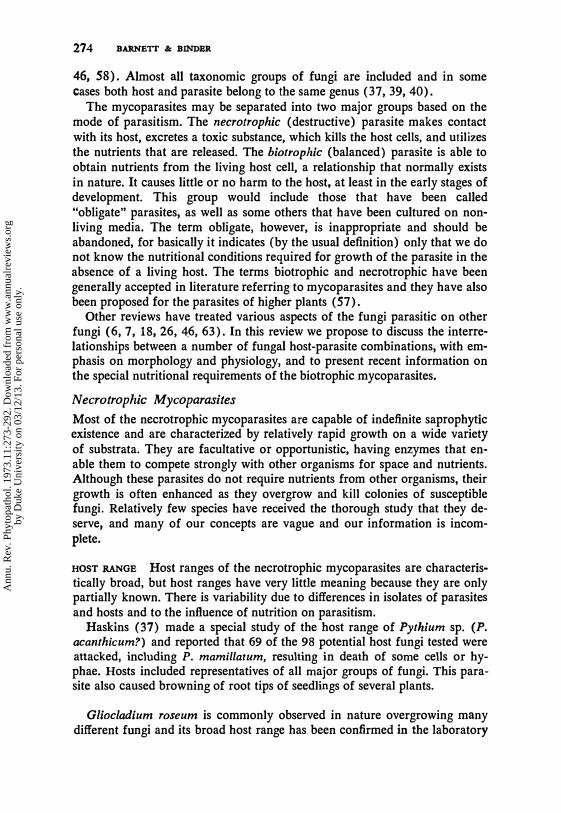
46, 58). Almost all taxonomic groups of fungi are included and in some cases both host and parasite belong to the same genus (37, 39, 40).
The mycoparasites may be separated into two major groups based on the mode of parasitism. The necrotrophic (destructive) parasite makes contact with its host, excretes a toxic substance, which kills the host cells, and utilizes the nutrients that are released. The biotrophic (balanced) parasite is able to obtain nutrients from the living host cell, a relationship that normally exists in nature. It causes little or no harm to the host, at least in the early stages of development. This group would include those that have been called "obligate" parasites, as well as some others that have been cultured on nonliving media. The term obligate, however, is inappropriate and should be abandoned, for basically it indicates (by the usual definition) only that we do not know the nutritional conditions required for growth of the parasite in the absence of a living host. The terms biotrophic and necrotrophic have been generally accepted in literature referring to mycoparasites and they have also been proposed for the parasites of higher plants (57).
Other reviews have treated various aspects of the fungi parasitic on other fungi (6, 7, 18, 26, 4.6, 63). In this review we propose to discuss the interrelationships between a number of fungal host-parasite combinations, with emphasis on morphology and physiology, and to present recent information on the special nutritional requirements of the biotrophic mycoparasites.
Necrotrophic Mycoparasites
Most of the necrotrophic mycoparasites are capable of indefinite saprophytic existence and are characterized by relatively rapid growth on a wide variety of substrata. They are facultative or opportunistic, having enzymes that enable them to compete strongly with other organisms for space and nutrients. Although these parasites do not require nutrients from other organisms, their growth is often enhanced as they overgrow and kill colonies of susceptible fungi. Relatively few species have received the thorough study that they deserve, and many of our concepts are vague and our information is incomplete.
HOST RANGE Host ranges of the necrotrophic mycoparasites are characteristically broad, but host ranges have very little meaning because they are only partially known. There is variability due to differences in isolates of parasites and hosts and to the influence of nutrition on parasitism.
Haskins (37) made a special study of the host range of Pythium sp. (P. acanthicum?) and reported that 69 of the 98 potential host fungi tested were attacked, including P. mamillatum, resulting in death of some cells or hyphae. Hosts included representatives of all major groups of fungi. This parasite also caused browning of root tips of seedlings of several plants.
Gliocladium roseum is commonly observed in nature overgrowing many different fungi and its broad host range has been confirmed in the laboratory
Ann
u. R
ev. P
hyto
path
ol. 1
973.
11:2
73-2
92. D
ownl
oade
d fr
om w
ww
.ann
ualr
evie
ws.
org
by D
uke
Uni
vers
ity o
n 03
/12/
13. F
or p
erso
nal u
se o
nly.
FUNGAL HOST-PARASITE RELATIONSIDP 275
(11). Severely attacked were species of Ceratocystis, Trichothecium roseum, Thamnidium elegans and others, and no species has been found to be completely immune in all stages of development.
Three unidentified species of Cephalosporium were similar in virulence on three species of Helminthosporium (41). Conidia, conidiophores, and hyphae of H. teres and H. vagans were killed by all three species but only a few cells of the mycelium of H. sativum were killed. One isolate of C. acremonium, causal agent of black bundle disease of corn, was similar in virulence.
We have observed a basidiomycete, identified by cultural characteristics as Polyporus adustus, attacking and destroying mycelium of many other fungi, including both Mucorales and the higher fungi. Isolates varied in virulence but both monokaryotic and dikaryotic isolates obtained as contaminants and those obtained from known fruit bodies of P. adustus were similar in behavior. A special study comparing several wood rotting basidiomycetes revealed that several additional species were similar to P. adustus in their ability to parasitize and destroy other fungi in culture (34). The more virulent species included Polyporous versicolor and Pleurotus ostreatus.
Rhizoctonia solani, the well-known pathogen of economic crops, is highly successful in competition with other soil fungi. An extensive study showed that a number of isolates of R. solani could destroy hyphae of several other fungi under favorable conditions in the laboratory (21). The most susceptible hosts were Rhizopus nigricans, Mucor spp., Helicostylum sp., Pythium spp., and Amblyosporium botrytis, the only host outside the PhYl<omycetes.
Ampelomyces quisqualis (Cicinnobolus cesati) is known in nature only as a parasite of powdery mildew fungi, although it is easily cultured on laboratory media. Recently it has been described as an internal parasite of mycelium, sporangia, and sporangiophores of several Mucorales, causing severe damage (45). Some species of Mortierella were resistant.
A number of other fungi are frequently found on aged mushrooms and other fleshy fungi. These include species of Verticillium, Cephalosporium, Mycogone, Dactylium, Didymocladium, and Sepedonium, some of which may cause serious losses in commercial mushroom beds. Calcarisporium arbuscula has been isolated from apparently healthy wild mushrooms (61). Little is known about the host-parasite relationship of these fungi.
MODE OF PARASITISM The mode of parasitism among necrotrophic myco-'
parasites is similar in many species but is known to vary with the different host-parasite combinations. The concept of parasitism implies prolonged contact with or without penetration of the host, but experimentally it is often difficult to determine what part diffusible antibiotics might play in the relationship.
Species of Trichoderma, principally T. viride, have long been known to be antagonistic to other soil and wood-inhabiting fungi. Weindling (62) was one of the first to point out the parasitic nature of this fungus but some of its
Ann
u. R
ev. P
hyto
path
ol. 1
973.
11:2
73-2
92. D
ownl
oade
d fr
om w
ww
.ann
ualr
evie
ws.
org
by D
uke
Uni
vers
ity o
n 03
/12/
13. F
or p
erso
nal u
se o
nly.
276 BARNETT & B�ER
antagonistic activity has since been attributed to antibiotics (42, 63). We may conclude that the degree of destruction by this parasite is dependent upon the isolate of the species and of the host, and upon nutrition and environment.
Gliocladium roseum and the wood-rotting basidiomycetes usually make contact by means of short branches, which touch or curl around the host hyphae or spores, the contents of which soon begin to disintegrate (11, 34). There is no evidence of any diffusible toxic substance in advance of the growing hyphae of these or other necrotrophic mycoparasites. Some other parasites of this group, Rhizoctonia solani and Papulospora stoveri, may form extensive loose coils of hyphae with few or no branches (21,60).
Penetration of host cells may folIow their death, as in Gliocladium roseum (11) and Pleurotus ostreatus, or may occur while the host is alive, as by Apelomyces quisqualis in nature (29) and Rhizoctonia solani on Mucor sp. and Rhizopus nigricans (21). The latter host sometimes shows a biological resistance by early digestion of parasite hyphae, or mechanical resistance by deposition of wall-like material surrounding the invading hypha (21), the latter being relatively common among the filamentous fungi.
A cytological study by Emmons (29) revealed that for a short time following the invasion of Erysiphe cichoracearum by a hypha of A. quisqualis, the appearance of the host cell remained unchanged. Apparently the parasite was obtaining nutrients from the living cell. After a short time the nuclear membrane of the host cell disappeared and the contents showed signs of death and disintegration as the parasite continued its growth and development. This appears to be a parasite that combines early biotrophic existence with later necrotrophic parasitism.
Although the details of the relationship between Darluca filum and its rust hosts are not well understood, there is some evidence that the parasite is destructive (1). Aecia may be infected at any stage of development and hyphae of the parasite ramify among but do. not penetrate aecial hyphae or spores, some of which show disintegration. The net result is to check the development of the rust pustule without complete destruction. D. filum, like the typical necrotrophic parasite, grows on common laboratory media and utilizes a variety of carbon and nitrogen sources (24, 49, 51). Isolates are very variable but there seems to be no nutritional deficiency that would account for the constant association of this parasite with rust fungi only.
Only a few studies of the fine structure of necrotrophic mycoparasites have been made. Although coiling of hyphae of Trichoderma longibrachiatum around hyphae of Pellicularia sasakii was common, a series of electron micrographs showed that the mycoparasite sometimes penetrated and invaded living cells of the host but caused little disorganization of the contents during early stages of infection (36). The first sign of infection was the formation of an internal infection papilla composed of wall material surrounding the invading hypha. The sheath-like papilla was pierced by the parasite hypha, which then continued its growth inside the host ceU.
Ann
u. R
ev. P
hyto
path
ol. 1
973.
11:2
73-2
92. D
ownl
oade
d fr
om w
ww
.ann
ualr
evie
ws.
org
by D
uke
Uni
vers
ity o
n 03
/12/
13. F
or p
erso
nal u
se o
nly.
FUNGAL HOST-PARASITB RELATIONSHIP 277
EFFECT OF NUTRITION AND ENVIRONMENT Isolates of Rhizoctonia solani differed widely in virulence under the same cultural conditions (21). Susceptibility of four fungi to R. solani was decidedly greater in darkness than in white light. Susceptibility of Mucor recurvus to R. solani was high at 25°C and decreased to zero at 15°C. In general, good mycelial growth of both the parasite (R. solani) and the host (M. recurvus or R. nigricans) was required for heavy parasitism, with certain sugars in a complete medium being more favorable than others (21).
On the other hand, a medium giving an imbalance of carbon to nitrogen, favored parasitism by Polyporus adustus and other wood-rotting basidiomycetes (34). This emphasizes the fact that the substrate that furnishes the same nutrients for both host and necrotrophic parasite may favor one fungus at the expense of the other.
It is interesting to speculate about the possibility of using the necrotrophic mycoparasites or their metabolic products as an aid in reducing growth or sporulation of plant pathogens. Although the results of most studies have been disappointing, it would seem desirable that some of these parasites should receive continued investigation. Yarwood (67) demonstrated that growth and conidium formation by colonies of clover powdery mildew may be checked following artificial inoculation with Ampe/omyces quisqualis under controlled conditions.
Biotrophic Mycoparasites
The biotrophic mycoparasites differ basically from the necrotrophic group in their ability to obtain nutrients from the living host cell. Throughout the many years of close association with the host, some of these parasites apparently have lost the ability to synthesize one or more required nutrients, and must depend on a host to furnish a continuous supply. The loss in synthetic ability has not always involved the same nutrient, so that the deficiencies of the parasites may differ. Furthermore, the susceptible host must not only synthesize and contain the required nutrient within its cells, but the parasite must have the ability to obtain the nutrient from the host. Thus, permeability of the host cell membrane may play an important role in determining the susceptibility of the host fungi and the success of the parasite.
Until recent years few of these mycoparasites that we now call biotrophic had been cultured successfully on nonliving media in the laboratory, and there was some justification for believing them to be obligate parasites, similar to the rusts, powdery mildews, and downy mildews on higher plants. However, our present knowledge based on research of the last two decades has indicated that most, and perhaps all, of these mycoparasites have the ability to make continuous near-normal growth on artificial laboratory media, provided that nutrients for which the parasite is deficient are added. Specific nutritional requirements for axenic growth of several biotrophic mycoparasites are discussed in a separate section of this review.
Three distinct types or modes of biotrophic mycoparasitism are known:
Ann
u. R
ev. P
hyto
path
ol. 1
973.
11:2
73-2
92. D
ownl
oade
d fr
om w
ww
.ann
ualr
evie
ws.
org
by D
uke
Uni
vers
ity o
n 03
/12/
13. F
or p
erso
nal u
se o
nly.
278 BARNBTr & BINDBR
(a) the internal parasites represented by the Chytrids develop within cells of other fungi; (b) the contact parasites do not produce any haustoria or other internal hyphae; (c) the haustorial parasites produce distinct haustoria within the host hyphae. The three types are so distinct in morphology and physiology that they must be discussed separately.
Internal Mycoparasites
The internal mycoparasites are tentatively placed in the biotrophic group because they appear to cause little or no harm to the host during early development, although they may destroy the host protoplasm prior to sporulation. Little is known of their nutritional requirements.
Karling (39, 40) has discussed taxonomy and morphology of a number of parasitic chytrids and their hosts, but very little is known about the nutritional relationship of parasite and host. Included here is a brief discussion of two parasitic chytrids which penetrate and grow within the host, completely surrounded by host cytoplasm. Rozella cladochytrii has a known host range of three species of Nowakowskiella and three of Cladochytrium (39). The parasite enters the rhizomycelium of N. profusum and develops as a naked protoplast surrounded by and scarcely distinguishable from the host protoplasm. In the early stages there is little change except a local swelling of the host, but later the host protoplasm is destroyed as the parasite forms a surrounding waIl and produces zoospores.
Olpidiopsis incrassata is a unicellular internal parasite of species of Saprolegnia and lsoachlya. These hosts �ere susceptible only before the mycelium produced sporangia or oogonia, and the period of susceptibility could be prolonged by delaying initiation of reproduction (56). It was proposed that a chemical precursor for reproduction is also essential for infection by the parasite. Such a mechanism for controlling susceptibility would be unique and this host-parasite relationship deserves more study.
Biotrophic Contact Mycoparasites
Although it seems probable that this mode of parasitism may be relatively common in nature, it was not described in detail until 1958 (10). Even now only five mycoparasites of this group are known and have been studied in the laboratory, although some of them have been recognized as being common associates of other fungi in nature. All are imperfect fungi and their hosts are ascomycetes or imperfects.
NATURAL OCCURRENCE AND HOST RANGE Calcarisporium parasiticum is known only from laboratory cultures in West Virginia. It has appeared five times (1954-1956) in isolations of Dothiorella quercinfl from red oak twigs and was described as a new species (5). The known host range is limited to several species of Physalospora and closely related fungi.
Ann
u. R
ev. P
hyto
path
ol. 1
973.
11:2
73-2
92. D
ownl
oade
d fr
om w
ww
.ann
ualr
evie
ws.
org
by D
uke
Uni
vers
ity o
n 03
/12/
13. F
or p
erso
nal u
se o
nly.
FUNGAL HOST-PARASITE RELATIONSHIP 279
Gonatobotrys simplex is widely distributed but is not commonly isolated. It is associated with Alternaria spp. in nature, and in culture parasitizes species of Alternaria and Cladosporium (64).
Gonatobotryum fuscum has been reported as a parasite on Ceratocystis in England (59) and from West Virginia growing on old mats of Ceratocystis jagacearum (53). In the laboratory the host range has been extended to additional species of Ceratocystis, Graphium, and Leptographium (53).
Gonatorrhodiella highlei is a common parasite on Nectria coccinea var. jaginata, causal agent of the beech bark disease in the New England states, and is not known to be associated with any other fungus in nature. In culture Tritirachium sp. and Cladosporium sp. are also parasitized (31).
Stephanoma phaeospora is known only from a culture with Fusarium sp. isolated from an orchid (22). The host range in culture has recently been extended to nine species of ascomycetes and imperfects and two species of Ustilago (50).
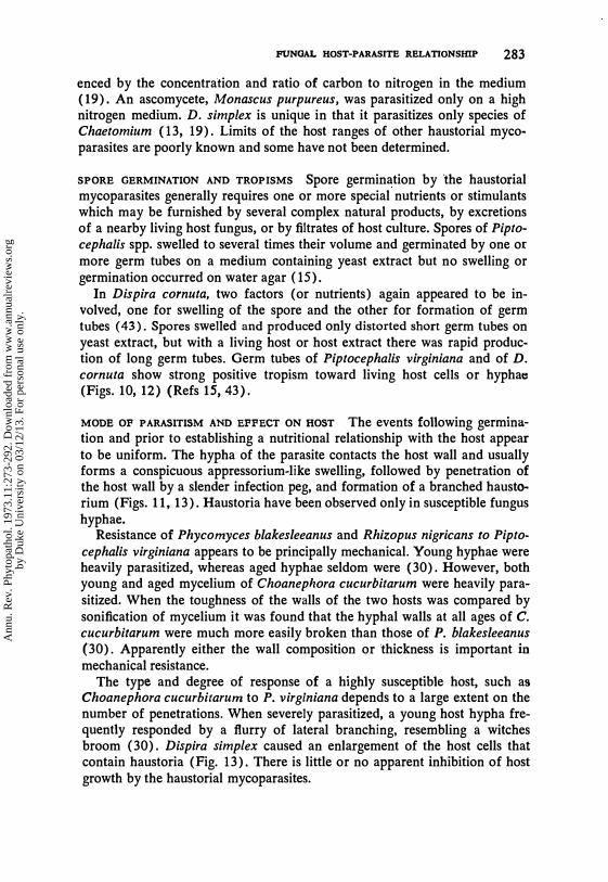
SPORE GERMINATION AND TROPISMS The conidia of Calcarisporium parasiticum and Gonatobotrys simplex do not germinate in distilled water or on a synthetic medium, but require a substance secreted by the host hyphae or furnished by some natural products such as yeast extract, malt extract, mycelium extract, or orange juice (10, 64). Germination on a yeast extract medium is typically by secondary spores or buds (Figs. I, 3). At this stage an attractant is apparently secreted by the germinating spores, for there is a positive tropism of the nearby host hyphae directly toward the parasite (Figs. 2, 4). The unusual ability to cause a tropism of the host is evidently a survival mechanism because the parasite at this stage does not produce long term tubes. In contrast, conidia of G. fuscum, G. highlei and S. phaeospora produce long germ tubes on germination and the host hyphae do not show a positive tropism.
MODE OF CONTACT Parasitism is by means of specialized branches, often no more than a few microns long, which contact the host hypha, may partially or completely surround it, or touch end-to-end a short branch of the host (Figs. 5-8). The small "buffer cell" at the point of contact is characteristic of Calcarisporium parasiticum (Fig. 5). The presence of these special absorptive branches or cells is considered as evidence of parasitism.
There is evidence that a required nutrient is normally held within the cells of most hosts and very little escapes into the substrate before autolysis (10, 64). The contact cells must, therefore, function in some way to increase the permeability of the host cell membrane to this nutrient. However, recently it was shown that several fungi excreted a growth factor into the medium that stimulated growth of Stephanoma phaeospora (50).
Ann
u. R
ev. P
hyto
path
ol. 1
973.
11:2
73-2
92. D
ownl
oade
d fr
om w
ww
.ann
ualr
evie
ws.
org
by D
uke
Uni
vers
ity o
n 03
/12/
13. F
or p
erso
nal u
se o
nly.
280 BARNETI & BINDER
� �
1
�5 : :-':� 8 p
(;? 0
rt 3
4 H
6
Figures 1-13 Spore germination, tropisms and morphology of host-biotrophic mycoparasite relationships. 1. Spore germination of Calcarisparium parasiticum by secondary spores (10). 2. Tropism of Sphaeropsis ma/arum hyphae toward C. parasilicum (10).3. Spore germination of Gonatobotrys simplex by budding (64). 4. Tropism of Alternaria hyphae toward G. simplex (64). 5. Mode of contact between C. parasiticum and S. malarum (10). 6. Mode of contact between G. simplex and Alternaria sp. (64). 7. Mode of contact between Gonatobotryum fuscum
Ann
u. R
ev. P
hyto
path
ol. 1
973.
11:2
73-2
92. D
ownl
oade
d fr
om w
ww
.ann
ualr
evie
ws.
org
by D
uke
Uni
vers
ity o
n 03
/12/
13. F
or p
erso
nal u
se o
nly.
FUNGAL HOST-PARASITE RELATIONSHIP 281
EFFECTS OF PARASITE ON GROWTH OF HOST The contact biotrophic parasite generally causes no apparent harm to its hosts except a reduction in rate of growth. Since there is no evidence of toxin production, the slower growth may be due to competition for nutrients. A typical example is that of Calcarisporium parasiticllm growing in liquid media on Physalospora obtusa. Less dry mycelium was produced by the combined host and parasite than by the host alone (10). This phase of the relationship between host and contact biotrophic parasite needs much more investigation.
EFFECTS OF HOST NUTRITION ON DEVELOPMENT OF THE PAR A SITE The composition of the host medium, i.e., the nutrients and their concentrations, is known to affect host metabolism which in turn may determine the relative success or failure of the parasite. Physalospora obtllsa, a highly susceptible species to Calearisporillm parasitieum, supported about equal growth of the parasite regardless of the carbon-nitrogen ratio, whereas highly resistant P. ilicis was more heavily parasitized on a medium high in available nitrogen (10).
Parasitic growth of GonatobotrYllm fuscum was greater on a medium with relatively high carbon-nitrogen ratio (17, 53). A complex nitrogen source favored the parasite , as did an excess of microelements , particularly manganese.
Gonatorrhodiella highlei showed distinct inhibition by increased concentration of asparagine before the host, Neetria eoeeinea var. faginata, was affected (31). This inhibition was attributed to accumulation of ammonia in the cultures containing high asparagine. Increased KN03 in the medium was not inhibitory and did not result in accumulation of ammonia. An excess supply of thiamine and biotin in the medium may be essential to good parasitism by G. highlei.
An unusual relationship involving biotin and pyridoxine occurs when G. fuscum is cultured with the host Graphium sp., which is deficient for pyridoxine (8). Parasitism is heavy in darkness on media containing adequate pyridoxine and thiamine, but in light the pyridoxine is destroyed so rapidly that the host growth is limited and the parasite is strongly inhibited. Biotin must be added when the concentration of pyridoxine is very low (ca 12 p.g/
and Graphium sp. (53). 8. Mode of contact between Stephanoma phaeospora and Tritirachium sp. 9. Resistance sheaths of G. fuscum preventing penetration by Graphium sp. 10. Spore germination and tropism of Piptocephalis virginiana toward hypha of Choanephora cucurbitarum (15). 1 1 . Haustorium of P. virginiana in hypha of C. cucurbitarum (15). 12. Spore germination and tropism of Dispira cornuta toward swollen spore of Cokeromyces recurvatus (43). 13. Contact branch and haustorium of Dispira simplex in hypha of Chaetomium sp. (19). H = host,
P == parasite.
Ann
u. R
ev. P
hyto
path
ol. 1
973.
11:2
73-2
92. D
ownl
oade
d fr
om w
ww
.ann
ualr
evie
ws.
org
by D
uke
Uni
vers
ity o
n 03
/12/
13. F
or p
erso
nal u
se o
nly.
282 BARNETT &: BINDER
liter or less). It is now known that G. fuscum is deficient for both thiamine and biotin (23).
A mutualistic symbiotic relationship can be demonstrated between G. luscum and Graphium sp. on a medium containing thiamine and only a trace of biotin and pyridoxine. After a very slow start G. luscum synthesizes pyridoxine needed by Graphium sp., which in turn synthesizes enough biotin for growth of the parasite (8). The rate of growth increases as the mass of mycelium increases.
Another unusual complex relationship between G. fuscum and Graphium sp., which may be related to nutrition, has been observed frequently in aging cultures. There is attempted penetration of hyphae and conidia of G. fuscum by hyphae of Graphium sp. The Graphium hypha forms a slight appressorium-like swelling and there is a deposition of dark wall material around the penetrating hypha. These resistance sheaths may extend partially or completely across the hyphal cavity (Fig. 9). Only occasionally is the sheath penetrated and internal hyphae of Graphium sp. become evident. Is this a reversal of parasitism in which the parasite becomes the reluctant host? The existence of resistance sheaths surrounding penetrating hyphae of mycoparasites has been reported for Rhizoctonia solani (21) and for Trichoderma longibrachiatum (36) and one may assume that they are common.
TEMPERATURE AND LIGHT In general, the favorable temperatures for both host and mycoparasite fall within the same range, and no striking effects of temperature on parasitism have been reported. The destruction of pyridoxine by light and the effects on the deficient host, Graphium sp., are reported above. It has also been reported that Gonatorrhodiella highlei on Nectria coccinea var. laginata and on Tritirachium sp. is completely inhibited by light (200fc) (31).
Biotrophic Haustorial Mycoparasites
The filamentous haustorial mycoparasites belong to the morphological group, merosporangiferous Mucorales, i.e., those that produce spores in rod-like sporangia (12, 13). The principal genera are Syncephalis and Piptocephalis, in the Piptocephalidaceae, and Dispira, Dimargaris and Tieghemiomyces, in the Dimargaritaceae. The species have received intensive taxonomic and morphological study by Benjamin (12,13).
HOST RANGES For the most part the hosts of these parasites belong to other families of the Mucorales. Piptocephalis virginiana parasitized 22 species of Mucorales (15). Most other species of Piptocepha!is have similar host ranges, except P. xenophila, which can parasitize several species of ascomycetes and imperfects (27).
Three species of Dispira vary in their host ranges. D. cornuta parasitizes only species of Mucorales (3, 14). Susceptibility to D. parvispora was influ-
Ann
u. R
ev. P
hyto
path
ol. 1
973.
11:2
73-2
92. D
ownl
oade
d fr
om w
ww
.ann
ualr
evie
ws.
org
by D
uke
Uni
vers
ity o
n 03
/12/
13. F
or p
erso
nal u
se o
nly.
FUNGAL HOST-PARASITE RELATIONSHIP 283
enced by the concentration and ratio of carbon to nitrogen in the medium (19). An ascomycete, Monascus purpureus, was parasitized only on a high nitrogen medium. D. simplex is unique in that it parasitizes only species of Chaetomium (13, 19). Limits of the host ranges of other haustorial mycoparasites are poorly known and some have not been determined.
SPORE GERMINATION AND TROPISMS Spore germination by 'the haustorial mycoparasites generally requires one or more special nutrients or stimulants which may be furnished by several complex natural products, by excretions of a nearby living host fungus, or by filtrates of host culture. Spores of Pip tocephalis spp. swelled to several times their volume and germinated by one or more germ tubes on a medium containing yeast extract but no swelling or germination occurred on water agar (15).
In Dispira cornuta, two factors (or nutrients) again appeared to be involved, one for swelling of the spore and the other for formation of germ tubes (43). Spores swelled and produced only distorted short germ tubes on yeast extract, but with a living host or host extract there was rapid production of long germ tubes. Germ tubes of Piptocephalis virginiana and of D. cornuta show strong positive tropism toward living host cells or hyphae (Figs. 10, 12) (Refs 15, 43).
MODE OF PARASITISM AND EFFECT ON HOST The events following germination and prior to establishing a nutritional relationship with the host appear to be uniform. The hypha of the parasite contacts the host wall and usually forms a conspicuous appressorium-like swelling, followed by penetration of the host wall by a slender infection peg, and formation of a branched haustorium (Figs. 11, 13). Haustoria have been observed only in susceptible fungus hyphae.
Resistance of Phycomyces blakesleeanus and Rhizopus nigricans to Piptocephalis virginiana appears to be principally mechanical. Young hyphae were heavily parasitized, whereas aged hyphae seldom were (30). However, both young and aged mycelium of Choanephora cucurbitarum were heavily parasitized. When the toughness of the walls of the two hosts was compared by sonification of mycelium it was found that the hyphal walls at all ages of C. cucurbitarum were much more easily broken than those of P. blakesleeanus (30). Apparently either the wall composition or thickness is important in mechanical resistance.
The type and degree of response of a highly susceptible host, such as Choanephora cucurbitarum to P. virginiana depends to a large extent on the number of penetrations. When severely parasitized, a young host hypha frequently responded by a flurry of lateral branching, resembling a witches broom (30). Dispira simplex caused an enlargement of the host cells that contain haustoria (Fig. 13). There is little or no apparent inhibition of host growth by the haustorial mycoparasites.
Ann
u. R
ev. P
hyto
path
ol. 1
973.
11:2
73-2
92. D
ownl
oade
d fr
om w
ww
.ann
ualr
evie
ws.
org
by D
uke
Uni
vers
ity o
n 03
/12/
13. F
or p
erso
nal u
se o
nly.
284 BARNETT & B�ER
FINE STRUCTURE OF THE HAUSTORIUM In general the fine structure of the haustorium of Piptocephalis virginiana, the only mycoparasite studied with the electron microscope, is similar to that of haustorial parasites of higher plants (2, 47). The haustorium formed within hyphae of Mycotypha microspora is bounded by an electron dense sheath with a convoluted surrounding membrane. The haustorial wall is continuous with the wall of the parasite hypha and there is evidence of a collar of host wall material at its base. The infection apparatus formed by P. virginiana in hyphae of Choanephora cucurbitarum, also a highly susceptible host, varied only in minor points from that described above (47).
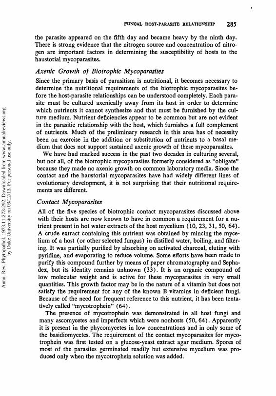
Table 1 The relation of amounts of glucose and yeast extract to percentage dry weight of TCA-soluble nitrogen in the mycelium of Morfierella pusil/a and degree of parasitism by Piptocephalis virginiana at the end of 7 days (54).
g Glucose-g Yeast Extract
5-1 25-1 5-5 25-5 Mg dry mycelium 82 185 138 256 % TCA-nitrogen 0.61 0.34 1.46 0.40 Parasite rating Poor None Excellent Trace
HOST NUTRITION AND GROWTH OF PARASITE Ayers (3) was a pioneer in investigations of effects of host nutrition on degree of parasitism by the haustorial mycoparasites. He demonstrated that media high in carbon were unfavorable to growth of Dispira cornuta on its host, whereas media rich in a usable nitrogen source favored parasitism. These results have been generally confirmed and the experiments extended (17, 43, 44). Cokeromyces recurvatus was heavily parasitized by D. cornuta on a medium with a low carbonnitrogen ratio but not at all on a medium high in carbon and low in nitrogen (17) .
A more extensive study was made on the effects of nitrogen sources and concentrations on parasitism by Piptocephalis virginiana (14). On most hosts alanine, asparagine, glutamic acid, and casein hydrolysate favored the development of the parasite, whereas glycine, urea, ammonium sulfate, and ammonium tartrate were poor nitrogen sources. The host, Mortierella pusilla, made about equal growth on media with glutamic acid and ammonium sulfate but there was no growth of the parasite on the host on the latter medium. M. pusilla was parasitized on a medium with high concentration of glucose and glutamic acid, whereas M. ramannianus was not (14).
The degree of parasitism of M. pusilla by P. virginiana was directly correlated with the amount of soluble nitrogen in the host mycelium (Table 1). Furthermore, it was possible to prevent the growth of this parasite in liquid media for 15 days or longer by maintaining a high concentration of glucose in the culture medium (54). In the same medium with no additional glucose
Ann
u. R
ev. P
hyto
path
ol. 1
973.
11:2
73-2
92. D
ownl
oade
d fr
om w
ww
.ann
ualr
evie
ws.
org
by D
uke
Uni
vers
ity o
n 03
/12/
13. F
or p
erso
nal u
se o
nly.
FUNGAL HOST-PARASITE RELATIONSHIP 285
the parasite appeared on the fifth day and became heavy by the ninth day. There is strong evidence that the nitrogen source and concentration of nitrogen are important factors in determining the susceptibility of hosts to the haustorial mycoparasites.
Axenic Growth of Biotrophic Mycoparasites
Since the primary basis of parasitism is nutritional, it becomes necessary to determine the nutritional requirements of the biotrophic mycoparasites before the host-parasite relationships can be understood completely. Each parasite must be cultured axenically away from its host in order to determine which nutrients it cannot synthesize and that must be furnished by the culture medium. Nutrient deficiencies appear to be common but are not evident in the parasitic relationship with the host, which furnishes a full complement of nutrients. Much of the preliminary research in this area has of necessity been an exercise in the addition or substitution of nutrients to a basal medium that does not support sustained axenic growth of these mycoparasites.
We have had marked success in the past two decades in culturing several, but not all, of the biotrophic mycoparasites formerly considered as "obligate" because they made no axenic growth on common laboratory media. Since the contact and the haustorial mycoparasites have had widely different lines of evolutionary development, it is not surprising that their nutritional requirements are different.
Contact Mycoparasites
All of the five species of biotrophic contact mycoparasites discussed above with their hosts are now known to have in common a requirement for a nutrient present in hot water extracts of the host mycelium (10,23,31,50,64). A crude extract containing this nutrient was obtained by mincing the mycelium of a host (or other selected fungus) in distilled water, boiling, and filtering. It was partially purified by absorbing on activated charcoal, eluting with pyridine, and evaporating to reduce volume. Some efforts have been made to purify this compound further by means of paper chromatography and Sephadex, but its identity remains unknown (33). It is an organic compound of low molecular weight and is active for these mycoparasites in very small quantities. This growth factor may be in the nature of a vitamin but does not satisfy the requirement for any of the known B vitamins in deficient fungi. Because of the need for frequent reference to this nutrient, it has been tentatively called "mycotrophein" (64).
The presence of mycotrophein was demonstrated in all host fungi and many ascomycetes and imperfects which were nonhosts (50,64). Apparently it is present in the phycomycetes in low concentrations and in only some of the basidiomycetes. The requirement of the contact mycoparasites for mycotrophein was first tested on a glucose-yeast extract agar medium. Spores of most of the parasites germinated readily but extensive mycelium was produced only when the mycotrophein solution was added.
Ann
u. R
ev. P
hyto
path
ol. 1
973.
11:2
73-2
92. D
ownl
oade
d fr
om w
ww
.ann
ualr
evie
ws.
org
by D
uke
Uni
vers
ity o
n 03
/12/
13. F
or p
erso
nal u
se o
nly.
28·6 BARNETI '" BINDER
Calcarisporium parasiticum, in liquid synthetic media containing mycotrophein, utilized common hexose sugars but little or no growth occurred on sorbose, xylose, or disaccharides. Several single amino acids were utilized as nitrogen sources, with glutamic acid being the best (33).
Gonatobotrys simplex utilized only the common hexose sugars, but failed to grow when single amino acids were the nitrogen source. Only yeast extract provided a good nitrogen source for growth and sporulation (64).
Gonatorrhodiella highlei differed from the two previous contact parasites in making no growth on a yeast extract medium, even with added mycotrophein (31). The requirement for a second nutrient was demonstrated when a water extract of beech bark was added, resulting in good axenic growth.
Gonatobotryum /uscum did not grow on any of the media that supported growth of other contact mycoparasites. It made substantial axenic growth only when the medium contained unusually high concentrations of thiamine (optimum 4-8 mgll) and biotin (optimum 0.2-0.4 mgll) in addition to high mycotrophein (23). Only the common hexose sugars were utilized, but a number of single amino acids were utilized as nitrogen sources. The rate of growth of G. /uscum on a favorable medium was greatly increased when 1.2 % dimethyl sulfoxide was added, suggesting that a problem of permeabil. ity may be involved.
Stephanoma phaeospora failed to grow on a malt extract medium unless a water extract of certain fungi was added (50). It seems likely that the fungus extract contains the same nutrient (mycotrophein) that is required by other biotrophic contact mycoparasites, but this cannot be determined with certainty until the nutrient is identified and tested further.
Haustorial Mycoparasites
Nutritional requirements for axenic growth of the haustorial mycoparasites are more complex than for the contact mycoparasites. None of the species studied were affected by the addition of the mycotrophein solution to the me· dium. Of the several species successfully cultivated axenically, only Dispira parvispora grew well and sporulated on a glucose-yeast extract medium, but no detailed study of this species has been made. Some species have an apparent inability to utilize glucose, or other common sugars as a carbon source.
Syncephalis spp. grew axenically on a complex medium containing beef liver (28). Dispira corn uta, Dispz'ra simplex, Dimargaris verticillata, and Tieghemiomyces parasiticus made only a trace of growth on a yeast extract or casein hydrolysate medium with glucose, but all grew well when glycerol replaced the glucose as the carbon source (9). Detailed studies in liquid culture have been made only with D. cornuta and T. parasiticus. Failure of
Ann
u. R
ev. P
hyto
path
ol. 1
973.
11:2
73-2
92. D
ownl
oade
d fr
om w
ww
.ann
ualr
evie
ws.
org
by D
uke
Uni
vers
ity o
n 03
/12/
13. F
or p
erso
nal u
se o
nly.
FUNGAL HOST-PARASITE RELATIONSHIP 287
these parasites to make substantial growth on a glucose medium could mean either that the glucose did not enter the mycelium or that the fungus lacked the necessary enzymes to catabolize glucose. One is tempted to use the latter explanation, except for certain experiments performed with T. parasitic us (16). Labeled 14C02 was produced from media containing HC-glucose or HC-glycerol as single carbon sources, but at a much higher rate from the latter source (16). This indicates a slow or limited entrance of glucose into the mycelium under these conditions. On the other hand, an increase in glucose concentration did not increase the rate of growth of the parasite. The addition of the surfactant Tween 80 to a glucose-casein hydrolysate medium resulted in much greater growth than did Tween 80 without glucose (180 mg vs. 35 mg dry wt per culture in 6 wks) (16). While some of the Tween 80 may have been used as a carbon source, the greater effect was on the rate of entrance of glucose. This action may be similar to that of glycols and Tweens reported as facilitating the entry of nutrients into mycelium of Claviceps paspali (48).
In further studies, all of the enzymes for the catabolism of glucose via the Embden-Meyerhof-Parnas and the hexose-monophosphate pathways were demonstrated in cell-free extracts of mycelium of Tieghemiomyces parasiticus grown on a glycerol medium (16). Glycerol may play a dual role of serving as an excellent carbon source and of lowering the surface tension of the medium.
The carbon metabolism of Dispira corn uta appears to be similar to that of T. parasitic us but no study of its enzymes has been made (4).
There seems little doubt that glucose can enter the mycelium of the haustorial parasites under some conditions, but it also appears that the vegetative hyphae serve as a barrier to certain nutrients in media that are readily absorbed by haustoria within the host cells. It is also possible that the presence of the host may influence the permeability of the parasite membranes to certain nutrients.
Growth of several of the haustorial mycoparasites was favored by an unusually high concentration of casein hydrolysate as the nitrogen source (20-40 glliter) (4,9, 16). This may be due to a high nitrogen requirement or to the increased concentration of a required amino acid present in low concentrations in casein hydrolysate. Utilization of nitrogen sources by Dispira cornuta and by Tieghemiomyces parasiticus has been studied and was found to be markedly different. T. parasiticus made good axenic growth on a glycerol medium with casein hydrolysate or a mixture of 18 amino acids, but no growth on any single amino acid as the nitrogen source. The conclusion reached from a special study of amino acid utilization was that L-cysteine, Lvaline, and L-Ieucine are extremely important for axenic growth of this fungus (16). The addition of certain other amino acids to these three did not increase the rate of growth greatly. Dispira cornuta utilized L-alanine, L-aspartie, and L-glutamic acid in a glycerol medium (4). Certain other amino acids and ammonium sulfate were utilized less efficiently and nitrate nitrogen
Ann
u. R
ev. P
hyto
path
ol. 1
973.
11:2
73-2
92. D
ownl
oade
d fr
om w
ww
.ann
ualr
evie
ws.
org
by D
uke
Uni
vers
ity o
n 03
/12/
13. F
or p
erso
nal u
se o
nly.
288 BARNETT & B�Ea
was not utilized. In general, increased concentrations of single amino acids or ammonium sulfate, to several times the concentrations used for most saprophytic fungi, resulted in increased growth rate. The reason for this is not at all clear and the problem should receive further careful study.
The haustorial mycoparasites frequently show deficiencies for thiamine and biotin (4,9, 16). High concentrations of thiamine (1 mg or more/liter) have been reported as favorable for Dispira cornuta and for T. parasitic us on agar media, but not for the latter in liquid media.
The success that has been enjoyed in the axenic culture of several of the haustorial mycoparasites has brought with it the promise that other parasites now considered as "obligate" may likewise be cultured axenically, provided that the appropriate nutritional and physical conditions are met by the medium. However, the fact that great differences exist among species or genera has been brought out by research with Piptocephalis spp. After many attempts using a great number of nutritional conditions, we have never succeeded in culturing Piptocephalis. except on a living susceptible host. Spores of Piptocephalis germinate readily on various media in the absence of a host fungus, but only a limited amount of mycelium is formed, which often sporulates before it ceases to grow (14). This problem remains as a real challenge to the interested researcher.
Discussion and Summary The success of the necrotrophic mycoparasites in destroying host cells or reducing populations of competing fungi depends on a number of external factors, of which nutrition is of high importance. Yet these parasites bave no specific nutritional requirement that is satisfied only by the host. We may conclude that this type of mycoparasitism is exceedingly common among filamentous fungi in nature. There is much to be learned from studies of the production of toxins and enzymes by these parasites and the mode of host destruction.
The mycelium of the susceptible host is known to furnish all nutrients required for growth and development of the biotrophic contact mycoparasites, but the mere presence of a required nutrient does not necessarily result in susceptibility. The parasite must be able, by means of special cells or branches, to absorb the essential nutrients from the living host cell, presumably by altering the permeability of the cell membrane. All five known mYCOparasites of this group have one essential nutritional deficiency in common, i.e. a water-soluble, heat-stable, vitamin-like growth factor (mycotrophein) present in aU host and some nonhost fungi. Its purification and identification are among the major unsolved problems related to nutrition of the biotrophic contact mycoparasites.
The rate and degree of development of the biotrophic haustorial mycoparasites on their hosts are greatly favored by a substrate high in nitrogen and relatively low in carbon. There is evidence that a high concentration of solu-
Ann
u. R
ev. P
hyto
path
ol. 1
973.
11:2
73-2
92. D
ownl
oade
d fr
om w
ww
.ann
ualr
evie
ws.
org
by D
uke
Uni
vers
ity o
n 03
/12/
13. F
or p
erso
nal u
se o
nly.
FUNGAL HOST-PARASITE. RELATIONSHIP 289
hIe nitrogen in the host mycelium is directly related to concentration of nitrogen in the medium and to host susceptibility.
It is of interest that the electron microscope has revealed that the haustorium of a mycoparasite (Piptocephalis virginiana) is strikingly similar in structure to that of the haustorial parasites of higher plants. One must conclude that its formation and structure are the response to a nutritionally compatible environment which leads to free passage of nutrients into the parasite.
The failure of Tieghemiomyces parasiticus to make good axenic growth on a glucose medium, even though all enzymes involved in glucose catabolism were shown to be present, suggests strongly that the rate of penetration of nutrients into vegetative mycelium may be a limiting factor in growth of the haustorial mycoparasites.
The presence and concentration of specific amino acids appear to be of prime importance to axenic growth of some of the haustorial mycoparasites. A similarity with the requirements of the rust, Melampsora lini, for a S-containing amino acid was indicated by recent work (25). Other similarities in nutrition of these two groups of parasites may be expected.
Ann
u. R
ev. P
hyto
path
ol. 1
973.
11:2
73-2
92. D
ownl
oade
d fr
om w
ww
.ann
ualr
evie
ws.
org
by D
uke
Uni
vers
ity o
n 03
/12/
13. F
or p
erso
nal u
se o
nly.
290 BARNElT &: BINDER
Literature Cited 1. Adams, J. F. 1920. Darluca on Per
idermium peckii. Mycologia 12: 309-15
2. Armentrout, V. N., Wilson, C. L. 1969. Haustorium-host interaction during mycoparasitism of Mycotypha microspora by Piptocephalis virginiana. Phytopathology 59 : 897-905
3. Ayers, T. T. 1 935. Parasitism of Dispira cornuta. Mycologia 27: 235-61
4. Barker, S. M., Barnett, H. L., 1973. Nitrogen and vitamin requirements for axenic growth of the haustorial mycoparasite, Dispira cornuta. Mycologia. In press
5. Barnett, H. L. 1958. A new Calcarisporium parasitic on other fungi. Mycologia 50:497-500
6. Barnett, H. L. 1963. The nature of mycoparasitism by fungi. Ann. Rev. Microbial. 17: 1-14
7. Barnett, H. L. 1964. Mycoparasitism. Mycologia 56: 1-19
8. Barnett, H. L. 1968. The effects of light, pyridoxine and biotin on the development of the mycoparasite, Gonatobotryum fuscum. Mycologia 60: 244-5 1
9. Barnett, H. L. 1970. Nutritional requirements for axenic growth of some haustorial mycoparasites. Mycologia 62:750-61
10. Barnett, H. L., Lilly, V. O. 1 958. Parasitism of Calcarisporium parasiticum on species of Physalospora and related fungi. West Va. Univ. Agr. Exp. Sta. Bull. 420T. 37 pp.
1 1. Barnett, H. L., Lilly, V. G. 1962. A destructive mycoparasite, Glioe/adium roseum. Mycologia 54 : 72-79
12. Benjamin, R. K. 1959. The merosporangiferous Mucorales. Aliso 4: 32 1-433
13. Benjamin, R. K. 1961. Addenda to "The merosporangiferous Mucorales." A liso 5 : 1 1-19
14. Berry, C. R. 1959. Factors affecting parasitism of Piptocephalis virginiana on other Mucorales. Mycologia 51 : 824-32
1 5. Berry, C. R., Barnett, H. L. 1 957. Mode of parasitism and host range of Piptocephalis virginiana. Mycologia 49 : 374-86
1 6. Binder, F. L. 1971. Carbohydrate metabolism and nitrogen nutrition 0/ Tieghemiomyces parasiticus.
Ph.D. Dissertation, Wesf Virginia University, Morgantown. 89 pp.
17. Bishop, R. H. 1964. Effects of nutrition on the mycoparasite, Gonatabotryum fuscum. Ph.D. Dissertation, West Virginia University, _
Morgantown. 134 pp. 1 8. Boosalis, M. O. 1964. Hyperpara
sitism. Ann. Rev. Phytopathol. 2: 263-76
19. Brunk, M. A., Barnett, H. L. 1 966. Mycoparasitism of Dispira simplex and D. parvispora. Mycologia 58 : 518-23
20. Bushnell, W. R. 1 968. In vitro development of an Australian isolate of Puccinia graminis f. sp. trifici. Phytopathology 58: 526-27
21. Butler, E. E. 1957. Rhizoctonia solani as a parasite of fungi. Mycologia 49 :354-73
22. Butler, E. E., McCain, A. H. 1968. A new species of Stephanoma. Mycologia 60:955-59
23. Calderone, R. A., Barnett, H. L. 1 972. Axenic growth and nutrition of Gonatobotryum fuscum. Mycologia 64: 153-60
24. Calpouzos, L., Theis, T., Batille, C. M. 1 957. Culture of the rust parasite, Darluca filum. Phytopathology 47 : 108-09
25. Coffey, M. D., Shaw, M. 1972. Nutritional studies with axenic cultures of the flax rust. Melampsora lini. Physiol. Plant Pathol. 2:37-46
26. De Yay, J. E. 1956. Mutual relationships in fungi. Ann. Rev. Microbiol. 10 : 1 15-40
27. Dobbs, C. G., English, M. P. 1 954. Piptocephalis xenophila sp. nov. parasitic on non-mucorine hosts. Trans. Brit. Mycol. Soc. 37: 375-89
28. Ellis, J. J. 1966. On growing Syncephalis in pure culture. Mycologia 58 :465-69
29. Emmons, C. W. 1 930. Cicinnobolus cesati, a study in host-parasite relationships. Bull. Torrey Bot. Club 57 :42 1-41
30. England, W. H. 1969. Relation of age of two host fungi to development of the mycoparasite, Piptocephalis virginiana. Mycologia 6 1 : 586-92
31 . Gain, R. E., Barnett, H. L. 1970. Parasitism and axenic growth of Gonatorrhodiella highlei. Mycologia 62: 1 122-29
Ann
u. R
ev. P
hyto
path
ol. 1
973.
11:2
73-2
92. D
ownl
oade
d fr
om w
ww
.ann
ualr
evie
ws.
org
by D
uke
Uni
vers
ity o
n 03
/12/
13. F
or p
erso
nal u
se o
nly.
FUNGAL HOST-PARASITE RELATIONSIDP 291
32. Gilman, J. C., Tiffany, L. H. 1952. Fungicolous fungi of Iowa. Proc. Iowa Acad. Sci. 59: 99-110
33. Goldstrohm, D. D. 1966. An investigation of the growth factor and other nutritional requirements of Calcarisporium parasiticum. Ph.D. Dissertation, West Virginia Univ., Morgantown. 132 pp.
34. Griffith, N. T., Barnett, H. L. 1 967. Mycoparasitism by basidiomycetes in culture. Mycologia 59: 149-54
35. Hansford, C. G. 1946. The foliicolous ascomycetes, their parasites and associated fungi. Commonwealth Mycol. Inst., Kew, England, Paper No. 15, 240 pp.
36. Hashioka, Y., Pukita, T. 1969. Ultrastructural observations on mycoparasitism of Trichoderma, Gliocladium and Acremonium to phytopathogenic fungi. Rept. Tottori Mycol. Inst. (Japan) 7 : 8-18
37. Haskins, R. H. 1963. Morphology, nutrition and host range of a species of Pythium. Can. J. Microbiol. 9 :45 1-57
38. Heim, R. 1967. Cle pour la determination des espices ban ales de champignons fongicoles. Rev. de Mycol. 3 1 : 393-99
39. Kariing, J. S. 1942. Parasitism among the chytrids. Am. J. Bot. 29 :24-35
40. Kariing, J. S. 1960. Parasitism among chytrids. II. Chytridiomyces verrucosus sp. nov. and Phlyetochytrium synchytrii. Bull. Torrey Bot. Club 87: 326-36
41 . Kenneth, R., Isaac, P. K. 1 963. Cephalosporium species parasitic On Helminthosporium. Can. J. Plant Sci. 44: 182-87
42. Komatsu, M., Hashioka, Y. 1964. Trichoderma viride, as an antagonist of wood-inhabiting Hymenomycetes. V. Lethal effects of the different Trichoderma forms on Lentinus edodes inside log-woods. Rept. Tottori Mycol. Inst. (Japan ) 4 : 1 1 .... 1 8
4 3 . Kurtjmlan, C . P. 1967. Parasitism, spore germination, and axenic growth of Dispira cornuta. Ph.D. Dissertation, West Virginia Univ., Morgantown. 127 pp.
44. Kurtzman, C. P. 1968. Parasitism and axenic growth of Dispira cornuta. Mycologia 60 : 9 1 5-23
45. Linnemann, G. 1968. Ampelomyces quisqualis Ces., ein Parasit auf Mu-
corineen. Archiv. Microbiol. 60: 59-75
46. Madelin, M. F. 1968. Fungi parasitic on other fungi and lichens, p. 253-59. In The Fungi. Ed Ainsworth, G. C., Vol. 3, Academic: New York
47. Manocha, M. S., Lee, K. Y. 1971. Host-parasite relations in mycoparasite. I. Fine structure of host, parasite, and their interface. Can. J. Bot. 49 : 1677-81
48. Mizrahi, A., Miller, G. 1969. Role of glycols and Tweens in production of ergot alkaloids by Claviceps paspali. J. Bacteriol. 97: 1 155-59
49. Nicolas, G., Villanueva, J. R. 1965. Physiological studies on the rust hyperparasite Darluca filum. Myeologia 5 7 : 782-88
SO. Rakvidhyasastra, V., Butler, E. E. 1972. Mycoparasitism by Stephanoma phaeospora. Mycologia. In press
51 . Rambo, G. W., Bean, G. A. 1970. Survival and growth of the mycoparasite Darluca filum. Phytopathology 60: 1436-40
52. Scott, K. J., MacLean, D. J. 1 969. Culturing of rust fungi. Ann. Rev. Phytopathol. 7: 123-46
53. Shigo, A. L. 1960. Parasitism of Gonatobotryum fuscum on species of Ceratocystis. Myc% gia 52: 584-98
54. Shigo, A. L., Anderson, C. D., Barnett, H. L. 1961. Effects of concentration of host nutrients on parasitism of Piptoeephalis xenophila and P. virginiana. Phytopathology 5 1 : 6l 6-20
55. Siebs, E. 1971. 'Ober die Kultivierbarkeit des Kronenrosts (Puccinia coronata Cda.) von Futtergrosern in vitro. Phytopathol. Z. 72:97-1 14
56. SIifkin, M. K. 1961. Parasitism of Olpidiopsis incrassata on members of the Saprolegniaceae. I. Host range and effects of light, temperature and stage of host infectivity. Mycologia 53: 1 83-93
57. Thrower, L. B. 1966. Terminology for plant parasites. Phytopathol. Z. 56:258-59
58. Tubaki, K. 1955. Studies on Japanese hyphomycetes. II. Fungicolous group. Nagaoa 5: 1 1-40
59. Vincent, M. 1953. A Chalaropsis on beech. Nature 172 :963-64
60. Warren, J. R. 1948. An undescribed species of Papulospora parasitic on
Ann
u. R
ev. P
hyto
path
ol. 1
973.
11:2
73-2
92. D
ownl
oade
d fr
om w
ww
.ann
ualr
evie
ws.
org
by D
uke
Uni
vers
ity o
n 03
/12/
13. F
or p
erso
nal u
se o
nly.
292 BARNETI' &: BINDER
Rhizoctonia solani. Myc% gia 40: 391-401
61. Watson, P. 1955. Caicarisporium arbuscula living as an endophyte in apparently healthy sporophores of Russula and Lactarius. Trans. Brit. Mycol. Soc. 3 8 : 409-14
62. Weindling, R. 1932. Trichoderma /ignorum as a parasite of other soil fungi. Phytopathology 22: 837-45
63. Weindling, R. 1959. Rol,= of parasitism in microbial antagonism. In Recent Advances in Botany. Int. Bot. Congr. IX. Montreal. 623-26
64. Whaley, J. W., Barnett, H. L. 1963. Parasitism and nutrition of Gona-
tobotrys simplex. Mycologia 55 : 199-2 10
65. Williams, P. G., Scott, K. J., Kuhl. J. L., MacLean, D. J. 1 967. Sporulation and pathogenicity of Puccinia graminis f. sp. tritici grown on artificial medium. Phytopathology 57: 326-27
66. Wong, A. L., Willetts, H. J. 1970. Observations on growth of selected Australian races of wheat stem rust in axenic culture. Trans. Brit. Mycol. Soc. 55:23 1-28
67. Yarwood, C. E. 1932. Ampelomyces quisqualis on clover mildew. Phytopathology 22 : 3 1
Ann
u. R
ev. P
hyto
path
ol. 1
973.
11:2
73-2
92. D
ownl
oade
d fr
om w
ww
.ann
ualr
evie
ws.
org
by D
uke
Uni
vers
ity o
n 03
/12/
13. F
or p
erso
nal u
se o
nly.