Embed Size (px)
Citation preview
6i2 .886.9 : 597-35
The function of the am pullae of Lorenzini, with some observations on the effect of
tem peratu re on sensory rhythm s
B y A. S a n d
Marine Biological Association, Plymouth
{Communicated by S. W. Kemp, F.R.S.— Received 2 May 1938)
[Plates 33-36]
Introduction
The recognition of the morphological and developmental relationship of the vertebrate auditory organ and the lateral-line system of fishes and aquatic Amphibia rests on the foundation of a large volume of comparative researches. The main outlines of this generalization were already laid down forty years ago, and Cole’s work on the cranial nerves and lateral sense organs of fishes (1898) contains a comprehensive treatm ent of the history of the subject. The acustico-lateral or neuromast system embraces, in addition to the labyrinth and the lateral-line canals, the pit organs found to a greater or less extent in most fishes, the vesicles of Torpedo, and the ampullary canal system of Selachians and Holocephali. Concerning these Cole w rote: “ The history of our knowledge of the phylogeny of the sensory canals is coincident with three discoveries—the discovery th a t the‘mucus’ canals contain sense organs, the discovery of Savi’s vesicles, and the discovery of the ampullae of Lorenzini.. . . We now know th a t all three types belong to the lateral line system, and I shall suggest th a t they represent three stages in the development of a canal—the most superficial condition, represented by the p it organs and Savi’s vesicles; the full development, represented by the canal; and the intermediate type, forming neither a Savi vesicle nor yet a canal, represented by the ampullae of Lorenzini” (p. 187).
This conception has remained valid to the present day. The ampullae of Lorenzini, with which I am here principally concerned, are briefly described in current text-books as transitional or specialized neuromasts, and the implication always is th a t structurally and functionally they do not differ
[ 524 ]
on September 5, 2018http://rspb.royalsocietypublishing.org/Downloaded from
significantly from the neuromasts of the lateral-line canals. For example, in their recent exhaustive treatise on the vertebrate nervous system Kappers, Huber and Crosby (1936) state with reference to the lateral-line canals, the Savi vesicles and the ampullae of Lorenzini: “ thus in the various animals there is a transition between an open and a closed system for perceiving vibrations” (p. 438). »
Histological structure. A careful study of the literature leaves one with the impression tha t the unqualified inclusion of the ampullae of Lorenzini in the lateral-line system is unwarranted. In contrast with the very extensive and unanimous evidence on the question of the identity of lateralline canal neuromasts and the organs of the labyrinth, the relatively few investigations devoted to the structure and innervation of the ampullae of Lorenzini have yielded controversial, and even contradictory results. In the ampulla there are two types of cells, and these were described by Merkel (1880) as pear-shaped sense cells with sense hairs, and supporting cells. I t was Merkel’s description which established the Lorenzini ampullae as members of the neuromast system. Peabody (1897) could find no sense hairs, and questioned the sensory function of the ampullae. Retzius (1898) agreed with Peabody concerning the absence of sense hairs, but regarded the pear-shaped cells as secondary sense cells, without, however, committing himself as to their probable function. Brandes (1898) advanced a totally different interpretation, describing the pear-shaped cells as gland cells concerned with the secretion of the jelly which fills the ampullary canals, while the pyramidal supporting cells of Merkel were, in his view, sensory. Metcalf (1915, 1916) found no sense hairs, but, like Retzius, ascribed *a sensory function to the large pear-shaped cells. In the most recent contribution to the subject Dotteiweich (1932) revives Brandes’ interpretation. A thorough and extensive histological study leads him to affirm with conviction tha t the pear-shaped cells secrete the jelly, while the pyramidal cells are the terminal structures of sensory fibres, and are therefore primary sense cells. Dotterweich’s work, in my opinion, carries great conviction, but, in view of so much disagreement, his conclusions must await independent confirmation before they can win final acceptance. If Dotterweich is right, the ampullae of Lorenzini are not neuromast organs at all, and our views concerning their morphological relationships will have to be profoundly modified.
Innervation. I t is generally stated tha t the ampullae of Lorenzini are innervated by lateralis fibres. A simple dissection suffices to show that their innervation is closely associated w ith that of the lateral-line canals. The superficial ophthalmic, buccal and hyomandibular branches of the
The function of the ampullae of Lorenzini 525 on September 5, 2018http://rspb.royalsocietypublishing.org/Downloaded from
526 A. Sand
facial nerve carry the nerve supply to all the groups of ampullae as well as to all the lateral-line canals of the head. Norris (1929) says: “ there was in no instance any doubt as to the exact innervation of an ampulla. Each ampulla is innervated by one or more latero-sensory fibres th a t enter the brain in the roots of the facial nerve, never the trigeminal nerve. They term inate in the lobus lineae lateralis and possibly in the acusticum also.’* Nevertheless Allis (1901), whose work on the lateralis system is of classical importance, and who is, so far as I know, the only author who has studied the development of the ampullae, gave it as his opinion th a t they are homologous, not with neuromasts, but with th a t other group of surface sense organs whose development in he had previously described(1889): the terminal buds of ganoids and teleosts. I t was Merkel (1880) who had first drawn a sharp distinction between these “ Endknospen” and the organs of the lateral-line system, and Herrick (1901) showed th a t in structure and innervation they are similar to the taste-buds of the mouth, and recognized them as constituting an entirely distinct sensory system, the taste-bud system, whose nerves form a part of the visceral sensory component, and term inate centrally in the fasciculus communis (1903). These organs Herrick (1902) showed experimentally to be gustatory receptors.
Herrick (1903) rejected Allis’s contention th a t the ampullae of Lorenzini of elasmobranchs are related to these superficial taste-buds of ganoids and teleosts. His objection was based, not on any positive evidence concerning the innervation of the ampullae, bu t on his belief in their histological identity with neuromast organs. His point of view is revealed in the wording of his conclusion: “ We can however advance with confidence a t least this negative conclusion, th a t the terminal buds have not been derived from ampullary organs or any other members of the acustico-lateralis system” (italics inserted). I f Dotterweich’s histological findings had been available to Herrick a t the time, it is probable th a t his reaction to Allis’s ideas would have been more favourable.
I t is not my purpose to advance any definite view as to the relationships of the ampullae of Lorenzini, for I have not studied them histologically or neurologically. But from the functional aspect, as will presently appear, they belong to an entirely different sense modality from the acustico- lateral system, and this fact, taken in conjunction with the uncertainty tha t characterizes the problems of their histological structure and innervation, calls for a profound revision of our conceptions concerning their morphological and physiological meaning.
Disposition and function. Quite apart from questions of histological
on September 5, 2018http://rspb.royalsocietypublishing.org/Downloaded from
The function of the ampullae of Lorenzini 527
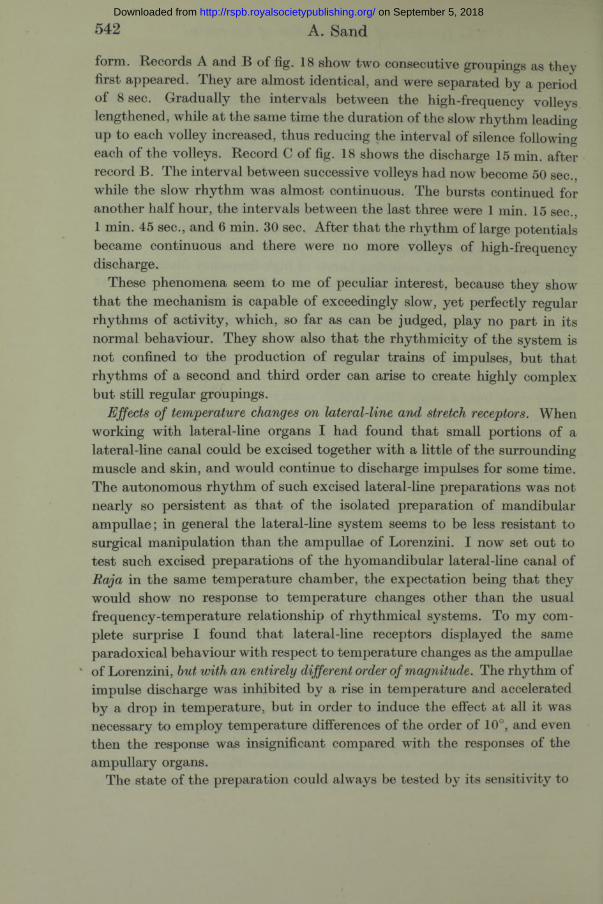
structure and innervation the appearance of the ampullary canals of Lorenzini is strikingly different from th a t of the lateral-line canals. The smooth thin-walled ampullary tubes, filled with a remarkably viscous jelly, lie free under the skin, and lengths of several centimetres can be easily withdrawn. The lateral-line canals lie in the skin, and cannot be freed from the connective tissue in which they are embedded. The ampullary canals radiate in all directions from five bilateral groups of ampullae, whose disposition in Laemargus has been figured by Ewart (1892), and this figure has found its way into numerous text-books and is reproduced also by Dotterweich (1932). In Raja batis Ewart and Mitchell (1892) described a similar arrangement, and I have found tha t Raja clavata, maculata, micro- cellata and brachyura, as well as Scyllium canicula display essentially thesame disposition. The ampullae lie together in large clusters around the arborizations of the nerve. They have been picturesquely described as “ bunches of grapes” . The whole group is contained in a capsule of fibrous connective tissue which is perforated by the ampullary tubes, and inside the capsule the ampullae are held in a delicate, almost fluid, stroma. Fig. 1 (Plate 33) is a photograph of a few of the hyomandibular ampullae of Raja, and shows their irregular contour, their connexion with the branches of the nerve, and the smooth tubes of jelly leading from them. The scale is 5 mm. The tubes lie side by side over the surface of the muscle and terminate peripherally a t small pores in the skin through which the jelly can be easily squeezed out. Most of the surface area of the head is covered with ampullary tubes.
When one contemplates the remarkable elaboration of the lateral-line canal system and of the ampullary canal system in a ray it is difficult, indeed I would say impossible, to believe th a t they do not perform entirely separate and distinct sensory functions. Those who have speculated as to the function of the ampullae of Lorenzini have for the most part been preoccupied with their histology and innervation, questions concerning which, as I have shown, no final statement is as yet possible. They have been guided by their belief in the homology of the ampullae with neuromast organs, and have assigned to them a vaguely mechanical function: a receptivity to vibrations, pressure and the like. Very few authors have a ttempted an experimental approach to the problem. Parker (1909) and Metcalf (1915) have observed muscular movements in response to mechanical stimulation of the ampullae, and Dotterweich (1932) injected distilled water subcutaneously into dogfish and inferred from the restless behaviour of the injected animals that the jelly in the ampullary canals had swelled and had stimulated pressure receptors in the ampullae. I t may
Vol. C X X V . B. 35
on September 5, 2018http://rspb.royalsocietypublishing.org/Downloaded from
528 A. Sand
safely be said th a t no satisfactory method has yet been applied to the investigation of the sensory function of the ampullae of Lorenzini.
Experimental investigation
My recent physiological study of the lateral-line canals (Sand 1937) disclosed the fundamental identity of the underlying mechanism of the lateral-line receptors and of the receptors of the semicircular canals of the labyrinth. I t demonstrated also the extreme vibrational sensitivity of the lateral-line system. W ith the experience gained in th a t work it was a relatively simple m atter to test the ampullae of Lorenzini, and to see whether their activity bore any relation to th a t of lateral-line receptors. To accomplish this it was necessary to separate the nerve fibres supplying the ampullae from the accompanying lateral-line fibres. The anatomical relations are such th a t although it is easy to find long slender nerves composed entirely of lateral-line fibres, every nerve branch which innervates a group of ampullae carries also one or more bundles of fibres which pass round or between the ampullae and continue their course to terminate in a lateral-line canal. This is so, a t least, in ray s ; Scyllium was less thoroughly examined. The mandibular ampullae are the most convenient to work with. They are a small compact group lying under the skin of the lower jaw just below the corner of the mouth. Their tubes extend in brush-like formation mesially, and term inate in pores scattered over the skin below the lower lip. Their nerve is an offshoot of the great hyomandibular branch of the facial which carries the nerve supply to the hyomandibular lateral-line canal, the large hyomandibular group of ampullae, as well as the motor innervation to some of the jaw muscles. This branch leaves the hyomandibular nerve a short distance above the hyomandibular ampullae, curves downward between the first branchial chamber and the jaw muscles, and. just before it enters the capsule of the mandibular ampullae, gives off a branch which runs along the edge of the capsule to innervate the mandibular lateral-line canal. One can isolate and cut this mandibular lateral-line branch without much difficulty, leaving a pure preparation of ampullary end-organs.
The electrical apparatus employed consisted of a five-stage condenser- coupled amplifier, Matthews oscillograph, moving-film camera and loudspeaker.
After pithing the ray, the nerve was dissected out and cut centrally, leaving a length of 2 or 3 cm. in connexion with the ampullae. This peripheral length of nerve was slung on platinum wire electrodes, the earth
on September 5, 2018http://rspb.royalsocietypublishing.org/Downloaded from
electrode being nearest the ampullae. Recording was monophasic, and afferent action potentials appeared on the records as upward displacements of the base-line.
Principal experimental facts. The main experimental results emerged with complete clarity from the very beginning of the investigation, and were confirmed without any exceptions on over forty preparations. These results were so unforeseen and altogether so extraordinary tha t it would be as well to enumerate them briefly a t this point before proceeding to a discussion of the detailed evidence.
The activity of the ampullae of Lorenzini is defined as follows:
(1) Like lateral-line receptors they display an autonomous rhythm of activity.
(2) They have no mechanical sensitivity whatever.(3) They are exceedingly sensitive to temperature.(4) Their response to cooling is an acceleration, their response to warming
is a retardation or inhibition of the rhythm of impulse discharge.
Qualitative experiments. Recording from the whole nerve one observes a massive asynchronous discharge in which it is impossible to identify the individual action potentials. The whole nerve, as I have described, contains several hundreds of fibres supplying the mandibular ampullae as well as a smaller bundle innervating the mandibular lateral-line canal. Thus the preparation at this stage contains both lateral-line and ampullary end- organs, and displays th a t extreme vibrational sensitivity so characteristic of active lateral-line receptors. Gentle tapping on the table on which the preparation is set up is adequate to augment and accelerate the asynchronous oscillations. And the response to direct mechanical stimulation of the animal’s skin is even more violent. These responses are completely abolished when the lateral-line branch of the nerve is cut. The asynchronous spontaneous discharge, now confined to the ampullary fibres, goes on as before, but no change in its volume can be detected when mechanical stimuli are applied. The complete absence of mechanical sensitivity is further confirmed when, after section of the lateral-line branch, the nerve bundles supplying the mandibular ampullae are also separated out and all except one are cut. One is left then with a preparation in which the number of active fibres has been sufficiently reduced to permit the recognition of individual impulse trains in the records and also in the sound from the loudspeaker. No kind of mechanical stimulation, such as touch, stroking or pressure, has any effect on the frequency of these discharges.
The response to temperature was first discovered when elasmobranch
The function of the ampullae of Lorenzini 529
35-2
on September 5, 2018http://rspb.royalsocietypublishing.org/Downloaded from
530 A. Sand
Ringer which had been cooled or warmed a few degrees below or above room temperature was dropped from a pipette on to the skin overlying the mandibular ampullae. A drop or two of cooled Ringer evoked an immediate acceleration, the frequencies of individual rhythms rising abruptly to something of the order of 70-100 per sec., and producing in the loudspeaker a clear musical note, whose pitch, after reaching its peak, declined gradually over a period of several seconds until finally the original frequency (8-16 per sec.) was re-established. Conversely, a drop or two of warmed Ringer caused an immediate cessation of the discharge, even in preparations containing a large number of active units. After a total inhibition lasting several seconds, one unit would begin discharging, then presently another, a t first slowly, then more rapidly, until before long the original rhythm had again returned. Such responses could be repeated over and over again.
The response to warming could also be evoked by means of a hot glass rod, held about 1 cm. above the mandibular ampullae. When the glass rod was brought near the discharge ceased, when the rod was withdrawn it gradually started up again. Applied to the back of my hand a t a similar distance, the rod produced a mild sensation of warmth.
Isolated preparation and temperature chamber. The mandibular ampullae are contained in a smooth membranous envelope which has very loose attachments with the surrounding tissues, so th a t the whole mandibular capsule with its nerve could be cleanly removed, thus providing a neat and small isolated preparation, which was capable of maintaining its activity for hours. Fig. 2 (Plate 33) is a photograph of the preparation, showing the mandibular capsule of a small ray with its brush of ampullary tubes cut across, its nerve, and the mandibular lateral-line branch by-passing the capsule. A few ampullae are dimly discernible by transparency inside the capsule. The nerve has been cut down so as to leave only a few intact fibres. The scale is 5 mm.
This preparation served for the quantitative study of the responses of individual ampullary end-organs to temperature changes. I t was installed in an apparatus which is illustrated in fig. 3. The mandibular capsule is laid in a little puddle of 2 or 3 drops of elasmobranch Ringer in the depression in the glass bulb B, and the nerve is slung on the platinum wire electrodes P. Ice-water G and warm water W are delivered through the two-way tap to the bulb and escape a t the outflow O. This glassware is mounted in a brass pot with a flanged, tight-fitting lid. The pot is filled with paraffin wax up to the level of the bulb. The wax serves as a support for the bulb and platinum wires, and also reduces the air space in the
on September 5, 2018http://rspb.royalsocietypublishing.org/Downloaded from
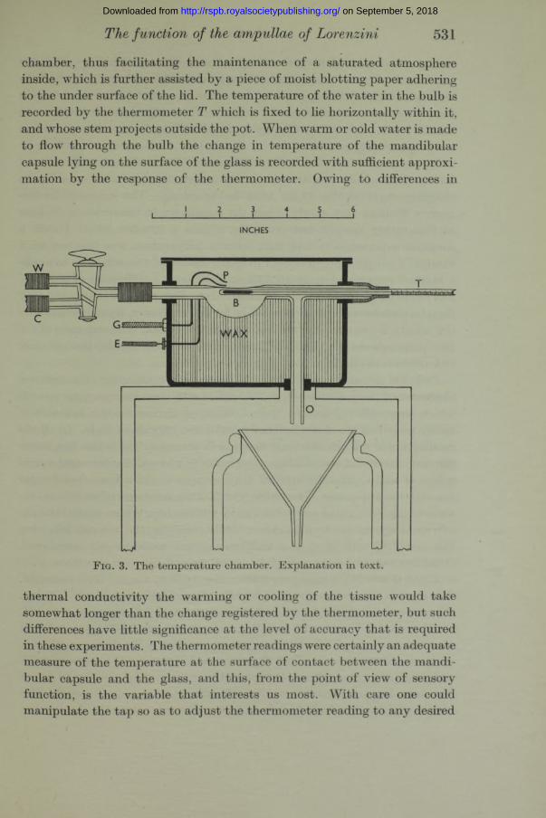
The function of the ampullae of Lorenzini 531
chamber, thus facilitating the maintenance of a saturated atmosphere inside, which is further assisted by a piece of moist blotting paper adhering to the under surface of the lid. The temperature of the water in the bulb is recorded by the thermometer T which is fixed to lie horizontally within it, and whose stem projects outside the pot. When warm or cold water is made to flow through the bulb the change in temperature of the mandibular capsule lying on the surface of the glass is recorded with sufficient approximation by the response of the thermometer. Owing to differences in
INCHES
F i g . 3. The temperature chamber. Explanation in text.
thermal conductivity the warming or cooling of the tissue would take somewhat longer than the change registered by the thermometer, but such differences have little significance at the level of accuracy that is required in these experiments. The thermometer readings were certainly an adequate measure of the temperature at the surface of contact between the mandibular capsule and the glass, and this, from the point of view of sensory function, is the variable tha t interests us most. With care one could manipulate the tap so as to adjust the thermometer reading to any desired
on September 5, 2018http://rspb.royalsocietypublishing.org/Downloaded from
532 A. Sand
value, and one could keep the temperature constant within ±0*1° C. for any length of time. A mechanical device was employed to signal the two- way operations of the tap, so tha t the beginning and end of a rise or drop in temperature was marked on the record. These signals were accurate to within 1 sec., and served admirably to identify a long series of random temperature changes.
A preparation installed in this apparatus will remain active for several hours. Two of my preparations continued overnight and were still discharging rhythmically 24 hr. after being set up. The autonomous frequency declines very slowly, while the sensitivity to temperature declines a t first rather steeply, but presently attains a steadier level. I t was a common experience to find th a t when a preparation was first set up a change in temperature of 0*1° C. in either direction was adequate to evoke a marked response, but about half an hour later a difference of about 1° was required to produce a comparable effect. This order of sensitivity may then be maintained with little change for several hours, though eventually the sensitivity to temperature changes becomes weak. The responses of the two preparations th a t had survived overnight were feeble, though their autonomous rhythm s were sufficiently vigorous.
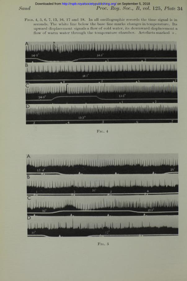
Response of single end-organs to temperature changes. The general character of the responses recorded is shown in the accompanying records which were selected from a large number of similar records taken from thirty preparations of the isolated mandibular capsule of Raja. In all the oscillographic records the time signal is in seconds. The white line below the base line is the temperature signal. Its downward displacement marks a flow of warm water, its upward displacement marks a flow of cold water through the apparatus. The initial and final temperatures of each temperature change were noted and are shown on the records. Where several lengths of record are shown to illustrate a single response, the seconds following the establishment of a new temperature are consecutively numbered. Artefacts caused by the manipulation of the tap are marked with a x .
Fig. 4 (Plate 34) is from preparation No. 24 from a small Raja maculata. The records were taken about 1^ hr. after the animal was killed by pithing. They show a response to a rise in temperature of 0-3° (records A and B, continuous), and a response to a drop in temperature of 0-7° (records C and JD, continuous). A single rhythm of large potentials is clearly seen, although at least one other train of small potentials is present. When these small potentials coincide with one of the large potentials, they give rise to one of the extra-large potentials occurring irregularly in records C and D. The rise in temperature from 14*0 to 14-3° causes a retardation in the rhythm
on September 5, 2018http://rspb.royalsocietypublishing.org/Downloaded from
extending over several seconds (record B). The drop from 14-0 to 13-3° causes a prolonged acceleration (records C and D).
Fig. 5 (Plate 34) is from a more complex preparation (No. 13). There are several rhythmically active units. The retardation resulting from a rise in temperature from 15*4 to 16-0° is striking (records A and B continuous), and so is the acceleration following the reverse change from 16-0 to 15-3° (record C). In record D a rise from 10 to 12° causes complete inhibition. This figure illustrates a fact which has been abundantly confirmed in numerous experiments, namely, that the behaviour of all the ampullary receptors is the same.
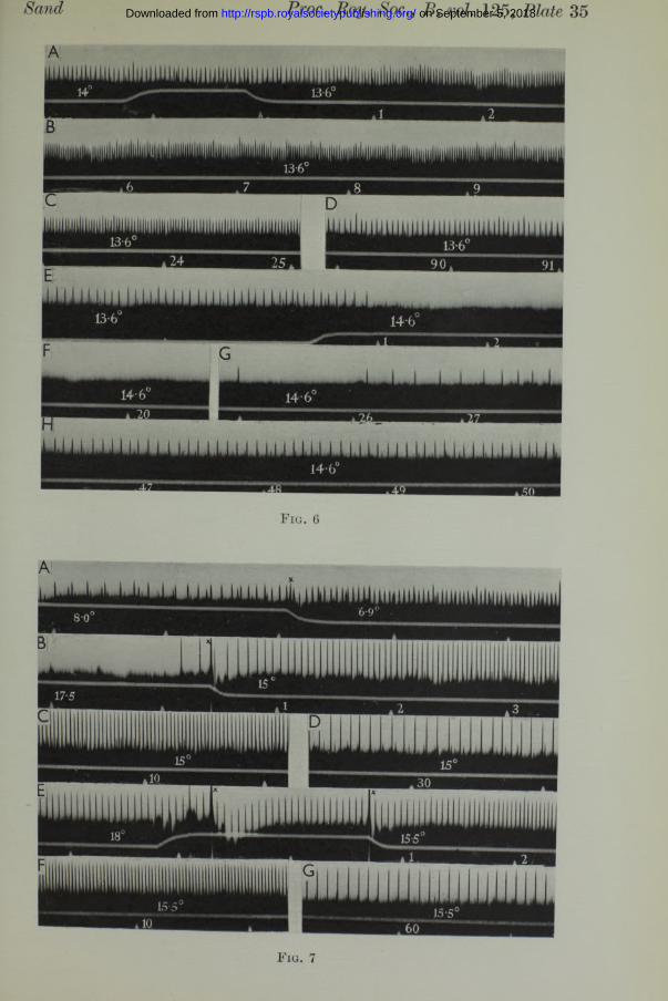
In the experiments of fig. 6 (Plate 35) the recording was continued long enough to show the adaptation of the end-organ to the new temperature. Records A-D show the prolonged response to a drop of 0-4° (14-0 to 13*6), and records E -H show the total inhibition lasting about 25 sec. after a rise of 1°.
These relations hold throughout the physiological temperature range.1 have not recorded at temperatures below 3° C., and above about 20° the receptor mechanism suffers irreversible damage, and the rhythm of discharge usually soon stops. But within these limits the acceleration in response to a fall in temperature and the inhibition in response to a rise can always be demonstrated, as fig. 7 (Plate 35) shows. Record A is a cooling response in the lower temperature range. Following this response the temperature was raised from 7 to 13° when there was a period of inhibition lasting 45 sec. The temperature was maintained at 13° for 5 min., during which time a discharge of uniform frequency continued. The temperature was then raised further from 13 to 18°. There was an immediate cessation of the discharge, and this time the inhibition was apparently permanent, for the preparation remained silent for 25 min., during which time the temperature was maintained at 17*5-18°. Then the temperature was dropped to 15°, and this cooling immediately revived the preparation. The response is shown in fig. 7 (Plate 35), records B, C and D. The temperature was then raised again to 18°, but this time in three steps of 1° each. This gradual warming produced only a temporary arrest a t each step, and at 18° the preparation was firing vigorously. Record E, fig. 7, begins after2 min. at 18°, and the frequency is 17 per sec. Rapid cooling down to 15*5° (records E, F and G) evoked a response almost identical with the preceding one.
All these records will suffice to illustrate the quality of the experimental results obtained in this investigation. They show that there was ample material for the counting of the frequencies of discharge from individual
The function of the ampullae of Lorenzini 533 on September 5, 2018http://rspb.royalsocietypublishing.org/Downloaded from
534 A. Sand
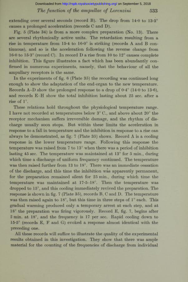
receptor units. For purposes of quantitative comparison the curves obtained when individual frequencies are plotted against time are far more illuminating than short lengths of oscillographic records. In the following figures the main characteristics of the temperature response are presented in this fashion. In fig. 8 are shown four responses to temperature changes of less than 0-5°. Two are warmings and two are coolings. The vertical line at 4 sec. marks the beginning of the change in temperature. The initial and
120 117
SECONDS
F ig. 8. Responses of single units to temperature changes of less than 0-5° C.
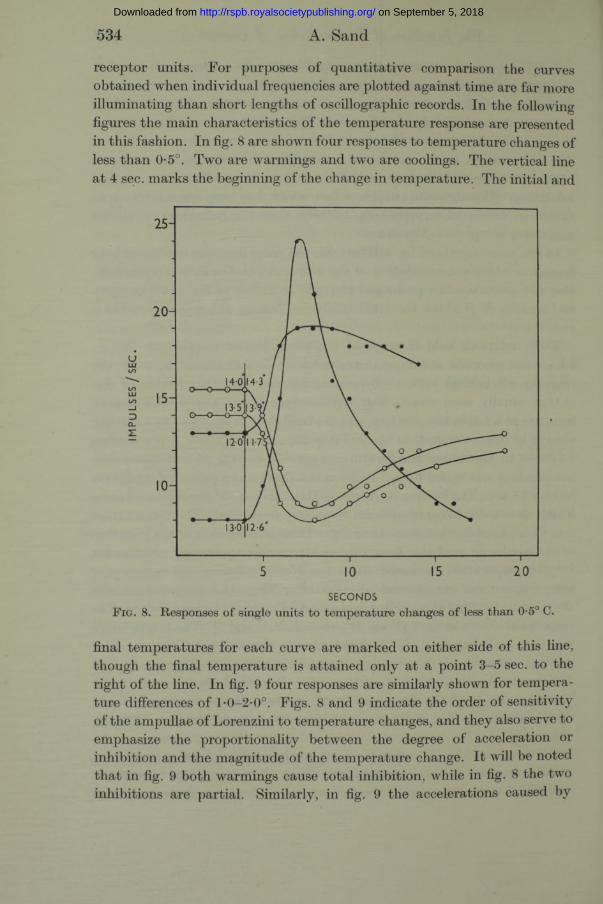
final temperatures for each curve are marked on either side of this line, though the final temperature is attained only at a point 3-5 sec. to the right of the line. In fig. 9 four responses are similarly shown for temperature differences of l*0-2-0°. Figs. 8 and 9 indicate the order of sensitivity of the ampullae of Lorenzini to temperature changes, and they also serve to emphasize the proportionality between the degree of acceleration or inhibition and the magnitude of the temperature change. I t will be noted tha t in fig. 9 both warmings cause total inhibition, while in fig. 8 the two inhibitions are partial. Similarly, in fig. 9 the accelerations caused by
on September 5, 2018http://rspb.royalsocietypublishing.org/Downloaded from
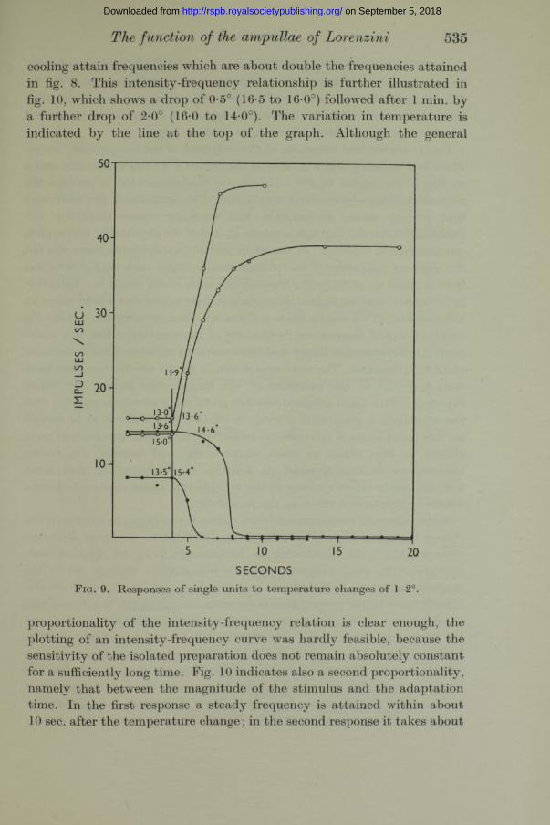
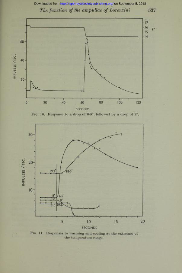
cooling attain frequencies which are about double the frequencies attained in fig. 8. This intensity-frequency relationship is further illustrated in fig. 10, which shows a drop of 0-5° (16-5 to 16-0°) followed after 1 min. by a further drop of 2-0° (16-0 to 14-0°). The variation in temperature is indicated by the line a t the top of the graph. Although the general
The function of the ampullae of Lorenzini 535
SECONDSF ig. 9. Responses of single units to temperature changes of 1-2°.
proportionality of the intensity-frequency relation is clear enough, the plotting of an intensity-frequency curve was hardly feasible, because the sensitivity of the isolated preparation does not remain absolutely constant for a sufficiently long time. Fig. 10 indicates also a second proportionality, namely that between the magnitude of the stimulus and the adaptation time. In the first response a steady frequency is attained within about 10 sec. after the temperature change; in the second response it takes about
on September 5, 2018http://rspb.royalsocietypublishing.org/Downloaded from
536 A. Sand
a minute. Even longer adaptation times (2-3 min.) have been recorded for large temperature changes. These are no doubt partly accounted for by differences in the time of thermal equilibration, though it is unlikely th a t this is the major factor, for the peak frequency is always rapidly attained whether the temperature difference be small or large.
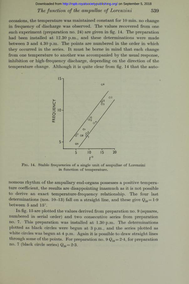
Fig. 11 shows four responses a t the extremes of the temperature range. There is a warming and a cooling in the region 5-8° and a warming and a cooling in the region 18-20°. This figure illustrates what is perhaps the most astonishing aspect of this very remarkable mechanism. The statement th a t cooling causes acceleration and warming causes inhibition undoubtedly holds for any temperature a t which the rhythmical mechanism can continue to function. In the whole series of experiments there was not the slightest indication of an optimum temperature such th a t a deviation from it either up or down the temperature scale would result in a reduction in frequency, and an approach towards it from either end would cause an acceleration. (This will be made still clearer when we come to consider the steady, adapted frequencies in function of temperature.) But this statement is not intended to imply th a t the sensitivity of the receptor is the same a t all temperatures. The responses shown in fig. 11 are the best th a t have been recorded a t the extremes of the temperature range. My whole experience with these preparations has given me a strong impression tha t the sensitivity is greatest in the region 10-15°. I t is difficult to be certain on this point because the extreme sensitivity of a fresh preparation must always be first tested a t room temperature, and because the drastic temperature change required to transfer the preparation from room temperature to either extreme of the temperature range appears to have a markedly deleterious effect on the sensitivity.
Stable frequency-temperature relation. After the response to a temperature change has taken place adaptation occurs, and the frequency of discharge returns again to the initial value, as, for example, in fig. 10. This, a t least, appears to be what happens when the temperature changes are of the magnitude th a t we have hitherto considered, i.e. 0*5-2°. When, however, the effects of larger temperature differences are examined, it is found tha t the frequency at the end of the response is not quite the same as it was at the beginning. After a rise in temperature of several degrees the rhythm, having recovered from a period of inhibition, gradually accelerates until it attains a steady frequency which is higher than the frequency before the response, and after a drop of several degrees and the resulting maximal acceleration, the rhythm adapts to a lower frequency than before the response. This is seen to be so in fig. 12, which illustrates a 20 min. experi-
on September 5, 2018http://rspb.royalsocietypublishing.org/Downloaded from
The function of the ampullae of Lorenzini 537
SECONDS
F ig. 10. Response to a drop of 0-5°, followed by a drop of 2°.
SECONDSF ig. 11. Responses to warming and cooling at the extremes of
the temperature range.
on September 5, 2018http://rspb.royalsocietypublishing.org/Downloaded from

538 A. Sand
ment in which the temperature was raised from 10 to 15°, maintained at 15° for 10 min., then lowered again to 10°, and maintained at 10° for another 10 min. The adapted frequency was 7 per sec. a t the beginning. After the first response it rose to 11 per sec., and after the second response it sank again to 8 per sec. I t is clear, therefore, that when we consider conditions of stable equilibrium, the frequency of discharge varies as a function of temperature, and displays a positive temperature coefficient. In this important respect our paradoxical system behaves in an orthodox manner, indeed it behaves like every other biological rhythm that has ever
840 1200240 600SECONDS
F i g . 12. Temperature raised from 10 to 15°, and, after 10 min., lowered again to 10°.
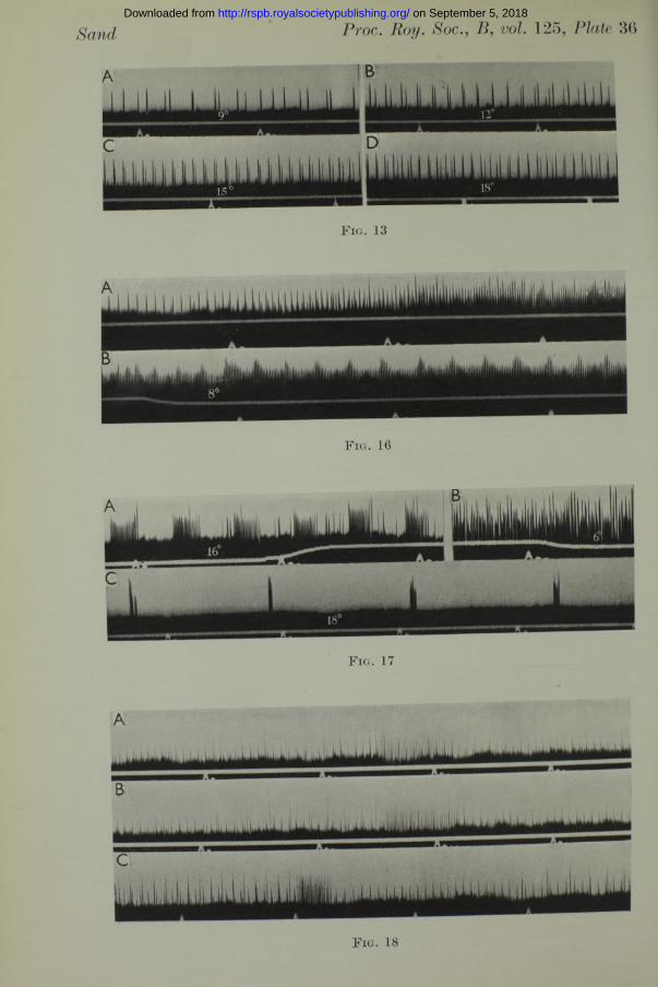
been studied. An experiment such as tha t of fig. 12 at once suggests the possibility of deriving a temperature curve for this sensory rhythm, and determining its temperature characteristics. In the oscillographic records of fig. 13 (Plate 36) the increased frequencies of two units in function of temperature are shown between 9 and 18° C. Similar records were taken at different temperatures from three preparations. In all cases the equilibration time allowed a t each temperature was 5 min., which was certainly sufficient for the system to attain a stable equilibrium, for when, on several
on September 5, 2018http://rspb.royalsocietypublishing.org/Downloaded from
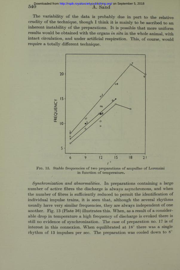
occasions, the temperature was maintained constant for 10 min. no change in frequency of discharge was observed. The values recovered from one such experiment (preparation no. 24) are given in fig. 14. The preparation had been installed a t 12.30 p.m., and these determinations were made between 3 and 4.30 p.m. The points are numbered in the order in which they occurred in the series. I t must be borne in mind th a t each change from one temperature to another was accompanied by the usual response, inhibition or high-frequency discharge, depending on the direction of the temperature change. Although it is quite clear from fig. 14 th a t the auto-
The f unction of the ampullae of Lorenzini 539
F ig. 14. Stable frequencies of a single unit of ampullae of Lorenzini in function of temperature.
nomous rhythm of the ampullary end-organs possesses a positive temperature coefficient, the results are disappointing inasmuch as it is not possible to derive an exact temperature-frequency relationship. The four last determinations (nos. 10—13) fall on a straight line, and these give Q10= 1*9 between 5 and 15°.
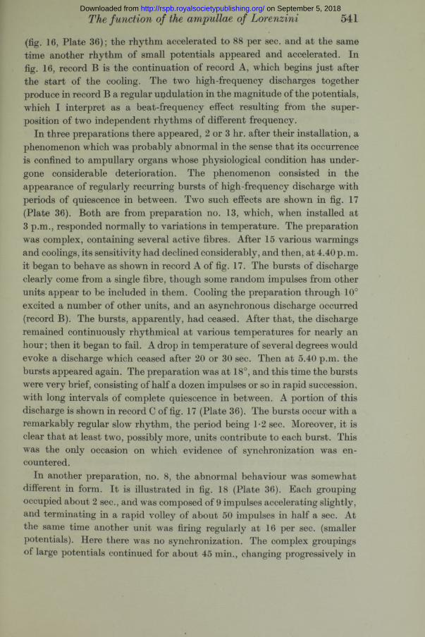
In fig. 15 are plotted the values derived from preparation no. 9 (squares, numbered in serial order) and two consecutive series from preparation no. 7. This preparation was installed a t 1.30 p.m. The determinations plotted as black circles were begun a t 3 p.m., and the series plotted as white circles was begun at 4 p.m. Again it is possible to draw straight lines through some of the points. For preparation no. 9 $ 10 = 2-4, for preparation no. 7 (black circle series) Q10=2-3.
on September 5, 2018http://rspb.royalsocietypublishing.org/Downloaded from
540 A. Sand
The variability of the data is probably due in part to the relative crudity of the technique, though I think it is mainly to be ascribed to an inherent instability of the preparations. I t is possible th a t more uniform results would be obtained with the organs in situ in the whole animal, with in tact circulation, and under artificial respiration. This, of course, would require a totally different technique.
F i g . 15. Stable frequencies of two preparations of ampullae of Lorenzini in function o f temperature.
/
Synchronization and abnormalities. In preparations containing a large number of active fibres the discharge is always asynchronous, and when the number of fibres is sufficiently reduced to permit the identification of individual impulse trains, it is seen that, although the several rhythms usually have very similar frequencies, they are always independent of one another. Fig. 13 (Plate 36) illustrates this. When, as a result of a considerable drop in temperature a high frequency of discharge is evoked there is still no evidence of synchronization. The case of preparation no. 17 is of interest in this connexion. When equilibrated a t 18° there was a single rhythm of 13 impulses per sec. The preparation was cooled down to 8L
on September 5, 2018http://rspb.royalsocietypublishing.org/Downloaded from
(fig. 16, Plate 36); the rhythm accelerated to 88 per sec. and at the same time another rhythm of small potentials appeared and accelerated. In fig. 16, record B is the continuation of record A, which begins just after the start of the cooling. The two high-frequency discharges together produce in record B a regular undulation in the magnitude of the potentials, which I interpret as a beat-frequency effect resulting from the superposition of two independent rhythms of different frequency.
In three preparations there appeared, 2 or 3 hr. after their installation, a phenomenon which was probably abnormal in the sense that its occurrence is confined to ampullary organs whose physiological condition has undergone considerable deterioration. The phenomenon consisted in the appearance of regularly recurring bursts of high-frequency discharge with periods of quiescence in between. Two such effects are shown in fig. 17 (Plate 36). Both are from preparation no. 13, which, when installed at 3 p.m., responded normally to variations in temperature. The preparation was complex, containing several active fibres. After 15 various warmings and coolings, its sensitivity had declined considerably, and then, at 4.40 p. m. it began to behave as shown in record A of fig. 17. The bursts of discharge clearly come from a single fibre, though some random impulses from other units appear to be included in them. Cooling the preparation through 10° excited a number of other units, and an asynchronous discharge occurred (record B). The bursts, apparently, had ceased. After that, the discharge remained continuously rhythmical at various temperatures for nearly an hour; then it began to fail. A drop in temperature of several degrees would evoke a discharge which ceased after 20 or 30 sec. Then at 5.40 p.m. the bursts appeared again. The preparation was at 18°, and this time the bursts were very brief, consisting of half a dozen impulses or so in rapid succession, with long intervals of complete quiescence in between. A portion of this discharge is shown in record C of fig. 17 (Plate 36). The bursts occur with a remarkably regular slow rhythm, the period being T2 sec. Moreover, it is clear that at least two, possibly more, units contribute to each burst. This was the only occasion on which evidence of synchronization was encountered.
In another preparation, no. 8, the abnormal behaviour was somewhat different in form. I t is illustrated in fig. 18 (Plate 36). Each grouping occupied about 2 sec., and was composed of 9 impulses accelerating slightly, and terminating in a rapid volley of about 50 impulses in half a sec. At the same time another unit was firing regularly at 16 per sec. (smaller potentials). Here there was no synchronization. The complex groupings of large potentials continued for about 45 min., changing progressively in
The function of the ampullae of Lorenzini 541 on September 5, 2018http://rspb.royalsocietypublishing.org/Downloaded from
542 A. Sand
form. Records A and B of fig. 18 show two consecutive groupings as they first appeared. They are almost identical, and were separated by a period of 8 sec. Gradually the intervals between the high-frequency volleys lengthened, while a t the same time the duration of the slow rhythm leading up to each volley increased, thus reducing the interval of silence following each of the volleys. Record C of fig. 18 shows the discharge 15 min. after record B. The interval between successive volleys had now become 50 sec., while the slow rhythm was almost continuous. The bursts continued for another half hour, the intervals between the last three were 1 min. 15 sec., 1 min. 45 sec., and 6 min. 30 sec. After tha t the rhythm of large potentials became continuous and there were no more volleys of high-frequency discharge.
These phenomena seem to me of peculiar interest, because they show that the mechanism is capable of exceedingly slow, yet perfectly regular rhythms of activity, which, so far as can be judged, play no part in its normal behaviour. They show also th a t the rhythmicity of the system is not confined to the production of regular trains of impulses, but that rhythms of a second and third order can arise to create highly complex but still regular groupings.
Effects of temperature changes on lateral-line and stretch receptors. When working with lateral-line organs I had found th a t small portions of a lateral-line canal could be excised together with a little of the surrounding muscle and skin, and would continue to discharge impulses for some time. The autonomous rhythm of such excised lateral-line preparations was not nearly so persistent as th a t of the isolated preparation of mandibular ampullae; in general the lateral-line system seems to be less resistant to surgical manipulation than the ampullae of Lorenzini. I now set out to test such excised preparations of the hyomandibular lateral-line canal of Raja in the same temperature chamber, the expectation being that they would show no response to temperature changes other than the usual frequency-temperature relationship of rhythmical systems. To my complete surprise I found tha t lateral-line receptors displayed the same paradoxical behaviour with respect to temperature changes as the ampullae of Lorenzini, but with an entirely different order of magnitude. The rhythm of impulse discharge was inhibited by a rise in temperature and accelerated by a drop in temperature, but in order to induce the effect a t all it was necessary to employ temperature differences of the order of 10°, and even then the response was insignificant compared with the responses of the ampullary organs.
The state of the preparation could always be tested by its sensitivity to
on September 5, 2018http://rspb.royalsocietypublishing.org/Downloaded from
Sand Proc. Boy. Soc., B, vol. 125, Plate 33
F ig . 1. A few o f the hyom andibular am pullae o f R aja . Scale 5 mm.
J ig . 2. Isolated preparation of the mandibular capsule. The nerve has been reduced to a few fibres. Ampullary canals cut across. Lateral-line branch can be seen by-passing the capsule. Scale 5 mm.
(Facing p . 512 )
on September 5, 2018http://rspb.royalsocietypublishing.org/Downloaded from
Sand Proc. Roy. Soc., B, vol. 125, Plate 34
F ig s . 4, 5, 6, 7, 13, 16, 17 and 18. In all oscillographic records the time signal is in seconds. The white line below the base line marks changes in temperature. Its upward displacement signals a flow of cold water, its downward displacement a flow of warm water through the temperature chamber. Artefacts marked x .
F ig . 4
F ig . 5
on September 5, 2018http://rspb.royalsocietypublishing.org/Downloaded from
Sand Proc. Roy. Soc., B, 125, Plate 35
mm
13-6°
imuiMmiuiuiiyMiiiiaiuiutyiUMiiuiiijiiiiiJiiiHiiUiiuiJiiiiiiUii M i l m uiu u i iiiiiiiiiiiiiilii Li 1 y ui u
.1.111 ill. 111ItLiiUJl Llilliiii 1 ikilli i m i l
■G
i _ ^ „i i i i i i i i j jB S mma o r a a wmmmmmmmwxmammmmmBmm
U ili.lilii.m i LI .1 i 1.1 ■! i i 1.11 i l . l . i i l i i l m i Li 1 ill i ill U i l l L
F ig . 6
F ig . 7
on September 5, 2018http://rspb.royalsocietypublishing.org/Downloaded from
Sand Proc. Roy. Soc.,B, vol. 125, Plate 36
F i g . 16
F i g . 17
F ig . 18
on September 5, 2018http://rspb.royalsocietypublishing.org/Downloaded from
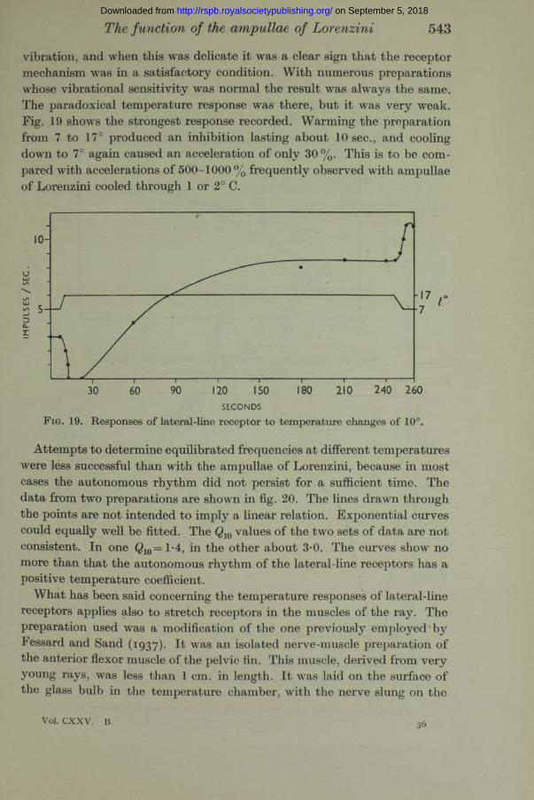
vibration, and when this was delicate it was a clear sign th a t the receptor mechanism was in a satisfactory condition. With numerous preparations whose vibrational sensitivity was normal the result was always the same. The paradoxical temperature response was there, but it was very weak. Fig. 19 shows the strongest response recorded. Warming the preparation from 7 to 17° produced an inhibition lasting about 10 sec., and cooling down to 7° again caused an acceleration of only 30 %. This is to be compared with accelerations of 500-1000 % frequently observed with ampullae of Lorenzini cooled through 1 or 2° C.
The function of the ampullae of Lorenzini 543
£ «;—f
240 260SECONDS
F ig. 19. Responses of lateral-line receptor to temperature changes of 10°.
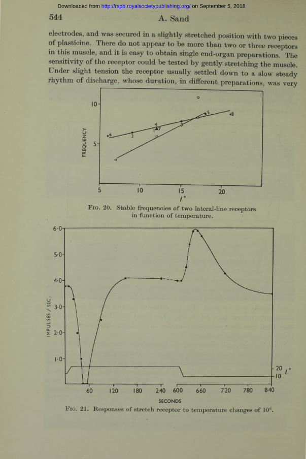
Attempts to determine equilibrated frequencies a t different temperatures were less successful than with the ampullae of Lorenzini, because in most cases the autonomous rhythm did not persist for a sufficient time. The data from two preparations are shown in fig. 20. The lines drawn through the points are not intended to imply a linear relation. Exponential curves could equally well be fitted. The $ 10 values of the two sets of data are not consistent. In one Q10= l-4 , in the other about 3*0. The curves show no more than tha t the autonomous rhythm of the lateral-line receptors has a positive temperature coefficient.
What has been said concerning the temperature responses of lateral-line receptors applies also to stretch receptors in the muscles of the ray. The preparation used was a modification of the one previously employed by Fessard and Sand (1937). I t was an isolated nerve-muscle preparation of the anterior flexor muscle of the pelvic fin. This muscle, derived from very young rays, was less than 1 cm. in length. I t was laid on the surface of the glass bulb in the temperature chamber, with the nerve slung on the
Vol. C X X V . B. 36
on September 5, 2018http://rspb.royalsocietypublishing.org/Downloaded from
IMPU
LSES
/ S
E<
544 A. Sand
electrodes, and was secured in a slightly stretched position with two pieces of plasticine. There do not appear to be more than two or three receptors in this muscle, and it is easy to obtain single end-organ preparations. The sensitivity of the receptor could be tested by gently stretching the muscle. Under slight tension the receptor usually settled down to a slow steady rhythm of discharge, whose duration, in different preparations, was very
F i g . 20. Stable frequencies of two lateral-line receptors in function of temperature.
240 600SECONDS
F i g . 2 1 . Responses of stretch receptor to temperature changes of 1 0 ° .
on September 5, 2018http://rspb.royalsocietypublishing.org/Downloaded from
variable. In most, the discharge continued only for 5-15 min. and then, in the manner so characteristic of these organs, stopped abruptly without preliminary retardation. A slight increase in tension would then start up a fresh discharge. In some preparations, however, the receptor continued to discharge a regular rhythm for an hour, or even longer. These were used to test the effects of variations in temperature. In no instance did small temperature changes of 1-2° produce any noticeable effect on the rhythm, but large changes of 10° or so evoked retardation or acceleration to an extent closely similar to the same phenomena in lateral-line receptors. One such experiment is illustrated in fig. 21. I t does not require further description.
The function of the ampullae of Lorenzini 545
Z 50-
F i g . 2 2 . Stable frequencies of two stretch receptors in function of temperature.
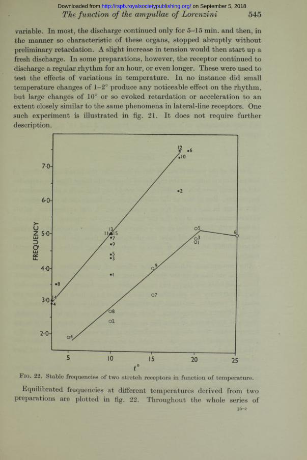
Equilibrated frequencies at different temperatures derived from two preparations are plotted in fig. 22. Throughout the whole series of
36-2
on September 5, 2018http://rspb.royalsocietypublishing.org/Downloaded from
546 A. Sand
determinations each muscle remained, of course, under constant tension. The fifteen determinations [plotted as black circles occupied about 80 min. I t will be noted that, as judged by the frequencies at 10°, the black-circle series shows a regular acceleration with time, for which I am unable to suggest an explanation. The white-circle series shows this also for the three values a t 20°, though determination no. 6, whose value indicates the harmful effect of so high a temperature as 25°, evidently served to depress the subsequent frequencies a t lower temperatures. The last five determinations of the black-circle series (nos. 11-15) show that the preparation had reached a state of complete stability, and these five points fall very accurately on a straight line. Between 5 and 15° Q10 = 1*9 for the upper curve and 2*1 for the lower.
Discussion
Nature of the mechanism. I f the evidence for the paradoxical effects of tem perature changes on the rhythm of impulse discharge from the ampullae of Lorenzini were less conclusive, one would say th a t such a phenomenon is impossible. But in face of the overwhelming evidence one is constrained to cast about in one’s mind for an explanation. A real explanation, in the experimental sense, is not, for the time being, to be expected. All tha t can be done with the material a t one’s disposal is to suggest a basis for a working hypothesis which would raise the phenomenon from the level of extreme improbability and place it among facts awaiting further experimental analysis.
Our conceptions concerning the effects of temperature on biological processes are based, in the first instance, on the experience of chemistry. But, as Belehradek (1935) has emphasized, many biological processes obey very imperfectly the quantitative laws which have been derived from the study of chemical reactions, as witness, for example, the inconstancy of Qxq a t different temperatures. Such inconstancies are due to the physicochemical complexity of even the simplest biological systems, and it is not inconceivable th a t the interrelationship of the component reactions of a given biological system may be such as to give rise to a form of behaviour with respect to temperature which appears to flout the fundamental laws of chemical action.
On the assumption tha t the paradoxical temperature effect is due to ascertainable physico-chemical causes, the following scheme is offered as a first approximation to a working hypothesis. I t is assumed that the frequency of impulse discharge is a function of the difference between the
on September 5, 2018http://rspb.royalsocietypublishing.org/Downloaded from
The function of the ampullae of Lorenzini 547
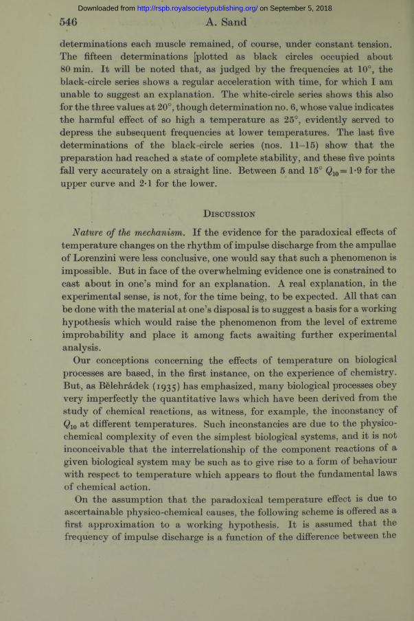
velocities of two processes. I t is further assumed that one of these processes, I, when subjected to a change in temperature, attains a new equilibrium rapidly, whereas the other process, E, attains a new equilibrium slowly. The consequences of these assumptions are illustrated by fig. 23. To give the illustration a concrete form the curves have been derived from arbitrary values such that the velocity of process is taken to be 4*0 at 0°, and its Q10 as 2-5 at all temperatures; and the velocity of process I is also 4-0 at 0°, and its Q10 2*0 at all temperatures. The curves E and I then
SECONDS
F ig. 23. Explanation in text.
represent the course in time of the two processes during the 100 sec. following a drop in temperature from 15 to 13°. They are calculated from the empirical formulae
and 1 = 115 +
where E1Z and E15, / 13 and J15 are the equilibrated velocities of E and I at 13 and 15° respectively, t = time after the temperature drop, and R and r have the significance of time-factors which govern the rate of kinetic equilibration of the reactions. The values I? =150 and r= l*5 have been
on September 5, 2018http://rspb.royalsocietypublishing.org/Downloaded from
548 A. Sand
arbitrarily taken. The curve for E - then has the form shown, and resembles the time course of the sensory discharge following a drop in temperature. The dashed curve is plotted for the values o iE — I following a greater temperature drop from 15 to 10°. As with the sensory discharge, the initial “ response” is greater and the final equilibrated value is lower than with the smaller temperature difference. Following a rise in temperature the curves would be mirror images of those here shown, and the shape o i E - I would bear a similar resemblance to the sensory effect.
The processes E and I may be conceived as chemical reactions resulting in the production of excitatory and inhibitory substances, or they may represent more physiological quantities like excitatory state and threshold, and no doubt other possibilities could be imagined. But whatever the underlying mechanism of the rhythmical discharge may turn out to be, I think it can be maintained with some confidence th a t if the observed phenomena are to be interpreted in terms of simple underlying processes whose course in time after a temperature change is monotonic, such an interpretation will involve the conception of a factor of kinetic equilibration, whose magnitude will be different for the different processes composing the system. The only instance of the occurrence of such a time factor th a t is known to me is in connexion with the viscosity of gelatin solutions, which, as Loeb (1922) showed, continues to increase a t certain temperatures for many minutes after thermal equilibrium has been established. Possibly the frequency of the sensory discharge from the ampullae of Lorenzini is connected with the viscosity of some component in the system.
Biological significance of the results. The qualitative similarity in the behaviour of the ampullae, lateral-line organs and stretch receptors in relation to temperature changes may be taken as further evidence of the relationship of the biophysical structure of their excitatory systems. They are already related by their ability to maintain persistent rhythms of discharge, rhythm s which in the intact animal apparently never cease throughout life, except when subjected to an inhibitory stimulus. (Stretch receptors in rays are always found to be rhythmically active when the muscles in situ are in the normal position of rest.) They are further relatedby the fact th a t their persistent rhythm is subject to inhibition or excitation by antagonistic phases of the appropriate physical disturbance (see Sand 1937; Fessard and Sand 1937). I t is possible tha t in the mechanism of their reaction to temperature they may be found to be related to rhythmical and tonic muscles, for Mangold (1926) has described a paradoxical slowing of the isolated Carcinus heart in response to warming, and
on September 5, 2018http://rspb.royalsocietypublishing.org/Downloaded from
549
Christ (1929), working with isolated strips of bovine carotids, showed th a t a sudden cooling through 10° resulted in a contraction followed by relaxation, while the contraction induced by a rise in tem perature was sometimes preceded by a brief relaxation. W hether these phenomena bear any relation to the behaviour of sensory rhythm s cannot a t present be decided.
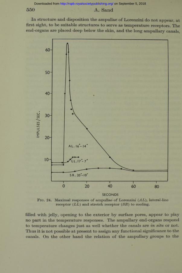
W hatever meaning is ultim ately assigned to the qualitative similarity in the behaviour of the three sensory rhythm s, there can be little doubt as to the significance of their quantitative differences. The magnitude of the responses of lateral-line receptors and stretch receptors to tem perature changes is minute compared with the responses of the ampullae of Lorenzini. Fig. 24 may serve for the comparison of the maximal responses of which the three sensory systems are capable. The three curves are reproduced to one scale from preceding figures. Two degrees is very nearly a maximal stimulus for ampullae of Lorenzini. The best preparations have shown marked responses to one or two tenths of a degree, tem perature differences which I myself could scarcely appreciate when I placed my finger in the position of the preparation, in contact with the surface of the glass bulb. Such sensitivity unquestionably raises the tem perature responses of the ampullary end-organs to the rank of a specific sensory function. Hoagland (1935) dem onstrated the acceleration of the discharge from lateral-line receptors with increasing tem peratures in a m anner similar to my experiments of fig. 20, and drew from this the conclusion th a t “ I t would seem, therefore, th a t one function of the lateral-line system is th a t of thermal reception” (p. 70). I t cannot be too strongly emphasized th a t such variation of a sensory rhythm in function of tem perature proves nothing more than th a t sensory rhythm s, like all kinds of other rhythm ical biological processes, m ust possess a positive tem perature coefficient. Hoagland’s reasoning would apply equally well to all three receptors th a t I have considered, and would lead to the absurd conclusion th a t ampullae of Lorenzini, lateral-line organs and stretch receptors are all concerned with the function of thermal reception. In the ampullae of Lorenzini we are confronted with a system whose variations in relation to steady temperatures are insignificant in comparison with its reactions to tem perature changes. Such changes produce enormous and almost instantaneous responses which have all the characteristics of specific sensory action. I t is a system which, like most sensory systems, is admirably designed to emphasize the effects of change. I t is common experience tha t, by virtue of their adap tation, our own temperature receptors are poor thermometers; they are specialized for the detection of relative changes in temperature. The activity of the ampullae of Lorenzini is of the same kind.
The function of the ampullae of Lorenzini on September 5, 2018http://rspb.royalsocietypublishing.org/Downloaded from
550 A. Sand
In structure and disposition the ampullae of Lorenzini do not appear, a t first sight, to be suitable structures to serve as tem perature receptors. The end-organs are placed deep below the skin, and the long am pullary canals,
SR. 20-10°
SECONDSF ig . 24. M aximal responses o f am pullae o f Lorenzini {AL) , lateral-line
receptor (LL) and stretch receptor (SR) to cooling.
filled w ith jelly, opening to the exterior by surface pores, appear to play no p a rt in the tem perature responses. The am pullary end-organs respond to tem perature changes ju s t as well w hether the canals are situ or not. Thus it is no t possible a t present to assign any functional significance to the canals. On the other hand the relation of the am pullary groups to the
on September 5, 2018http://rspb.royalsocietypublishing.org/Downloaded from
respiratory water current is striking. In the ray, the superficial ophthalmic, inner and outer buccal ampullae lie in the thin flat rostrum, the mandibular group (my preparation) is just under the skin below the corner of the mouth, and the large hyomandibular group lies close up against the anterior wall of the branchial chamber. In respiration these surfaces are bathed by the stream sucked in through the m outh and spiracle and expelled through the gills, and partly through the mouth. I have satisfied myself th a t the ophthalmic, buccal and mandibular ampullae can respond in situ to small tem perature changes a t the surface of the skin, the only difference being th a t there is possibly a slight delay in their response. There do not appear to be any cutaneous thermo-sensory endings in rays. I have explored skin-nerve preparations from various parts of the body, and have failed to find any evidence of receptors sensitive to tem perature, although these preparations contained numerous tactile endings. I t m ust be concluded, therefore, th a t the ampullae of Lorenzini are the only tem perature receptors th a t these animals possess.
I t is unlikely th a t the tem perature receptors of higher vertebrates, even of teleostean fishes, will be found to be related in any way to the ampullae of Lorenzini of elasmobranchs and holocephali. Typical Lorenzini ampullae have been described, by Friedrich-Freska (1930), in one teleost, the tropical marine siluroid, Plotosus anguillaris.* But their presence in teleostean fishes is certainly very rare, and it seems probable th a t in the evolution of the land vertebrates the ampullae of Lorenzini have entirely disappeared.
P art of the apparatus was provided by a grant from the Government Grant Committee of the Royal Society.
Summary
The histological structure of the ampullae of Lorenzini is still in dispute, though there is strong evidence th a t they are not neuromasts. The central connexions of their innervation have not been thoroughly studied. Their functional activity, as revealed by the present oscillographic investigation, is entirely distinct from tha t of lateral-line organs. For these reasons their unqualified inclusion in the acustico-lateralis system is unwarranted.
Their occurrence in this form is interesting because Plate (1924) has supported his view that the ampullae of Lorenzini are pressure receptors by pointing out that they occur only in groups which have no swim-bladders. Plotosus, however, like all siluroids, has a swim-bladder, in addition to its ampullae of Lorenzini.
The function of the ampullae of Lorenzini 551 on September 5, 2018http://rspb.royalsocietypublishing.org/Downloaded from
552 A. Sand
Like acoustico-lateral receptors they possess an autonomous rhythm of activity, bu t in contrast w ith the former, they are unaffected by any form of mechanical stim ulation. Their sensory sensitivity is confined to therm al stim ulation.
Their responses to tem perature changes are highly peculiar. They respond to cooling by an acceleration, to warming by an inhibition of the rhythm of impulse discharge. Such responses have been shown to occur throughout the physiological tem perature range, from 3 to 20° approximately, though it is probable th a t they are most sensitive in the region 10-15°. In this region clear responses have been recorded from isolated preparations to tem perature differences of one or two tenths of a degree.
After a response to a tem perature change, adaptation occurs, and a regular slow rhythm of discharge is re-established. Such adapted frequencies p lotted against tem perature yield the familiar type of tem perature curve w ith a positive tem perature coefficient.
Lateral-line and stretch receptors of show a similar paradoxicalbehaviour w ith respect to tem perature changes, bu t on an entirely different scale. Stimuli of the order of 10° are required to produce an effect, and their maximal responses, compared with those of the ampullae, are minute. This is in terpreted as meaning th a t the paradoxical effect of tem perature changes is common to sensory rhythm s in general, and possibly to some tonic muscles as well, b u t th a t in the ampullae of Lorenzini the mechanism has become specialized and sensitized for the performance of a specific thermo-sensory function.
The general form of a hypothesis to account for the paradoxical tem perature effect is suggested. I t involves the assumption of underlying reactions with different tim e factors of kinetic equilibration.
No cutaneous thermo-sensory endings have been found in rays, and it is concluded th a t the ampullae of Lorenzini are the only tem perature receptors of elasmobranch fishes.
R eferences
Allis, E . P . 1889 J . M orph. 2 , 463.— 1901 Quart. J . M icr. Sci. 45, 87.
Belehradek, J . 1935 Temperature and Living Matter. Protoplasma Monographs, 8, Berlin.
Brandes 1898 Verh. dtsch. zool. Ges. p. 179.Christ, W. 1929 £ . Biol. 89, 465.Cole, F . J . 1898 Trans. L inn. Soc. 7, 115.Dotterweich, H. 1932 Zool. Jb. 50, 347.
on September 5, 2018http://rspb.royalsocietypublishing.org/Downloaded from
The function of the ampullae of Lorenzini 553
Ewart, J. C. 1892 Trans. Roy. Soc. Edinb. 37, 59.Ewart, J. C. and Mitchell, J. C. 1892 Trans. Roy. Soc. Edinb. 37, 87.Fessard, A. and Sand, A. 1937 J- Exp. Biol. 14, 383.Friedrich-Freska, H. 1930 Zool. A nz. 87, 49.Herrick, C. J . 1901 J . Comp. Neurol. 11, 177.
— 1902 Bull. U .S. Fish Comm. 22, 237.— 1903 J . Comp. Neurol. 13, 121.
Hoagland, H. 1935 “ Pacemakers in Relation to Aspects o f Behaviour.” New York. Kappers, C. U . A., Huber, G. C. and Crosby, E. C. 1936 “ The Comparative
Anatom y o f the Nervous System in Vertebrates, including Man.” New York. Loeb, J. 1922 “ Proteins and the Theory o f Colloidal Behaviour,” p. 215. New York. Mangold, E. 1926 Z. vergl. Physiol. 3, 521.Merkel, F . 1880 “ Ueber die Endigungen der sensiblen Nerven in der H aut der
W irbeltiere.” Rostock.Metcalf, H. E. 1915 Trans. Amer. M icr. Soc. 34, 131.
— 1916 Trans. Amer. M icr. Soc. 35, 167.Norris, H . W. 1929 J . Comp. Neurol. 47, 449.Parker, G. H. 1909 Bull. U.S. Bur. Fish. 29, 45.Peabody, J. E. 1897 Zool. Bull. 1, 163.Plate, L. 1924 “ Allgemeine Zoologie und Abstam m ungslehre,” 2 . Jena.Retzius, G. 1898 Biol. Untersuch. N .F . 8 , 75.Sand, A. 1937 Proc. Roy. Soc. B, 123, 472.
D escription of Plates 33-36.(See p. 542)
F ig . 1. A few o f the hyomandibular ampullae of Raja. Scale 5 mm.
F i g . 2 . Isolated preparation o f the mandibular capsule. The nerve has been reduced to a few fibres. Ampullary canals cut across. Lateral-line branch can be seen by-passing the capsule. Scale 5 mm.
F i g s . 4, 5, 6, 7, 13, 16, 17 and 18. In all oscillographic records the tim e signal is in seconds. The white line below the base line marks changes in temperature. Its upward displacement signals a flow o f cold water, its downward displacement a flow of warm water through the temperature chamber. Artefacts marked x . Further explanations in text.
on September 5, 2018http://rspb.royalsocietypublishing.org/Downloaded from