Embed Size (px)
Citation preview
611
http://journals.tubitak.gov.tr/veterinary/
Turkish Journal of Veterinary and Animal Sciences Turk J Vet Anim Sci(2013) 37: 611-614© TÜBİTAKdoi:10.3906/vet-1206-31
The first report of morbidity and mortality in Golden Pheasant, Chrysolophus pictus,due to a mixed infection of Heterakis gallinarum and H. isolonche in Iran
Ali HALAJIAN1,*, John Michael KINSELLA2, Pejman MORTAZAVI3, Mohammad ABEDI3
1Department of Biodiversity (Zoology), University of Limpopo, Turfloop Campus, Private Bag X1106, Sovenga 0727 Polokwane, South Africa2HelmWest Laboratory, Missoula, Montana, USA
3Department of Pathology, Faculty of Specialised Veterinary Sciences, Science and Research Branch, Islamic Azad University, Tehran, Iran
* Correspondence: [email protected]
1. IntroductionThere are approximately 512 species of birds in Iran (1). Occasionally nonendemic birds are imported into the country and are a point of interest, especially for the diseases that they may bring into the country. The lack of previous reports on Heterakis isolonche infections in any birds in Iran raises the concern of possible transmission of this pathogenic Heterakis through imported birds to domestic poultry. Therefore, there is a significant need for clinicians to be aware of nonendemic birds and their potential parasites.
2. Case historyAfter observing a number of unexplained deaths in a small flock (50 adult birds) of Golden Pheasant (Chrysolophus pictus) in the city of Nashtarood, Mazandaran Province, Iran (36°44′58.98″N, 51°01′26.75″E), the flock was checked. Numerous sick birds were revealed to be weak and emaciated, with depressed appetite and inactivity. Some were also affected with diarrhoea. All the birds were adults with weights of 325–500 g.
The carcasses (10 in total) were taken to the laboratory and a necropsy of the entire body was undertaken; from mouth to vent, all parts of the gastrointestinal tract, abdominal cavity, internal organs, and skin were assessed separately.
In all the dead birds, caeca were swollen. In dissection, numerous nematodes were found free inside the caeca or attached to the mucosa. Worms were collected carefully, cleaned and relaxed in saline, and stored in 70% ethanol until examination. In the laboratory, nematodes were cleared and studied in temporary mounts of lactophenol and then returned to the preservative. Voucher specimens were deposited in the United States National Parasite Collection, Beltsville, Maryland, USA, under accession numbers 105626 for Heterakis gallinarum and 105627 for H. isolonche. Identification was done morphologically based on the factors listed by Soulsby et al. (2).
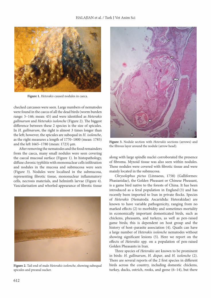
Since all the dead bird’s caeca had gross nodules on the mucosal surface (Figure 1), several tissue samples were taken from the caeca and placed in 10% formalin for histopathology examination. Sections were stained with hematoxylin and eosin stain.
3. Results and discussionIn dissection, all the carcasses had swollen caeca in gross (Figure 1). Numerous nematodes were found free and attached to mucosa in all the caeca. Nematodes were collected macroscopically and then under a stereomicroscope. Gross existence of nodules on the mucosal surface of caeca was also noticed and no signs of bacterial infections, pus, or any other parasites in the
Abstract: Following deaths in a flock of Golden Pheasants, Chrysolophus pictus, in Mazandaran Province, northern Iran, during June 2008, necropsies of dead birds were undertaken and revealed a heavy mixed infection of Heterakis isolonche and H. gallinarum in the caeca, which were heavily swollen with gross nodules on the surface. Infected caeca were studied histopathologically and surviving birds were treated with fenbendazole 2.5% (20 mg/kg), with a second course 3 weeks later. No further mortality was seen in the flock. This is the first documented report of Heterakis isolonche in Iran, and also the first report of a concurrent infection of 2 species of Heterakis in birds in Iran.
Key words: Heterakis isolonche, Heterakis gallinarum, Iran, pheasant
Received: 25.06.2012 Accepted: 12.01.2013 Published Online: 26.08.2013 Printed: 20.09.2013
Case Report
612
HALAJIAN et al. / Turk J Vet Anim Sci
checked carcasses were seen. Large numbers of nematodes were found in the caeca of all the dead birds (worm burden range: 5–146; mean: 45) and were identified as Heterakis gallinarum and Heterakis isolonche (Figure 2). The biggest difference between these 2 species is the size of spicules. In H. gallinarum, the right is almost 3 times longer than the left; however, the spicules are subequal in H. isolonche, as the right measures a length of 1770–1800 (mean: 1785) and the left 1665–1780 (mean: 1723) µm.
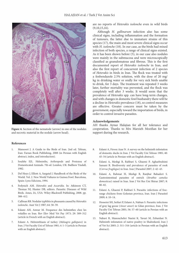
After removing the nematodes and the food remainders from the caeca, many small nodules were seen covering the caecal mucosal surface (Figure 1). In histopathology, diffuse chronic typhlitis with mononuclear cells infiltration and nodules in the mucosa and submucosa were seen (Figure 3). Nodules were localised in the submucosa, representing fibrotic tissue, mononuclear inflammatory cells, necrosis materials, and helminth larvae (Figure 4). Vascularisation and whorled appearance of fibrotic tissue
along with large spindle nuclei corroborated the presence of fibroma. Myxoid tissue was also seen within nodules. These nodules were covered with fibrotic tissue and were mainly located in the submucosa.
Chrysolophus pictus (Linnaeus, 1758) (Galliformes: Phasianidae), the Golden Pheasant or Chinese Pheasant, is a game bird native to the forests of China. It has been introduced as a feral population in England (3) and has recently been imported to Iran in private flocks. Species of Heterakis (Nematoda: Ascaridida: Heterakidae) are known to have variable pathogenicity, ranging from no marked effects (2) to morbidity and sometimes mortality in economically important domesticated birds, such as chickens, pheasants, and turkeys, as well as pen-raised game birds; this is dependent on host group and the history of host–parasite association (4). Quails can have a large number of Heterakis isolonche nematodes without showing significant lesions (5). Here we report on the effects of Heterakis spp. on a population of pen-raised Golden Pheasants in Iran.
Three species of Heterakis are known to be prominent in birds: H. gallinarum, H. dispar, and H. isolonche (2). There are several reports of the 2 first species in different birds across the country, including domestic chickens, turkey, ducks, ostrich, rooks, and geese (6–14), but there
Figure 1. Heterakis caused nodules in caeca.
Figure 2. Tail end of male Heterakis isolonche, showing subequal spicules and preanal sucker.
Figure 3. Nodule section with Heterakis sections (arrows) and the fibrous layer around the nodule (arrow head).
613
HALAJIAN et al. / Turk J Vet Anim Sci
are no reports of Heterakis isolonche even in wild birds (9,10,15,16).
Although H. gallinarum infection also has some clinical signs, including inflammation and the formation of tumours, the latter due to immature strains of this species (17), the main and most severe clinical signs occur with H. isolonche (18). In our case, as the birds had mixed infection of both species, a range of clinical signs existed. As it has been shown before (5), in our case also nodules were mainly in the submucosa and were microscopically classified as granulomatous and fibrous. This is the first documented report of Heterakis isolonche in Iran, and also the first report of concurrent infection of 2 species of Heterakis in birds in Iran. The flock was treated with a fenbendazole 2.5% solution, with the dose of 20 mg/kg in drinking water or orally for very sick birds unable to drink, for 3 days. The treatment was repeated 3 weeks later, further mortality was prevented, and the flock was completely well after 3 weeks. It would seem that the prevalence of Heterakis spp. can have long-term changes, and with changes in domestic fowl husbandry there will be a decline in Heterakis prevalence (18), so control measures are effective. Greater concern must be taken by the government, especially toward the importation of birds, in order to control invasive parasites.
AcknowledgementsAH thanks Aynaz Halajian for all her tolerance and cooperation. Thanks to Mrs Marzieh Mozifian for her support during the research.
Figure 4. Section of the nematode (arrow) in one of the nodules and necrotic material in the nodule (arrow head).
References
1. Mansoori J. A Guide to the Birds of Iran. 2nd ed. Tehran, Iran: Farzan Book Publishing, 2008 (in Persian with English abstract, index, and introduction).
2. Soulsby EJL. Helminths, Arthropods and Protozoa of Domesticated Animals. 7th ed. London, UK: Bailliere Tindall, 1982.
3. Del Hoyo J, Elliott A, Sargatal J. Handbook of the Birds of the World. Vol. 2. New World Vultures to Guinea Fowl. Barcelona, Spain: Lynx Edicions, 1994.
4. Fedynich AM. Heterakis and Ascaridia. In: Atkinson CT, Thomas NJ, Hunter DB, editors. Parasitic Diseases of Wild Birds. Ames, IA, USA: Wiley-Blackwell Publishing, 2008. pp. 388–412.
5. Callinan RB. Nodular typhlitis in pheasants caused by Heterakis isolonche. Aust Vet J 1987; 64: 58–59.
6. Eslami AH, Anwar M. Frequence des helminthes chez les volailles en Iran. Rev Elev Med Vet Pay 1973; 29: 309–312 (article in French with an English abstract).
7. Eslami A. Helminthiasis of turkey (Meleagris gallopavo) in Iran. J Vet Faculty Uni of Tehran 1981; 4: 1–5 (article in Persian with an English abstract).
8. Eslami A, Firooz Azar N. A survey on the helminth infestation of domestic ducks in Iran. J Vet Faculty Uni Tehran 1985; 40: 45–54 (article in Persian with an English abstract).
9. Eslami A, Meshgi B, Rahbari S, Ghaemi P, Aghaebrahimi Samani R. Biodiversity and prevalence of parasites of rook (Corvus frugilegus) in Iran. Iran J Parasitol 2007; 2: 42–43.
10. Eslami A, Rahmat H, Meshgi B, Ranjbar Bahadori S. Gastrointestinal parasites of ostrich (Struthio camelus domesticus) raised in Iran. Iran J Vet Res Uni Shiraz 2007; 8: 80–82.
11. Eslami A, Ghaemi P, Rahbari S. Parasitic infections of free-range chickens from Golestan province, Iran. Iran J Parasitol 2009; 4: 10–14.
12. Hosseini SH, Seifuri P, Eslami A, Nabian S. Parasitic infections of gray lag goose (Anser anser) in Gilan province, Iran. J Vet Faculty Uni Tehran 2001; 56: 57–60 (article in Persian with an English abstract).
13. Nabavi R, Manouchehri Naeini K, Yavari M, Zebardast N. Helminth infestation of native poultry in Shahrekord, Iran J of Vet Sci 2005; 2: 311–316 (article in Persian with an English abstract).
614
HALAJIAN et al. / Turk J Vet Anim Sci
14. Nabavi R, Abdollah-Poor M, Abdi Zadeh R. Study on the gastrointestinal helminths of native fowls of Gatvand, Khuzestan, Iran. In: The 6th National and the First Regional Congress on Parasitology and Parasitic Diseases. Karaj, Iran: NRCP; 2008.
15. Eslami A, Moradi M, Rahbari S, Meshgi B. Parasites in woodpeckers (Jynx torquilla, Dendrocopos syriacus) of Iran. J Vet Parasitol 2004; 18: 85–86.
16. Halajian A, Eslami A, Mobedi I, Amin O, Mariaux J, Mansoori J, Tavakol S. Gastrointestinal helminths of magpies (Pica pica), rooks (Corvus frugilegus) and carrion crows (Corvus corone) in Mazandaran province, north of Iran. Iran J Parasitol 2011; 6: 38–44.
17. Menezes RC, Tortelly R, Gomes DC, Pinto RM. Nodular typhlitis associated with the nematode Heterakis gallinarum and Heterakis isolonche in pheasants: frequency and pathology with evidence of neoplasia. Mem I Oswaldo Cruz 2003; 98: 1011–1016.
18. Griner LA, Migaki G, Penner LR, McKee AE. Heterakidosis and nodular granulomas caused by Heterakis isolonche in the ceca of gallinaceous birds. Vet Pathol 1977; 14: 582–590.
19. Potts GR. Long-term changes in the prevalence of caecal nematodes and histomonosis in gamebirds in the UK and the interaction with poultry. Vet Rec 2009; 164: 715–718.