Embed Size (px)
Citation preview
333
The Fine Structure and Morphological Organization of the
Peripheral Nerve-fibres and Trunks of the Cockroach(Periplaneta americana)
By ARTHUR HESS
(From the Department of Anatomy, Washington University School of Medicine, St. Louis,Missouri, U.S.A.)
With three plates (figs, i to 3)
SUMMARY
Sections of the peripheral nerve-trunks of the metathoracic leg of the cockroach(Periplaneta americana) were studied with the electron microscope. Paraffin sectionswere also prepared and stained. Protargol succeeds in staining the nerve-fibres.Osmium tetroxide, a modified Weigert procedure, and Luxol fast blue stain the myelinsheaths, as does mercuric bromphenolblue, a protein stain. The axoplasm is relativelyfree of formed elements; it contains mitochondria. The myelin sheath, when presenton the largest and also some smaller fibres, consists of about two or three loose over-lapping processes of Schwann cells, covered by their plasma membranes, enclosinglipid-like droplets and having a beaded appearance. Between the nerve-fibres in thenerve-trunk is Schwann-cell cytoplasm, which arises from Schwann cells that sur-round the whole nerve-trunk. The same fold of Schwann-cell membrane may enterinto the formation of the myelin sheath around more than one nerve-fibre. Severalsmall non-myelinated fibres, which may be as small as 0-3 n in diameter or less, maybe enclosed in the same fold of Schwann-cell membrane. Outside of the Schwann-celllayer and surrounding the nerve-trunk is a thin layer of connective tissue, which doesnot send trabeculae into the interior of the nerve. Tracheae and tracheoles accompanythe nerve but are not included within the sheaths surrounding a nerve-trunk, evennear the termination of the nerve-fibres in muscle. The structure of the cockroachperipheral nerve is compared with that described by previous investigators, with thatof other insects, and with invertebrate and vertebrate nerve.
INTRODUCTION
MUCH information has recently been accumulated on the physiologicalmechanisms of nerve and muscle in insects (Hoyle, 1954, a, b; 1957).
Progress in knowledge of the structure of the nerves of insects has not yetparalleled the advances in physiological information. Pringle (1939) has con-tributed much information on the anatomy and physiology of the leg musclesand peripheral nerve of the cockroach. In the present investigation, the struc-ture of the peripheral nerve-trunks and fibres of the cockroach has beeninvestigated by staining techniques for the light-microscope and by electronmicroscopy. Some information has been obtained about the ultrastructure ofthe nerve-fibres and the relations of the nerve-fibres to each other and theirsurrounding sheaths.
[Quarterly Journal of Microscopical Science, Vol. 99, part 3, pp. 333-340, Sept. 1958.]2421.3 Z
334 Hess—Peripheral Nerve-fibres of the Cockroach
MATERIAL AND METHODS
The metathoracic leg of Periplaneta americana was used. Whole legs, piecesof leg, and pieces of muscle were fixed in alcohol of varying concentrations,osmium tetroxide, or 50% alcohol with 3 g of chloral hydrate per 100 ml.Cross and longitudinal paraffin sections were prepared. Protargol stains andhaematoxylin and eosin were used on the tissues fixed in alcohol. The tissuesfixed in alcohol / chloral hydrate were stained by a Cajal block impregnationprocedure. Sections were also stained for myelin by a modified Weigert stain(Erhart, 1951) and by Luxol fast blue (Kliiver and Barrera, 1953), a sulphonatedcopper salt of phthalocyanine, generously donated by E. I. du Pont deNemours & Co. Attempts were also made to impregnate nerve-fibres byformic acid / gold chloride techniques. In addition, the mercuric bromphenolblue stain, a procedure specific for proteins (Mazia and others, 1953), wasapplied to the paraffin sections. Nerve-fibres were also examined under thepolarizing microscope.
For electron microscopy, small pieces of metathoracic leg or of musclewere fixed for 10-30 min in Dalton's fluid (Dalton and Felix, 1955), a solutionwhich contains 1% OsO4, 1% K2Cr2O7 at pH 7-2, and 0-85% NaCl. Afterdehydration, specimens were embedded in methyl (1 part) and butyl (6 parts)methacrylate and ultrathin cross and longitudinal sections (200-300 A thick)were cut with the Servall Porter-Blum microtome. The sections were insertedinto an RCA-EMU-type electron microscope. Micrographs were taken ata magnification of 2,000 to 6,000 and enlarged to the desired size. The finalmagnifications are approximate. Thick plastic sections were cut and viewedunder the phase-contrast microscope to provide orientation for the subsequentelectron microscopy.
RESULTSAxoplasm
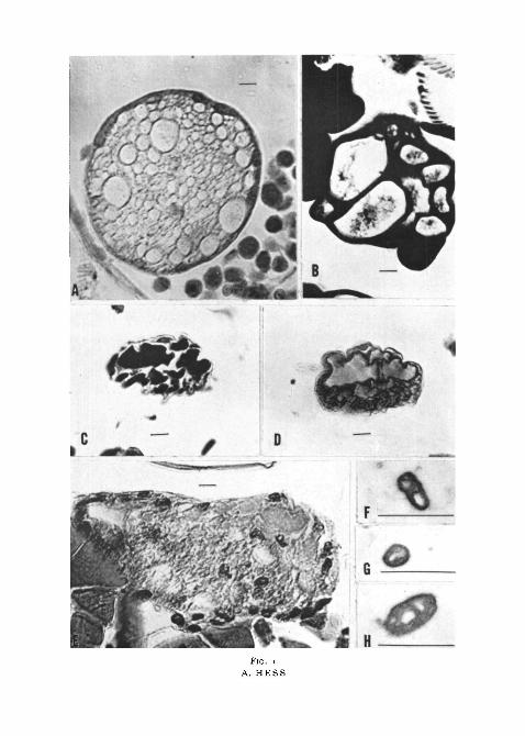
The diameters of the nerve-fibres encountered in cockroach nerve varyfrom about 10/LI to about 0-3 /JL or even less. The axons of insect nerve-fibresare usually described as structureless. After the treatment of sections withosmium tetroxide (fig. 1, B) or after gold or silver impregnation, the axonusually remains unstained. At times a granular precipitate can be seen. Afterstaining with mercuric bromphenol blue, the axoplasm is coloured a pale blue(fig. 1, D). Protargol succeeds in impregnating the axons. They are coloured
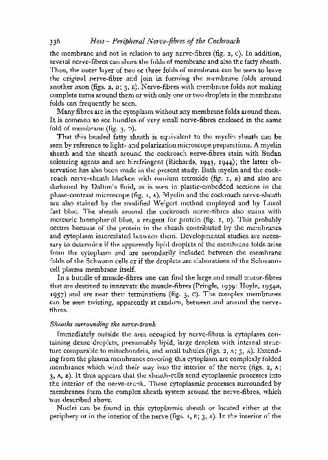
FIG. 1 (plate). Sections of cockroach nerve. In A-E (photomicrographs) the scale represents10n; in F-H (electron micrographs) it represents 1 p. Magnifications are approximate.
A, fixed in Dalton's fixative, embedded in plastic. The sheath around the individual nerve-fibres is seen.
B, fixed in osmium tetroxide solution. The sheath around individual nerve-fibres isblackened.
C, protargol preparation. The axons are impregnated and have a crumpled border.D, stained with mercuric bromophenol blue. The axoplasm was pale blue. The sheaih
around individual nerve-fibres was also stained.E, silver impregnation, to show the nuclei in the nerve.F-G, mitochondria within the nerve-fibres.
sch
Hess—Peripheral Nerve-fibres of the Cockroach 335
dark blue or black and frequently appear to lose their round shape and havea crumpled border (fig. 1, c).
Electron micrographs reveal the elements present in axons. The mostobvious structures seen are electron-dense bodies (figs. 2, A; 3, A) having aninternal structure of folded membranes and frequently having round areasof light density (figs. 1, F, G, H). These dark bodies may be mitochondria. Incross-sections of nerve, they usually appear round, which indicates that theyare longitudinally oriented in the axon. The rest of the axoplasm usuallypresents an amorphous granular appearance (figs. 2, A; 3, A). In cross-sections, some granules have an interior less dense than the periphery, whichindicates that they are small tubules longitudinally oriented in the axoplasm.In general, the rather sparse content of organelles and fibrils and light densityof the axoplasm are striking, especially when compared with the axons ofvertebrates.
Sheaths of the nerve-fibres
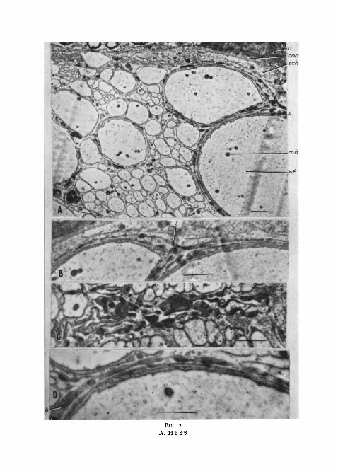
The nerve-fibres rest in the cytoplasm of Schwann cells which enclose thewhole nerve-trunk, a relationship that will be described in more detail below.Processes of these cells, covered by their plasma membranes, twist and turnthrough the interior of the nerve-trunk. Some entwine themselves around anerve-fibre so that it may be surrounded by two or three loosely imbricatedprocesses with cytoplasm and its organelles enclosed between the plasmamembrane layers (figs. 2, A, B, D). Electron-dense droplets, apparently of alipid nature, are frequently seen. These are enclosed between the membranes.These droplets thus enter into the formation of a beaded fatty sheath aroundsome nerve-fibres (figs. 2, A, B, D), which may be equivalent to the myelinsheath of other forms. Most commonly, the largest nerve-fibres are surroundedby this beaded fatty sheath. However, this relationship is not invariableas it is not uncommon to see large nerve-fibres without any membrane foldsor lipid or to see small nerve-fibres surrounded by membrane folds andbeaded droplets. The membranes appear to course at random through theinterior of the nerve-trunk. At times lipid droplets can be seen enclosed in
FIG. 2 (plate). Electron micrographs of cockroach nerve. The scales represent i ft. Magni-fications are approximate.'auuiis cue tip^iuAuiiaic.
A, cross-section of cockroach nerve-trunk. The axoplasm has an amorphous granular
lipid nature. The wall of a trachea is at the top of the photograph.B, enlargement of A showing the beaded membrane folds, the enclosed cytoplasm, and
droplets forming a sheath on one nerve-fibre and passing on to form a sheath on anothernerve-fibre.
C, section of a nerve-trunk showing folds of membrane with droplets and not surroundingor forming a sheath on any nerve-fibre.
D, section showing the membrane folds enclosing cytoplasm around a nerve-fibre and thebeaded sheath of the nerve-fibre.
b, beaded sheath of the nerve-fibre; con, connective tissue layer; /, beaded membrane foldpassing on from one nerve-fibre to the other; mit, mitochondrion; n, nucleus of wall oftrachea; nf, nerve-fibre; s, sheath of nerve-fibre; sch, cytoplasm of Schwann cell.
336 Hess—Peripheral Nerve-fibres of the Cockroach
the membrane and not in relation to any nerve-fibres (fig. 2, c). In addition,several nerve-fibres can share the folds of membrane and also the fatty sheath.Thus, the outer layer of two or three folds of membrane can be seen to leavethe original nerve-fibre and join in forming the membrane folds aroundanother axon (figs. 2, B; 3, E). Nerve-fibres with membrane folds not makingcomplete turns around them or with only one or two droplets in the membranefolds can frequently be seen.
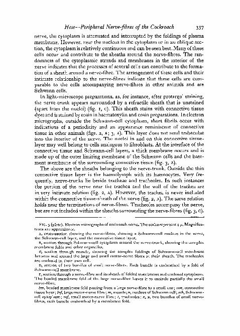
Many fibres are in the cytoplasm without any membrane folds around them.It is common to see bundles of very small nerve-fibres enclosed in the samefold of membrane (fig. 3, D).
That this beaded fatty sheath is equivalent to the myelin sheath can beseen by reference to light- and polarization microscope preparations. A myelinsheath and the sheath around the cockroach nerve-fibres stain with Sudancolouring agents and are birefringent (Richards, 1943, 1944); the latter ob-servation has also been made in the present study. Both myelin and the cock-roach nerve-sheath blacken with osmium tetroxide (fig. 1, B) and also aredarkened by Dalton's fluid, as is seen in plastic-embedded sections in thephase-contrast microscope (fig. 1, A). Myelin and the cockroach nerve-sheathare also stained by the modified Weigert method employed and by Luxolfast blue. The sheath around the cockroach nerve-fibres also stains withmercuric bromphenol blue, a reagent for protein (fig. 1, D). This probablyoccurs because of the protein in the sheath contributed by the membranesand cytoplasm intercalated between them. Developmental studies are neces-sary to determine if the apparently lipid droplets of the membrane folds arisefrom the cytoplasm and are secondarily included between the membranefolds of the Schwann cells or if the droplets are elaborations of the Schwann-cell plasma membrane itself.
In a bundle of muscle-fibres one can find the large and small motor-fibresthat are destined to innervate the muscle-fibres (Pringle, 1939; Hoyle, 1954a,1957) and are near their terminations (fig. 3, c). The complex membranescan be seen twisting, apparently at random, between and around the nerve-fibres.
Sheaths surrounding the nerve-trunk
Immediately outside the area occupied by nerve-fibres is cytoplasm con-taining dense droplets, presumably lipid, large droplets with internal struc-ture comparable to mitochondria, and small tubules (figs. 2, A; 3, A). Extend-ing from the plasma membranes covering this cytoplasm are complexly foldedmembranes which wind their way into the interior of the nerve (figs. 2, A;3, A, B). It thus appears that the sheath-cells send cytoplasmic processes intothe interior of the nerve-trunk. These cytoplasmic processes surrounded bymembranes form the complex sheath system around the nerve-fibres, whichwas described above.
Nuclei can be found in this cytoplasmic sheath or located either at theperiphery or in the interior of the nerve (figs. 1, E; 3, A). In the interior of the
Hess—Peripheral Nerve-fibres of the Cockroach 337
nerve, the cytoplasm is attenuated and interrupted by the foldings of plasmamembrane. However, near the nucleus in the cytoplasm or in an oblique sec-tion, the cytoplasm is relatively continuous and can be seen best. Many of thesecells occur and contribute to the sheaths around the nerve-fibres. The ran-domness of the cytoplasmic strands and membranes in the interior of thenerve indicates that the processes of several cells can contribute to the forma-tion of a sheath around a nerve-fibre. The arrangement of these cells and theirintimate relationship to the nerve-fibres indicate that these cells are com-parable to the cells accompanying nerve-fibres in other animals and areSchwann cells.
In light-microscope preparations, as, for instance, after protargol staining,the nerve-trunk appears surrounded by a refractile sheath that is unstained(apart from the nuclei) (fig. 1, c). This sheath stains with connective tissuedyes and is stained by eosin in haematoxylin and eosin preparations. In electronmicrographs, outside the Schwann-cell cytoplasm, short fibrils occur withindications of a periodicity and an appearance reminiscent of connectivetissue in other animals (figs. 2, A; 3, A). This layer does not send trabeculaeinto the interior of the nerve. The nuclei in and on this connective tissue-layer may well belong to cells analogous to fibroblasts. At the interface of theconnective tissue and Schwann-cell layers, a thick membrane occurs and ismade up of the outer limiting membrane of the Schwann cells and the base-ment membrane of the surrounding connective tissue (fig. 3, A).
The above are the sheaths belonging to the nerve-trunk. Outside the thinconnective tissue layer is the haemolymph with its haemocytes. Very fre-quently, nerve-trunks lie beside tracheae and tracheoles. In such instancesthe portion of the nerve near the trachea and the wall of the trachea arein very intimate relation (fig. 2, A). However, the trachea is never includedwithin the connective tissue-sheath of the nerve (fig. 2, A). The same relationholds near the terminations of nerve-fibres. Tracheoles accompany the nerve,but are not included within the sheaths surrounding the nerve-fibres (fig. 3, c).
FIG. 3 (plate). Electron micrographs of cockroach nerve. The scales represent I y.. Magnifica-tions are approximate.
A, cross-section showing the nerve-fibres, showing a Schwann-cell nucleus in the nerve,the Schwann-cell layer, and the connective tissue layer.
B, section through Schwann-cell cytoplasm around the nerve-trunk, showing the complexmembrane folds and other organelles.
c, section through muscle, showing the complex foldings of Schwann-cell membranebetween and around the large and small motor-nerve fibres in their sheath. The tracheolesare enclosed in their own cell.
D, section of two bundles of small nerve-fibres. Each bundle is ensheathed by a fold ofSchwann-cell membrane.
E, section through a nerve-fibre and its sheath of folded membranes and enclosed cytoplasm.The beaded membrane fold of the large nerve-fibre leaves it to encircle partially the smallnerve-fibre.
bm, beaded membrane fold passing from a large nerve-fibre to a small one; con, connectivetissue layer; Inf, large motor-nerve fibre; m, muscle; n, nucleus of Schwann cell; sch, Schwann-cell cytoplasm; snf, small motor-nerve fibre; t, tracheoles; x, x, two bundles of small nerve-fibres, each bundle ensheathed by a membrane fold.
338 Hess—Peripheral Nerve-fibres of the Cockroach
DISCUSSION
The description of the structure of cockroach nerve in electron micrographsconforms very well to that of previous investigators who employed light- andpolarization microscopy. The structure of the sheath around the individualnerve-fibres described in the present investigation may well be responsible forthe optical properties of this sheath, as described by Richards (1944). Thatthe protein of the individual nerve-sheaths of insects may be collagenous(Richards, 1944) seems to be incorrect.
The sheaths around the nerve-trunk also require comment. The nerve-trunk is usually described as ensheathed by an outer homogeneous layercalled the 'neural lamella' and an inner cellular sheath denoted as the 'peri-lemma' (Hoyle, 1952). The 'perilemma' is equivalent to the Schwann-celllayer and the 'neural lamella' appears to be the connective tissue-sheath.Richards (1944) suggests that the neural lamella does not seem to be collagen'since it does not swell, dissolve or even lose its birefringence in dilute aceticacid (3 days) and since immersion experiments give different results for theneural lamella and the presumably collagenous sheaths around individualnerves'. However, as was mentioned above, the sheaths around individualnerve-fibres are probably not collagenous; the neural lamella appears to be so.The descriptions of the nerve-sheaths of insects by Scharrer (1939) andTwarog and Roeder (1956) are restricted to the ganglia. Contrary to generalbelief, the composition of the sheaths on the ganglia and those around thenerves differ (Hess, 1958); hence discussion of the sheaths around the gangliawill be reserved.
There appear to be many differences in structure between the peripheralnerve of locusts, as described by Hoyle (1954a), and that of cockroaches, asdescribed here. The locust nerve apparently has some kind of connectivetissue in its interior, the cockroach has Schwann-cell cytoplasm between itsnerve-fibres. The locust nerve-trunk is surrounded by a tracheolated mem-brane and a fatty envelope, that of the cockroach is not. Hoyle (1957) includesa trachea within the outer sheath of locust nerve near a motor end-plate.Although I have not yet studied neuromuscular endings, the tracheoles nearnerve terminations are not included within the outer sheaths of the nerve.Whether these differences in structure between locust and cockroach nerveare real or whether they will break down upon more detailed study remainsto be seen.
In lobster and squid nerve-fibres, osmiophil dense-edged layers, similar tothe membrane foldings of the Schwann cell described here in insect nerve,occur at the axon interface and in the cytoplasm of the Schwann cell (Gerenand Schmitt, 1955). However, the insect nerve usually has in addition lipid-like droplets enclosed or intercalated between the membrane folds of Schwanncell that surround the nerve-fibres; thus the insect myelin sheath is commonlybeaded in appearance.
The differences in the relation between cockroach nerve-fibres and Schwann
Hess—Peripheral Nerve-fibres of the Cockroach 339
cells and between vertebrate axons and Schwann cells are interesting. Invertebrate nerves, each myelinated nerve-fibre is enclosed by a Schwann cell,while several non-myelinated fibres share a Schwann cell (Gasser, 1955; Hess,1956). In the cockroach, Schwann cells surround the whole nerve-trunk.Each vertebrate non-myelinated fibre is suspended in the cytoplasm of thesame Schwann cell by a mesentery of Schwann-cell membrane called a'mesaxon' (Gasser, 1955; Hess, 1956). The 'mesaxon' of insect nerve-fibrescan surround several small non-myelinated fibres, which are included as abundle in the same Schwann-cell membrane. The myelin sheath of mammaliannerve-fibres consists of concentric lamellae wrapped around the axon (Hessand Lansing, 1953). It has been suggested that these lamellae are formed bywrappings of the Schwann-cell membrane (Geren, 1954). The insect fattysheath is also apparently formed by wrappings of the Schwann-cell membrane,which form lamellae on the nerve-fibre. The insect lamellae are not so tightlypacked and cytoplasm intervenes between them. In this respect, the insectfatty sheath bears a striking resemblance to a developing immature vertebratemyelin sheath. The fatty sheath of insects includes droplets apparently ofa lipid nature in addition to the wrappings of Schwann-cell membrane and isbeaded in appearance. The lamellae of the vertebrate myelin sheath are of eventhickness and consist only of wrappings of the Schwann-cell membrane. Thewrappings of the Schwann-cell membrane in vertebrates are restricted toindividual nerve-fibres so that most commonly two myelinated axons do notshare a myelin sheath and are not included within the same Schwann cell,although rarely the latter has been seen to occur (Hess, 1956). In insects,several myelinated and non-myelinated fibres share the same Schwann cellsand the wrappings of the same Schwann-cell membrane can frequently beseen to leave one nerve-fibre and join in the formation of the beaded fattysheath of other fibres, so that several nerve-fibres can be seen to share in thewrappings of the same Schwann-cell membrane and fatty sheath. The appar-ent randomness of the foldings of the Schwann-cell membrane in insect nerveis striking. Perhaps a study of the development of insect peripheral nerve-trunks will reveal that the sheath system of their constituent nerve-fibres isnot as random as it appears in mature forms.
The sheaths of the cockroach nerve-trunk and those of vertebrate nerve arecomparable. The connective tissue layer surrounding the whole nerve-trunk•of the cockroach nerve is analogous to that of the epineurium of vertebratenerves. However, this layer does not send trabeculae into the interior of thenerve and hence, at least for cockroach nerve, there is no peri- or endo-neurium. The thick membrane at the interface of Schwann-cell and connectivetissue layers and composed of the outer limiting membrane of the Schwanncells and the basement membrane of the connective tissue is reminiscent ofthe neurilemma or Schwann sheath of vertebrate nerve, which is made up ofsimilar membranous layers (Hess, 1956). There are also close functionalparallels in the sheaths investing insect and vertebrate nerve tissue (Hoyle,1953; Twarog and Roeder, 1956).
34° Hess—Peripheral Nerve-fibres of the Cockroach
I wish to thank Mrs. Dorothy Goldstein for her aid in preparation of slidesand Dr. C. N. Sun for his assistance in all phases of electron microscopy.Dr. A. J. De Lorenzo also very kindly consented to take some of the electronmicrographs.
REFERENCES
DALTON, A. J., and FELIX, M.D., 1955. In Fine structure of cells, p. 274. Groningen (Noordhoff).ERHART, E. A., 1951. Z. wiss. Mikr., 60, 155.GASSER, H. S., 1955. J. Gen. Physiol., 38, 709.GEREN, B. B., 1954. Exp. Cell Res., 7, 558.
and SCHMITT, F. O., 1955. In Fine structure of cells, p. 251. Groningen (Noordhoff).HESS, A., 1956. Proc. Roy. Soc. B, 144, 496.
1958. In preparation.and LANSING, A. I., 1953. Anat. Rec, 117, 175.
HOYLE, G., 1952. Nature, 169, 281.J953. J- exp. Biol., 30, 121.1954a. Proc. Roy. Soc. B, 143, 281.19546. Ibid., 143, 343.1957. In Recent advances in invertebrate physiology, p. 73. Eugene (University of Oregon
Publications).KLOVER, H., and BARRERA, E., 1953. J. Neuropath, exp. Neurol., 12, 400.MAZIA, D., BREWER, P. A., and ALFERT, M., 1953. Biol. Bull., 104, 57.PRINGLE, J. W. S., 1939. J. exp. Biol., 16, 220.RICHARDS, A. G., 1943. J. N.Y. Ent. Soc., 51, 55.
1944. Ibid., 52, 285.SCHARRER, B. C. J., 1939. J. comp. Neurol., 70, 77.TWAROG, B. M., and ROEDER, K. D., 1956. Biol. Bull., 111, 278.