Embed Size (px)
Citation preview

London, Berlin, Chicago, Tokyo, Barcelona, Beijing, Istanbul, Milan, Moscow,New Delhi, Paris, Prague, São Paulo, Seoul, Singapore and Warsaw
The FacePictorial Atlas of Clinical Anatomy
Ralf J. Radlanski Karl H. Wesker
The_Face.indb 3 05.09.2012 16:28:14

Throughout an individual’s lifetime, there is a fateful connec-tion between the face and the person. With the face, we rec-ognize each other; we communicate with our facial expression. When we meet for the first time, it is the face that makes the first, unrepeatable visual impression.
We dress ourselves according to our mood, and according to the role we intend to play. In order to signal a certain identity, we are set, however, with the appearance of our face.
Purely morphologically, each face shows very individual char-acteristics and proportions.
Faces differ in countless nuances in relation to skin character-istics, the form and color of the eyes, the eye spacing, the out-line of the eyebrows, the prominence of the cheeks, the con-tour of the nose, and the cut of the mouth and the chin, just to mention a few examples. From these features, not only Johann Caspar Lavater, the eighteenth century promoter of physi-ognomy, but also long before this Aristotle and peasant public opinion developed misguided theories of types.
Many people are not aware why they perceive a certain face as nice, friendly, personable and attractive – but not another one. However, the importance of our face is evident even at birth. There are studies that show that caresses of an infant by the mother are much more numerous and more intense when the mother finds her baby attractive.
So, very early, there is an intensive interaction between the ex-pectations and fulfillments to the response by people – or a deep disappointment.
Prologue: The Face
Moods are reflected in our facial expression; thus the face be-comes a mirror of our soul.
Things like the luster of the eyes can hardly be influenced by the individual but are undoubtedly perceived by those around. The tension and activity of the muscles of facial expression contribute to the overall impression. It is not surprising that a complete lifetime can be engraved into the face by the per-manent activity of the muscles of facial expression and, with this, the position, orientation and depth of the wrinkles and creases. Consequently, the face shows not only a spontaneous impression of the person but also a lived identity.
The interdependency between the face and the identity of a person has to be acknowledged not only by the individual but also by the therapist. In particular, severe malformations or traumatic facial damage, which cannot be hidden by clothing, causes the affected person to be mercilessly exposed during in-teractions with the world around. These individuals need the most advanced specialist efforts of medical science and tech-nology. However, individuals who feel that their person, their identity, is not or is no longer congruent with their face also require special medical attention. The face can be subtly modi-fied therapeutically, with a high degree of responsibility to bal-ance the self-perception and the desires against the feasible changes. The detailed knowledge of the anatomy of the face is one of the many foundations for such interventions. This is the use that is intended for this atlas.
The_Face.indb 9 05.09.2012 16:28:14

1 The face . . . . . . . . . . . . . . . . . . . . 1
1.1 Introduction . . . . . . . . . . . . . . . . . . . . 2
1.1.1 General remarks . . . . . . . . . . . . . . . . . . 2
1.1.2 Regions of the face . . . . . . . . . . . . . . . . 4
1.1.3 Morphometry and proportions of the face . . . . 6
1.2 The face in anterior view . . . . . . . . . . . . . . 18
1.2.1 Fat compartments of the face
in anterior view . . . . . . . . . . . . . . . . . . . 18
1.2.2 Muscles of the face in anterior view . . . . . . . . 25
1.2.3 Vascular and nerve supply of the face
in anterior view . . . . . . . . . . . . . . . . . . . 38
1.3 The face in lateral view . . . . . . . . . . . . . . . 54
1.3.1 Fat compartments of the face
in lateral view . . . . . . . . . . . . . . . . . . . . 54
1.3.2 Muscles of the face in lateral view . . . . . . . . . 62
1.3.3 Vascular and nerve supply of the face
in lateral view . . . . . . . . . . . . . . . . . . . . 78
1.4 The head in vertical view . . . . . . . . . . . . 106
1.5 The head in dorsal view . . . . . . . . . . . . . 116
1.6 The neck . . . . . . . . . . . . . . . . . . . . . 122
1.6.1 The neck in anterior view . . . . . . . . . . . . 123
1.6.2 The neck in lateral view . . . . . . . . . . . . . 128
1.6.3 The neck in dorsal view . . . . . . . . . . . . . 133
1.7 Facial expression . . . . . . . . . . . . . . . . . 136
1.8 The facial skeleton . . . . . . . . . . . . . . . . 148
1.9 Sectional anatomy . . . . . . . . . . . . . . . . 162
1.10 Schematic representations of pathways
in the face . . . . . . . . . . . . . . . . . . . . 172
2 The orbital region . . . . . . . . . . . . 180
2.1 Surface topography of the orbital region . . . . 182
2.2 Preseptal muscles and fat layers . . . . . . . . . 185
2.3 The orbital septum and the eyeball . . . . . . . 189
2.4 Vascular and nerve supply in the orbital region . . . 196
Contents
2.5 Vascular and nerve supply in the orbital region
in relation to the muscles . . . . . . . . . . . . 205
2.6 Sectional anatomy of the orbital region . . . . . 210
3 The nasal and midfacial region . . 216
3.1 Surface topography of the nasal region . . . . . 218
3.2 The nose in anterior view . . . . . . . . . . . . 222
3.3 The nose in lateral view . . . . . . . . . . . . . 230
3.4 The nose in caudal view . . . . . . . . . . . . . 244
3.5 The nasal cavity . . . . . . . . . . . . . . . . . 245
3.6 The sinuses . . . . . . . . . . . . . . . . . . . 258
4 The mouth . . . . . . . . . . . . . . . . . 262
4.1 Extraoral topography of the oral region . . . . . 264
4.2 Topographical anatomy of the oral region . . . . 265
4.3 Vascular and nerve supply of the oral region . . . 267
4.4 The oral cavity . . . . . . . . . . . . . . . . . . 278
4.5 Anatomy of the lips, teeth, periodontium
and alveolar bone in sections . . . . . . . . . . 283
4.6 The anterior oral vestibule . . . . . . . . . . . . 289
4.7 Anatomy in the area around the
mandibular ramus . . . . . . . . . . . . . . . . 290
4.8 The temporomandibular joint . . . . . . . . . . 303
4.9 Anatomy of the oral region in sections . . . . . 308
4.10 Pathways of odontogenic spread of infections . . 314
5 The ear . . . . . . . . . . . . . . . . . . . . 316
6 The skin and aging of the face . . . . 330
Appendix . . . . . . . . . . . . . . . . . . . . . 345
References . . . . . . . . . . . . . . . . . . . . . . . 347
Index . . . . . . . . . . . . . . . . . . . . . . . . . . 349
The_Face.indb 11 05.09.2012 16:28:15
Galeaaponeurotica
Occipitofrontalis,frontal belly
Procerus
Orbicularis oculi
ROOF (retroorbicularisoculi fat)
Levator labii superiorisalaeque nasi muscle
Nasalis
Zygomaticus minor
Levator labii superioris
Levator anguli oris
Zygomaticus major
Orbicularis oris
Masseter
Depressor labiiinferioris
Mentalis
Temporoparietal
SOOF (suborbicularisoculi fat)
ROOF (retroorbicularisoculi fat)
Zygomatic bone,zygomatic arch
Mandible
Parotid gland
Buccal fat pad(of Bichat)
Parotid duct
Submentalfat compartment
Glabella fat pad
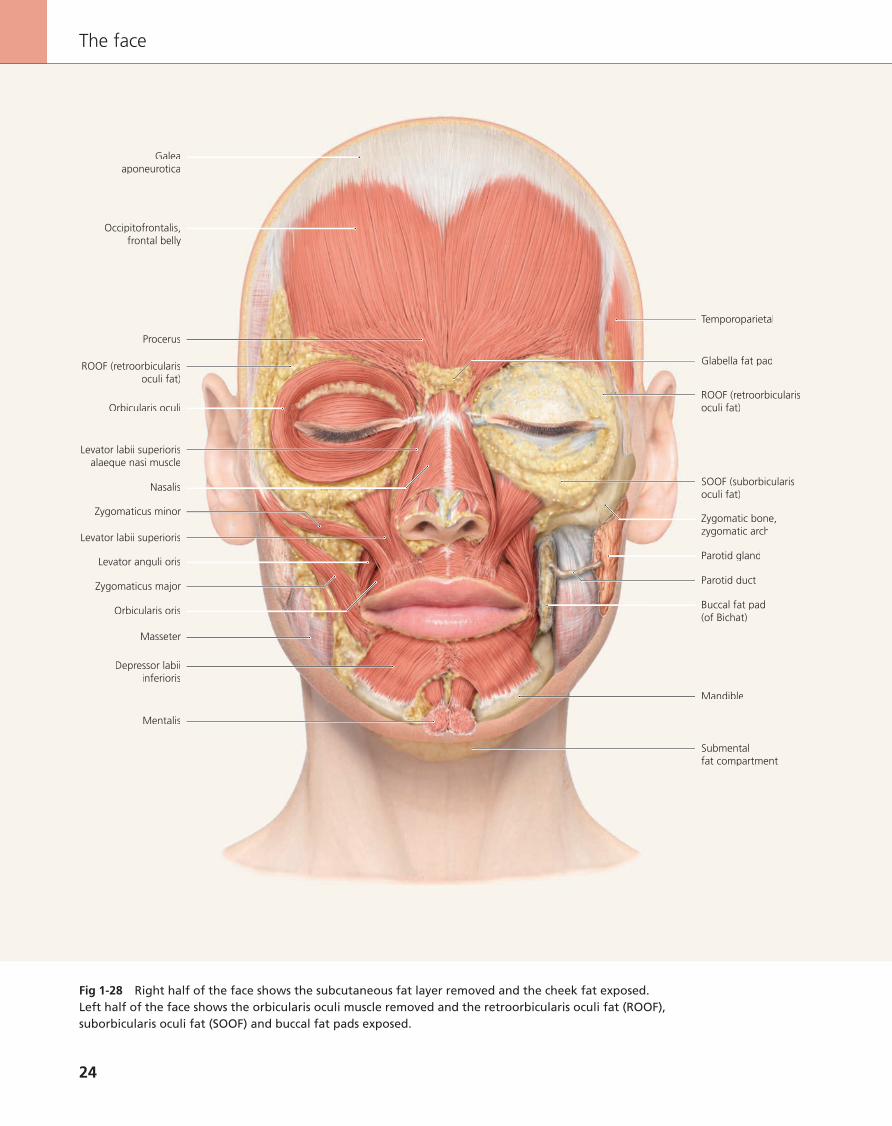
Fig 1-28 Right half of the face shows the subcutaneous fat layer removed and the cheek fat exposed. Left half of the face shows the orbicularis oculi muscle removed and the retroorbicularis oculi fat (ROOF), suborbicularis oculi fat (SOOF) and buccal fat pads exposed.
The face
24
The_Face.indb 24 05.09.2012 16:30:21

The relationship between the fat compartments and the facial muscles was the main topic in the previous section. In the fol-lowing figures, the facial muscles themselves will be addressed directly, starting again with the most superficial muscle layers of the face.
Fig 1-29 The left side of the face shows the superficial facial muscles. The occipitofrontalis muscle (seen here: frontal belly) is connected to the sturdy galea aponeurotica. Fiber tracts of the depressor supercilii muscles originate from the glabella re-gion and become concomitant with the muscle fibers in the eyebrow region. Some muscle fibers merge with the orbicu-laris oculi muscle. In the region of the glabella, the procerus muscle stretches out and corresponds with the fibers of the un-derlying occipitofrontalis muscle. The outer cartilaginous nasal skeleton is covered by the nasalis muscle, the anterior dilator naris muscle and the compressor narium minor muscle. In the border between the orbicularis oculi muscle and the nose, the levator labii superioris alaeque nasi muscle takes a narrow but long course. In the lower lip region, the orbicularis oris muscle is covered completely by the depressor anguli oris and depres-sor labii inferioris muscles. The upper lip is partly overlapped by the levator labii superioris alaeque nasi, levator labii su-perioris and zygomaticus minor muscles. In the corner of the mouth, the zygomaticus major muscle inserts together with the risorius muscle, with fibers that preferentially run horizon-tally. Still further posteriorly, some extensions of the platysma muscle run across the margin of the jaw. The tip of the chin is dominated by the mentalis muscle. Large parts of the lower cheek muscles and the temporal region are still covered with solid fascia. The chiasm of the facial muscles at the corners of the mouth is called the modiolus. It is formed by the orbicula-ris oris, buccinator, levator anguli oris, depressor anguli oris, zygomaticus major, risorius and platysma muscles.
Fig 1-30 As soon as the platysma, the risorius muscle and the fascia in the deep cheek region are removed in the right part of the face, the parotid gland, the parotid duct, the mas-seter muscle and the buccal fat pad (of Bichat) become ex-posed.
Fig 1-31 After removal of the peripheral portion of the or-bicularis oculi muscle in the left half of the face, the insertion of the levator anguli oris muscle in the maxilla becomes vis-ible. Furthermore, in the left half of the face, the zygomaticus minor and major muscles and the depressor anguli oris mus-cles are removed. As a result, the course of the parotic duct, which crosses over the masseter muscle, can be traced. Also, some parts of the mandible become visible.
Fig 1-32 In the left half of the face, the depressor supercilii muscle has been removed to expose some parts of the corru-
gator supercilii muscle. Although most parts of this muscle run underneath the frontal belly of the occipitofrontalis muscle, its fibers must eventually penetrate this muscle. The complete removal of the orbicularis oculi muscle exposes the orbital sep-tum. At its caudal margin, the infraorbital foramen becomes visible as soon as the levator labii superioris muscle has been elevated. This also allows the levator anguli oris muscle to be completely visible. Removal of the depressor labii inferius mus-cle exposes the lower lip portion of the orbicularis oris mus-cle. The fascia wrapping the parotid gland has also been re-moved.
Fig 1-33 When the temporal fascia is removed (left half of the face), the temporalis muscle becomes exposed. In addi-tion, the temporal process of the buccal fat pad becomes visi-ble. The chin region parts of the orbicularis oris muscle run un-derneath the depressor labii inferioris muscles and above the mentalis muscle.
Fig 1-34 The corrugator supercilii muscle runs underneath the frontal belly of the occipitofrontalis muscle. However, its fibers eventually penetrate the frontal belly in order to in-sert into the subcutaneous connective tissue. Portions of the procerus muscle, which runs on top of the frontal belly, have been kept visible in the left half of the face. Also in the left half of the face, the fascia of the masseter muscle has been re-moved.The parotid duct perforates the buccal fat pad and the buc-cinator muscle close to the anterior margin of the masseter muscle. The nasalis muscle, dorsal part, has been removed in the left half of the face to expose the upper lateral cartilage of the nose.
Fig 1-35 In the right half of the face, parts of the procerus muscle, which runs above the corrugator supercilii muscle, are preserved. All muscles that radiate into the perioral region, such as the levator anguli oris muscle (which still is visible in the right half of the face), have connections with the fibers of the orbicularis oris muscle.
Fig 1-36 The orbicularis oris and the buccinator muscles form a functional unit that embraces the oral cavity. As well as running around the oral cavity in a circular pattern, fibers of the orbicularis oris muscle also radiate into the buccinator muscle.
Fig 1-37 The oral vestibulum is formed by the buccinator muscle in the maxilla and mandible.
Fig 1-38 The right half of the face is shown with the bucci-nator muscle and gingiva maintained.
1.2.2 Muscles of the face in anterior view
The face in anterior view
25
The_Face.indb 25 05.09.2012 16:30:21

Articular capsule
Lateral ligament
Temporalis
Masseter
Body(of mandible)
Zygomatic arch
Infraorbitalforamen
Buccal fat pad(of Bichat)
Parotid duct
Orbicularis oris
Buccinator
Mental foramen
Mentalis
Fig 1-58 Zygomatic arch and masseter muscle partly removed to reveal the exten-sion of the buccal fat pad with its temporal process.
Fig 1-58 The continuity of the buccal fat pad into the tem-poral region becomes visible when the zygomatic arch and the masseter muscle are partly removed.
Fig 1-59 In the cheek region, all muscles are more or less tightly, but continuously, connected together and with the skin by means of interwoven connective tissue, the SMAS. From here, strands run toward the skin (false retaining liga-ments) and form the septa of the compartments. There are also strands of connective tissue that insert into bone; these are called true retaining fibers.
The connective tissue may contain some fat, the amount vary-ing from individual to individual.
This aponeurotic system is manipulated during facial cosmetic surgery, particularly rhytidectomy (facelift).
Fig 1-60 The SMAS is continuous with the facial muscles and allows facial expression. These connections between mus-cles and the connective tissue of the skin, or between muscles, are called false retaining ligaments.
Fig 1-61 There are also strands of connective tissue that in-sert into bone; they are called true retaining ligaments.
The face
60
The_Face.indb 60 05.09.2012 16:33:14

Parotid gland
Buccal branches(CN VII)
Accessoryparotid gland
Zygomatico-cutaneous ligament
Zygomaticusmajor and minor
Parotid duct
Platysma
Fig 1-60 False retaining ligaments of the superficial musculo-aponeurotic system.
Fig 1-61 True retaining ligaments of the superficial musculo-aponeurotic system.
Fig 1-59 Detail of the superficial musculo-aponeurotic system (SMAS) in the cheek region.
The face in lateral view
61
The_Face.indb 61 05.09.2012 16:33:23

Frontal boneFrontal bone
ProcerusProcerus
Nasal boneNasal bone
Levator labii superiorisLevator labii superiorisalaeque nasi musclealaeque nasi muscle
OrbicularisOrbicularisoculi
MedialMedialnasal conchanasal concha
InferiorInferiornasal conchanasal concha
Angular a. and v.Angular a. and v.
MaxillaMaxilla
LevatorLevatoranguli orisanguli oris
TongueTongue
Levator labiisuperioris
Depressoranguli oris
Orbicularisoris
Facial v.
Perpendicularlamina
Corrugatorsupercilii
Fig 3-54 Frontal section through the nasal cavity at the level of the incisor teeth.
Fig 3-54 Frontal section through the nasal cavity at the level of the incisor teeth.
Fig 3-55 Frontal section through the nasal cavity at the level of the first molar teeth.
Fig 3-56 Frontal section through the nasal cavity at the level of the second molar teeth.
Fig 3-57 Horizontal section through the nasal cavity at the level of the medial nasal concha.
Fig 3-58 Horizontal section through the nasal cavity at the level of the superior nasal concha.
Fig 3-59 Horizontal section through the nasal cavity at lower-eye level.
Fig 3-60 Horizontal section through the nasal cavity at mid-eye level.
The nasal and midfacial region
254
The_Face.indb 254 05.09.2012 16:48:12

Medial nasalconcha
Perpendicularlamina
Inferior nasalconcha
Tongue
Vomer
Maxillarysinus
Maxilla
Middle nasalmeatus
Ethmoidalcells
Medial nasalconcha
Perpendicularlamina
Olfactorybulb (CN I)
Inferior nasalconcha
Tongue
Vomer
Maxillarysinus
Maxilla
Crista galli
Ethmoidalcells
Fig 3-55 Frontal section through the nasal cavity at the level of the first molar teeth.Fig 3-56 Frontal section through the nasal cavity at the level of the second molar teeth.
255
The nasal cavity
The_Face.indb 255 05.09.2012 16:48:16

Fig 4-21 The mucosa at the ventral surface of the tongue is as thin as the one on the floor of the mouth. The thickness of the epithelium is only 0.2 mm. The blood vessels can be clearly seen through this thin epithelium; in some older individuals varicose veins may protrude.
Fig 4-22 The tongue is a body with varying motility that completely fills the space palatal of the dental arches when the mouth is closed. The tip of the tongue can reach almost every point of the oral mucosa. The dorsal mucosa of the tongue is completely different from the epithelium at the ven-tral side. The epithelium of the dorsum of the tongue is kerat-inized. Underneath is a tough lamina propria, rich in vessels and nerves. The superficial layer of the lamina propria carries a large number of papillae, which are covered by epithelium. According to their form and size, they are distinguished into thready (filiform papilla), mushroom-shaped (fungiform papil-lae), leaf-shaped (foliated papillae) and wall-like (circumvallate papillae) papillae. The papillae greatly enlarge the surface of the tongue. An enormous number of taste buds are embedded into the epithelium of the papillae, each taste bud bearing re-ceptors for specific taste sensations. Consequently, each taste is perceptible at any place of the tongue but there are maxima of specific flavor perception at certain regions of the tongue.
Fig 4-23 Much of the epithelium of the cheek is not kerati-nized. However, along a horizontal line, parallel to the occlusal plane, there can be a white line (linea alba) in some individu-als, which represents a line of keratinization of the epithelium. The cheek epithelium is the thickest epithelium of the oral mu-cosa (0.5–0.6 mm).
Fig 4-21 Sublingual mucosa.
Fig 4-22 Papillae of the tongue.
Fig 4-23 Cheek mucosa and pharyngeal mucosa.
The mouth
282
The_Face.indb 282 05.09.2012 16:49:27

Alveolarprocess
Alveolarpart
Glosso-alveolar sulcus
Genio-glossus
Maxilla
Orbicularisoris
Oralvestibule
Upper lip
Lower lip
Orbicularisoris
Oralvestibule
Body(of mandible)
Fig 4-24 Sagittal section through the anterior region of the mouth, slightly lateral of the midline.
Fig 4-24 The alveolar processes and the teeth are bordered by the tongue on the inside and the lips (and cheeks laterally) on the outside. The physiologically correct alignment of the upper to the lower incisors is an overlap of the incisal ridge of the upper incisor over that of the lower incisor (overbite). A bi-omechanically ideal support would be given if the lower incisal ridge rests at the transition between the palatal concavity and the convexity of the tubercle. This would result in an overlap
of the upper incisal ridge anterior to the labial surface of the lower incisor (overjet).
The angulation of the dental axis is influenced by the forces exercised by the tongue and the lips. Swallowing and speak-ing, however, is of minor importance and the permanent pres-sure exercised by the tongue and by the lips is more signifi-cant.
4.5 Anatomy of the lips, teeth, periodontium and alveolar bone in sections
283
Anatomy of the lips, teeth, periodontium and alveolar bone in sections
The_Face.indb 283 05.09.2012 16:49:31

A
Adipose tissue of the orbit 191, 211Agger nasi 250Alar– lobe 223ff, 235, 237, 240, 243f, 266– – lobular connective tissue 223ff, 235,
237, 240, 243f, 266– sidewall 218fAlar-facial junction 218fAlveolar– crest 287– part (of mandible) 283, 286ff– process (of maxilla) 283– yokes 35f, 77, 157f, 226, 238Angle– mandibular 159, 340– of eye– – lateral 183– – medial 183, 186, 194– of mouth 264Ansa cervicalis, superficial 125f, 131Anterior– nasal spine (Point Spa) 17, 35, 157,
226, 244– septal angle 244Anthelix 318ffAntitragus 318ffArch, venous, jugular 126Arterial palpebral arch– inferior 39, 79– superior 39, 49, 79Arteries– nasal septal, posterior 248– superior labial 228, 240– temporal, deep 79, 84, 86, 93, 294Artery– angular 39, 79, 173, 198, 228, 239,
257, 275ff– anterior– – auricular 323– – ethmoidal 248, 251f– – – external nasal branches 240– – nasal 44, 198, 201, 206, 252– basilar 168– buccal 39, 79, 84, 272, 291– common carotid 39, 84, 167, 173– dorsal nasal 39, 79, 173, 228, 239– external carotid 39, 79, 84, 126, 173– facial 39, 79, 84, 173, 260ff, 311ff– frontal 39, 44, 79, 84, 96, 98, 100, 102,
Index
173, 191, 197f, 201ff– inferior– – alveolar 84, 164f, 291, 311ff– – labial 39, 79, 84, 173, 268, 273ff,
285f– infraorbital 39, 79, 84, 86, 93, 198,
201, 239, 268– infratrochlear 191, 197f, 201f, 206– internal carotid 84, 167, 173, 310ff– labial superior 39, 79, 84, 173, 268,
273ff– lacrimal 198, 201ff– lingual 84– masseteric 86, 91, 273, 294– – anterior 39, 47, 79, 268– – posterior 39, 79, 268, 291– maxillary 39, 79, 84, 268, 252, 291– meningeal, middle 294– mental 39, 79, 84, 173, 268, 272ff,
289– nasal septal, posterior 248– nasopalatine 252, 281– occipital 79, 86, 129ff– – posterior branch 109, 115, 119f– ophthalmic 198f– palatine– – descending 252– – greater 260, 281– – lesser 252– palpebral– – lateral 173, 198, 201– – medial 173, 198, 201– posterior– – auricular 79, 84, 119, 322ff– – cerebral 168– – ethmoidal 248, 252– – superior alveolar 291, 294– sphenopalatine 84, 248, 252– sublingual 281– submental 39, 79, 86, 173, 268, 272ff– superficial cervical 131– supraorbital 39, 79, 84, 173, 191, 197,
201, 206– supratrochlear 39, 44, 79, 84, 173,
191, 197f, 201ff– temporal 102, 206– – deep 100, 102, 115, 170, 228, 239– – superficial 39, 79, 84, 113ff, 173,
322– – – frontal branch 39, 79, 115, 173,
239
– – – parietal branch 39,79, 109, 173– transverse facial 39, 79, 86, 173, 268,
272f– zygomaticofacial 39, 191, 197f, 201ff,
206f, 272ff– zygomaticoorbital 39, 79, 86, 198, 201Articular – capsule, 58ff, 300ff, 307– disc 166, 296, 305f, 309f, 340– fossa 160, 306– tubercle 293, 304, 306, 309Articulare (Point Ar) 17Atlas 153Auditory tube 169, 247, 249, 310Axis 153
B
Bipupillar line (PP) 9Body (of mandible) 32ff, 69ff, 94f, 150f,
300ff, 340Bone– ethmoid 75, 226, 238, 247ff– – orbital plate 35, 75, 157, 159– – perpendicular plate 35, 157, 163– frontal 30ff, 74ff, 150ff, 188ff, 224ff,
236ff, 247ff– – orbital part 35, 157– hyoid 126– lacrimal 35, 75, 157f, 190, 225f, 238– nasal 30ff, 75, 150f, 157ff, 190ff,
224ff, 236ff, 247ff– occipital 77, 151ff, 159ff– – basilar part 296f– palatine 247ff– – pyramidal process 160– parietal 34ff, 73ff, 150ff, 161– sphenoid 35, 247ff– – greater wing 34, 73ff, 150f, 161,
296– – – orbital surface 35, 157– temporal 34f, 77, 150f, 157ff, 190,
296, 310– – mastoid process 33ff, 77, 150ff,
300ff, 319f– – petrous part 152f– – squamous part 73ff, 151ff, 160– – tympanic part 159– – zygomatic arch 28ff, 60, 73ff, 77,
151, 157, 159, 165f, 188ff, 207, 293, 300ff, 314
349
Index
The_Face.indb 349 05.09.2012 16:52:46

– zygomatic 28ff, 60, 73ff, 77, 151, 159, 165f, 188ff, 207, 293, 300ff, 314
B-point, soft tissue (Point B’) 7, 11, 17Branch– cervical (CN VII) 42, 82, 89, 92ff, 125f,
131, 270– external nasal (CN V2) 49ff, 82, 84f,
89ff, 176f, 208f, 227ff, 241ff– marginal mandibular (CN VII) 42, 47ff,
82, 89, 101ff, 270ff– mental of inferior alveolar v. 44, 48,
86, 91, 93, 174Branches– anterior auricular 322, 327– auricular (CN X) 178, 325, 329– buccal (CN VII) 42, 82, 89, 92ff, 270,
277– glandular, of submental a. 100– inferior labial branches (CN V3) 177– lateral posterior nasal (CN V1) 251– medial superior alveolar (CN V2) 291ff– parotid (CN V3) 297– posterior superior alveolar (CN V2)
291ff– superior– – labial (CN V2) 85, 176f, 241ff– – posterior alveolar 89, 92, 177, 292– temporal (CN VII) 42, 47ff, 82, 89ff,
101ff, 111ff– zygomatic (CN VII) 42, 82, 89ff, 101ff,
177, 203, 270Brow fat, prolapse 336ffBuccal fat pad (of Bichat) 22, 27ff, 30ff,
59ff, 164ff, 314
C
Canal, optic 35, 157, 226Canthus– lateral 183– medial 183, 186, 194Capsule, articular 58ff, 300ff, 307Caput mandibulae, head of mandible
74f, 169, 310Cartilage– alar 150f, 168, 223ff, 231ff, 239ff, 266– – lateral crus 235ff– – medial crus 225, 232f, 235ff, 240ff– quadrangular cartilage 168, 224f,
235f, 240ff, 249, 253, 256f– sesamoid 225, 233ff, 240, 253– upper lateral 224f, 233ff, 251Caruncle– lacrimal 183, 193ff– sublingual 280fCauda of helix 319ff
Cavity, nasal 213, 256ffCells, ethmoidal 164f, 255, 257ffCementum 288Cervical– fascia– – pretracheal lamina 126– – prevertebral layer 131, 135Cervicale (Point C) 7, 13, 17Cheek 264, 278f– fat compartment– – lateral-temporal 21, 56f, 105– – medial 21, 57, 105– fat pad, deep 23fChin fat compartment 22, 57, 105Ciliae 183, 186, 211Ciliar body 210fColumella (Point Cm) 7, 12, 218ffCommissure– lateral palpebral 183– medial palpebral 183, 186, 194Concha 318ff– nasal– – inferior 35, 157, 163ff, 169, 237,
250ff, 314– – medial 35, 157, 163ff, 237, 250ff,
296Condylar process 151, 159, 160, 304ff,
340Condyle 304, 309Condylion (Point Co) 17Corium 333Cornea 210f, 339Coronoid process 74f, 159, 169, 304ff,
340Corpus callosum 168Crest, alveolar 287Crista galli 255Crow’s feet 337Crus helicis 318ffCymba conchae 318ff
D
Darwin’s tubercle (helical tubercle) 318Dental pulp 287fDentine 288Dentogingival fibers 288Dermis 333Disc, articular 166, 296, 305f, 309f, 340Dome– lateral angle of 244– medial angle of 244Dorsum nasi (Point DN) 6fDuct– nasolacrimal 170f, 193, 213ff, 226,
256f
– parotid 27ff, 58ff, 65ff, 89ff, 102, 272, 293
– submandibular 281
E
Ear lobe 318ffEminentia– cavitatis conchae 321, 326f– fossae triangularis 321– scaphae 321Enamel 288Erb’s point (punctum nervosum) 129ffEthmoid bone– orbital plate 75– perpendicular lamina 168, 226, 236f,
253ffEthmoidal cells 164f, 255, 257ffEyelid– upper 183, 190– lower 183, 190
F
Facet 218fFalx cerebri 168, 249Fascia– cervial– – pretracheal lamina 126– – prevertebral layer 131, 135– – superficial lamina, investing layer
124ff, 135Fat compartment– cervical 57, 130– cheek– – lateral-temporal 21, 56f, 105– – medial 21, 57, 105– chin 22, 57, 105– jowl 21f, 57, 105– nasolabial 57, 105– submental 22ff, 57f, 124ffFat pad– buccal (of Bichat) 22, 27ff, 30ff, 59ff,
164ff, 314– cheek, deep 23fFibers– dentogingival 288– olfactory 248– periosteogingival 288Fissure– inferior orbital 35, 157– superior orbital 35, 157, 226Fold– labiomental 336– marionette 336– mentolabial 336
Index
350
The_Face.indb 350 05.09.2012 16:52:47

Foramen– apical 286– – accessory 287– incisive 152, 160– infraorbital 28ff, 70ff, 150f, 157,
190ff, 208, 300ff– mandibular 152f, 160– mastoid 118, 152f– mental 29ff, 70ff, 150f, 157, 159, 293,
340– nasal 240– parietal 154– supraorbital 34ff, 150, 157, 192f, 226,
238– transverse 153– zygomaticofacial 192f, 208Fornix– inferior conjunctival 210– superior conjunctival 211Fossa– antitragicohelicina 319ff– articular 160, 306– infratemporal 213– jugular fossa 5– major supraclavicular 5– minor supraclavicular 5– retromandibular fossa 4– triangular 318ffFrenulum – of tongue 278ff– of upper lip 278f, 289Frenum, buccal 280fFrontal notch 35f, 42, 157, 192f, 226
G
Galea aponeurotica 22, 58, 95, 107, 114, 120
Ganglion, pterygopalatine 84f, 295Gingiva 278f, 288f– attached 286fGingival– margin 286ff– sulcus 288Glabella– fat pad 22ff, 58, 105, 186ff– (Point Gl) 6f, 9ff, 17, 218f– soft tissue (Point Gl’) 17Gland– accessory parotid 58, 61, 65, 66, 102ff– lacrimal 164, 171, 191ff, 213– – orbital lobe 197, 226– – palpebral lobe 197, 226– parotid 27f, 52, 58, 102, 126, 130ff,
165ff, 277, 309ff– – accessory 58, 61, 65, 66, 102ff
– sebaceous 333f– sublingual 164f, 281, 313f– submandibular 104, 126, 165, 167,
312ffGlands– ciliar (Moll glands) 211– labial 285f– meibomian (tarsal glands) 211– palatine 281– tarsal 211Gonial angle, soft tissue (point Go’) 15Gonion (Point Go) 15Granular layer (of Tomes) 288Gray line 183
H
Hair– bulb 334– cortex 334– follicle 333– medulla 334Helix 318ffHorner-muscle 171, 195, 213, 257– deep insertions 190 Hypophysis 168, 247, 249
I
Incisure, intertragic 319ffInferior– arterial palpebral arch 39, 79– orbital fissure 35, 157– palpebral branch (CN V2) 42, 44, 47ff,
82, 101ff, 176f, 231, 270Infratip lobule 218fIntertragic incisure 319ffInvesting layer, cervical fascia,
superficial lamina 124ff, 135Iris 183, 211, 213
J
Jowl fat compartment 21f, 57, 105Jugular venous arch 126Junction– alar-facial 218f– osseocartilaginous (Rhinion) 218
L
Labiomental fold 336Labrale– inferior (Point Lb inf) 7, 11– superior (Point Lb sup) 7, 11, 13Lacrimal
– canaliculus– – inferior 193f, 226– – superior 193f, 226, 236f, 247ff– caruncle 183, 193ff– lake 193– punctum 194, 226– sac 190ff, 226– trough 336Lateral palpebral commissure 183Lens 210, 213, 339Ligament– of levator palpebrae 192f– orbicularis retaining 190– palpebral– – lateral 191ff, 214– – medial 191ff, 214– periodontal 287f– sphenomandibular 307– stylomandibular 304, 307– zygomaticocutaneous 61, 190Limen nasi 250Line, oblique 157, 159, 340Lip– lower 264f, 278f, 283– upper 264f, 278f, 283– vermilion zone 285Lip seal 264fLobe– alar 223ff, 235, 237, 240, 243f, 266– ear 318ffLobular connective tissue (at alar lobe)
223ff, 235, 237, 240, 243f, 266Locus Kiesselbachi 248Lower lip 264f, 278f, 283Lymph nodes– anterior superficial cervical 130– deep– – cervical 132, 175– – parotid 130, 175– facial 175– lateral superficial cervical 126, 130,
132– mastoid (retroauricular) 132, 175– occipital 132, 175– submandibular 126, 132, 175– submental 132, 175 M
Malar bag 211, 337Mandible 23f, 28ff, 48, 77, 152ff, 163ff,
312ff– head of (Caput mandibulae) 74f, 169,
310– ramus of 32ff, 73ff, 150ff, 160, 266,
272, 300ff
Index
351
The_Face.indb 351 05.09.2012 16:52:47

Mandibular angle 159, 340Mandibulare (Point Md) 15Marionette fold 336Maxilla 28ff, 75, 150f, 159ff, 188ff,
206ff, 236ff, 272ff, 281, 283, 300ff– frontal process 32ff, 77, 150f, 157,
224ff, 237– orbital surface 35, 157, 226, 238– palatine process 152Maxillare (Point Mx) 15Meatus– acoustic, external 77, 151, 159, 293,
300ff– inferior nasal 237, 250, 253– middle nasal 237, 250, 253, 255– superior nasal 237, 250, 253Medial– palpebral commissure 183, 186, 194– pterygoid 85, 166ff, 261, 291ff, 307ffMedietas dentium (Point MD) 6fMeibomian glands 211Meissner corpuscles 333Mental– protuberance 37, 157– tubercle 340Mentolabial fold 336Menton– (Point Me) 15– soft tissue (Point Me’) 7, 9, 10, 12f, 15,
17Merkel cell 333Moll glands (ciliar glands) 211Mucosa, alveolar 278f, 286Muscle– alar nasalis 47f, 58f, 63ff, 91ff, 223f,
231ff, 239f– anterior auricularis 58f, 101ff, 321,
327f– antitragicus 321, 327, 329– arrector pili 333– buccinator 30ff, 47ff, 63ff, 89ff, 164ff,
223f, 260f, 272ff, 311ff– compressor narium minor 36, 47f, 58f,
63ff, 91ff, 228ff– corrugator supercilii 28ff, 47f, 71, 91f,
138ff, 254– depressor– – anguli oris 22ff, 36, 50ff, 76, 99ff,
137, 146, 254, 265f, 276f– – labii inferioris 22f, 50ff, 96ff, 137,
146, 265f, 275ff– – septi nasi 36, 47f, 76, 223f, 231ff– – supercilii 26f, 49f, 70, 137, 187f– digastric– – anterior belly 126, 164f, 293, 314– – posterior belly 166, 293, 309ff
– dilator naris anterior 36, 47f, 58ff, 91ff, 223f, 231f
– genioglossus 85, 164f, 168, 281, 296f, 314
– genohyoid 85, 164f, 168f, 281, 296f, 314
– helicis– – major 321– – minor 321– Horner- 171, 195, 213, 257– – superficial insertions 190– hyoglossus 167– inferior– – oblique 170, 192f, 190, 210, 214f,
257– – pharyngeal constrictor 85– – rectus 165, 170, 192f, 199, 210, 215 – – tarsal 190, 211– lateral– – pterygoid 85, 166, 169, 296, 309f– – – lower head 261, 296f, 305f– – – upper head 261, 293ff, 305f– – rectus 165, 171, 192f, 199, 213, 257 – levator– – anguli oris 22ff, 36, 49ff, 76, 97f,
137ff, 254, 266, 275f– – labii superioris 22ff, 36, 50ff, 96ff,
137, 140ff, 261, 266, 276f– – – alaeque nasi 22ff, 36, 51, 58ff,
76, 100ff, 137, 141ff, 223f, 228ff, 254ff
– longissimus capitis 76– masseter 23ff, 36, 47, 58ff, 104, 164ff,
273, 277, 291ff, 309ff– – deep part 48f, 91ff, 104, 300ff– – superficial part 50f, 91ff, 104, 274ff,
300ff– medial– – pharyngeal constrictor 167– – rectus 165, 171, 199, 213, 257 – mentalis 22ff, 47ff, 58ff, 91ff, 137,
142ff, 163, 265f, 273ff, 286, 313– mylohyoid 126, 164ff, 281, 296f,
313f– nasalis 22ff, 49ff, 76, 137, 142ff, 169f,
206ff, 223ff, 231ff, 256f– oblique– – auriculae 321– – capitis superior 76– occipitofrontalis– – frontal belly 23ff, 48ff, 58ff, 93ff,
114f, 137f, 163ff, 187f, 206, 210, 339
– – occipital belly 58ff, 93ff, 107f, 114f, 117, 120, 134f
– omohyoid 84f, 126
– – inferior belly 131f– – superior belly 132– orbicularis– – oculi 22ff, 49ff, 96ff, 137ff, 186f,
206ff, 254, 257, 275, 309– – – lacrimal part 36, 76, 187– – – orbital part 22, 36, 76, 187, 190,
209ff, 339– – – palpebral part 187, 190, 209ff,
339– – oris 23ff, 47ff, 58ff, 91ff, 137, 144f,
223ff, 247ff, 266, 273ff, 283ff, 311ff– posterior auricularis 58f, 101ff, 321,
327ff– procerus 22ff, 49ff, 76, 96ff, 137ff,
186ff, 206ff, 223f, 231ff– rectus capitis posterior– – major 76– – minor 76– risorius 22, 26, 52, 63ff, 137, 140ff,
266, 277– semispinalis capitis 76, 135– splenius capitis 76, 135– sternocleidomastoid 76, 104, 131ff,
166, 312f– sternohyoid 84f, 126, 132– sternothyroid 85, 132– styloglossus 76– stylohyoid 293– stylopharyngeus 167, 312, 315– superior– – auricularis 58ff, 99ff, 321, 327ff– – pharyngeal constrictor 167– – rectus 165, 193, 199, 210 – – tarsal 190, 210– temporalis 27ff, 47ff, 59ff, 91ff, 108,
165ff, 206ff, 261, 296f, 300ff, 309f– temporoparietal 58f, 63ff. 98ff, 209– thyrohyoid 84f, 132– tragicus 321– transverse auriculae 321, 327, 329– trapezius 76, 131, 135– uvulae 247, 249f– zygomaticus– – major 23ff, 36, 51ff, 76, 100ff, 137,
140f, 147, 266, 277, 312– – minor 22ff, 36, 51f, 63ff, 76, 100ff,
137, 140ff, 260, 266, 277, 309
N
Nasal– cavity 213, 256ff– septum 164f, 247, 256f, 314Nasion– (Point N) 15, 17, 218f
Index
352
The_Face.indb 352 05.09.2012 16:52:47

– soft tissue (Point N’) 7, 12f, 17Nasolabial fold 264, 336fNasolacrimal duct 170f, 193, 213ff, 226,
256fNerve– 3rd occipital 135– abducent (CN VI) 199– accessory, external branch (CN XI)
131f, 135– anterior ethmoidal 248, 251– auriculotemporal 42, 82, 84f, 89, 94ff,
104, 111ff, 176ff, 291ff, 324f– buccal (CN V3) 47ff, 82, 84f, 176f,
272ff, 291ff– deep temporal 47, 82, 84f, 89, 94ff,
177, 295– facial (CN VII) 82, 84f, 92ff, 101, 273ff,
291ff, 313, 324f– glossopharyngeal (CN IX) 167, 312f– greater– – auricular 82, 103f, 119f, 124ff, 324f– – – anterior branch 178, 325– – occipital 82, 103, 111ff, 119f, 132ff– – palatine 251, 281– hypoglossal (CN XII) 281, 312– inferior– – alveolar 284f, 176f, 272f, 291ff, 307,
311ff– – oculomotor (CN III) 199– infraorbital (CN V2) 42, 47ff, 82, 84f,
176, 191, 197ff, 270ff– infratrochlear 42, 47ff, 84f, 176f,
197ff, 227ff, 241ff– lacrimal 197ff, 208– lateral pterygoid 84f, 291ff, 307– lesser occipital 82, 94ff, 111ff, 119,
129ff, 178, 324f– lingual (CN V3) 82, 281, 291ff, 307– mandibular (CN V3) 82, 85, 177f,
291ff, 307, 309– masseteric (CN V3) 94ff, 176f, 291ff– maxillary (CN V2) 178, 261– medial pterygoid 177, 294ff, 307– mental (CN V3) 42, 47f, 82, 84f, 89,
176f, 270ff, 289– mylohyoid 297, 299, 307– nasociliar 199– nasopalatine 81– ophthalmic (CN V1) 178– optic (CN II) 199, 213, 248f, 257, 310– posterior auricular 82, 103f, 111ff,
135, 324f– superior oculomotor 199– supraorbital– – lateral branch 42, 47ff, 82, 84f, 92ff,
176f, 197ff, 227ff, 241ff
– – medial branch 42, 47ff, 82, 84f, 93ff, 101f, 176f, 197ff, 227f, 241ff
– supratrochlear 42, 47ff, 82, 94ff, 176, 197, 201ff, 227ff, 241ff
– transverse cervical 124ff, 130f, 178– vagus (CN X) 167, 313– zygomaticofacial 42, 47ff, 82, 84, 94ff,
176f, 201ff, 208f, 272ff, 294fNostril sill 218f Notch, frontal 35f, 42, 157, 192f, 226
O
Oblique line 157, 159, 340Occipital condyle 152Odontoblasts 288Olfactory– bulb 250, 255– fibers 248Optic chiasm 257Oral vestibule 34f, 281, 283, 286fOrbit 35, 157– soft tissue (Point Or’) 7, 11, 14Orbital– plate, ethmoid bone 75– septum 29ff, 70ff, 86, 89ff, 188ff, 203,
339Orbitale (Point Or) 15, 17Osseocartilaginous junction, Rhinion,
218fOtobasion inferius 318
P
Palatine rugae (Transverse palatine folds) 280f
Palbebronasal sulcus 183Palpebral– inferior sulcus 183– superior sulcus 183Palpebromalar groove 336Papilla– dermal 334– incisive 280f– interdental gingival 286fParotid duct 27ff, 58ff, 65ff, 89ff, 102,
272, 293Periodontal ligament 287fPeriosteogingival fibers 288Perpendicular lamina of ethmoid bone
168, 226, 236f, 253ffPhiltrum (Point Ph) 6f, 218f, 264Piriform aperture 35, 157, 226Platysma 22, 58ff, 103f, 124, 129ff, 137,
145f, 164ff, 266, 277, 312ffPlexus
– infraparotidoid 82– pterygoid (deep temporal vv.) 81, 169,
174, 310Plica semilunaris 183, 194Pogonion, soft tissue (Point Pg’) 7, 11ffPorion (Point Por) 7, 11, 17Posterior nasal spine posterior (Point
Spp) 17Process– alveolar 283– condylar 151, 159, 160, 304ff, 340– coronoid 74f, 159, 169, 304ff, 340– spinous 153– styloid 151ff, 159, 300ff– transverse 153Prolapse– of brow fat 336ff– of lower lid fat 336f, 339– of the postseptal orbital fat body 339– of the postseptal preaponeurotic fat
body 339Protuberance– external occipital 117f, 152f, 160– mental 37, 157Pulp, dental 287fPunctum nervosum (Erb’s point) 129ffPupil 183
R
Ramus of mandible 32ff, 73ff, 150ff, 160, 266, 272, 300ff
Region– auricular 4f– buccal 4f– frontal 4– infraorbital 4f– infratemporal 4– lateral cervical 4f– mastoid 4f– mental 4f– nasal 4– occipital 4f– oral 4f– orbital 4f– parietal 4f– parotideomasseteric 4f– posterior cervical 4f– sternocleidomastoid 4f– temporal 4f– zygomatic 4fRetina 210fRhinion (osseocartilaginous junction)
218fROOF (retroorbicularis oculi fat) 24, 58,
187f, 213
Index
353
The_Face.indb 353 05.09.2012 16:52:47
S
Scapha 318ffSclera 183Sella turcica (Point S) 17Sinus– ethmoidal – frontal 167f, 236f, 247ff, 258f– maxillary 164f, 167ff, 210f, 255ff, 314– sphenoid 168, 170, 215, 247ffSOOF (suborbicularis oculi fat) 22ff, 58,
187ff, 215Sphenoid, greater wing 34f, 73ff,
77,150f, 159, 296Spine– anterior nasal (Point Spa) 17, 35, 157,
226, 244– posterior nasal (Point Spp) 17Stomium (Point St) 7, 9, 11Stratum– basale 334– corneum 334– granulosum 334– papillare 334– spinosum 334Subcutis 333fSublingual caruncle 280fSubmandibular duct 281Subnasale (Point Sn) 7, 9ff, 14, 17, 220Sulcus, palpebromalar 183Supercilium 183, 186Superfical ansa cervicalis 125f, 131Superior– arterial palpebral arch 39, 49, 79– nasal concha 237, 250, 253– oblique m. 165, 192f, 197, 199– orbital fissure 35, 157, 226– palpebral branch (CN V2) 52f, 82, 101,
103, 177, 231Supraorbitale (Point SOr) 15Supratip– dorsum 218– – dorsum nasi (Point DN) 6fSuture– coronal 73ff, 151, 154, 159, 161– frontal 157, 226– intermaxillary 35, 157, 226– internasal 226– lambdoid 74f, 118, 151ff, 159ff– nasofrontal 236f, 253– sagittal 118, 152ff, 160f
– sphenofrontal 73ff, 151, 159– sphenosquamous 73ff, 151, 159– squamous 73ff, 151, 159
T
Tarsus– inferior 192f, 210ff, 339– superior 192f, 210ff, 339Tip-defining point, alar cartilage (Point
AN) 6f, 11, 13, 17Tongue 163f, 254f, 260, 314Tonsil, pharyngeal 248fTragion (Point Trg) 7, 10, 13fTragus 318ffTriangle– carotid 4f– omotracheal 4f– submandibular 4f– submental 4fTrichion (Point Tri) 7, 9Trochlea 192ff, 197Tubercle– articular 293, 304, 306, 309– mental 340Tuberculum helicis (Darwin’s tubercle)
318ff
U
Upper lip 264f, 278f, 283
V
Vein– angular 41, 50f, 171, 174, 202, 239,
269, 276f– anterior– – auricular 323– – ethmoidal, external nasal branch 240– deep facial 81, 91, 174– external– – jugular 41, 98, 124ff, 129ff– – nasal 41, 49ff, 81, 174, 202– facial 41, 47ff, 81, 132, 174, 202, 239,
269, 272ff, 309ff– inferior– – alveolar 164f, 311ff– – – mental branch 269, 272ff, 289– – labial 41, 47ff, 81, 174, 269, 273ff– – ophthalmic 199
– infraorbital 41, 47, 81, 174, 201ff, 239f, 269, 272ff
– internal jugular 41, 126, 167, 312f– lacrimal 202– masseteric 86, 91– maxillary 81, 174, 323– occipital 81, 110, 129ff, 174– – posterior branch 113ff, 119f– posterior auricular 86, 113ff, 323, 328– retromandibular 81, 132, 174, 309ff,
323– right subclavian 131f– submental 41, 47ff, 81, 125f, 174, 269,
272ff– superficial– – cervical 131– – temporal 41, 47ff, 81, 113ff, 174,
202, 272ff– – – frontal branch 51f, 81, 115, 174, 239– – – parietal branch 81, 115, 174, 207– superior– – labial 41, 47ff, 81, 174, 239f, 269,
273ff– – ophthalmic 191, 197, 199– supraorbital 44, 81, 174, 201f, 239– supratrochlear 41, 44, 47ff, 49ff, 81,
110, 113, 174, 197, 228f– transversal facial 81, 102, 174, 269– zygomaticofacial 41, 47ff, 81, 174,
201ff, 269, 272ff– zygomaticoorbital 86, 91Veins– deep temporal 47, 81, 174– inferior palpebral 41, 81, 174, 202– masseteric 41, 81, 174, 269– superior palpebral 41, 81, 174, 201ffVermilion zone of lip 285Vertrebra prominens (C7) 4f, 153Vomer 35, 152, 157, 160, 168ff, 226,
236f, 249, 253ff, 260, 296
Y
Yokes, alveolar 35f, 77, 157f, 226, 238
Z
Zonula fibers 211Zygomatic arch (zygomatic bone) 28ff,
60, 73ff, 77, 151, 159, 165f, 188ff, 207, 293, 300ff, 314
Index
354
The_Face.indb 354 05.09.2012 16:52:47