Embed Size (px)
Citation preview
Published online: 12 January 2016© The Author(s) 2016. This article is published with open access at Springerlink.com
The extent of the raphe in bicuspid aortic valves is associated with aortic regurgitation and aortic root dilatation
W.M.C. Koenraadt · N. Grewal · O.Y. Gaidoukevitch · M.C. DeRuiter · A.C. Gittenberger-de Groot · M.M. Bartelings · E.R. Holman · R.J.M. Klautz · M.J. Schalij · M.R.M. Jongbloed
Neth Heart J (2016) 24:127–133DOI 10.1007/s12471-015-0784-4
(37.74 vs. 36.01, p = 0.031). Patients with CoA and type 1A BAV had significantly less valve regurgitation (13.6 vs. 55.8 %, p < 0.001) and smaller diameters of the ascending aorta (33.7 vs. 37.8 mm, p < 0.001) and aortic arch (25.8 vs. 30.2 mm, p < 0.001) than patients with isolated BAV.Conclusions Type 1A BAV with complete raphe is asso-ciated with more aortic regurgitation and root dilatation. The majority of CoA patients have incomplete raphes, as-sociated with smaller aortic root diameters and less valve regurgitation.
Keywords Aortic diseases · Echocardiography · Heart defects, Congenital · Valvular heart disease
Introduction
Bicuspid aortic valve (BAV) is the most common congenital cardiac malformation, with a clear male predominance and an estimated prevalence of 0.5− 2 % [1–3]. BAV can occur in an isolated form or in association with other congenital malformations, such as coarctation of the aorta (CoA). The prevalence of BAV in CoA patients is reported to be as high as 60 % [4, 5].
Although some patients with isolated BAV remain asymp-tomatic throughout their lifetime, others develop severe cardiac complications from an early age onwards, such as aortic valve stenosis, aortic insufficiency and/or endocardi-tis. However, the first presentation can also be a clinically relevant aortic wall abnormality, including ascending aor-tic dilatation (reported to occur in 45 % of patients [6]) or rupture of the ascending aorta. Identifying patients who are prone to develop complications is a major challenge [7].
It is now recognised that BAV should not be considered to be one single entity, but that distinct morphological phe-
AbstractBackground The clinical course of bicuspid aortic valves (BAVs) is variable. Data on predictors of aortopathy and valvular dysfunction mainly focus on valve morphology.Aim To determine whether the presence and extent of the raphe (fusion site of valve leaflets) is associated with the degree of aortopathy and valvular dysfunction in patients with isolated BAV and associated aortic coarctation (CoA).Methods Valve morphology and aortic dimensions of 255 BAV patients were evaluated retrospectively by echocardiography.Results BAVs with a complete raphe had a significantly higher prevalence of valve dysfunction (especially aortic regurgitation) than BAVs with incomplete raphes (82.9 vs. 66.7 %, p = 0.01). Type 1A BAVs (fusion of right and left coronary leaflets) and complete raphe had larger aortic sinus diameters compared with the rest of the population
Drs. Koenraadt and Grewal contributed equally to this work.
M.R.M. Jongbloed () · N. Grewal · O.Y. Gaidoukevitch · M.C. DeRuiter · A.C. Gittenberger-de Groot · M.M. BartelingsDepartment of Anatomy & Embryology, Leiden University Medical Center,PO Box 9600, 2300 RC Leiden, The Netherlandse-mail: [email protected]
W.M.C. Koenraadt · A.C. Gittenberger-de Groot ·E.R. Holman · M.J. Schalij · M.R.M. JongbloedDepartment of Cardiology, Leiden University Medical Center,Albinusdreef 2,2333 ZA Leiden, The Netherlands
N. Grewal · R.J.M. KlautzDepartment of Cardiothoracic Surgery, Leiden University Medical Center,Leiden, The Netherlands
ORIGINAL ARTICLE - DESIGN STUDY ARTICLE
128 Neth Heart J (2016) 24:127–133
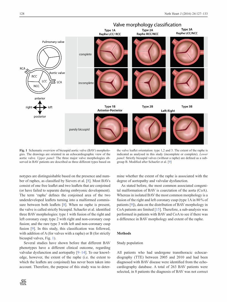
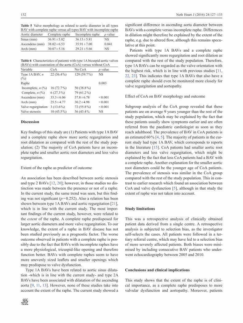
notypes are distinguishable based on the presence and num-ber of raphes, as classified by Sievers et al. [8]. Most BAVs consist of one free leaflet and two leaflets that are conjoined (or have failed to separate during embryonic development). The term ‘raphe’ defines the conjoined area of the two underdeveloped leaflets turning into a malformed commis-sure between both leaflets [8]. When no raphe is present, the valve is called strictly bicuspid. Schaefer et al. identified three BAV morphologies: type 1 with fusion of the right and left coronary cusp; type 2 with right and non-coronary cusp fusion; and the rare type 3 with left and non-coronary cusp fusion [9]. In this study, this classification was followed, with addition of A (for valves with a raphe) or B (for strictly bicuspid valves, Fig. 1).
Several studies have shown before that different BAV phenotypes have a different clinical outcome, regarding valvular dysfunction and aortopathy [9–14]. To our knowl-edge, however, the extent of the raphe (i.e. the extent to which the leaflets are conjoined) has never been taken into account. Therefore, the purpose of this study was to deter-
mine whether the extent of the raphe is associated with the degree of aortopathy and valvular dysfunction.
As stated before, the most common associated congeni-tal malformation of BAV is coarctation of the aorta (CoA). Whereas in isolated BAV the most common morphology is a fusion of the right and left coronary cusp (type 1A in 80 % of patients [9]), data on the distribution of BAV morphology in CoA patients are limited [15]. Therefore, a sub-analysis was performed in patients with BAV and CoA to see if there was a difference in BAV morphology and extent of the raphe.
Methods
Study population
All patients who had undergone transthoracic echocar-diography (TTE) between 2005 and 2010 and had been diagnosed with BAV disease were identified from the echo-cardiography database. A total of 263 BAV patients were selected, in 8 patients the diagnosis of BAV was not correct
Fig. 1 Schematic overview of bicuspid aortic valve (BAV) morpholo-gies. The drawings are oriented in an echocardiographic view of the aortic valve. Upper panel: The three major valve morphologies ob-served in BAV patients are described as three different types based on
the valve leaflet orientation: type 1,2 and 3. The extent of the raphe is indicated as analysed in this study (incomplete or complete). Lower panel: Strictly bicuspid valves (without a raphe) are defined as a sub-group B. Modified after Schaefer et al. [9]
129Neth Heart J (2016) 24:127–133
(EPD-Vision®, LUMC) and the echocardiography database, respectively. For this analysis of clinically acquired data, the Institutional Review Board waived the need for patient writ-ten informed consent.
Classification of valve morphology
As from a developmental point of view a morphological spectrum may exist in which the extent of the raphe can be regarded as a continuum, in the current study we chose to describe the major valve morphologies observed in BAV patients as 3 different types (type 1A, 2A and 3A, based on valve leaflet orientation, modified after Schaefer et al. [9]), in which the extent of the raphe can vary (Fig. 1). We defined fusion of the right and left coronary cusp as type 1A BAV, fusion of the right and non-coronary cusp as type 2A BAV and fusion of the left and non-coronary cusp as type 3A BAV. Strictly bicuspid valves (i.e. a valves without a raphe) were defined as being either type 1, 2 or 3, based on the orienta-tion of their leaflets, and referred as a subgroup B (Fig. 1).
Echocardiography
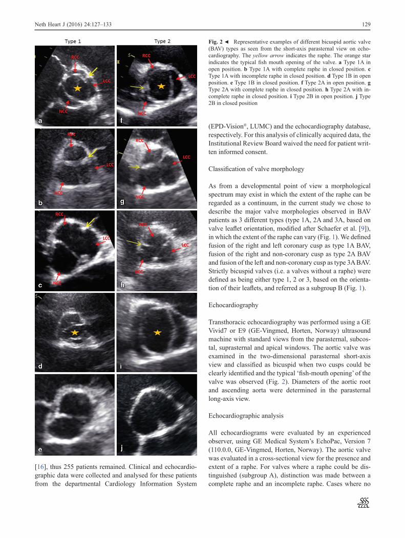
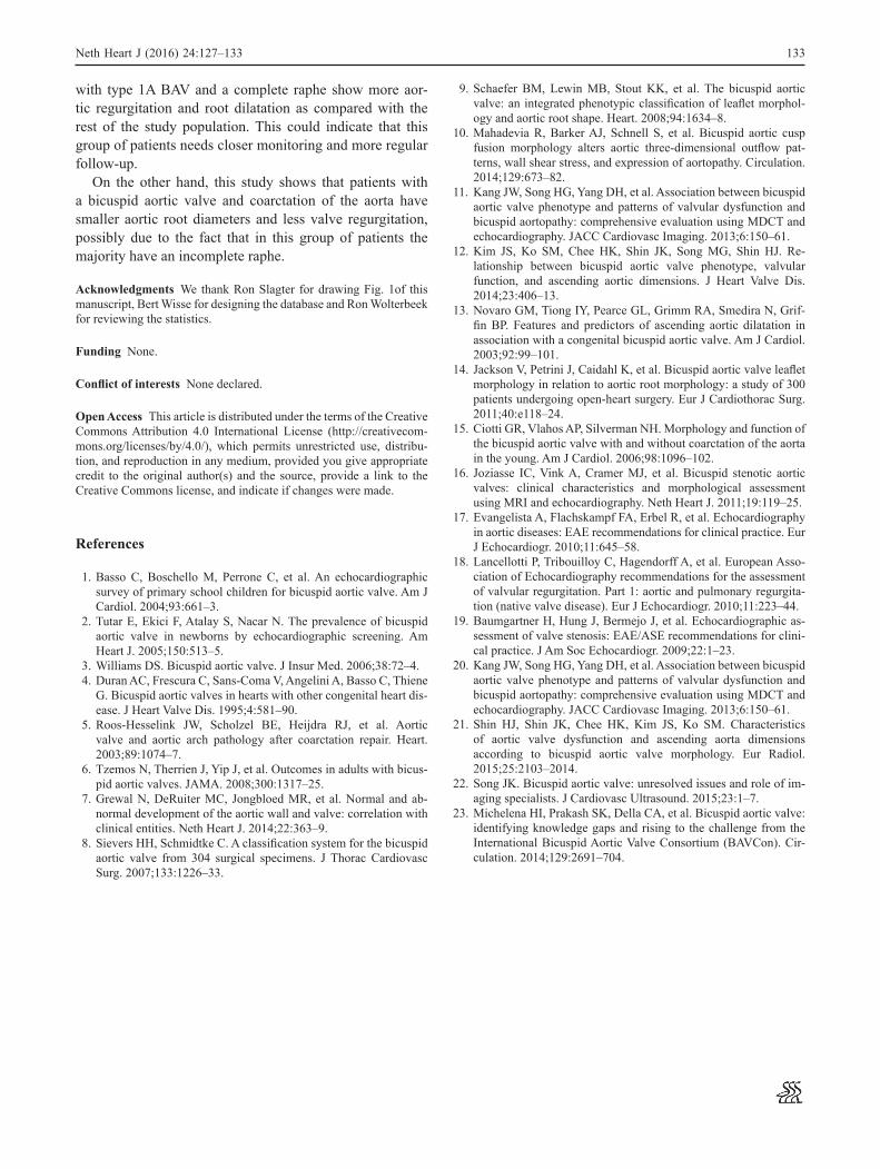
Transthoracic echocardiography was performed using a GE Vivid7 or E9 (GE-Vingmed, Horten, Norway) ultrasound machine with standard views from the parasternal, subcos-tal, suprasternal and apical windows. The aortic valve was examined in the two-dimensional parasternal short-axis view and classified as bicuspid when two cusps could be clearly identified and the typical ‘fish-mouth opening’ of the valve was observed (Fig. 2). Diameters of the aortic root and ascending aorta were determined in the parasternal long-axis view.
Echocardiographic analysis
All echocardiograms were evaluated by an experienced observer, using GE Medical System’s EchoPac, Version 7 (110.0.0, GE-Vingmed, Horten, Norway). The aortic valve was evaluated in a cross-sectional view for the presence and extent of a raphe. For valves where a raphe could be dis-tinguished (subgroup A), distinction was made between a complete raphe and an incomplete raphe. Cases where no
[16], thus 255 patients remained. Clinical and echocardio-graphic data were collected and analysed for these patients from the departmental Cardiology Information System
Fig. 2 9 Representative examples of different bicuspid aortic valve (BAV) types as seen from the short-axis parasternal view on echo-cardiography. The yellow arrow indicates the raphe. The orange star indicates the typical fish mouth opening of the valve. a Type 1A in open position. b Type 1A with complete raphe in closed position. c Type 1A with incomplete raphe in closed position. d Type 1B in open position. e Type 1B in closed position. f Type 2A in open position. g Type 2A with complete raphe in closed position. h Type 2A with in-complete raphe in closed position. i Type 2B in open position. j Type 2B in closed position
130
(Table 1). Mean age was not significantly different in the dif-ferent BAV subgroups (p = 0.515, Table 2) or in patients with incomplete and complete raphes (p = 0.357). There were no significant differences in valvular function between the dif-ferent morphological subtypes. However, BAVs with a com-plete raphe had a significantly higher prevalence of valvular
raphe was detected (subgroup B) were defined as strictly bicuspid valves. Diameters of aortic sinus, ascending aorta and aortic arch were measured from leading edge to leading edge in end-diastole according to the European Association of Echocardiography recommendations [17]. Aortic annu-lar diameter was measured from inner edge to inner edge during systole. All measurements were in mm, rounded to 2 significant figures. The ascending aorta was considered dilated at a diameter of > 38 mm.
Valvular dysfunction was defined as aortic stenosis or regurgitation. European Association of Echocardiography (EAE) recommendations were used for determining sever-ity of aortic stenosis and regurgitation, grading from mild to severe [18, 19]. Subgroup analysis was performed in patients with a history of CoA, the same protocol was fol-lowed in this group.
Statistical analysis
All collected data were registered in a Microsoft Office Access 2003 database. The database was exported into IBM SPSS Statistics Version 20 for computing variables and sta-tistical analysis. Independent samples T-tests were used to compare means of numerical data in two categories. One-way ANOVA tests were used for comparing numerical data in more than two categories. Cross-tabulations were made for binary categorical data, on which chi-square goodness-of-fit-tests were performed to test for independence. For sets of independent numerical data linear regression analy-sis was used to evaluate trends. Similarly, trends for binary categories were evaluated with binary logistic regression to correct for possible confounding factors such as age and gender. All statistical analyses were two-tailed and consid-ered significant if p < 0.05.
Results
Patient characteristics
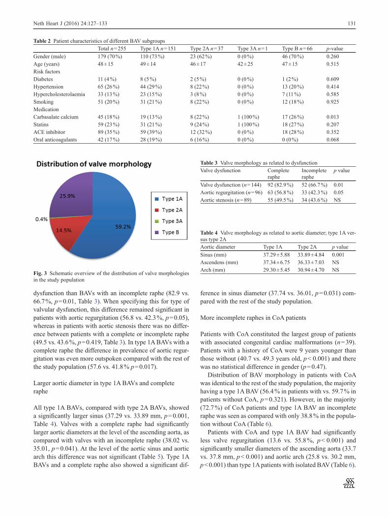
A total of 255 patients with BAV (age 18–85 years, mean 48 ± 15 years) were identified and analysed [16]. Of these, 179 were male (70.2 %) and 76 female (29.8 %). Patient characteristics and echocardiographic data are summarised in Table 1. Baseline characteristics were not significantly different between the BAV subtypes (Table 2). The distribu-tion of valve morphology is shown in Fig. 3.
More valvular dysfunction in BAVs with complete raphe
A total of 120 patients were diagnosed with aortic stenosis, the same number of patients showed aortic regurgitation
Table 1 Patient characteristicsVariable n (%)Gender
Male 179 (70.2)Female 76 (29.8)
Bicuspid aortic valve morphologyType 1A 151 (59.2)Type 2A 37 (14.5)Type 3A 1 (0.4)Strictly bicuspid (type B) Type 1B Type 2B
66 (25.9) 42 (63.6) 24 (36.4)
Extent of the rapheComplete 111 (43.5)Incomplete 78 (30.6)Valve surgery 66 (25.9)Mitral valve replacement 1 (0.39)Mitral valve repair 2 (0.78)Aortic valve replacement 50 (19.6)Aortic valve repair 10 (3.9)Bentall 22 (8.6)Tricuspid valve repair 1 (0.39)Pulmonary valve replacement 1 (0.39)
Congenital defects 51 (20.0)Aortic coarctation 39 (15.3)Atrial septal defect 1 (0.39)Ventricular septal defect 11 (4.3)Marfan 2 (0.78)Common truncus 1 (0.39)Patent ductus arteriosus 5 (2.0)Valvular dysfunction 189 (74.1)Aortic valve stenosis 120 (47.1)Aortic valve regurgitation 120 (47.1)Mitral valve regurgitation 16 (6.3)Tricuspid valve regurgitation 8 (3.1)Pulmonary valve stenosis 1 (0.39)Pulmonary valve regurgitation 1 (0.39)
Medication 140 (54.9)ACE inhibitors 91 (35.7)Statins 61 (23.9)Carbasalate calcium 45 (17.6)Oral anticoagulants 43 (16.9)Antiarrhythmic drugs 33 (12.9)
Risk factors 117 (45.9)Hypertension 65 (25.5)Smoking 54 (21.2)Diabetes 11 (4.3)Cerebrovascular accident 14 (5.5)Hypercholesterolaemia 33 (12.9)Peripheral arterial disease 7 (2.7)
Neth Heart J (2016) 24:127–133
131
ference in sinus diameter (37.74 vs. 36.01, p = 0.031) com-pared with the rest of the study population.
More incomplete raphes in CoA patients
Patients with CoA constituted the largest group of patients with associated congenital cardiac malformations (n = 39). Patients with a history of CoA were 9 years younger than those without (40.7 vs. 49.3 years old, p < 0.001) and there was no statistical difference in gender (p = 0.47).
Distribution of BAV morphology in patients with CoA was identical to the rest of the study population, the majority having a type 1A BAV (56.4 % in patients with vs. 59.7 % in patients without CoA, p = 0.321). However, in the majority (72.7 %) of CoA patients and type 1A BAV an incomplete raphe was seen as compared with only 38.8 % in the popula-tion without CoA (Table 6).
Patients with CoA and type 1A BAV had significantly less valve regurgitation (13.6 vs. 55.8 %, p < 0.001) and significantly smaller diameters of the ascending aorta (33.7 vs. 37.8 mm, p < 0.001) and aortic arch (25.8 vs. 30.2 mm, p < 0.001) than type 1A patients with isolated BAV (Table 6).
dysfunction than BAVs with an incomplete raphe (82.9 vs. 66.7 %, p = 0.01, Table 3). When specifying this for type of valvular dysfunction, this difference remained significant in patients with aortic regurgitation (56.8 vs. 42.3 %, p = 0.05), whereas in patients with aortic stenosis there was no differ-ence between patients with a complete or incomplete raphe (49.5 vs. 43.6 %, p = 0.419, Table 3). In type 1A BAVs with a complete raphe the difference in prevalence of aortic regur-gitation was even more outspoken compared with the rest of the study population (57.6 vs. 41.8 % p = 0.017).
Larger aortic diameter in type 1A BAVs and complete raphe
All type 1A BAVs, compared with type 2A BAVs, showed a significantly larger sinus (37.29 vs. 33.89 mm, p = 0.001, Table 4). Valves with a complete raphe had significantly larger aortic diameters at the level of the ascending aorta, as compared with valves with an incomplete raphe (38.02 vs. 35.01, p = 0.041). At the level of the aortic sinus and aortic arch this difference was not significant (Table 5). Type 1A BAVs and a complete raphe also showed a significant dif-
Neth Heart J (2016) 24:127–133
Table 2 Patient characteristics of different BAV subgroupsTotal n = 255 Type 1A n = 151 Type 2A n = 37 Type 3A n = 1 Type B n = 66 p-value
Gender (male) 179 (70 %) 110 (73 %) 23 (62 %) 0 (0 %) 46 (70 %) 0.260Age (years) 48 ± 15 49 ± 14 46 ± 17 42 ± 25 47 ± 15 0.515Risk factorsDiabetes 11 (4 %) 8 (5 %) 2 (5 %) 0 (0 %) 1 (2 %) 0.609Hypertension 65 (26 %) 44 (29 %) 8 (22 %) 0 (0 %) 13 (20 %) 0.414Hypercholesterolaemia 33 (13 %) 23 (15 %) 3 (8 %) 0 (0 %) 7 (11 %) 0.585Smoking 51 (20 %) 31 (21 %) 8 (22 %) 0 (0 %) 12 (18 %) 0.925MedicationCarbasalate calcium 45 (18 %) 19 (13 %) 8 (22 %) 1 (100 %) 17 (26 %) 0.013Statins 59 (23 %) 31 (21 %) 9 (24 %) 1 (100 %) 18 (27 %) 0.207ACE inhibitor 89 (35 %) 59 (39 %) 12 (32 %) 0 (0 %) 18 (28 %) 0.352Oral anticoagulants 42 (17 %) 28 (19 %) 6 (16 %) 0 (0 %) 0 (0 %) 0.068
Fig. 3 Schematic overview of the distribution of valve morphologies in the study population
Table 3 Valve morphology as related to dysfunctionValve dysfunction Complete
rapheIncomplete raphe
p value
Valve dysfunction (n = 144) 92 (82.9 %) 52 (66.7 %) 0.01Aortic regurgitation (n = 96) 63 (56.8 %) 33 (42.3 %) 0.05Aortic stenosis (n = 89) 55 (49.5 %) 34 (43.6 %) NS
Table 4 Valve morphology as related to aortic diameter; type 1A ver-sus type 2AAortic diameter Type 1A Type 2A p valueSinus (mm) 37.29 ± 5.88 33.89 ± 4.84 0.001Ascendens (mm) 37.34 ± 6.75 36.33 ± 7.03 NSArch (mm) 29.30 ± 5.45 30.94 ± 4.70 NS
132
significant difference in ascending aorta diameter between BAVs with a complete versus incomplete raphe. Differences in dilation might therefore be explained by the extent of the raphe, e.g. due to altered flow, although this remains specu-lative at this point.
Patients with type 1A BAVs and a complete raphe showed significantly more regurgitation and root dilation as compared with the rest of the study population. Therefore, type 1A BAVs can be regarded as the valve orientation with the highest risk, which is in line with previous studies [11, 22, 23]. This indicates that type 1A BAVs that also have a complete raphe should even be monitored more closely for valve regurgitation and aortopathy.
Effect of CoA on BAV morphology and outcome
Subgroup analysis of the CoA group revealed that these patients are on average 9 years younger than the rest of the study population, which may be explained by the fact that these patients usually show symptoms earlier and are often referred from the paediatric cardiologist as soon as they reach adulthood. The prevalence of BAV in CoA patients is an estimated 60 % [4, 5]. The majority of patients in the cur-rent study had type 1A BAV, which corresponds to reports in the literature [15]. CoA patients had smaller aortic root diameters and less valve regurgitation, which might be explained by the fact that less CoA patients had a BAV with a complete raphe. Another explanation for the smaller aortic root diameters could be the younger age of CoA patients. The prevalence of stenosis was similar in the CoA group compared with the rest of the study population. This in con-trast to earlier research which found an association between CoA and valve dysfunction [5], although in that study the extent of raphe was not taken into account.
Study limitations
This was a retrospective analysis of clinically obtained patient data derived from a single centre. A retrospective analysis is subjected to selection bias, as the investigator self-selects the cases. All patients were followed in a ter-tiary referral centre, which may have led to a selection bias of more severely affected patients. Both biases were mini-mised by including consecutive BAV patients who under-went echocardiography between 2005 and 2010.
Conclusions and clinical implications
This study shows that the extent of the raphe is of clini-cal importance, as a complete raphe predisposes to more valvular dysfunction and aortopathy. Moreover, patients
Discussion
Key findings of this study are (1) Patients with type 1A BAV and a complete raphe show more aortic regurgitation and root dilatation as compared with the rest of the study pop-ulation; (2) The majority of CoA patients have an incom-plete raphe and smaller aortic root diameters and less valve regurgitation.
Extent of the raphe as predictor of outcome
An association has been described between aortic stenosis and type 2 BAVs [12, 20]; however, in those studies no dis-tinction was made between the presence or not of a raphe. In the current study, the same trend was seen, but this find-ing was not significant (p = 0.252). Also a relation has been shown between type 1A BAVs and aortic regurgitation [21], which is in line with the current study. The most impor-tant findings of the current study, however, were related to the extent of the raphe. A complete raphe predisposed for larger aortic diameters and more valve regurgitation. To our knowledge, the extent of a raphe in BAV disease has not been studied previously as a prognostic factor. The worse outcome observed in patients with a complete raphe is pos-sibly due to the fact that BAVs with incomplete raphes have a more physiological, tricuspid-like opening and therefore function better. BAVs with complete raphes seem to have more unevenly sized leaflets and smaller openings which may predispose to valve dysfunction.
Type 1A BAVs have been related to aortic sinus dilata-tion -which is in line with the current study- and type 2A BAVs have been associated with dilatation of the ascending aorta [9, 11, 13]. However, none of these studies take into account the extent of the raphe. The current study showed a
Table 5 Valve morphology as related to aortic diameter in all types BAV with complete raphe versus all types BAV with incomplete rapheAortic diameter Complete raphe Incomplete raphe p valueSinus (mm) 36.91 ± 5.82 36.13 ± 5.81 NSAscendens (mm) 38.02 ± 6.53 35.91 ± 7.00 0.041Arch (mm) 30.07 ± 5.16 29.21 ± 5.66 NS
Table 6 Characteristics of patients with type 1A bicuspid aortic valves (BAVs) with coarctation of the aorta (CoA) versus without CoAVariable CoA No CoA p valueType 1A BAV, n (%)
22 (56.4 %) 129 (59.7 %) NS
Raphe 0.003 Incomplete, n (%) 16 (72.7 %) 50 (38.8 %) Complete, n (%) 6 (27.3 %) 79 (61.2 %)Ascendens (mm) 33.3 ± 6.06 37.8 ± 6.78 < 0.001Arch (mm) 25.5 ± 4.77 30.2 ± 4.98 < 0.001Valve regurgitation 3 (13.6 %) 72 (55.8 %) < 0.001Valve stenosis 10 (45.5 %) 56 (43.4 % NS
Neth Heart J (2016) 24:127–133
133
9. Schaefer BM, Lewin MB, Stout KK, et al. The bicuspid aortic valve: an integrated phenotypic classification of leaflet morphol-ogy and aortic root shape. Heart. 2008;94:1634–8.
10. Mahadevia R, Barker AJ, Schnell S, et al. Bicuspid aortic cusp fusion morphology alters aortic three-dimensional outflow pat-terns, wall shear stress, and expression of aortopathy. Circulation. 2014;129:673–82.
11. Kang JW, Song HG, Yang DH, et al. Association between bicuspid aortic valve phenotype and patterns of valvular dysfunction and bicuspid aortopathy: comprehensive evaluation using MDCT and echocardiography. JACC Cardiovasc Imaging. 2013;6:150–61.
12. Kim JS, Ko SM, Chee HK, Shin JK, Song MG, Shin HJ. Re-lationship between bicuspid aortic valve phenotype, valvular function, and ascending aortic dimensions. J Heart Valve Dis. 2014;23:406–13.
13. Novaro GM, Tiong IY, Pearce GL, Grimm RA, Smedira N, Grif-fin BP. Features and predictors of ascending aortic dilatation in association with a congenital bicuspid aortic valve. Am J Cardiol. 2003;92:99–101.
14. Jackson V, Petrini J, Caidahl K, et al. Bicuspid aortic valve leaflet morphology in relation to aortic root morphology: a study of 300 patients undergoing open-heart surgery. Eur J Cardiothorac Surg. 2011;40:e118–24.
15. Ciotti GR, Vlahos AP, Silverman NH. Morphology and function of the bicuspid aortic valve with and without coarctation of the aorta in the young. Am J Cardiol. 2006;98:1096–102.
16. Joziasse IC, Vink A, Cramer MJ, et al. Bicuspid stenotic aortic valves: clinical characteristics and morphological assessment using MRI and echocardiography. Neth Heart J. 2011;19:119–25.
17. Evangelista A, Flachskampf FA, Erbel R, et al. Echocardiography in aortic diseases: EAE recommendations for clinical practice. Eur J Echocardiogr. 2010;11:645–58.
18. Lancellotti P, Tribouilloy C, Hagendorff A, et al. European Asso-ciation of Echocardiography recommendations for the assessment of valvular regurgitation. Part 1: aortic and pulmonary regurgita-tion (native valve disease). Eur J Echocardiogr. 2010;11:223–44.
19. Baumgartner H, Hung J, Bermejo J, et al. Echocardiographic as-sessment of valve stenosis: EAE/ASE recommendations for clini-cal practice. J Am Soc Echocardiogr. 2009;22:1–23.
20. Kang JW, Song HG, Yang DH, et al. Association between bicuspid aortic valve phenotype and patterns of valvular dysfunction and bicuspid aortopathy: comprehensive evaluation using MDCT and echocardiography. JACC Cardiovasc Imaging. 2013;6:150–61.
21. Shin HJ, Shin JK, Chee HK, Kim JS, Ko SM. Characteristics of aortic valve dysfunction and ascending aorta dimensions according to bicuspid aortic valve morphology. Eur Radiol. 2015;25:2103–2014.
22. Song JK. Bicuspid aortic valve: unresolved issues and role of im-aging specialists. J Cardiovasc Ultrasound. 2015;23:1–7.
23. Michelena HI, Prakash SK, Della CA, et al. Bicuspid aortic valve: identifying knowledge gaps and rising to the challenge from the International Bicuspid Aortic Valve Consortium (BAVCon). Cir-culation. 2014;129:2691–704.
with type 1A BAV and a complete raphe show more aor-tic regurgitation and root dilatation as compared with the rest of the study population. This could indicate that this group of patients needs closer monitoring and more regular follow-up.
On the other hand, this study shows that patients with a bicuspid aortic valve and coarctation of the aorta have smaller aortic root diameters and less valve regurgitation, possibly due to the fact that in this group of patients the majority have an incomplete raphe.
Acknowledgments We thank Ron Slagter for drawing Fig. 1of this manuscript, Bert Wisse for designing the database and Ron Wolterbeek for reviewing the statistics.
Funding None.
Conflict of interests None declared.
Open Access This article is distributed under the terms of the Creative Commons Attribution 4.0 International License (http://creativecom-mons.org/licenses/by/4.0/), which permits unrestricted use, distribu-tion, and reproduction in any medium, provided you give appropriate credit to the original author(s) and the source, provide a link to the Creative Commons license, and indicate if changes were made.
References
1. Basso C, Boschello M, Perrone C, et al. An echocardiographic survey of primary school children for bicuspid aortic valve. Am J Cardiol. 2004;93:661–3.
2. Tutar E, Ekici F, Atalay S, Nacar N. The prevalence of bicuspid aortic valve in newborns by echocardiographic screening. Am Heart J. 2005;150:513–5.
3. Williams DS. Bicuspid aortic valve. J Insur Med. 2006;38:72–4. 4. Duran AC, Frescura C, Sans-Coma V, Angelini A, Basso C, Thiene
G. Bicuspid aortic valves in hearts with other congenital heart dis-ease. J Heart Valve Dis. 1995;4:581–90.
5. Roos-Hesselink JW, Scholzel BE, Heijdra RJ, et al. Aortic valve and aortic arch pathology after coarctation repair. Heart. 2003;89:1074–7.
6. Tzemos N, Therrien J, Yip J, et al. Outcomes in adults with bicus-pid aortic valves. JAMA. 2008;300:1317–25.
7. Grewal N, DeRuiter MC, Jongbloed MR, et al. Normal and ab-normal development of the aortic wall and valve: correlation with clinical entities. Neth Heart J. 2014;22:363–9.
8. Sievers HH, Schmidtke C. A classification system for the bicuspid aortic valve from 304 surgical specimens. J Thorac Cardiovasc Surg. 2007;133:1226–33.
Neth Heart J (2016) 24:127–133