Embed Size (px)
Citation preview
The Expression of SPARC in Human Urothelial Cells (UROtsa) Exposed to or
Malignantly Transformed by Cadmium or Arsenite
Jennifer LarsonDoctoral Candidate
University of North DakotaOctober 5, 2011

Outline
• Background Info• Purpose• Methods • Results• Conclusions• Future directions
Bladder Cancer• Transitional cell carcinoma of the bladder is the 9th most common cancer worldwide and the
4th in U.S (Bischoff & Clark, 2010)
• Highest cost per patient of all cancers (Sullivan et al., 2010; Jacobs et al., 2010)
• Bladder cancer was the first cancer in which industrial carcinogens were found to play the major role in disease causation Link between exposure to aromatic amines and development of bladder cancer in factory
workers (Rehn, 1895)
• Cd⁺² and As⁺³ are human carcinogens (IARC, 1993; IARC, 1980) and linked to the development of bladder cancer (Steinmaus et al., 1994 & 2000)
• Association between cigarette smoking and bladder cancer 2-4 times increased risk (Clavel et al., 1989; Morrison et al., 1984; Silverman et al., 1992)
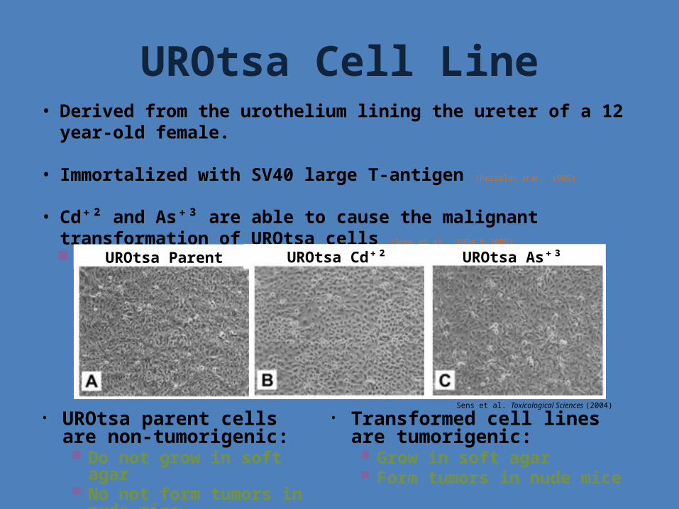
UROtsa Cell Line• Derived from the urothelium lining the ureter of a 12 year-old female.
• Immortalized with SV40 large T-antigen (Perzoldt et al., 1995)
• Cd⁺² and As⁺³ are able to cause the malignant transformation of UROtsa cells (Sens et al., 1994 & 2000)
Generated 7 different Cd+2 & 6 different As+3 cell lines.
• UROtsa parent cells are non-tumorigenic: Do not grow in soft agar No not form tumors in nude
mice
• Transformed cell lines are tumorigenic: Grow in soft agar Form tumors in nude mice
UROtsa Parent UROtsa Cd⁺² UROtsa As⁺³
Sens et al. Toxicological Sciences (2004)
SPARC• Secreted Protein, Acidic and Rich in Cysteine
Also known as: Osteonectin & BM-40
• Belongs to the Matricellular group of proteins Secreted macromolecules that interact with cell-surface receptors, ECM,
and/or growth factors and proteases, but do NOT have structural roles Known to bind to structural matrix proteins
• Collagen and Vitronectin
Found in tissues undergoing repair/remodeling (Salonen et al. 1990)
Upregulated during embryological development (Sage et al., 1989)
Other matricellular proteins include:• Thrombosponbin 1 & 2• Tenascin C & X• Osteopontin• CTGF• Testican 1, 2, & 3 (SPARC-related protein)• Hevin (SPARC-related protein)
SPARC• 3 General functions of SPARC:
1. De-adhesion: • Exogenous SPARC inhibits cell spreading and cell attachment (Sage et al. 1989)
• Disassemble focal adhesions (Murphy-Ullrich et al. 1995, Murphy-Ullrich 2001)
• Reorganizes actin stress fibers to periphery of cell (Murphy-Ullrich et al. 1995)
• May play a role in cell migration
2. Anti-proliferation: • Cell cycle inhibitor (Funk & Sage 1991, 1993; Sage et al. 1995)
• Promoted prostate cancer cell migration (De et al. 2003) and stimulated fibroblasts during myocardial infraction (Wu et al. 2006)
• May play a role in cell migration
3. Regulation of ECM & growth factors: • Binds to many ECM components (Brekken & Sage 2000)
• Known activator of MMP-2 (Tremble et al. 1993)
• Regulates PDGF, VEGF, & TGF-β1 expression (Raines et al, 1992, Kuppricon et al. 1998, Abe et al. 2004, Hasselaar & Sage 1992, Lane & Sage 1994)
• After exogenous treatment, capable of inducing the expression of SPARC
• May play a role in the progression of metastatic tumors
Purpose
• To determine the role SPARC plays in the formation and progression of bladder cancer.
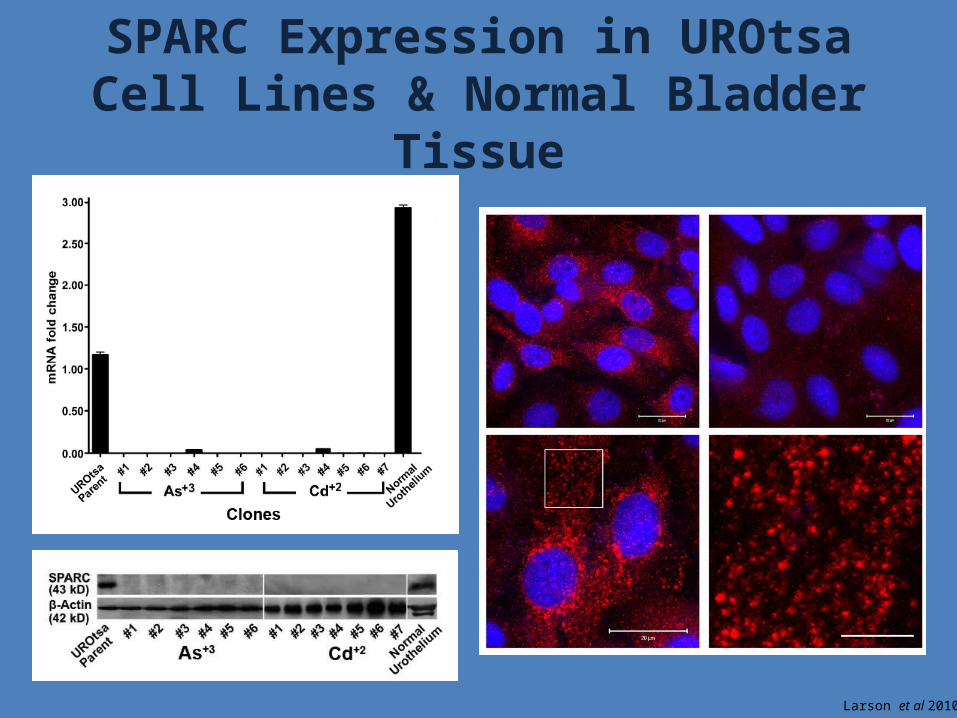
SPARC Expression in UROtsa Cell Lines & Normal Bladder Tissue
Larson et al 2010
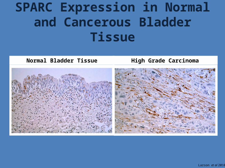
SPARC Expression in Normal and Cancerous Bladder Tissue
Normal Bladder Tissue High Grade Carcinoma
Larson et al 2010
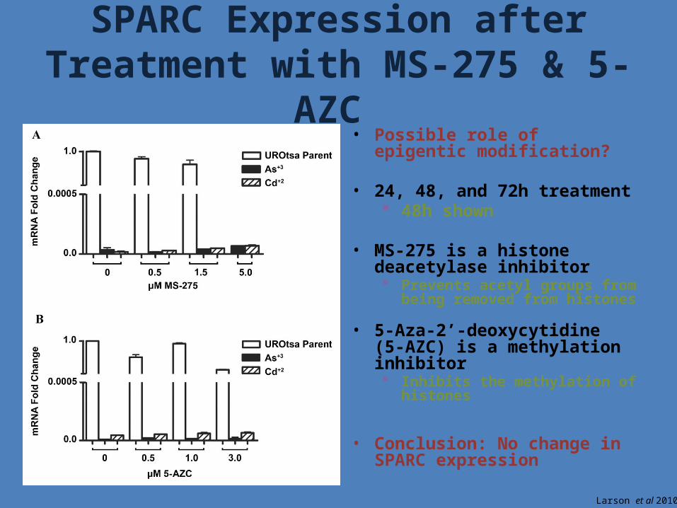
• Possible role of epigentic modification?
• 24, 48, and 72h treatment 48h shown
• MS-275 is a histone deacetylase inhibitor Prevents acetyl groups from being
removed from histones
• 5-Aza-2’-deoxycytidine (5-AZC) is a methylation inhibitor Inhibits the methylation of
histones
• Conclusion: No change in SPARC expression
SPARC Expression after Treatment with MS-275 & 5-AZC
Larson et al 2010
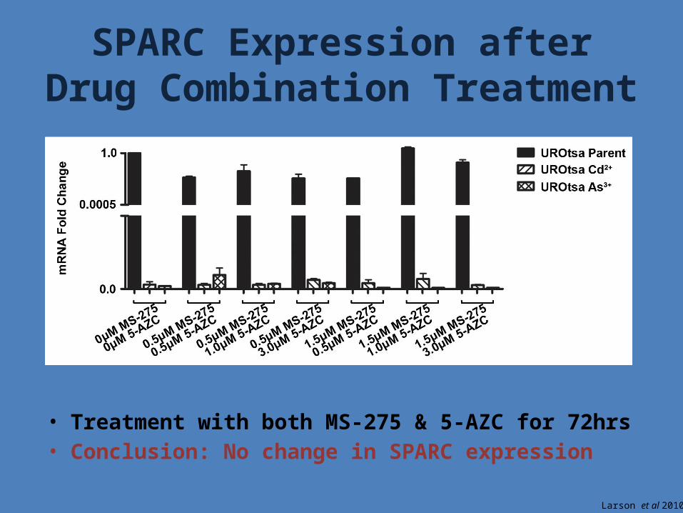
• Treatment with both MS-275 & 5-AZC for 72hrs• Conclusion: No change in SPARC expression
SPARC Expression after Drug Combination Treatment
Larson et al 2010
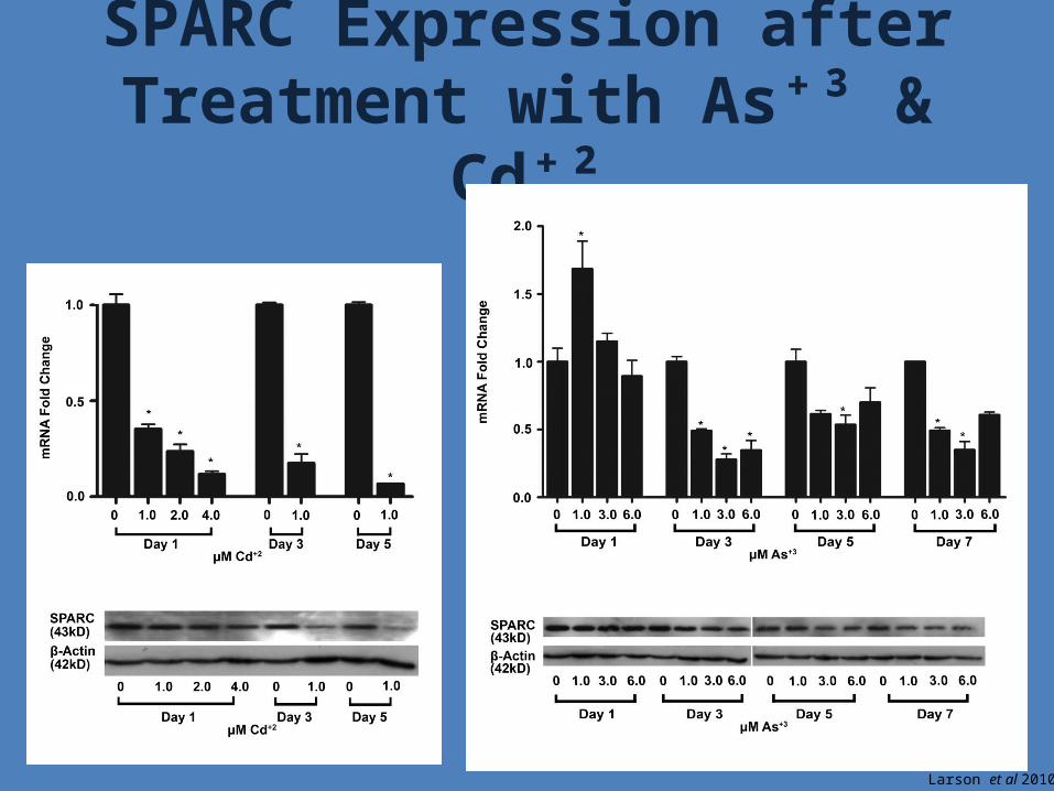
SPARC Expression after Treatment with As⁺³ & Cd⁺²
Larson et al 2010
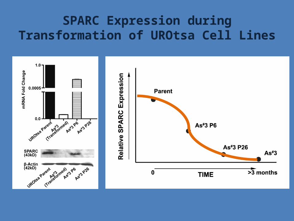
SPARC Expression during Transformation of UROtsa Cell Lines

Summary: SPARC Expression
• Moderate SPARC expression in Normal Tissue• Low or undetectable SPARC expression in Cancerous/Tumor Tissue
• SPARC expression does not appear to be regulated epigenetically in our system
• SPARC mRNA and protein expression decreases with increasing concentrations of As3+ and Cd2+
SPARC Transfection
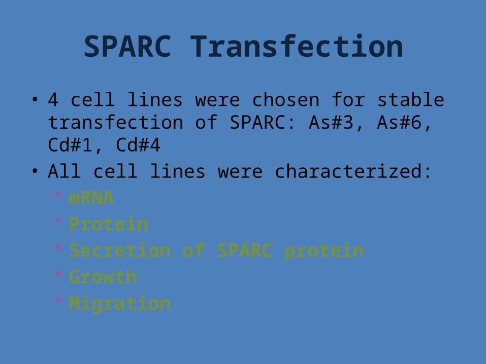
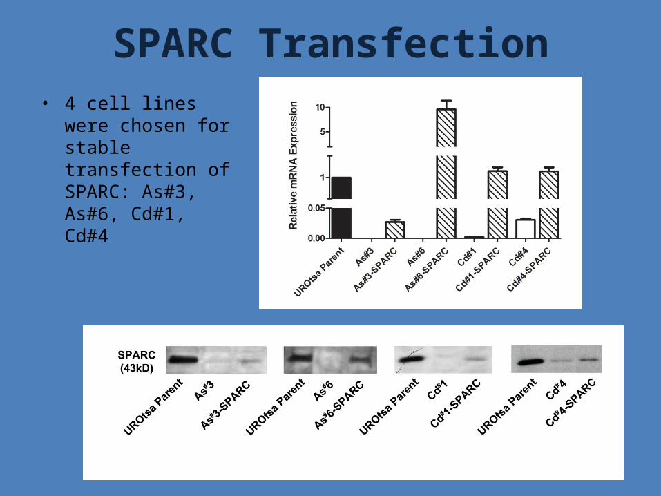
• 4 cell lines were chosen for stable transfection of SPARC: As#3, As#6, Cd#1, Cd#4
• All cell lines were characterized: mRNA Protein Secretion of SPARC protein Growth Migration
SPARC Transfection• 4 cell lines were
chosen for stable transfection of SPARC: As#3, As#6, Cd#1, Cd#4
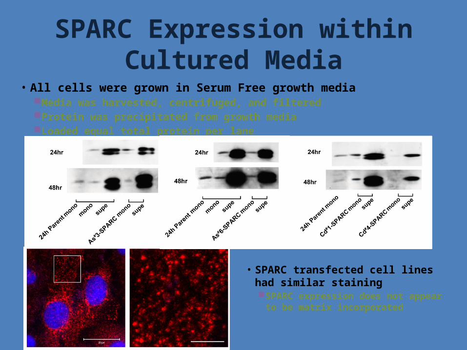
• All cells were grown in Serum Free growth media Media was harvested, centrifuged, and filtered Protein was precipitated from growth media Loaded equal total protein per lane
SPARC Expression within Cultured Media
• SPARC transfected cell lines had similar staining SPARC expression does not appear
to be matrix incorporated
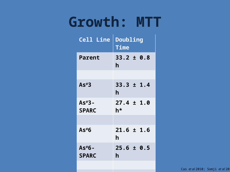
Growth: MTTCell Line Doubling Time
Parent 33.2 ± 0.8 h
As#3 33.3 ± 1.4 h
As#3-SPARC 27.4 ± 1.0 h*
As#6 21.6 ± 1.6 h
As#6-SPARC 25.6 ± 0.5 h
Cd#1 27.8 ± 0.6 h
Cd#1-SPARC 23.8 ± 0.6 h*
Cd#4 20.7 ± 1.1 h
Cd#4-SPARC 22.0 ± 0.6 h
Cao et al 2010; Somji et al 2010
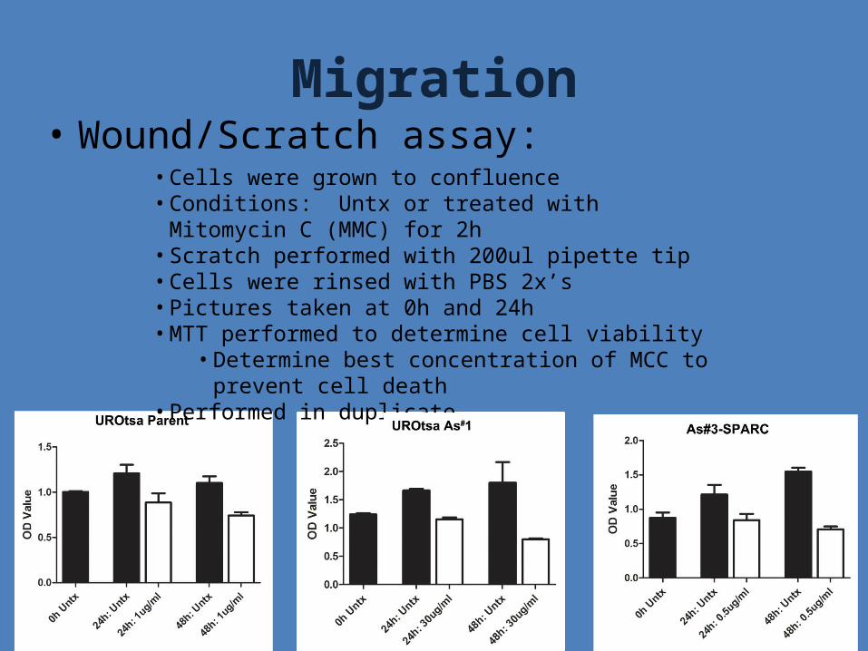
• Wound/Scratch assay: Migration
• Cells were grown to confluence• Conditions: Untx or treated with Mitomycin C (MMC) for 2h• Scratch performed with 200ul pipette tip• Cells were rinsed with PBS 2x’s• Pictures taken at 0h and 24h• MTT performed to determine cell viability
• Determine best concentration of MCC to prevent cell death
• Performed in duplicate
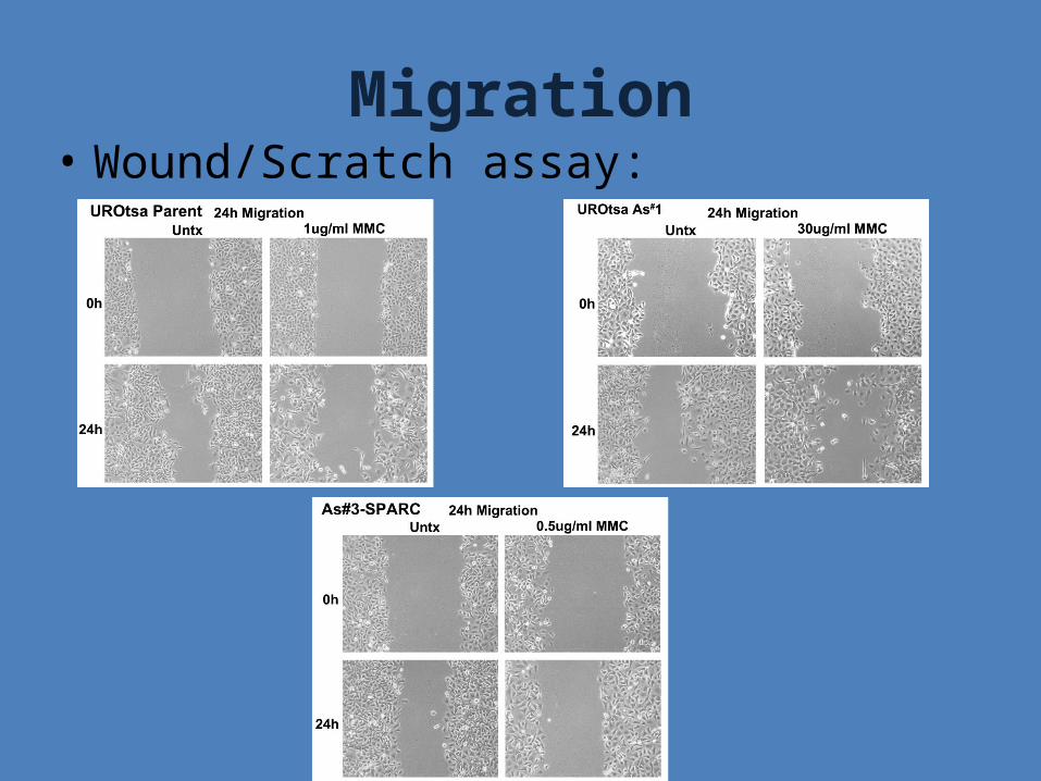
• Wound/Scratch assay: Migration
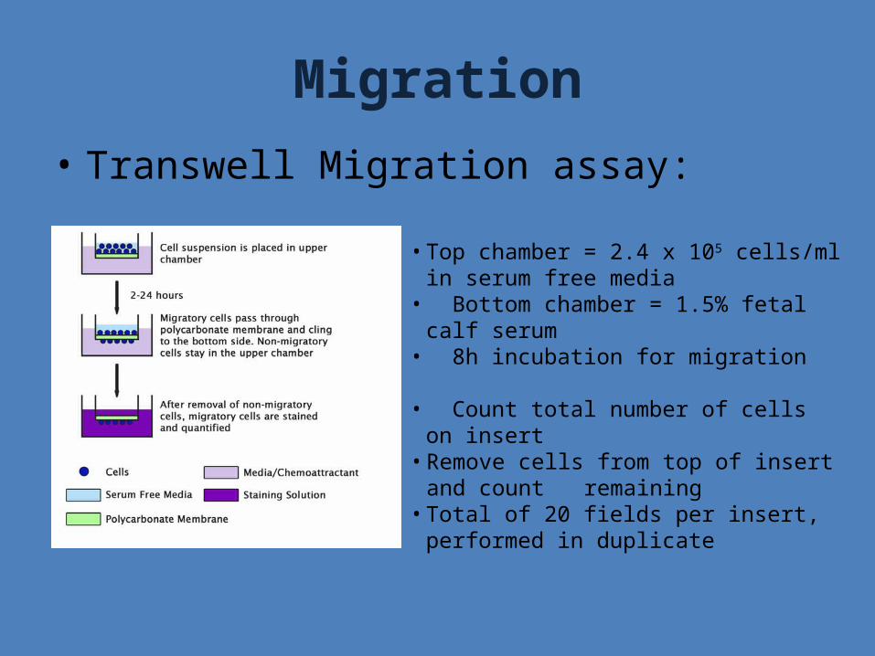
Migration• Transwell Migration assay:
• Top chamber = 2.4 x 10⁵ cells/ml in serum free media
• Bottom chamber = 1.5% fetal calf serum• 8h incubation for migration
• Count total number of cells on insert• Remove cells from top of insert and count
remaining• Total of 20 fields per insert, performed in
duplicate

Migration• Transwell Migration assay:
** **
*** *
*
Conclusions
• Expression of SPARC is not epigenetically regulated in our system
• SPARC expression decreased with treatment with Cd2+ & As3+
• SPARC tranfections: Secrete SPARC into media 2 transfected cell lines had decreased migration
Future Directions
• Assess the ability of SPARC transfected cell lines to form tumors in nude mice Tumor size Subcutaneous vs. intraperitoneal tumor formation Expression of SPARC in tumor
• Assess the structural differences of SPARC between UROtsa Parent and transfected cell lines Glycosylation Post-translational modifications
• Characterization of the promoter elements that turn off SPARC expression with treatment of Cd2+ & As3+

Acknowledgements
• Dr Jane Dunlevy Holly Hewitt
• Dr Don Sens• Dr Mary Ann Sens• Dr Seema Somji• Dr Scott Garrett• Dr XuDong Zhou

Questions?


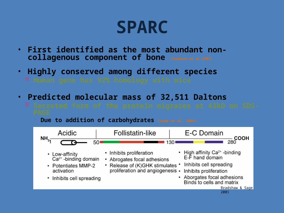
SPARC• First identified as the most abundant non-collagenous component of
bone (Termine et al.1981)
• Highly conserved among different species Human gene has 92% homology with mice
• Predicted molecular mass of 32,511 Daltons Secreted form of the protein migrates at 43kD on SDS-PAGE
• Due to addition of carbohydrates (Sage et al. 1984)
Bradshaw & Sage 2001
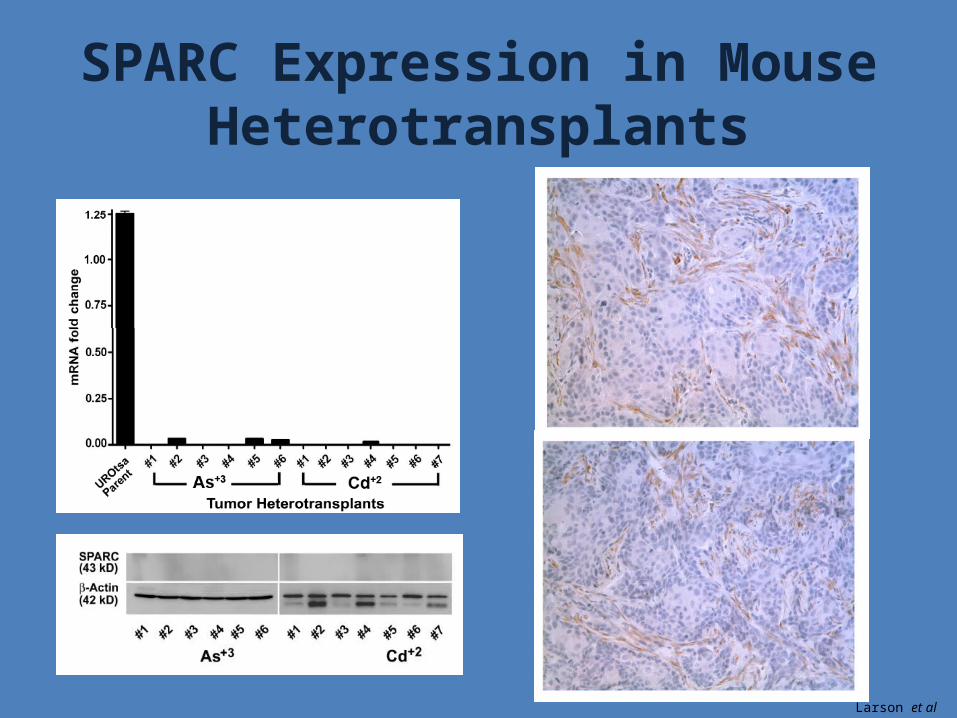
SPARC Expression in Mouse Heterotransplants
Larson et al 2010
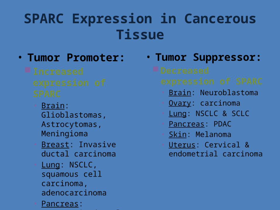
SPARC Expression in Cancerous Tissue
• Tumor Promoter: Increased expression of
SPARC• Brain: Glioblastomas,
Astrocytomas, Meningioma• Breast: Invasive ductal
carcinoma• Lung: NSCLC, squamous cell
carcinoma, adenocarcinoma• Pancreas: Pancreatic ductal
adenocarcinoma• Skin: Melanoma
• Tumor Suppressor:Decreased expression of
SPARC• Brain: Neuroblastoma• Ovary: carcinoma• Lung: NSCLC & SCLC• Pancreas: PDAC• Skin: Melanoma• Uterus: Cervical &
endometrial carcinoma
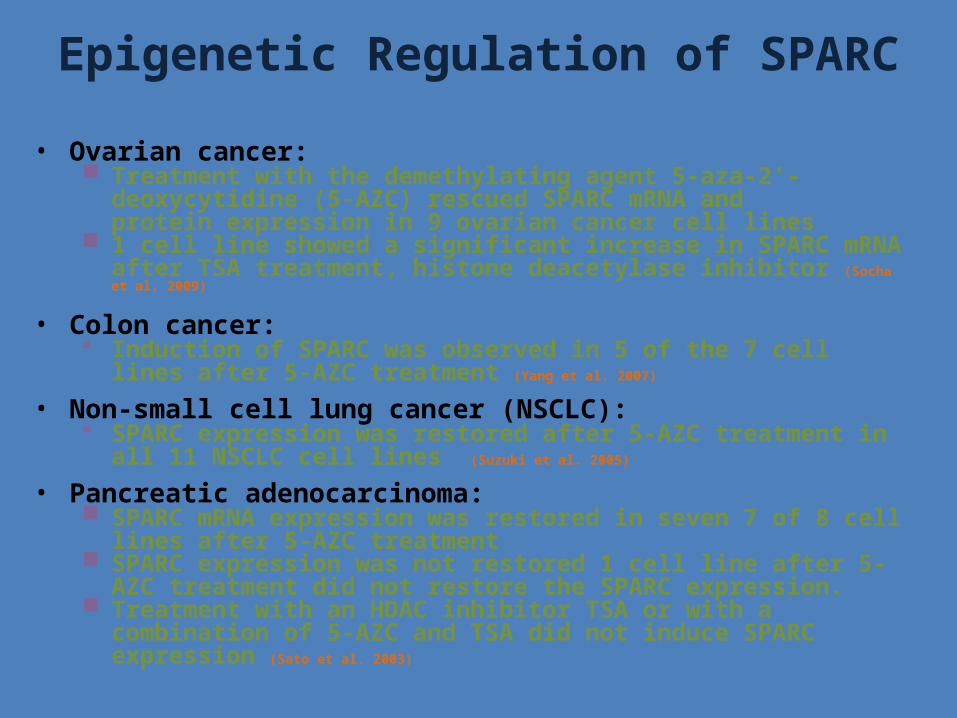
Epigenetic Regulation of SPARC• Ovarian cancer:
Treatment with the demethylating agent 5-aza-2'-deoxycytidine (5-AZC) rescued SPARC mRNA and protein expression in 9 ovarian cancer cell lines
1 cell line showed a significant increase in SPARC mRNA after TSA treatment, histone deacetylase inhibitor (Socha et al. 2009)
• Colon cancer: Induction of SPARC was observed in 5 of the 7 cell lines after 5-AZC
treatment (Yang et al. 2007)
• Non-small cell lung cancer (NSCLC): SPARC expression was restored after 5-AZC treatment in all 11
NSCLC cell lines (Suzuki et al. 2005)
• Pancreatic adenocarcinoma: SPARC mRNA expression was restored in seven 7 of 8 cell lines after
5-AZC treatment SPARC expression was not restored 1 cell line after 5-AZC treatment
did not restore the SPARC expression. Treatment with an HDAC inhibitor TSA or with a combination of 5-AZC
and TSA did not induce SPARC expression (Sato et al. 2003)