Embed Size (px)
Citation preview

MAY 2020
Upfront Businesses step up to fight
COVID-19
07
In My ViewPandemic preparations in
the lab
10
In PracticeVaping: is it really safer
than smoking?
27 – 29
Sitting Down WithPathologists’ assistant
Sarah Garner
50 – 51
65#
Official society partner of The Pathologist
The Evolution of the LabAs guardians of population health, the laboratory’s role is more important than ever
16 – 25

Editor ia lTrying TimesIn the midst of a pandemic, it’s not just physical health that matters
www.thepathologist.com
A round the world, new traditions have formed. In
New York City, for instance, people open their doors
and windows at seven o’clock every night, look out
of their homes, and applaud the healthcare workers
who are keeping the country running. The UK has a similar tradition
on Thursday evenings. The “balcony concerts” given by Italians in
lockdown have struck international fame via the Internet. Although
media coverage focuses on doctors and nurses on the front lines (and,
in a rare move, occasionally mentions those involved in laboratory
testing), the conversations on social media and in local hubs include
other essential workers; for instance, the people who ensure that
hospitals and laboratories are kept clean, or those who answer the
COVID-19 telephone hotlines.
Cheery news abounds: the 100-year-old man who has
raised £32 million for the National Health Service; the
children setting up “take what you will” stands outside their
homes; the people sewing face masks for bus drivers, retail
workers, and other vital (and often overlooked) professions.
But on social media, I see a different story. Every day,
another pathologist or laboratory medicine professional on
Twitter posts that they’re taking a break for their mental
health. Every day, another voice on Facebook or YouTube
or Instagram goes silent to focus inward, rather than deal
with the seemingly endless flow of (not always good, often
politically motivated) news.
In the midst of one health crisis, are we missing another?
COVID-19 may be grabbing the spotlight at the moment,
but it is perhaps more important than ever to be aware of
the emotional and psychological effects this pandemic is
having – not just on the doctors and nurses patrolling the
intensive care wards, but also everyone else in the chain, from
the driver who transports the swabs to the pathologist who
writes the reports. In the latest installment of our online-only
“Pandemic Perspectives” (1), Marisa Saint Martin shares her
own approach to maintaining balance and wellness in the
midst of a storm. Whether you prefer mindfulness, a workout,
a social media break, or something else entirely, remember to
pause and take a moment for your own health. Fit your oxygen
mask first – before helping anyone else with theirs.
Michael SchubertEditor
References1. M Schubert, "Keeping Pace With the
Pandemic", The Pathologist (2019).
Available at: https://bit.ly/3buP867.

27
07
On The Cover
Artist’s representation of the
laboratory defending the population
against diseases such as COVID-19.
03 Editorial Trying Times
by Michael Schubert
Upfront
06 The latest COVID-19 business
news, a catalog of NUDT15 gene
variants, and medical terms you
may have missed during training.
08 Case of the Month
In My View
10 Andrew Jaeger highlights
the unexpected demands a
pandemic can place on the
laboratory and explains how
labs can flex to accommodate
changes and crises.
11 Adoption of digital pathology
and artificial intelligence
has been slow, says Liron
Pantanowitz – but if
pathologists step up, they can
move their laboratories into the
future.
12 David Fenstermacher asks: is
your laboratory ready for a rapid
rise in diagnostic testing? If
not, try his “informatics health
check” to stay up to date.
From The ASCP
13 At the Eye of the Storm
With COVID-19 in every
headline, more and more eyes are
turning to the laboratory – and
now, pathologists and laboratory
medicine professionals are in
the spotlight.
On The CoverOn The Cover
Artist’s representation of the
laboratory defending the population
against diseases such as COVID-19.
news, a catalog of NUDT15 gene
variants, and medical terms you
may have missed during training.
08 Case of the Month
13 At the Eye of the Storm
With COVID-19 in every
headline, more and more eyes are
turning to the laboratory – a
now, pathologists and labora
medicine professionals are in
the spotlight.
nd
atory
n
Contents

Sitting Down With
50 Sarah Garner, Pathologists’
Assistant and Director of the
Pathologists’ Assistant Program at
Tulane University, New Orleans,
Louisiana, USA.
Profession
41 The Pathfinder of Pathology
Christopher D.M. Fletcher,
in an interview with Pallavi
A. Patil, shares the wisdom
he has accumulated over a 35-
year career in pathology, how to
encourage new students into the
lab, and what he foresees for the
discipline’s future.
Feature
16 The Evolution of the Lab
The laboratory not only serves
individual patients, but also
safeguards the health of the
population as a whole. Never
before has this role been so
significant as in the midst of a
pandemic – so experts weigh in
on Clinical Lab 2.0, COVID-19,
and laboratorians’ increasing role
as population health protectors.
In Practice
27 Smoke and Mirrors
Many ex-smokers have taken up
vaping in the belief that it is a
safer practice – but is it? Recent
pulmonary pathology research
says this may not be the case.
I S S U E 6 5 - M AY 2 0 2 0
Feel free to contact any one of us: [email protected]
Content TeamEditor - Michael Schubert
Luke Turner (Associate Editor) Charlotte Barker (Associate Editorial Director)
Kirstie Anderson (Commercial Editor)
Commercial TeamPublisher - Lee Noyes
Sally Loftus (Associate Publisher)Danny Crowe (Business Development Executive
North America)
Design TeamHead of Design - Marc Bird
Hannah Ennis (Senior Designer) Charlotte Brittain (Designer)
Digital Team
Digital Team Lead - David RobertsPeter Bartley (Digital Producer Web/Email)
Abygail Bradley (Digital Producer Web/App)
Audience TeamAudience Growth Strategy Manager
– Brice Agamemnon
CRM & ComplianceCRM & Compliance Manager - Tracey Nicholls
Hayley Atiz (CRM Assistant)
Commercial Support TeamInternal Systems Manager - Jody Fryett
Dan Marr (Campaign Reporting Analyst) Jennifer Bradley (Production Assistant)
Lindsey Vickers (Project Manager - Webinars)
Events TeamEvents Manager - Alice Daniels-Wright
Jess Lines (Events Coordinator)
Marketing TeamMarketing Manager - Katy PearsonJo Baylay (Marketing Executive)
Kevin O’Donnell (Marketing Executive) Matt Everett (Social Media Manager)
Accounts TeamKerri Benson (Accounts Assistant),
Emily Scragg (Accounts Apprentice)
Human ResourcesHuman Resource Manager - Tara Higby
Management TeamChief Executive Officer - Andy DaviesChief Operating Officer - Tracey Peers
Senior Vice President (North America) - Fedra Pavlou Financial Director - Phil Dale
Commercial Director - Richard HodsonContent Director - Rich Whitworth
Change of address [email protected]
Hayley Atiz, The Pathologist, Texere Publishing Limited, Booths
Park 1, Chelford Road, Knutsford, Cheshire, WA16 8GS, UK
General enquiries
www.texerepublishing.com | [email protected]
+44 (0) 1565 745 200 | [email protected]
Distribution: The Pathologist (ISSN 2055-8228),
is published monthly by Texere Publishing Limited, Booths
Park 1, Chelford Road, Knutsford, Cheshire, WA16 8GS, UK.
Single copy sales $15 (plus postage, cost available on request
[email protected]). Non-qualified annual subscription
cost is available on request.
Reprints & Permissions – [email protected] opinions presented within this publication are those of the authors
and do not reflect the opinions of The Pathologist or its publishers, Texere Publishing. Authors are required to disclose any relevant
financial arrangements, which are presented at the end of each article, where relevant. © 2020 Texere Publishing Limited. All rights
reserved. Reproduction in whole or in parts is prohibited.
50
NextGen
32 Improving Interoperability
When thinking of digital
pathology, few consider image
metadata – but without this key
factor and its interoperability, many
aspects of digital pathology remain
out of reach.
36 Think SMRT
When next-generation sequencing
fails to diagnose a genetic disorder,
where do you turn? SMRT long-
read sequencing may be able to spot
previously undetectable mutations.
31
Sitting Down With
500 Sarah Garner, Pathologists’
Assistant and Director of the
Pathologists’ Assistant Program at
Tulane University, New Orleans,
LoL uiisiiana, UUSAA.
encourage new students into the
lab, and what he foresees for the
discipline’s future.
When thinking of digital
pathology, few consider image
metadata – but without this key
factor and its interoperability, many
aspects of digital pathology remain
out of reach.
36 Think SMRT
When next-generation sequencing
fails to diagnose a genetic disorder,
where do you turn? SMRT long-
ad sequencing may be able to spot
rreeviooususlyly uundndetetecectatablble mumutat tiions.
rea
prr
Reports
14 Testing for Gene Fusions
30 Pathologists: In-House Experts

6 Upfront
The Pandemic To Date A timeline of the most significant moments in the COVID-19 pandemic so far
Thiopurine drugs serve a wide variety
of purposes – from treating childhood
leukemias to managing autoimmune
disorders. However, not all patients tolerate
thiopurines equally well. Until recently,
doctors couldn’t predict how individual
patients might react to treatment – but
a research group at St. Jude Children’s
Research Hospital in Memphis, Tennessee,
recently catalogued almost every variant in
the NUDT15 enzyme to better understand
the potential for side effects (1). Senior author
Jun Yang tells us more…
What prompted you to investigate
NUDT15?
We first discovered that NUDT15
regulates drug toxicity in 2015 – and the
evidence that the NUDT15 gene can
predict side effects of thiopurines continues
to grow. There are many variants in this
gene, but the vast majority have been not
studied carefully, so we do not know if they
cause drug toxicity. A couple of years ago,
we launched a major effort to map every
possible pharmacogenetic variant in the
NUDT15 gene – a lofty goal, but one
whose results are very exciting.
What does NUDT15 do?
NUDT15 breaks down thiopurine drug
metabolites; its activity is important to keep
toxicity in check. Some genetic variants
disrupt the protein’s function. Patients
with these loss-of-function genetic variants
cannot break down thiopurine drugs and
have excessive toxicities.
Tell us about your new assay…
There are lots of NUDT15 variants in
humans. Traditional characterization
requires the creation of each variant
protein one at a time – extremely tedious
and obviously not scalable. Our new high-
throughput method studies the function
of thousands of variants simultaneously
by introducing each one into a single cell
and then characterizing tens of thousands
of cells.
Our results offer a comprehensive
reference of potential pharmacogenetic
variants in NUDT15. Most diagnostic
laboratories currently test only a couple
of variants in the context of thiopurine
pharmacogenetics – but there are many more
variants equally likely to cause thiopurine
toxicities. The data is particularly
relevant when NUDT15 is sequenced and
novel variants identified. In the past, these
variants would have been considered of
“unknown significance.” Now, labs can
look up their variants in our data for an
improved understanding of their results.
Reference1. CC Suiter et al., Proc Natl Acad Sci USA, 117,
5394 (2020). PMID: 32094176.
UpfrontResearch
Innovation Trends
No More Unknowns? Scientists have catalogued almost every variant of the NUDT15 gene to understand its pharmacogenetic effects
opurine
any more
opurine
macog
e – a
re ver
UDT15 do?
aks down t
activity is im
ck. Some g
ggg
a
r
gg
a
r
y
genetic
o
exc
genetic
a lofty
ry
genet
a lofty
y exci
dia
y a
h
agnostic
a couple
h
i
ts
nction. Patients
i i
sults
of po
n NUs cu
of variants in the context of th
pharmacogenetics – but there are m
i ll lik l h
?
th
mp
genetic variant
cc
y
it
c
y
iti
s off
otent
UDT15. Most d
urrently test only
cing
hara
g each
acteri
h
izeriz
sult
db ii i h
g
p
ts
rence o
variants in
laboratories
of variants
troduc
hen ch
lls.
Our res
refere
hiopurine drug
portant to keep
enetic variant
e
e
by int
and th
of cel
Ou
variant in the
oal, but one
g.
o
ng
va
go
ing.
res
eference
var
al, bu
reh
co
dia
th
one into a single
th
ngle
housands
hens
ogen
agnostic
e
c
stic
si
e
ive
etic
stic
rehen
h
ffer a compr
tial pharma
T15. Most d
one into a
ing tens of t
h
zing tzi
e into
ns of thousa
e celle cell
ands
NS
TrendsT
Research novation
pfrontUR
Inn
UppfronUpUUp tearc
Uppffr
T I M E L I N E
January 23, 2020: WHO’s Emergency Committee meets to consider the outbreak, with multiple countries now reporting cases.
February 11, 2020: The disease caused by the novel coronavirus is given a name: COVID-19.
December 31, 2019:Pneumonia of unknown cause is detected in the city of Wuhan and reported to the WHO. March 2, 2020:
WHO says the virus is capable of community transmission, but can still be contained.

7Upfront
Information ExchangeThe UK’s National Health Service
has issued an adoption directive
instructing all laboratories to use
the National Pathology Exchange
(NPEx) for electronic transfer of
COVID-19 test requests and results.
The aim of 100 percent adoption is
to minimize manual processes and
the resulting potential for errors and
delays (1).
A Digital Blood CountA new “dry” analyzer needs only
two drops of blood to return a
complete blood count. OLO first
digitizes blood samples by taking
over 1,000 images, then automates
cell identification and counting. The
device is well-suited to quarantine
settings in which it can be used to
assess the health of COVID-19
patients (2).
Aid From AIA new artificial intelligence tool that
analyzes X-ray images and helps
healthcare professionals manage patients
with COVID-19 can be accessed for free
by hospitals and academic institutes
around the globe. Thirona and Delft
Imaging launched CAD4COVID to
help triage infected patients by indicating
affected lung tissue with an abnormality
score between 0 and 100 (3).
Trial TrackerA new COVID-19 global clinical trial
tracker has been launched to improve
collaboration between researchers,
clinicians, philanthropists, policymakers,
and other critical stakeholders. The live
dashboard details hundreds of ongoing
trials, indicating the most promising
efforts and helping decision-makers to
channel resources appropriately (4).
Visit tp.txp.to/COVID19/BiB to
read the full collection of COVID-19
business news!
References1. NPEx (2020). Available at:
https://bit.ly/2SccEOk.
2. ight Diagnostics (2020). Available at:
https://bit.ly/2UFqeeK.
3. Delft Imaging (2020). Available at:
https://bit.ly/3eMIj2v.
4. Global Coronavirus COVID-19 Clinical
Trial Tracker (2020). Available at:
https://bit.ly/3aAWTqq.
B U S I N E S S I N B R I E F How is the business world reacting to the pandemic?
SPECIAL SERIESInfectious Disease
Aichmophobia
(e km fo bi )
A fear of sharp objects, including needles.
Approximately 10 percent of adult
Americans are scared of needles and
will refuse injections they feel are
unnecessary, such as an annual flu shot.
Dysbiosis
(d sba o s s)
An imbalance between bacteria
forming the normal resident commensal
microbiome in certain parts of the human
body and new bacteria colonizing that
part of the body for pathological reasons.
More than 10,000 fecal microbiotal
transplants, colloquially known as “stool
transplants,” were performed in the
US in 2019 to correct large intestinal
bacterial dysbiosis in diseases such as
ulcerative colitis or recurrent C. diff-related pseudomembranous colitis.
UUUUpp 7f rontpp t
Why Didn’t They Teach This in Med School? A series on new (and not-so-new) medical terms and diagnoses that most of us (probably) missed in training
Curated by Ivan Damjanov
B U S I N
How is t
March 11, 2020:COVID-19 is declared a pandemic by WHO, with the number of global cases now past 100,000.
March 13, 2020:Europe is the new epicenter of the pandemic, with more cases and deaths than the rest of the world combined. May 13, 2020:
Over 290,000 people have now died globally from COVID-19.
MCOWHOnow p
April 13, 2020:An expert group of scientists, physicians, funders, and manufacturers forms to collaborate on vaccine development.

8 Upfront
To register your guess, please go to http://tp.txp.to/0520/case-of-the-month We will reveal the answer in next month’s issue!
Case of the Month is curated by Anamarija M. Perry, University of Michigan, USA.
A 61-year-old woman presents with a 1
cm left postauricular mass that she had
initially noticed three months earlier. On
imaging, the lesion appears to be well-
circumscribed. A fine needle aspiration
was performed and yielded cellular smear
preparations of which representative
findings are displayed in the images below.
What is the most likely diagnosis?
a) Cellular pleomorphic adenomab) Basal cell adenomac) Adenoid cystic carcinomad) Polymorphous adenocarcinomae) Epithelial-myoepithelial carcinoma
Answer to last issue’s Case of
the Month…
d) T-cell prolymphocytic leukemia
T-cell prolymphocytic leukemia (T-PLL) is
an aggressive T-cell leukemia that involves
peripheral blood, bone marrow, lymph
nodes, spleen, liver, and occasionally
skin. On peripheral blood smear, the
prolymphocytes are small to medium-sized,
with round to irregular (and occasionally
cleaved) nuclei, visible nucleoli, and
cytoplasmic blebs. Immunophenotypically,
T-PLL cells are positive for CD3, CD2,
CD5, CD7, TCL1, and CD52. Most cases
are CD4-positive (rarely CD8-positive);t
double CD4/CD8 expression (as observed
in this case) is seen in around 25 percent of
cases. This “double-positive” phenotype is
not usually observed in other peripheral/
post-thymic T-cell lymphomas. The most
common cytogenetic abnormality, seen in
80 percent of patients, is inv(14)(q11.2q32.1)
or, rarely, variant t(14;14)(q11.2q32.1),
leading to juxtaposition of the T-cell
receptor TRA at 14q11.2 with the TCL1A
and TCL1B genes at 14q32.1.
Differential diagnosis of T-PLL
includes T-lymphoblastic leukemia/
lymphoma, an immature neoplasm, and
other mature T-cell leukemias/lymphomas
can be challenging due to overlapping
morphologic and immunophenotypic
features. Morphologic evaluation, in
combination with different ancillary studies
and clinical correlation, is usually needed.
Contributed by Laura Baugh and Lina Shao, Department of Pathology University of Michigan, Ann Arbor, Michigan, USA. Reference1. SH Swerdlow et al., WHO Classification of
Tumours of Haematopoietic and Lymphoid
Tissues, 4th edition. IARC: 2017.
C A S E O F T H E M O N T H

Advance Your Career and Improve Your Skills with ASCP Certificate Programs
ASCP offers a broad selection of online certificate
programs that have been developed to support the ongoing
professional needs of pathologists, laboratory professionals
and residents. These programs are a convenient way to stay
sharp and learn new skills. Review the certificate programs
and see how they can help advance your career!
www.ascp.org/certificate-programs
Comprehensive and Inclusive Leadership
Training for the Laboratory Medicine Team
Hone your leadership skills at your own pace with 12 on-demand courses offering
CME/CMLE/SAMs credit! This program teaches participants to adapt their
behavior, communication skills,s and leadership styles to be effective in a variety of
workplace situations.Through an advanced self-assessment program, participants
gain insight into their current viewpoints on leadership topics, identify areas of
growth and use the knowledge gained to develop advanced leadership skills.
The Most Complete Pathology Informatics
Education Program You Will Find
Take the lead to improve lab productivity and move your lab to the next level with
this unique self-paced online certificate program. The program is designed for
individuals participating in laboratory informatics initiatives to enhance quality
diagnosis, throughput and patient safety.
The Best Training Resource in Laboratory Management Today
Created for pathologists, residents and laboratory professionals, LMU is a
customizable program for individuals planning on moving into management
positions or seeking to advance their management skills.
All certificate programs
on sale through June 30!
Save $50 with promo code LI20JA.
VALID UNTIL 6/30/20
Save up to $200 with promo code LMU20A.
VALID UNTIL 6/30/20
Save up to $150 with promo code UPI2020.
VALID UNTIL 6/30/20
A JOINT COLLABORATION BETWEEN ASCP AND APF
5_200354_LP _Certificate Programs_FP Ad_The Pathologist.indd 1 4/23/20 10:50 AM

10 In My V iew
Is your laboratory prepared to handle the
COVID-19 pandemic? The vast majority
of laboratories have been seriously thinking
about and making plans for a “pandemic
situation” for some time, after a surge
of renewed interest during the recent
Ebola outbreak. However, labs typically
need space, staff, and funds to implement
their plans – all of which are in short
supply. There always seem to be more
pressing demands on hospitals’ and health
systems’ limited funding: patient rooms,
operating rooms, imaging equipment,
emergency department treatment bays,
nurses, and more. Another barrier to
laboratory expansion is that the equipment
and analyzers specifically purchased
to provide rapid response testing in a
pandemic situation sit idle when they
are not needed – and must therefore
be revalidated and certified before they
can be brought into service. Often, they
require unique reagents whose limited
shelf life means that they expire before
they are needed and must then be restocked.
There has been a fair amount of
criticism regarding the slow response of
the laboratory community to the need
for COVID-19 testing. It’s important
to keep in mind that, for this (as with
all new pathogens), there were no “off
the shelf ” tests available – and the
vast majority of hospital labs don’t
have the infrastructure or trained staff
required to self-develop new, non-FDA-
approved molecular testing. Not every
lab has facilities for DNA and RNA
extraction, “Master Mix” creation, or the
development of other specialty reagents
not used in routine clinical testing.
But that’s not to say that labs can’t
optimize their available space. Open
planning concepts – such as “floors-free”
services (power, data, water, and gas from
overhead) and end-user reconfigurable
mobile benches and workstations – offer
the ability to quickly reconfigure areas
of the laboratory. The traditional fixed
casework and “honeycomb” of rooms
found in most laboratories, in contrast,
are inherently inflexible and require
time, funds, and construction to adapt.
Most clinical labs are adept at flexing
their workflows – a daily occurrence
in pretty much every hospital-based
laboratory. Staffing levels expand and
contract with every shift and with the seasons,
as do testing volumes. But what happens
when the resources run out? “Lean” supply
chains and “just in time” delivery models save
institutions money by reducing the capital
tied up in inventory and can free up valuable
laboratory space – but recent emphasis on
these approaches has contributed to today’s
chronic supply shortages. When supplies
become scarce, these models tend to crack…
or fail completely. If even one link in the
supply chain is disrupted, labs can’t access
the materials they need.
Many labs have also been struggling for
years with serious staff shortages. Some
have resorted to hiring staff from overseas
(at a fairly high cost) or opening their own
training schools, but these initiatives require
time and space. Investing in a higher degree
of automation can compensate for a lack of
staff, but only goes so far toward solving
a problem that runs much deeper than
individual laboratories.
It’s fortunate that laboratory staff are
both dedicated and versatile – and, in
today’s climate, equally good that they are
knowledgeable about protecting themselves.
Ultimately, however, what laboratories
need – especially in times of crisis – is
the same as what every other medical
department needs: enough supplies, enough
staff, and enough flexibility to cope with
unprecedented demand.
In My View
Experts from across the world share a single strongly held opinion
or key idea.
Experts from across the world share a single strongly held opinion
or key idea.
Preparing for a PandemicThe best way to be ready for COVID-19 is to be ready for anything
By Andrew Jaeger, Senior Medical Planner and Principal, HKS Architects, Detroit, Michigan, USA
“The vast majority
of hospital labs
don’t have the
infrastructure or
trained staff required
to self-develop new,
non-FDA-approved
molecular testing.”

www.thepathologist.com
11In My V iew
“Our ultimate aim
is to implement AI
for primary
diagnosis in
2020.”
“When will we see mainstream adoption
of artificial intelligence (AI) in pathology?”
It’s a question I hear at every digital
pathology meeting across the world – but
one that has no definite answer. I know
of a handful of labs that use AI every
day; some use it prospectively before the
pathologist sees the slides, whereas others
use algorithms retrospectively to confirm
diagnoses. Although small in number,
these labs demonstrate that there is a real
appetite for AI in pathology and that it can
be used successfully. But exactly how long
do I think widespread adoption will take?
In my view, at least five years. Many people
speak of a “third revolution” in pathology;
however, before labs can capitalize on AI,
it’s crucial to first have a proper digital
pathology platform – a key obstacle for
many slow adopters.
Our advantage at the University of
Pittsburgh Medical Center (UPMC) is
that we already have a digital pathology
platform in place. Although the journey
to a fully digital workflow is difficult for
any lab (not least because the chance of
reimbursement is initially minimal),
one of AI’s benefits is that it gives clear
justification for the implementation of a
fully digital platform. The combination of
AI and digital pathology makes it a much
more exciting journey and increases the
chance of buy-in from both pathologists
and non-pathologists.
The UPMC healthcare system has
41 hospitals and travel between them
can take up to five hours. We run a very
distributed operation, with generalists
on the periphery and academic medical
centers in the heart of Pittsburgh, so we
often move difficult cases to the central
hospitals. AI will support our distributed
model by providing expertise to community
pathologists, preventing the need to routinely
send cases elsewhere. I am particularly
excited by the increased accuracy that AI
promises for every single case – and the
potential cost savings that result.
As an academic medical center, AI’s
potential in research is an appealing
opportunity for our staff. We’ve already
started to probe images with AI tools to
make discoveries not possible through
human investigation alone. We have also
experienced the reputational benefits of
AI. We now commonly have prospective
residents ask us whether we are implementing
AI. Doing so makes ours a highly attractive
program and increases enrolment. Residents
want to be trained for the future.
Pathologists are often scared to be the first
to do something because of the possible risks
(bad press, medico-legal risk, and potential
patient harm). For these reasons, we tend to
follow the flock. And that’s where I think
pathology colleges – both in the US and
around the world – could do more in terms of
setting guidelines for the use of AI. Leading
the charge in this way would endorse the
adoption of AI and prevent it from being
perceived as a rogue activity. And, crucially, it
would provide some much-needed clearance
on regulatory methods.
Because there isn’t a wealth of experience
to draw upon when using AI, it’s important
to have guidance on validation – something I
struggled with when validating an algorithm
for clinical use at UPMC. I was unable to
seek advice from other labs, so I contacted
various organizations to ask what guidelines I
should follow. There were none. By applying
good scientific and laboratory practice, we
successfully ensured that the algorithm
is safe to use at UPMC – but it certainly
delayed the validation process. College
guidelines would undoubtedly simplify and
expedite the adoption of AI and ensure that
everything is standardized for safe practice.
We faced a similar hurdle when whole-slide
imaging first became commercially available;
many colleges stepped up and provided
guidelines for validation, which improved
adoption and made pathologists feel more
comfortable using the technology. That
should act as a precedent for AI.
Our ultimate aim is to implement AI
for primary diagnosis in 2020. We know
that will be a challenge – not least because
some of our pathologists are determined
to stick to traditional microscopes – but
we have noticed an increase in requests
for digital pathology tools. Instead of
a top-down approach, where digital
pathology would be forced upon our
staff, we’ve instead opted for a bottom-
up strategy that allows champions to rise
up, request digital pathology, and start
using AI for routine diagnosis. Such an
approach will allow a much smoother
transition with stepwise changes across
different subspecialties – and we hope to
reap its rewards in the near future!
Bottom-Up to 2020How can digital champions rise up and lead the way with AI for primary diagnosis?
By Liron Pantanowitz, Professor of Pathology and Biomedical Informatics and Vice Chairman for Pathology Informatics at the University of Pittsburgh Medical Center, Pittsburgh, Pennsylvania, USA

12 In My V iew
Next-generation sequencing (NGS)-based
diagnostics are overtaking traditional
approaches in a wide variety of indications,
including infectious diseases, rheumatology,
transplants, human hereditary disorders,
and non-invasive prenatal testing (1).
Increasingly, NGS technologies are also
used for the molecular characterization
of tumor subtypes, thereby unlocking
the use of targeted therapies in early- and
late-stage cancers. Add to this the fact
that NGS sequencing costs continue to
drop, reimbursement is improving, and
patients are gaining more education on
what’s available to them, and it’s easy to see
why the number of available NGS tests is
only expected to grow (2).
NGS has reached a turning point in
diagnosing and treating rare and inherited
diseases, which are often difficult to identify
clinically. In these complex diagnostic
cases, performing whole exome or genome
sequencing can detect these rare diseases
sooner and direct care appropriately. As the
cost decreases and performance improves,
more payors are seeing the benefit of
preventing expensive and unnecessary tests
and avoiding treatment delays. Indeed,
diagnostic testing for rare diseases is one of
the fastest-growing market segments (3).
Perhaps you have already had to respond
to the rapid rise in genetic testing. But
are you satisfied with your solutions? Are
you confident that they will scale to meet
future demands for turnaround time,
quality, and accuracy?
The first step for any NGS provider,
if you haven’t already taken it, leverages
another major technological advance: the
cloud. Moving your NGS analysis to a
cloud-based informatics platform provides
an environment that can flexibly scale to
meet the demand for increased test volume,
while saving time and money. Cloud-based
systems enable you to optimize analysis
pipelines for quality, speed, runtime, and
cost, and can help eliminate bottlenecks in
processing queues and server capacity.
If you are looking to expand your
footprint, either locally or globally, the cloud
can help bring your production pipelines into
a single, unified environment, with version-
controlled updates rolled out simultaneously
across your locales. The cloud-based
systems also allow you to decentralize your
sequencing among multiple sites or global
lab partners, and ensures compliance
and intellectual property (IP) protection
by keeping your proprietary pipelines
centralized and secure in your home region.
Of course, IP is not the only thing you
have to protect. Make sure your cloud-based
informatics platform has version-controlled
tools that allow team members to share large
datasets and analyses securely and efficiently,
with encryption and tracking to ensure
auditability and reproducibility. Ideally, the
platform will also facilitate easy compliance
with the industry’s strict privacy regulations,
which are constantly changing and often
vary by region. However you choose to
develop your informatics approach, you
will always have to consider security and
compliance – so keep them in mind from the
start to avoid trouble down the line.
For a quick “informatics health check,”
ask yourself the following questions:
• How will the changing genetic
testing landscape impact my
operations and support needs?
• Is my informatics system sufficient?
• Is it scalable?
• Does it give me the flexibility I need?
• How does it handle quality,
security, and compliance?
• Can it help me improve my sample
turnaround time or pipeline
development?
With the coming deluge of genomic tests
in the next five years or less, how can you
ensure you’re making the right investments
today? Should you continue building
your own infrastructure or upgrade to a
purpose-built NGS informatics platform?
Consider the growth you anticipate in
the next few years and the impact on your
existing systems. Also consider the hidden
costs associated with managing your own
NGS informatics infrastructure. For
instance, slow compute times, backlogged
queues, and cumbersome processes for
accessing test data create bottlenecks that can
increase your turnaround time. And don’t
forget the opportunity costs you lose by tying
up your resources and headcount on software
development. Do you want to continue to
invest time, money, and people in operating,
scaling, maintaining, securing, and providing
support on genomic analysis infrastructure?
There are clear benefits to keeping the process
in-house – but don’t underestimate your own
need for support, and don’t hesitate to call
on it when necessary. By keeping pace with
technology and industry innovations in the
NGS and genomics field, you can ensure that
you are not only ahead of the tide, but making
your own waves.
References
1. 1. C Di Resta et al., EJIFCC, 29, 4 (2018).
PMID: 29765282.
2. 2. KA Phillips, MP Douglas, (2018).
Available at: bit.ly/37k2nWi.
3. 3. Concert Genetics, (2018). Available at:
bit.ly/2KDmKEc.
Precision Medicine: The Next GenerationPreparing your laboratory for a rising tide of diagnostic tests
By David Fenstermacher, Vice President of Precision Medicine and Data Services at DNANexus, Mountain View, California, USA

13From the ASCP
At the start of the COVID-19 outbreak,
before most of the United States was
encouraged to shelter in place, I had the
opportunity to visit some of the major
laboratories handling the initial cases and
see firsthand how they were navigating this
uncharted territory. These labs were the
epicenters of testing – healthcare leaders
getting the right tests to the patients who
needed them most. My conversations
with their laboratory directors and the
teams of medical laboratory professionals
highlighted their unprecedented levels of
dedication to getting answers for patients
and health officials alike. They selflessly
put the needs of others ahead of their own,
knowing that safe, rapid testing was critical
to stopping the spread of COVID-19. That
attitude will be essential to fighting our way
through to the other side of this pandemic.
The question we face now is, “What does
that other side look like?” Unfortunately,
we can’t know what the world will look
like six months – or even one month –
from now. What we do know is that we, as
pathology and laboratory professionals, are
the ones who can shape how the COVID-19
pandemic is handled and, ultimately, how
it ends. As testing increases, we have the
data that informs health systems and
government officials about the crisis and
directs care for both individual patients and
the population as a whole. And we know
that the research we discover and share
during this time is essential to moving off
the precarious path that COVID-19 has
set before us and onto safer ground.
It is medical laboratory scientists’ skills
and expertise that make us critical to the
fight against COVID-19. And it is on
behalf of the urgent and demanding work
being done by the labs across the country
that we at the American Society for Clinical
Pathology are pushing the laboratory into
the spotlight, emphasizing the integral
role the lab plays before, during, and after
a crisis.
Throughout the pandemic, we have
continued to promote and advocate on the
behalf of the medical laboratory community.
We’ve sent Action Alerts urging the federal
government to expand testing capacity and
allow for remote pathology services. We’ve
called for a national testing strategy. Our
subject matter experts have given countless
interviews for print, radio, and television
discussing the essential nature of the
laboratory. We have published multiple
editorials around COVID-19 discoveries
in our journals. When I visited the medical
centers in those early days of the outbreak,
we were lucky enough to film people hard
at work; from that, we created a docuseries
that takes a behind-the-scenes look at the
work our laboratories are doing to fight
this pandemic. I invite you to see for
yourself just how much dedication the
laboratory is pouring into the fight against
the coronavirus pandemic at ascp.org/
COVID-19. We continue to update
this page as more resources become
available, so we can keep the laboratory
community informed.
We are working in challenging times with
many unknowns. But we do know this: the
pandemic will end. The laboratory will not.
At the Eye of the Storm COVID-19 has thrust the laboratory into the spotlight
By E. Blair Holladay
www.ascp.org
“We have the data
that informs health
systems and
government
officials about the
crisis and directs
care for both
individual patients
and the population
as a whole.”

Sponsored Feature14
Genetic rearrangements with contiguous, but unrelated, nucleic acid sequences – so-called fusion genes – often drive malignant
is important and growing in precision oncology research, but can present some challenges. First, it can be time-consuming. Second, the trend toward smaller biopsies and limited tumor content makes high demands on assay performance, including starting input and limit of detection (LoD). Finally, the continuous discovery of new oncogenic rearrangements means that
fusion genes.
Can recent advances in NGS instrumentation and solutions address
NGS-based tests that use RNA (rather than DNA) as the input analyte are particularly useful for fusion detection. These RNASeq assays enable us to both detect the fusion and also to measure its expression level. Furthermore, unlike DNA-based tests, RNASeq assays can accommodate sequences with large intronic regions (for example, NTRK genes). But how do these tests
for a gene fusion assay to be useful, it must meet specif ic requirements – not least the following:
• Rapid turnaround time to permit simultaneous return of both NGS and immunohistochemistry data (ideally in a fully integrated report)
• Minimal input requirement to consistently accommodate very small biopsies and low tumor cell counts; the ability to detect gene fusions from low transcript levels and retain high sensitivity and
level of false positives• Reliably identify all known gene
fusions, as well as novel fusion isoforms
How do existing RNASeq technologies
The Song laboratory has compared the ability of three different NGS-based RNASeq tests – the FusionPlex™ Solid Tumor (FPST) from ArcherDx, the TruSight™ Oncology 500 (TSO500), and the Oncomine™ Comprehensive Assay v3 (OCPv3) – to detect NTRK fusions. First, Song’s team showed that all three assays successfully detected the fusions present in the SeraSeq RNA standards for which they had compatible probes (FPST and TSO500 detected 15/15 SeraSeq FFPE NTRK fusion samples, and OCPv3 detected 13/13). Next, they assayed 16 clinical research specimens from glioblastoma patients, all bearing either an NTRK1 or NTRK3 fusions, except one with an NTRK2 fusion. Although the TSO500 detected all fusions, FPST and OCPv3 did not detect
the NTRK2 rearrangement. Finally, again using the SeraSeq RNA standards, Song’s lab compared the assays’ LoDs using a dilution-based approach. Notably, LoD varied substantially between platforms; for example, to detect the TPM3-NTRK1 fusion, the TSO500 required ~20 copies, whereas FPST and OCPv3 required only one copy; for ETV6-NTRK3, the FPST and TS500 required ~50 copies and the OCPv3 needed only one copy; and lastly, for LMNA-NTRK1, the TSO 500 needed ~20 copies, the FPST two copies, and OCPv3 only one copy. Finally, although these examples highlight that LoD is a major differentiator between the three assays, another critical parameter enhancing the differences between them is the assay input requirements. The FPST requires 25–250 ng input RNA, TSO500 requires 45–85 ng, and OCPv3 needs only 1–20 ng.
Jermann’s team evaluated the recently launched Oncomine™ Precision Assay, in combination with the latest Ion Torrent™ sequencer, the Genexus™ System. This new panel, unlike the OCPv3 assessed by Song’s team, uses novel FusionSync™ technology to identify both known and novel gene fusions (see sidebar: Detecting known and novel fusions with FusionSync). By testing SeraSeq RNA standards, Jermann showed that the Oncomine™ Precision
Figure 1. LoD comparison. OCPv3 enables the detection of low abundant fusion events, requiring
lower fusion copies to generate higher read counts compared with FPST or TSO500.
SSpSppoononononsoooored FeFF atatattururuuu ee1444444444444
Testing for Gene FusionsA comparison exercise among commonly used NGS assays
Based on an educational webinar with Wei Song and Phillip Jermann

www.thermofisher.com
Assay detected all known driver gene fusions and NTRK rearrangements. The subsequent analysis of clinical research samples showed 100 percent detection concordance (see Table 1) with reference methods (FISH or FPST assays) used for specimen characterization, along with demonstrating the added value of both methodological elements constituting the FusionSync™ technology – the targeted part and the tiling imbalance – with respect to their ability to identify both the driver gene and the break point of the rearrangement.
Could this be the future of
Song’s evaluation shows that OCPv3 has the best-performing LoD of the three tested assays, along with lower input requirements. Nonetheless, the OCPv3 could not detect unknown novel fusions, representing a drawback for investigating tumor types in which the landscape of gene fusions has not yet been thoroughly investigated. This limitation does not
apply to FPST or TS500. Conversely, Jermann’s experiments show that the new Oncomine™ Precision Assay on the Genexus™ System overcomes this limitation – without compromising the typical Oncomine™ characteristics. The Oncomine™ Precision Assay requires only 10 ng of input nucleic acid, which helps laboratories achieve minimal sample rejection rates while working with increasingly small biopsies. The unprecedented level of automation in the Genexus™ System has a direct impact on time to results; the 5–10 days usually required for manual NGS systems can now be reduced to 1–2 days. Such a substantial reduction of
laboratories to provide both NGS and immunohistochemistry results at the same time as part of a single integrated report. Lastly, but not least, the fully automated NGS technology embodied in the Genexus™ System considerably lowers a major NGS uptake barrier by removing the need to employ highly
specialized NGS technicians exclusively for NGS-related operations, extending the audience of laboratories that could embrace this technology.
Wei Song, Director of the Clinical Genomics Laboratory at Weill Cornell Medical College, works on novel methods for analyzing the mutation
Phillip Jermann, Head of Molecular Assay Development at the Institute of Medical Genetics and Pathology, University Hospital, Basel, has helped establish NGS-based diagnostic laboratories throughout Europe and works on development and evaluation of novel molecular diagnostic assays.
Reference
1. Q Gao et al., “Driver fusions and their
implications in the development and treatment
of human cancers”, Cell Rep, 23, 227 (2018).
PMID: 29617662.
Detecting known and novel fusions with FusionSync™FusionSync™ consists of two underlying technologies: i. Detection of known fusion
isoforms (as per previous Oncomine™ assays) by reverse transcription of sample RNA into cDNA, followed by multiplexed
fusion genes;ii. Detection of fusions involving
known driver genes (ALK, RET, FGFR1, 2 and 3 and NTRK1, 2 and 3) and unknown partners, by means of the tiling imbalance assay; such fusion events would have been missed by previous versions of Oncomine assays.
The tiling assay detects expression imbalances between the 5' and 3' ends of the gene; such imbalances indicate that part of the gene has been translocated and is being controlled by a different promoter, as part of a chimeric
gene product. The assay generates an imbalance score and a T value for deviation of expression from baseline. The resulting values are fed into an algorithm, which then returns an estimate of the probability of rearrangement.
Figure 2. NTRK3 fusion analysis by FusionSync™. Exon expression levels in the test sample (blue line) are
compared with baseline expression of non-rearranged DNA (grey lines; several readings are taken to
imbalance (right) indicates rearrangement; here, the expression is below baseline until exon 15, and jumps
above baseline thereafter. Red dotted line = predicted breakpoint.
www.thermomomomomomoomomomommoom fifififisfififi heheheheheheher.coooooooommmmmmmm
Hospital, Basel, has helped GS-based diagnostic s throughout Europe and evelopment and evaluation
olecular diagnostic assays.
er fusions and their
ment and treatment
ep, 23, 227 (2018).
ated e fully
bodied siderably
barrier by ploy highly
Reference
1. Q Gao et a
implication
of human c
PMID: 296
ww
al., “Driver fus
ns in the developme
cancers”, Cell Rep
617662.


Feature 17
THE EVOLUTION of
How a grassroots movement is positioning the laboratory at the forefront of healthcare
By Khosrow Shotorbani
the Lab
Feature 17
HHow a grassroots movement is posittioning tthe laboratory at the forefront of heaalthhcare
By Khosrow Shotorbani
www.thepathologist.com
ith an increasingly global society – not to mention a
growing pandemic – the idea of population health is
at the forefront of many medical minds. But who is responsible
for population health? Is it the epidemiologists, the sociologists,
or the politicians? A new movement, termed “Clinical Lab 2.0,”
suggests that the laboratory is an integral part of population
heath – and that laboratory medicine professionals can be
leaders in the move from volume- to value-based healthcare.
But what is Clinical Lab 2.0, and how does it position the
laboratory at the apex of population health?
T H E M E A N I NG OF CL I N ICA L L A B 2 .0
An initiative of the Project Santa Fe Foundation, the Clinical
Lab 2.0 movement is a grassroots effort to transform the role
of the diagnostic laboratory to better support the objectives
of population health and value-based healthcare. The effort,
launched in 2016, is designed to promote more effective
utilization of laboratory data in pursuit of the lab’s enormous
potential for improving patient and population outcomes,
reducing the total cost of care, and strengthening the patient
and clinician experience.
The movement was born from a realization among a select
group of laboratory leaders that our industry had reached a
major inflection point. In other words, the past was no longer
reflective of the future. We understood that the diagnostic
lab’s value proposition needed to evolve dramatically to align
with, and support, healthcare’s transition from volume to value.
At the same time, it was clear that longstanding business
models and conventional industry wisdom had not provided
much room for innovation. Finally, as the commoditization
of clinical testing has accelerated, it has become evident that
hospital-based laboratories are at increasing risk of being sold
or replaced by outsourced laboratory providers. And that’s why
developing ways to add value to the lab have become critical.
In the simplest terms, Clinical Lab 2.0’s mission is to position
the lab as the center of value-based care by promoting new
strategies, models, and ideas to empower laboratory leaders –
pathologists and management alike – to harness the data we
collect in pursuit of population-level initiatives. These efforts
can lead to substantial improvements in both outcomes and
the cost of care. Underpinning this mission is a recognition
that, although in vitro diagnostics account for just two cents
of every dollar spent on US healthcare, lab results serve as
the basis for over two-thirds of all medical decisions. Given
the ubiquity of clinical testing, we believe the laboratory can
positively impact virtually all aspects of healthcare and 100
percent of spending.
It has been four years since those laboratory leaders first
met in Santa Fe (hence the name of the organization), and our
message continues to gain traction both in the US and globally.
We’ve created a nonprofit organization, launched four multi-
institutional demonstration projects, hosted three additional
closed-door colloquia, and produced three public workshops
(all of which have been sold-out events) – and there is more to
come. Our meetings continue to be critical to our movement
by providing forums for a range of stakeholders to discuss
the opportunities presented by the Clinical Lab 2.0 concept.
E X T E N DI NG T H E L A BOR ATORY
Clinical Lab 2.0 represents an extension of the laboratory’s
existing transactional model (Clinical Lab 1.0) to incorporate
and reflect quantitative value around the total cost of delivery
and cost avoidance. Whereas 1.0 is reactive and focused
on “sick care” and de-escalation, 2.0 concentrates on early
detection, early escalation, intervention, and prevention (see
Tables 1 and 2).
In 2017, we authored an article that we hoped would change
the conversation about the potential of the clinical lab (1).
We asserted that, in traditional business and care models,
the clinical lab has been viewed primarily as an ancillary and
increasingly commoditized departmental function. In the 2.0
model, the lab’s aggregated data provides vital longitudinal
touchpoints to support the full spectrum of integrated health
care. Because the lab generates data regardless of where, when,
or how the patient receives care, we can serve as a repository
of actionable information across the entire care continuum.
Clinical Lab 2.0 can support pre-diagnostic identification
and closure of care gaps, as well as deliver post-diagnostic
computations of aggregated longitudinal data to enable a range
of insights and actions. These include clinical prevention,
programmatic clinical interventions, and optimization
of diagnostic and therapeutic management. Our goals?
Improved patient and population outcomes and management
of population risk.
In effect, Clinical Lab 2.0 views lab personnel as “first
responders.” They’re the first to see these critical important
data and the best-equipped to understand the implications.
As such, they’re optimally positioned to manage population
health in value-based care.
M Y CLIN ICA L LA B 2 .0 STORY
Clinical Lab 2.0 has no borders – it’s truly a global movement.
I’ve been excited to see the level of interest and engagement
our efforts have elicited in diverse healthcare settings around
Feature18
W
developing ways to add value to the lab have become critical.
In the simplest terms, Clinical Lab 2.0’s mission is to position
the lab as the center of value-based care by promoting new
strategies, models, and ideas to empower laboratory leaders –
pathologists and management alike – to harness the data we
collect in pursuit of population-level initiatives. These efforts
can lead to substantial improvements in both outcomes and
the cost of care. Underpinning this mission is a recognition
that, although in vitro diagnostics account for just two cents
of every dollar spent on US healthcare, lab results serve as
the basis for over two-thirds of all medical decisions. Given
the ubiquity of clinical testing, we believe the laboratory can
positit vely impact virtually all aspects of healthcare and 100
percene t ofof spenddiing.
of diagnostic and therapeutic management. Our goals?
Improved patient and population outcomes and management
of population risk.
In effect, Clinical Lab 2.0 views lab personnel as “first
responders.” They’re the first to see these critical important
data and the best-equipped to understand the implications.
As such, they’re optimally positioned to manage population
health in value-based care.
M Y CLIN ICA L LA B 2 .0 STORY
Clinical Lab 2.0 has no borders – it’s truly a global movement.
I’ve beee n excited to see the level of interest and engagement
ouur efffforts have ellicitted iin diiverse hehealtht carer settingn s around
l aders first
n) and our
www.thepathologist.com
the world. I’ve heard about the concerns and challenges faced
by healthcare systems globally – and what I’ve learned is that,
regardless of the setting, the fundamental principles of Lab 2.0
are universal in their application. Labs can play a critical role by
providing population risk stratification relative to the known
prevalence of chronic conditions, identifying care gaps and
predicting clinical risk, identifying high-risk patients before they
are admitted into emergency room or hospital, and facilitating
early intervention between care providers and patients. These
capabilities and their implications resonate globally.
The Lab 2.0 integrative model cannot exist without a solid Lab
1.0 foundation. The models are iterative and interconnected. In
envisioning the lab as the first responder, we’re saying that the
lab is the first to become aware of a clinical need and therefore
in the best position to provide leadership in addressing that
need. Reducing the time to diagnosis can help with diagnostic
optimization and appropriate laboratory test utilization, which,
in turn, leads to care optimization, therapeutic optimization,
and appropriate screening and surveillance.
If we don’t get the first step – identifying actionable clinical
information at the point at which it is generated – right, the entire
continuum of care becomes suboptimal, and that can cause significant
patient harm. The lab can be the catalyst for improving population
health outcomes, reducing the overall cost of care and, importantly,
empowering health systems to successfully manage the financial risk
of providing value-based care. The central advantage we possess is
the ability to produce scientifically measured, structured data at each
touchpoint on the care continuum. That means the information we
generate is clinically actionable with zero latency.
FROM OBSTACLE S TO OPPORTU N ITI E S
We’ve identified a number of barriers or obstacles that can
impact the transition to Lab 2.0. These include:
• Lack of a common language among providers, data
analysts, health systems, and payers with respect to
certain clinical conditions and lab results
• Lack of models for comparison and benchmarking
• The inability of existing laboratory information systems to
integrate data or provide information for clinical decision
support; current systems tend to support only revenue
cycle and contract pricing data
• Lack of outcomes-based evidence for laboratory-led
innovation
• Difficulty integrating laboratory insights into the existing
clinician workflow
• Lack of aligned incentives
• Inadequate leveraging of laboratory data into actionable
information, including the absence of detailed data-
sharing agreements
• Lack of access to capital for in-system laboratories versus
the for-profit sector of laboratory industry
• Lack of access to new and necessary skill sets
• Limited understanding of the laboratory’s potential
among health system leaders and inadequate engagement
of same
• No playbook for providing Lab 2.0 leadership
The Lab 2.0 initiative helps the industry overcome these barriers
by emphasizing three fundamental pillars of transformation:
• Leadership: Helping clinical lab leaders embrace a new
leadership mindset that extends beyond the four walls of
the laboratory.
• Standards: Measuring what matters – that is, the
development of new measurements and benchmarks that
support a new clinical value proposition.
• Evidence: Developing multi-institutional demonstrations
to show how laboratory medicine and pathology affect
population health and align with the drivers of value-
based care. Our projects focus on providing outcomes-
based evidence and producing roadmaps that all labs
can follow.
Feature 19
“A LTHOUGH I N V ITRO
DI AGNOSTICS ACCOU N T
FOR J UST T WO CEN TS
OF EV ERY DOLLA R
SPEN T ON US
H EA LTHCA R E, L A B
R ESU LTS SERV E AS TH E
BASIS FOR OV ER T WO-
TH IR DS OF A LL
M EDICA L DECISIONS.”
wwwwwwwwwwww.th.th.th.thepaepaepaepathothothothologlogloggististisst.co.co.cocommm
1.0 foundation. The models are iterative and interconnected. In
envisioning the lab as the first responder, we’re saying that the
lab is the first to become aware of a clinical need and therefore
in the best position to provide leadership in addressing that
need. Reducing the time to diagnosis can help with diagnostic
optimization and appropriate laboratory test utilization, which,
in turn, leads to care optimization, therapeutic optimization,
and appropriate screening and surveillance.
If we don’t get the first step – identifying actionable clinical
information at the point at which it is generated – right, the entire
continuum of care becomes suboptimal, and that can cause significant
patient harm. The lab can be the catalyyst for improving population
health outcomes, reducing g the overall cost of care and, impop rtantly,y
empoweringhhealth systems to successfufully manan gee thefinancial riskk
by emphasizing three fundamental pillars of transformation:
• Leadership: Helping clinical lab leaders embrace a new :leadership mindset that extends beyond the four walls of
the laboratory.
• Standards: Measuring what matters – that is, the:development of new measurements and benchmarks that
support a new clinical value proposition.
• Evidence: Developing multi-institutional demonstrations :to show how laboratory medicine and pathology affect
population health and align with the drivers of value-
based care. Our projects focus on providing outcomes-
baseed evidence and pproducingg roadmapps thhat all labbs
can n follow.

Feature20 FeFeatat ruree2020
The question you may be asking is: as
guardians of public health, what is the
lab’s role in the COVID-19 pandemic?
Obviously, our ability to serve as leaders
goes beyond our duty to provide timely,
accurate testing.
The four points we need to highlight
– and illustrate by our actions – are:
• The laboratory is the first to know
with real-time results.
• Laboratories are the first
responders providing
recommendations and developing
new strategies.
• Laboratories are the “epicenter
of informatics.” with insights
around disease patterns and
predicting outbreaks.
• Laboratories should serve as
the “command center” managing
this pandemic by developing
guidance as to who should be
tested and when.
In the COVID-19 pandemic
response, the lab takes center stage. I
have been humbled as I’ve witnessed
my colleagues across the country rise
to the challenge. In my opinion, it’s
impossible to overstate the impact of
the laboratory at this time.
I’ve been asked, “What is the role
of Clinical Lab 2.0 in managing this
pandemic?”
The Clinical Lab 2.0 model is based
on three key actionable pillars:
• Leadership outside the clinical
laboratory
• Clinical Lab 2.0 new standards:
measuring what matters to
provide actionable data that can
lead to objective key results
• The science of laboratory
medicine: focusing on not just
the analytical components of lab
medicine, but also the pre- and
post-analytical stages
The Clinical Lab 2.0 model argues
that laboratory medicine professionals
must assume a leadership role outside
the lab and engage their health system’s
stakeholders and public health agencies.
Obviously, we have to set up testing
to keep up with demand – a key task
that, at this point, remains challenging.
Clinical Lab 2.0 can then potentially
mine longitudinal data (laboratory
results, patient demographics, and
any pre-existing or past conditions) to
proactively determine which patients
are potentially at risk of comorbidities.
Labs can help their health systems
risk-stratify their populations based on
historical conditions, such as respiratory
syndromes or infections, chronic
diseases like diabetes mellitus, or cancer
leading to immunosuppression.
It’s important to remember that
a negative COVID-19 test result
doesn’t entirely eliminate a patient’s
risk. Not only are false-negative results
possible, but any patient who has not
yet been infected remains vulnerable.
Labs can identify a high-risk patient
pool, then partner with providers and
state agencies to develop targeted
isolation strategies for prevention and
intervention focused on outcome.
COVID-19 has undoubtedly raised
the critical, urgent, and quantitatively
relevant value of the clinical lab and its
clinical assets globally. The lab is the
centerpiece of healthcare delivery and
provides a method to triage care, as
opposed to being an ancillary cost center.
The clinical lab is the catalyst managing
population health, helping to flatten the
curve of not only COVID-19, but also
chronic conditions.
We also cannot forget the role of the
lab in returning infected patients – and,
indeed, the population as a whole – to
normal life. Who is infectious? Who
is immune? Who can go back to work
and who must remain in lockdown?
This is an especially vital function as
it relates to healthcare workers and
first responders on the front lines.
Furthermore, the data we gather – and
the tests we conduct – are critical to
further evaluating the effectiveness
of treatments and vaccines, and to
detecting (and ideally preventing)
future waves if COVID-19 becomes
a seasonal affliction. It’s our job to
provide global surveillance so that not
just individual patients, but the entire
population, can be protected.
In this pandemic, global healthcare
faces the ultimate challenge. Now,
more than ever, the tangible value
of the clinical laboratory – and the
unsung heroes who keep it running
every day – is self-evident. The
lab’s potential impact doesn’t
end when we release a result;
rather, that’s where it begins!
L a b s , P o p u l a t i o n H e a l t h
a nd C OV I D -19 By Khosrow Shotorbani

www.thepathologist.com
Feature 21
Our Project Santa Fe colleagues and participants have
encountered many of the opportunities generated by a more
engaged and integrated lab. Here are just a few that have
been presented in the recent literature:
Diabetic patients (with comorbidity): In most countries,
attempts to de-escalate the impact of diabetes don’t occur until
morbidity is severely advanced, typically when the patient’s
A1C level is over 9 and the kidney function (EGFR) is below
60. Generally, this means irreversible stage 3 kidney failure.
However, if at-risk patients are identified early – when their
A1C is 5–7 and EGFR is between 90 and 60 – we can manage
their care to improve outcomes and reduce downstream cost.
The ability to identify at-risk patients in the pre-diabetic
stage can help avoid the progression of the disease which, if
uncontrolled, can cost an average of US$10,970 per case (2).
Urinary tract infections: Laboratory-provided insights into
urinary tract infections managed in the emergency room (ER)
not only diagnose the acute condition, but also offer clues
to improving treatment and identifying patients with recurrent
infections. These insights “could result in more appropriate drug
treatment, improved resource allocation, and decreased ER costs
Table 1. Contrasting Clinical Lab 1.0 with Clinical Lab 2.0
eeaturu e 21
wwwwwwwwwwww.th.th.th.thepaepaepaepathothothothologlogloggististisst.co.co.cocommm
Diabetic patients (with como bidity): In most countries,
attempts to de-escalate the impact of diabetes don’t occur until
morbidity is severely advanced, typically when the patient’s
A1C level is over 9 and the kidney function (EGFR) is below
60. Generally, this means irreversible stage 3 kidney failure.
However, if at-risk patients are identified early – when their
A1C is 5–7 and EGFR is between 90 and 60 – we can manage
their care to improve outcomes and reduce downstream cost.
The ability to identify at-risk patients in the pre-diabetic
stage can help avoid the progression of the disease which, if
uncontrolled, can cost an average of US$10,970 per case (2).
Urinary tract infections: Laboratoory-provided insights into
urinary y tract infections managged in the emergeg ncy room ((ER) )
not only diagnose the acutte conddition, bbut aalso o offer cluess
to improving treatment and identifying patients with recurrent
infectioons. These insigghts “could result in more appprpropopriate e drrugu
treatmene t, iimproveed resource allocatioon, aand ddecreeasedd ER costs
Fe
“OU R PROJ ECTS FOCUS
ON PROV I DI NG
OU TCOM ES-BASED
EV I DENCE A N D
PRODUCI NG
ROA DM A PS.”

Feature22 FeFeatatururee222
During the early part of the pandemic,
laboratorians mobilized to provide
timely, accurate testing for individual
patients. In places where testing was
limited, lab personnel sometimes
enforced prioritization criteria.
Fortunately, in many (though not all)
parts of the world, tests are now available
in greater quantities and rationing is no
longer an issue. So it’s natural to ask:
what lies ahead for the lab as the next
stages of the pandemic unfold?
First, let’s acknowledge that none
of us know for certain how the next
few months will go. Different regions
are at different points on their case
growth curves – and the shapes of
those curves are dependent on the
circumstances. Will there be a rebound
in places where an apparent peak has
been reached? Will we see COVID-19
take on seasonal characteristics like
influenza? Will containment efforts
evolve or corrode? What role will herd
immunity play? When will there be a
viable vaccine? We just don’t know the
answer to these questions.
Despite the uncertainty, the laboratory
will continue to play a central role – but
the nature of that role will evolve. Here
are some of the potential future use cases
– and how the lab may fit in:
• Patient triage and population
health efforts. For patients
who have already tested
positive, laboratorians are in a
strong position to provide risk
stratification to guide disposition
and follow-up protocols.
• Contact tracing – a best practice
in epidemiology, but also a
resource-intensive one. Not every
COVID-19 positive patient will
have complete contact tracing.
Because clinicians operate
1:1, they often can’t see the
connections between events.
Labs can see all the data and
map the temporal and geospatial
relationships between events.
There is a long history of labs
reporting this data to public health
agencies for surveillance purposes,
but they can do more. Even
tracing within a health system or
locality can help prioritize contact
tracing and mitigate
disease spread.
• Antibody testing. It’s fraught with
challenges, but has been used for
other infectious diseases and will
be used for COVID-19 to assay
immunity at both the individual
and population level. Given the
interpretation pitfalls, lab expertise
will be needed to guide policy and
implementation.
• Vaccine prioritization. An effective
COVID-19 vaccine will be critical
to long-term containment. But,
in the early days of any vaccine,
access is often limited and we
will have to decide who goes first.
Essentially, it’s a risk/benefit ratio,
and labs – the center of the care
data flow – are in an excellent
position to help.
• Return to routine. We all know
that routine care, including cancer
screening and chronic disease
management, is being delayed and
deprioritized for this phase of the
pandemic. How do we get back
to par? As
routine
care efforts
rebound,
the lab can
again play a
central role in
helping providers
understand who needs
care most urgently. Who
should be at the front of
the cue for a colonoscopy
or a diabetes check-up? Labs
provide critical clues.
Central to all of these use cases is the
careful and informed interpretation of
data familiar to the lab. Of course, lab personnel cannot alone be responsible
for surveying all data available to them
for the purposes of powering these use
cases – but they don’t need to. Newer
technologies, including machine
learning, are maturing just in time to
help. Newer algorithms (full disclosure:
including some developed by my
company) can systematically analyze
structured data and lab results to flag the
patients at highest risk for COVID-19
complications, cancers, chronic disease
complications, and more. The natural
place for running these complications
is in the lab – why? Because labs
have both the data and the natural
expertise to translate insights back
to providers.
Jeremy Orr is a practicing, board-certified family physician and Chief Executive Officer and Chief Medical Officer of Medial EarlySign, Aurora, Colorado, USA.
A C l i n i c i a n ’ s P e r s p e c t i v e :
C OV I D -19 a nd t he L a b ’s E v o l v i ng Ro l e By Jeremy Orr

www.thepathologist.comwwwwwwwwwwww.th.th.th.thepaepaepaepathothothothologlogloggististisst.co.co.cocommm
Feature 23
for integrated health systems (3).”
Cost-effective drug therapy regimens for chronic disease: Laboratories are beginning to understand the value of having
pharmacists on their staff. These experts can help with
antibiotic stewardship and identify appropriate treatments
for chronic diseases – especially those that, like rheumatoid
arthritis and chronic reoccurring infections, require long-
term, high-cost therapy. “To manage these diseases, the cost
of drug treatment, monitoring of drug therapy regimens,
and treatment adjustments for empiric therapy require post-
analytic interpretation of laboratory results, along with drug
therapeutics knowledge (3).”
Pregnancy: For women who don’t receive routine care –
for instance, those on low incomes or without insurance –
laboratories can identify pregnancies early, avoid treatment
options that could present a pregnancy risk, and monitor prenatal
testing patterns and results to identify high-risk pregnancies and
women in need of more intensive prenatal care (3).
Opioids and benzodiazepines: We need innovative approaches to
tackle the ongoing opioid crisis. “As stewards of health analytic
data, laboratories are uniquely poised to approach the opioid
crisis differently,” says one study (4). The pilot study aimed to
“bridge laboratory data with social determinants of health data,
which are known to influence morbidity and mortality
of patients with substance use disorders.” The study
found that co-use is largely determined by the
patient’s providers, with increasing age and
geographic area also predicting
co-use. “The prominent
geographic distr ibution
of co-use suggests that
targeted educat iona l
initiatives may benefit the
communities in which
co-use is prevalent. This study exemplifies the Clinical Lab
2.0 approach by leveraging laboratory data to gain insights
into the overall health of the patient.”
The future of Clinical Lab 2.0 is somewhat academic, but
with a sense of agility and urgency. Our vision is to share
knowledge through publications and key partnerships, to
continue to build the evidence base with expanded multi-
institutional demonstration projects, and to continue to
host annual scientific colloquia and produce educational
workshops. Project Santa Fe Foundation is a member-
driven organization. Obviously, our movement cannot
achieve its objectives alone – so key partnerships are
a critical component of our future activities. These
relationships will help us broaden our reach,
engage industry partners in the in vitro
diagnostics and informatics space, and
potentially help influence policies that
Table 2. Testing the utility of lab services and the value of Project Santa Fe recommendations.
urure 2323FeFeFee tatuFF tt
Clinical Lab 1.0 – Transactional Clinical Lab 2.0 – Integrative
• Did the (new) laboratory testing cause harm?
• Was the laboratory testing able to distinguish between patients
who had disease versus those who did not?
• Was the innovation in laboratory testing able to provide
better patient diagnostic information than previous options
for such testing?
• Did patients benefit from such testing having been done?
• Do innovations, introduced by laboratory leadership, cause
harm to patients or populations?
• Are populations of individuals subjected to such innovations
measurably different from populations not subject to innovation?
• Are the differences in a favorable direction?
• Did patients (or populations) benefit from such innovation
having been introduced
ent
on
d mortality
The study
by the
continue t
institut
host a
work
drive
ac
“OU R MOV EM EN T
CA N NOT ACH I EV E ITS
OBJ ECTI V ES A LON E –
SO K EY PA RTN ERSH I PS
A R E A CR ITICA L
COM PON EN T OF OU R
F UTU R E ACTI V IT IES.”
en
d

How well is the pandemic being handled globally?That’s a very tricky question because each
country has a different approach. We
don’t have a global health department
to coordinate efforts worldwide. The
closest authority we have is the World
Health Organization, but their function
is to make recommendations, rather than
to take action.
I think the biggest thing we need
to learn from this pandemic is how to
cooperate internationally on health. We
need to understand that viruses do not
recognize borders. If we want to stop
the next outbreak before it becomes
a pandemic, we all need to work
together – regardless of a country’s size,
population, politics…
How are “behind the scenes” healthcare professionals contributing?There’s a lot of work being done that
isn’t very visible. We all know about the
doctors and nurses on the front lines. We
see pictures of them in the media every
day, wearing masks (if they’re lucky)
and treating patients. But there are a
lot of people you don’t see: pathologists,
laboratory staff, radiologists, technicians,
epidemiologists, cleaners, and more. All
of us are working together to mitigate
the impact of COVID-19.
How do you think the move from volume- to value-based care will affect COVID-19 testing and management?In Israel, we are under massive public
pressure to increase the volume of
testing. The public want us to test as
many people as we can, as often as
we can, regardless of how sensitive or
specific the tests are. As you can imagine,
I have a little bit of a problem with that!
I think the general public views
testing as very black-and-white. Many
common medical tests are presented to
patients as either positive or negative;
you either have the condition or you
don’t. Of course, expert diagnosticians
know that isn’t the case – and
COVID-19 is no exception. Someone
who tests negative for SARS-CoV-2
today might test positive tomorrow.
The result might be a false positive or
negative; many currently available tests
have low sensitivity or specificity. We
don’t need more volume; we need more
value. We need better tests.
That’s not to say we shouldn’t be
testing more people – it’s just that I
don’t think we should be taking that
step right now. First, we need accurate
tests – for both disease diagnosis
and antibody screening – and then
we should roll those tests out to the
population. Only then can we begin
lifting the precautionary restrictions
and returning to our normal lives.
In your opinion, what does the near future look like?Nobody knows what even the near
future holds. We first heard about
SARS-CoV-2 in December. The virus
didn’t even have a name until February.
We’ve only been acquainted with it for
four months. There are a lot of theories
and guesses at the moment, but
there’s no such thing as an
accurate projection right now.
I would caution everyone
to be wary of people who
claim to know what’s going
to happen, because no such
thing is possible.
What I can tell you is that,
at some point, the immediate
threat of the pandemic will die
out, the restrictions will be lifted
(although, again, each country has
a different approach to achieving
that), and we will go back to living
our lives. But SARS-CoV-2 won’t
disappear. We will have to learn
to live with the virus. There will be a
“new normal,” although it’s impossible
to say how that might look.
We will also need to truly grasp the
fact that there will be another pandemic
– we just don’t know when. The only
way to mitigate the impact of a future
pandemic is to coordinate health efforts
around the world as soon as a new
disease emerges. If we take one positive
from the COVID-19 pandemic, I hope
it’s a truly global health system.
See an extended version of this
interview online at at tp.txp.to/clinlab2.
Keren Landsman is a public health specialist. She is a member of Mida’at, a public health non-governmental organization, and works as an epidemiologist in the Israeli Ministry of Health and as a physician in the Levinsky Clinic, Tel Aviv, Israel.
P r o t e c t i n g
t he P opu l a t i o n How “behind the scenes” workers are safeguarding the world’s health during the COVID-19 pandemic
Michael Schubert interviews Keren Landsman
P r o t e c t i n gg
t he P opu l a t i o nt he P opu l a t i o nHow “behind the scenes” workers are safeguarding the world’shealth during the COVID-19 pandemic
Michael Schubert interviews Keren Landsman
P

www.thepathologist.comwwwwwwwwwwwwww .th.th.th.thepaepaepaepathothothothologlogloglogististisist.co.co.cocommmm
Feature 25
determine the direction of healthcare. Ultimately, our goal is
to create a tipping point that elevates the value of the clinical
lab, domestically and globally, as healthcare transitions from
volume to value and from sick-care to well-care.
HOW TO I MPLEM ENT CLIN ICA L LA B 2 .0
Become stewards of data. Create patient-centric longitudinal data
sets that make clinical sense. This will support population risk
stratification, identification of care gaps, and early identification
of high-risk patients. Laboratories enable actionable signals
to manage the risks of unfavorable outcomes and inordinate
financial resource use. This fundamental step, combined with
domain knowledge of pathology, will begin the conversation
with key stakeholders outside of the lab.
Get outside the four walls of the lab. Lab leaders must be able
to engage the C-suite in discussions about how the lab can
impact enterprise-level initiatives and play a pivotal role in
population health, value-based care, and mitigating financial
risk. We must be able to use C-suite language and address
organizational imperatives.
Take a seat at the table. Actively engage in helping design
future healthcare delivery models that use the predictive value
of the clinical lab data for clinical intervention, prevention,
and cost avoidance. The lab must be at the table – perhaps
even at the head of the table – to achieve better, more cost-
effective care.
Demonstrate value. We need to prove to the C-suite that
the lab’s value extends beyond simply costs per unit. Look for
short-term wins. We must align ourselves with key enterprise
objectives by demonstrating the value we can deliver in areas
that hospital leaders care about: outcomes, total cost, clinical
risk, financial risk, affordability, and increased access. If we
fail, we become a target of outsourcing.
We need to think big but act small. We don’t have to boil the
ocean to add new value – just getting out of the lab and telling
a different story is mission-critical. To start, you can mobilize
clinical data in a way that makes sense for your local healthcare
needs. For example, you could add basic delta checks on some
critical assays, start reporting that change to your clinical
colleagues, and seek their input.
But we can’t do this in a vacuum. Lab 2.0 requires strategic
and operational planning that can demonstrate the tangible
value of the clinical lab for customers we may never have served
before. Don’t encumber the Lab 2.0 way of valuation with old
ways of doing business. Enterprise health organizations must
find ways to improve clinical outcomes, reduce financial risks,
and improve patient satisfaction. Otherwise, we’ll be at the
mercy of inadequate reimbursement models.
A quote from James Crawford, our Project Santa Fe
Foundation Chairman of the Board, sums it up best: “There
has never been a better time to demonstrate the value of
laboratory medicine and pathology in the delivery of healthcare
– but it must be quantitatively proven and attributable to the
lab’s contribution.”
Khosrow Shotorbani is President and Executive Director of Project Santa Fe Foundation, CEO and founder of Lab 2.0 Strategic Services, and a Clinical Laboratory 2.0 industry advocate.
We would also like to acknowledge the Project Santa Fe Foundation Board of Directors’ institutions: Geisinger Health System, Henry Ford Health System, Intermountain Healthcare Central Laboratory, Kaiser Permanente Northern California, Mayo Clinic Laboratories, Northshore University HealthSystem, Northwell Health, TriCore Reference Laboratories, and The Robert Larner, M.D. College of Medicine, University of Vermont.
References
1. JM Crawford et al., “Improving American healthcare through ‘Clinical Lab 2.0’:
a Project Santa Fe report”, Acad Pathol, 4, 2374289517701067 (2017). PMID:
28725789.
2. TM Dall et al., “The economic burden of elevated blood glucose levels in 2012:
diagnosed and undiagnosed diabetes, gestational diabetes mellitus, and
prediabetes”, Diabetes Care, 37, 3172 (2014). PMID: 25414388.
3. K Swanson et al., “Improving the delivery of healthcare through clinical
diagnostic insights: a valuation of laboratory medicine through ‘Clinical Lab
2.0’”, J Appl Lab Med, 3, 487 (2018).
4. JS Warrington et al., “Integrating social determinants of health and laboratory
data: a pilot study to evaluate co-use of opioids and benzodiazepines”, Acad
Pathol, 6, 2374289519884877 (2019). PMID: 31700992.
eeaturu e 25
fail, we become a target of outsourcing.
We need to think big but act small. We don’t have to boil the
ocean to add new value – just getting out of the lab and telling
a different story is mission-critical. To start, you can mobilize
clinical data in a way that makes sense for your local healthcare
needs. For example, you could add basic delta checks on some
critical assays, start reporting that change to your clinical
colleagues, and seek their input.
But we can’t do this in a vacuum. Lab 2.0 requires strategic
Fe
“W E DON’T H AV E TO
BOI L TH E OCEA N TO
A DD N EW VA LU E – J UST
GETTI NG OU T OF TH E
LA B A N D TELLI NG A
DI FFER EN T STORY IS
M ISSION-CR ITICA L.”

6
www.global-engage.com
New York City
ANIL PARWANI
ANANT MADABHUSHIYUKAKO YAGI
Memorial Sloan

InPractice
Technologies and techniquesQuality and compliance
Workflow
28–29Smoke and Mirrors
Vaping, the use of e-cigarettes, is
becoming increasingly popular. Many
choose it over traditional smoking
because they believe it is safer – but
research reveals that vaping can
cause acute lung injuries not seen
in smoking. More data is needed to
determine its long-term effects, but
the practice (especially if it involves
illicit tetrahydrocannabinol) carries
clear risks even in the short term.

A new craze has been sweeping the
smoking world: “vaping,” or the use of
electronic products designed to act like
cigarettes. These e-cigarettes are popularly
believed to be safer than traditional tobacco
products and can be used to deliver not
only nicotine, but also flavored products,
marijuana, and other drugs. Because they
are relatively new to the market, not much
is known about the health risks of using
e-cigarettes – but most contain nicotine
and all contain other substances that are
potentially harmful when aerosolized.
Recent research has raised the concerning
issue of e-cigarette or vaping product
use-associated lung injury (EVALI) –
damage caused to the lungs by vaping.
At the moment, little is known about
this phenomenon or its development,
so we spoke to Sanjay Mukhopadhyay
– Director of Pulmonary Pathology at
the Cleveland Clinic and lead author of
a recent study into the pathology of the
disease – to find out more.
What inspired you to investigate the
potential dangers of vaping?
In September 2019, as the first large
reports on the outbreak of EVALI were
coming out in the medical literature, we
began to receive lung biopsies from patients
who had fallen ill after vaping. We quickly
realized that there was no systematic study
in the literature on the pathology of this
condition. Also, the few descriptions out
there were based on bronchoalveolar lavage
(BAL) cytology, not lung biopsies, and
were leading to an incorrect label (“lipoid
pneumonia”) for this entity. And that’s why
we decided to describe the pathology of this
disease in lung biopsies.
When we did, we found acute lung injury
patterns known as “organizing pneumonia”
and “diffuse alveolar damage” (see Figure 1).
Lung pathology experts see these patterns
frequently in daily practice, because they are
common ways in which the lung reacts to
injury, regardless of the exact cause.
How does vaping damage the lungs?
Most cases of EVALI are caused by vaping
illicit tetrahydrocannabinol (THC)-
containing oils. The CDC has reported
that vitamin E acetate, which is used to
“cut” THC-containing oils and mislead
customers, is the prime suspect (1). It has
been found in counterfeit THC cartridges
used by patients with EVALI, as well as
in BAL fluid from the lungs of these
patients. This chemical probably injures
the lung when inhaled (see Figure 2), and
it may be especially toxic when heated to
high temperatures.
The appearance of these injuries under
the microscope is very different to those
caused by traditional smoking (see Figure
3). Smoking causes accumulation of a fine,
brown pigment within macrophages in the
lung, whereas vaping does not. Smoking
also causes chronic lung damage in the
form of emphysema and fibrosis (“smoking-
related interstitial fibrosis”) or Langerhans
cell histiocytosis, none of which are caused
by vaping. In emphysema, the substance
of the lung is gradually destroyed over
several years and loses elastic recoil, like
an inflated balloon that has turned into an
empty brown paper bag. In smoking-related
interstitial fibrosis, the walls of the lung
sacs are thickened by collagen. Pulmonary
Langerhans cell histiocytosis results in the
formation of numerous tiny collections of
abnormal cells within the lungs and can
cause scarring and cyst formation in the
long term. In contrast, the lung injuries
seen in vaping resemble injuries that result
from inhaling toxic chemicals like bleach
or mustard gas, or from taking drugs like
amiodarone or bleomycin.
Overall, the changes caused by vaping
develop rapidly (and we consider them
acute), whereas those caused by smoking
generally develop over several years (and
are considered chronic). Unfortunately, we
don’t yet have any good data on the lung
pathology of long-term vaping. This will
In Pract ice28
Smoke and MirrorsA new study reveals the dangers of smoking’s “safer” cousin: vaping
Michael Schubert interviews Sanjay Mukhopadhyay
Figure 1. A CT scan showing the lungs of an
individual who had vaped THC.

www.thepathologist.com
require examination of lung biopsies or
other lung specimens from patients who
have vaped for several years. Nevertheless, I
would strongly advise anyone who is a non-
smoker to stay away from vaping.
Is vaping actually safer than smoking?
It’s hard to say at this point because,
other than nicotine addiction, the
health effects of vaping store-bought,
nicotine-containing e-cigarettes are not
well understood. In contrast, we know
that smoking is extremely dangerous –
probably the worst thing you can do to
your lungs. However, vaping illicit THC
is very dangerous, can lead to EVALI, and
should be strongly discouraged.
We know that children, teenagers, young
adults, pregnant women, and individuals
who do not currently use tobacco products
should never vape. Whether smokers should
vape to quit cigarettes is a more difficult
question – more research is needed to
determine if vaping is “safer” than smoking.
What should other pathologists know
about this new type of respiratory injury?
The oil red O stain is not required for the
diagnosis of EVALI, and a positive oil
red O does not prove “lipoid pneumonia.”
Lung biopsies have not confirmed
a single case of exogenous lipoid
pneumonia in EVALI. We are currently
conducting a study involving the oil red
O stain, with a focus on specificity. From
all accounts, pathologists are extremely
skeptical of the utility of this stain for
the diagnosis of EVALI.
The biggest limitation of pathology is that
it does not involve testing for chemicals.
Biopsies are helpful only in that they
show the type and severity of damage the
chemical is causing in the lung. Chemical
testing is the way forward; some of this
work has already been done by the CDC
(2). Specifically, the CDC conducted
chemical testing by isotope dilution mass
spectometry on BAL fluid samples from
the lungs of 29 patients with EVALI in
10 different states. All 29 of the samples
tested contained vitamin E acetate; THC
was found in 23 of 28 samples tested; and
nicotine was found in 16 of 26 samples
tested. None of the samples contained
plant oil, mineral oil, medium-chain
triglyceride oils, or terpenes. No other
potential toxins were found (3).
Vitamin E acetate was already a prime
suspect in the causation of EVALI based
on testing of product samples (vape
cartridges) used by EVALI patients.
Finding it in biologic samples such as
BAL fluid has added another piece
of evidence to build the case that this
chemical might be causing lung damage
in EVALI.
Sanjay Mukhopadhyay is Director of Pulmonary Pathology at the Cleveland Clinic and Associate Editor (Pulmonary) for the American Journal of Clinical Pathology, Cleveland, Ohio, USA.
References
1. Centers for Disease Control and Prevention,
“Outbreak of lung injury associated with the use of
e-cigarette, or vaping, products” (2019). Available
at: https://bit.ly/2DCaq34.
2. Centers for Disease Control and Prevention, “For
Healthcare Providers” (2019). Available at:
https://bit.ly/2YfM9JB.
3. BC Blount et al., “Evaluation of bronchoalveolar
lavage fluid from patients in an outbreak of
e-cigarette, or vaping, product use-associated lung
injury – 10 states, August-October 2019”,
MMWR Morb Mortal Wkly Rep, 68, 1040
(2019). PMID: 31725707.
In Pract ice 29
Figure 2. Histology of acute lung injury caused by vaping THC (20X).
Figure 3. A comparison of smoking-related interstitial fibrosis (left) with acute lung injury in EVALI (right).

Sponsored Feature30
Pathology is developing at light speed
– and one of the fastest-developing areas is precision medicine. I think every pathologist deals with “precision pathology” – it’s an integral part of the
a case of breast cancer, we report on tumor grade, tumor size, and receptor status. Separating molecular pathology is
of the examination process and complete
Why do you test in-house at
Israel has a “national health basket” of drugs and diagnostic tests. Hospital laboratories perform all tests included in the basket. Initially, we did a lot of immunohistochemistry and PCR; now, we do much more of our testing with next-generation sequencing (NGS) because it’s
It’s important for me as a pathologist to
For example, if I have a breast cancer case that looks like a low-grade lobular or ductal carcinoma and HER2 comes back strongly positive, I have to ask myself some important questions: Do the results
Ultimately, I might choose to repeat the
that’s something that can only really be done if you test in-house.
Also, we must not forget to develop our pathologists. Molecular pathology is an increasingly vital part of our profession and, if pathologists and laboratory medicine professionals aren’t given the opportunity to practice, we won’t be able to use those tools when we need them. We play a key role in patient care, and we owe it to them to keep our abilities honed.
Have you had experience with
A few years ago, my hospital chain tried to centralize our laboratory testing. Unfortunately, the lab was not equipped to handle our testing needs or connected to a pathology department and, to make a long story short, it failed. Over the few years of our centralization, a wide gap developed between our capabilities and those of hospitals that had not been centralized. Fortunately, since our testing moved back in-house, we’ve closed that gap.
Last year, there was an initiative to move all non-small cell lung cancer (NSCLC) NGS testing in Israel to a large commercial laboratory from overseas. The Israeli Association of Pathologists and other experts, including oncologists, strongly opposed this for many reasons: long turnaround times, loss of the ability to coordinate the patient care in cross-disciplinary tumor boards on-site, and more. As a result of this opposition, from July 2020 onward, local pathology departments will perform all DNA/RNA NGS analysis for NSCLC patient samples – the best possible outcome for patients, pathologists, and the healthcare system as a whole.
First, reducing turnaround time to results; some cancer patients have a very rapid clinical course and need test results right away – especially for companion diagnostics. If we send material abroad, it
can take weeks to get the results. Patients can’t wait that long to start treatment, so they may receive ineffective or even harmful chemotherapy. Turnaround time is critical in pathology in general, and especially in molecular pathology for cancer patients.
Second, preserving precious sample. Many hospitals use smaller panels for their precision testing. In lung cancer, for instance, you can assay a few dozen genes or you can assay hundreds. The more genes you test, the more biopsy tissue you need – and the less remains for future tests. When we test in-house, we carefully select our tests based on the available material. By only asking the most important questions, we make sure there’s enough material to get answers.
And third, keeping tissue in-house. As noted, biopsy material is precious; sending it out risks loss or damage. Even if the sample reaches its destination safely, we may not receive any material back because other labs may test less conservatively, forcing patients to undergo another biopsy if they need further testing. It’s far safer to avoid sending tissue out at all.
It’s the pathologist – the expert – who selects the appropriate assay based not only on how much tissue is available, but also its quality. In-house, that decision can be made on a case-by-case basis, but central labs often apply the same large panels to all material – and those panels are “all-or-nothing,” so if there isn’t enough material, you can’t prioritize the most important genes. That means
must undergo a repeat biopsy or risk having no answers at all – an unacceptable outcome that makes in-house testing vital for true precision oncology.
Ruthy Shaco-Levy is Professor and Head of Pathology at Soroka Medical Center, Clalit Health Services, and Head of the Israeli Pathologists Association, Beer-Sheva, Israel.
Pathologists: In-House ExpertsThe in-house laboratory is a valuable resource for both clinicians and patients
An interview with Ruthy Shaco-Levy
www.thermofisher.com
SpSpSpSpononnonsosososorerereeddd FeFeeFeeeatatataaturururu eeee30
Also, we must not forget to our pathologists. Molecular pathan increasingly vital part of our prand, if pathologists and labmedicine professionals aren’t gopportunity to practice, we won’
h l h
PPPPathhhhhooolllooogggiiisstts: IIIIInnn-HHHouse ExpertsThe in-house laboratory is
l bl f b th
hology at Soroka Medical Center, Clalit alth Services, and Head of the Israeli hologists Association, Beer-Sheva, Israel.
www.thermofisher.com
that’s something that can only really be dodododd nene iif f yoyou test in-house.
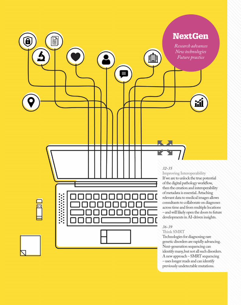
NextGenResearch advancesNew technologiesFuture practice
32-35Improving Interoperability
If we are to unlock the true potential
of the digital pathology workflow,
then the creation and interoperability
of metadata is essential. Attaching
relevant data to medical images allows
consultants to collaborate on diagnoses
across time and from multiple locations
– and will likely open the doors to future
developments in AI-driven insights.
36-39Think SMRT
Technologies for diagnosing rare
genetic disorders are rapidly advancing.
Next-generation sequencing can
identify many, but not all such disorders.
A new approach – SMRT sequencing
– uses longer reads and can identify
previously undetectable mutations.

NextGen32
After attending a recent digita l
pathology event, I became excited
about the interoperability commitments
indust r y vendors a re mak ing.
Breakthroughs from standards-based
working groups, such as Digital
Imaging and Communications in
Medicine (DICOM) and the Integrated
Healthcare Enterprise (IHE), along
with updated guidance from the US
Food and Drug Administration,
have set the stage for a more
sustainable and innovative approach t
digital pathology.
The recent increase of activity in the
DICOM standards community, for
example, is a clear sign of progress.
The f irst meeting of DICOM
Working Group 26 (DICOM WG-
26) took place in 2005, but the scope
of their Connectathon interoperability
demonstrations at this year’s Pathology
Visions conference reached new levels.
For the uninitiated, both the DICOM
organization and IHE are standards
development organizations, with the
goal of improving the interoperability of
health information technology (HIT)
systems – addressing specific clinical
needs in support of optimal patient
care. The DICOM organization was
established in 1983 to develop a specific
standard for the communication of
medical imaging information. DICOM
WG-26 is a working group within the
DICOM organization that focuses on
the development of a DICOM imaging
standard for whole-slide imaging (WSI)
in digital pathology. IHE promotes
the coordinated use of established
standards to achieve interoperability
between HIT systems and effective use
of electronic health records (EHRs) –
and has a strong relationship with
DICOM. Pathology and laboratory
medicine (PaLM) is a domain of
the IHE that addresses information
sharing and workflow related to in
vitro diagnostic testing in anatomic
pathology, clinical laboratories, and
at the point of care. The standards
and use cases have been defined and
vendors are starting to adopt them –
first in academic research organizations
Improving InteroperabilityFor an efficient AI-powered diagnostic workflow, it is crucial to ensure that pathology images and associated metadata are connected at the source
By David Dimond
“[DICOM] was
established in 1983
to develop a specific
standard for the
communication of
medical imaging
information.”

www.thepathologist.com
NextGen 33
a n d t h e n w i t h i n t r a d i t i o n a l
healthcare providers.
I’ve evangelized DICOM and IHE
standards for Radiology and Cardiology
PACS for years. I’ve built teams who
used the standards in both advisory
and consulting work to help select,
design, and implement some of the
largest PACS systems in the US. These
experiences taught me to appreciate
the nuances of technology integration
in healthcare.
Mining metadata
Although the increase in interest is
exciting, there’s a key feature at the
center of these developments that
requires careful consideration. From
my observations, what we need going
forward are “metadata-enhanced
workflows.”
Digital pathology has the potential
to become the new standard of care,
but digitization is only one part of the
equation. Digital pathology’s biggest
strength comes from metadata – the
information that can be attached
to digital images to become part of
an efficient medical workflow. This
includes everything relevant to the
images: annotations, medical notes,
patient identification, clinical order
data, modality protocols, and date and
time stamps. It is the composite of all
information that is associated with a
medical image, whether collected at the
time of imaging or added after the fact,
to create a “packet” needed for workflow
enablement, data integrity, provenance,
and security.
Without metadata, an archive of
WSI files is like storing files on a hard
drive using just file and folder names
– cumbersome to categorize, search,
and use. Conversely, if the pathology
images come with embedded data
and (more importantly) DICOM-
standardized tags, there is a greater
opportunity to generate insight during
the clinical diagnostic workflow and
research phases. Subsequent patient
care also becomes interconnected as
the metadata builds, ensuring that
each WSI file retains an audit trail,
and giving diagnosticians and clinicians
access to a holistic, longitudinal patient
Figure 1: Chord diagram illustrating connections between cancer subtypes using image search and associated slide metadata. Image courtesy of Kimia
Lab and Huron Digital Pathology.
“Digital pathology’s
biggest strength
comes from metadata
– the information
that can be attached
to digital images to
become part of an
efficient medical
workflow.”

NextGen34
view. Researchers also benefit because
metadata enhances their ability to
harvest high-quality data from biobanks
and distributed research archives.
The ability to “line up” the WSI
tags with diagnostic images, case
histories, and electronic health record
(EHR) data also allows pathologists to
effectively collaborate with virtual teams,
such as tumor boards, for difficult-to-
diagnose cases. This has the potential to
significantly impact patient survival rates
because consultations happen rapidly and
collectively. Furthermore, peer reviews
and multidisciplinary meetings can
become more seamless. Considering
the global shortage of pathologists,
streamlining their workflow will yield an
enormous efficiency benefit, helping them
use their valuable time more effectively.
Putting the pieces together
Combining data with images is a
complex task that relies heavily on
improved data interoperability. When
radiologists made the switch to digital,
for instance, it took most radiology
departments up to 15 years to realize the
benefits of enterprise Picture Archiving
and Communication Systems (PACS).
The prior lack of interoperability
inhibited productivity, slowing down
adoption rates. The good news is that,
in comparison, progress in digital
pathology technology is happening
at light speed – largely thanks to the
groundwork radiology has laid. For
example, DICOM Working Group
26 is leveraging DICOM standards by
combining WSI images and patient data
into one format. It’s essential to have
a standard like this so that equipment
and software from different vendors can
interoperate. DICOM Working Group
26 is also collaborating with teams
from the IHE initiative in pathology
and laboratory medicine to define how
data relating to specimens, diagnostic
observations, and documentation should
be structured.
For pathologists to take full advantage
of digital pathology, they must look at
it from a workflow-first perspective
and determine what can be improved
when moving to a digital workflow.
The ideal digital workflow includes
access to all case-related whole-slide
images via an image viewer integrated
with the laboratory information
system (LIS). Pathologists can then
capture regions of interest – preferably

NextGen 35
using some automated capabilities
such as barcodes and other forms of
metadata – and seamlessly export
these to the LIS report as needed.
They should then be able to share this
data with colleagues around the world
for secondary consults, using vendor-
agnostic communication protocols for
improved collaboration. One of the key
goals is to invest in an interoperable
digital pathology solution, avoiding
the pitfalls of potentially obsolete
proprietary systems in the future – a
problem currently seen with some of the
post-PACS radiology Vendor Neutral
Archive (VNA) implementations.
I currently work with medical
institutions to develop and implement
this type of workflow. By partnering
with leading digital pathology scanner
and PACS vendors, we gather bundled
solutions to enable data portability and
accessibility. For example, we provide
the technology and infrastructure
solutions to enable one vendor’s unique
indexing technologies, which adds
metadata in the form of hardware-
generated barcodes at the point of
scanning to link the relevant data to the
patient EHR from the outset. We did
this by developing specialized storage
solutions to maintain the link between
images and metadata to span multiple
public and private clouds in various
geographic locations. By implementing
this approach, healthcare organizations
not only gain the data mobility and
accessibility needed, but also avoid
the “data gravity” problem, in which
the sheer size of data impedes its use
outside its original repository.
As the quantity of digital pathology
data grows, increasingly sophisticated
analytical platforms are needed to glean
new insights, opening doors to machine
learning and artificial intelligence
(AI). There is growing awareness –
particularly from IT customers – that
there’s a wealth of future benefits
in store for patient treatment when
insights from digital pathology images
and data can be combined with other
disciplines like genomics and radiology.
By systematical ly tagging WSI
f iles with metadata, organizations
can categorize whole-slide images
by patient name, diagnostic site,
institution name, and more. They can
integrate images with patient reports
by cross-referencing demographic
data, automatically control access to
clinical data based on level of network
access, label images for rules-based
data retention purposes, and generate
end-to-end custodial audit trails from
the moment of making the scan. The
resultant metadata-enhanced digital
pathology files allow healthcare and
life sciences organizations to enhance
clinical collaboration, streamline
reporting, strengthen patient data
security, and simplify data management.
Within this highly specialized
area of digital pathology, research
organizations and industry must work
together on solutions that incorporate
the standards referenced in this article.
By doing so, they will be able to share
pathology data seamlessly with other
departments, both internally and with
other healthcare and life sciences
organizations. Once the integration of
disparate systems with well-formulated
and standardized data tags has been
established, digital pathology can take
the next step toward an enhanced
workflow across the continuum of care.
From there, a whole new world of AI-
driven, federated data analytics tools
will present opportunities that we’ve
only just begun to imagine.
David Dimond is Chief Innovation Officer, Global Healthcare – Life Sciences at Dell Technologies, Cambridge, Massachusetts, USA.
“The ideal digital
workflow includes
access to all case-
related whole-slide
images via an
image viewer
integrated with the
laboratory
information system.”

NextGen36
We are in a golden age of rare disease
research. Never before have our
laboratory techniques been so successful at
identifying rare diseases and elucidating
their underlying biological causes. The
knowledge we obtain today opens
the door to new treatments, giving
hope to people who suffer from these
rare disorders.
Many of the recent advances in rare
disease research stem from technology
innovations in DNA sequencing. Falling
costs have increased access to whole exome
and whole genome sequencing as tools
to assess the genetic basis of individual
rare disease cases. And in a relatively
short time, the genome-scale data these
methods produce has transformed the
community’s understanding of how these
diseases arise through rare genetic
mutations. There are now more than
7,000 known rare and Mendelian genetic
diseases identified – with more added
to the databases every year – providing
an invaluable information resource for
genome-wide screening and exploration.
But even with these next-generation
sequencing (NGS) tools, clinical research
teams have been unable to explain the
genetic basis behind a large percentage
of rare disease cases. Solve rates range
from 25 to 50 percent, leaving many
individuals and their clinical teams
without an answer to end the diagnostic
odyssey. With an estimated 400 million
people worldwide affected by a rare
disease, there is still a large underserved
population and a pressing need to improve
our diagnostic yield.
To that end, scientists have begun
deploying a higher-resolution DNA
sequencing technology known as single
molecule, real-time (SMRT) sequencing.
SMRT sequencing differs from previous
NGS tools by providing longer reads and
even higher accuracy (1). In just the past
few years, researchers have used SMRT
whole genome sequencing to solve
previously intractable rare diseases – and
other significant efforts are now underway.
The long and the short of it
NGS tools use a variety of approaches
to generate sequence data. What they
have in common, though, is that they
all produce short-read data. Massively
parallel short-read sequencing platforms
have a low cost per run, but generate
sequence reads that are typically only
50–350 base pairs long. To identify
genetic abnormalities, these short
reads are either mapped to a reference
genome or bioinformatically “stitched back
together” in a complex assembly process.
Short reads are useful for detecting
certain variant types known to occur
in the human genome, such as single
nucleotide variants (SNVs) and insertions
or deletions (indels) less than 10 base
pairs long. But, for larger variants, short
reads are of limited utility. The challenge
lies in mapping across larger structural
variants and indels; for instance, those
that occur in repeat expansion disorders,
such as fragile X syndrome, amyotrophic
lateral sclerosis, and schizophrenia.
Mapping issues associated with read
length also limit short-read sequencing’s
ability to call variants across an entire
genome. This includes variants in 193
medically relevant genes in the exome.
For example, a 200-base sequence read
may align to many different regions of the
reference genome, leading to sequence
gaps and conflating similar regions, such
as pseudogenes, repetitive regions, and
mobile elements (see Figure 1).
Short reads also tend to defy variant
phasing efforts, making it impossible to
distinguish maternally from paternally
derived haplotypes. Why does this
matter? It can be important when
determining whether an individual has
a functional copy of a gene in cases where
multiple mutations are present. Perhaps
most importantly, short-read sequence
information often misses the structural
variants (50 base pairs or longer; see Figure
2) that comprise most of the sequence
variation between any two individuals’
genomes (2). These larger variants can
be called incorrectly or excluded entirely
from genomes sequenced with short-
read data alone.
In contrast, longer individual reads
can fully cover even large structural
Think SMRTSolving rare disease with single molecule, real-time sequencing
By Luke Hickey
Figure 1. Short-read sequencing produces reads of 50–350 bp, which can lead to sequence gaps and
incomplete coverage of disease-causing gene regions. Long-read sequencing produces reads tens of
kilobases long, providing high-quality mapping across a genome for comprehensive variant detection.
NeNeNextxtx Geeeennnnn36
TThink SMRTSolving rare disease withsingle molecule, real-timesequencing
By Luke Hickey

www.thepathologist.com
NextGen 37
variants, removing assembly ambiguity
problems and revealing several times
more structural variants, with higher
precision and recall, than short reads (3).
Genomic dark matter
As short-read systems became more
affordable, scientists were eager to
apply these new genetic tools to rare
disease cases. Exome sequencing and,
eventually, whole genome sequencing
with these platforms turned out to be a
game-changer. Diseases that had long
resisted explanation were suddenly
understandable thanks to DNA sequence
data. It seemed that there was finally
an approach that could discover as-yet
unknown pathogenic variants in disease-
causing genes, giving affected families
long-sought answers.
Certainly, short-read NGS tools have
significantly increased the diagnostic
yield for rare disease cases, but they
cannot provide answers for every
situation. In fact, scientists estimate that
NGS platforms leave more than half of
rare disease cases still unsolved.
Could these remaining diseases
be caused by something other than
genetic mutations? Unlikely, given
what we already know about rare
diseases; based in part on hereditary
patterns and syndromic features, the
vast majority appear to be driven by
genetic mechanisms (4). What seems
more likely is that over 50 percent of
genetic mutations accountable for these
diseases are invisible to short-read
technologies. We do know that several
genetic variants can be pathogenic –
including repeat expansions, large
deletions, complex rearrangements,
transposable elements, and more. Now,
with growing awareness that short-read
NGS tools cannot accurately detect most
pathogenic structural variants, scientists
are turning to long-read sequencing –
and answers have begun to emerge.
Identifying pathogenic
structural variants
In one of the first examples of SMRT
sequencing on a rare disease, scientists
from Stanford University reported the
discovery of a disease-causing mutation
in an individual who had suffered a series
of benign tumors over the course of two
decades (5). Although the patient met
the clinical criteria for Carney complex,
a rare genetic disorder, experts had spent
eight years performing various types
of genetic analyses without success.
Because they could not f ind the
underlying mutation, they were unable
to provide a confirmed diagnosis.
Ultimately, Stanford scientists decided
to try SMRT sequencing, which led to
Figure 2. The types of sequence variants found in a human genome. Variants range in size from 1 bp
(single nucleotide variant), to >50 bp for larger structural variants such as deletions, insertions,
duplications, inversions translocations, and copy number variants (3).
Figure 3. A workflow for identifying pathogenic mutations in rare disease cases. Adapted from (5).
NextGen 37
causing genes, giving affected families
long-sought answers.
Certainly, short-read NGS tools have
significantly increased the diagnostic

NextGen38 NeeextxtxtxtGeGGG n38

www.thepathologist.com
NextGen 39NeNeNeeeNeeeextxxxttx GGGeeeennnnnnnnn 39
the answer – a disease-causing deletion,
stretching more than two kilobases,
that affects the gene associated with
Carney complex. The team sequenced
the same region in the individual ’s
parents, finding that neither carried
the same mutation, allowing them to
classify the mutation as de novo in the
affected patient.
In another case, researchers at Yokohama
City University and other institutes in Japan
deployed SMRT sequencing to investigate
the genetic mechanism responsible for the
progressive myoclonic epilepsy affecting
two siblings in a family (6). But it was
not their first attempt to find an answer;
previous efforts, including exome
sequencing with short-read sequencing
tools, had proven unsuccessful. SMRT
sequencing allowed the scientists to
focus on potentially causative structural
variants. A quick filter of the over 17,000
structural variants found across the genome
led to a homozygous 12.4 kilobase deletion
in a gene known to be associated with
a disease that causes similar clinical
symptoms to those found in the siblings.
Notably, the deletion fell in a region with
high GC content, which poses processing
challenges to short-read sequencers.
Follow-up testing confirmed the deletion
and proved that it was pathogenic.
There have been many other rare disease
diagnostic victories based on SMRT
sequencing (see Figure 3). Among them,
advances in repeat expansion disorders
stand out. These large runs of repetitive
sequence are associated with a wide
range of conditions, and the number
of repeats is often closely tied to the
severity of disease. Long-read sequence
data can fully span these large regions
and deliver direct, countable results – an
approach that has been used for ataxias,
fragile X syndrome, myotonic dystrophy,
and other disorders (7–11).
Large-scale efforts to solve rare disease
As the number of reported successes ramps
up, scientists engaged in rare disease research
are adopting SMRT sequencing technologies
more readily.
In Europe, the SOLVE-RD consortium
consists of nearly two dozen institutions
in 10 countries. Funded by a €15 million
award from the European Union, SOLVE-
RD works to improve the diagnosis and
treatment of rare diseases that have evaded
explanation. The program will sequence 500
whole genomes with long-read sequencing
tools to find disease-causing variants and
increase solve rates.
In the USA, the National Institutes
of Health-funded Clinical Sequencing
Exploratory Research program uses SMRT
sequencing as part of a large effort to increase
the diagnostic success rate for pediatric cases
that have proven challenging with other
approaches. Scientists at the HudsonAlpha
Institute for Biotechnology are generating
whole genome sequences for hundreds of
children with intellectual and developmental
disabilities for which the genetic cause has
not yet been found.
Large-scale programs like these should
contribute a significant amount of new
knowledge about the genetic mechanisms
underlying rare disease, filling in many
of the gaps in our understanding today.
As SMRT sequencing helps to explain more
rare diseases and increase overall diagnostic
yield, it should have a profound effect on
our ability to diagnose, understand,
and ultimately improve treatment for
rare disease cases.
Luke Hickey is Senior Director of Strategic Marketing at Pacific Biosciences, Menlo Park, California, USA.
References
1. AM Wenger et al., “Accurate circular consensus
long-read sequencing improves variant detection
and assembly of a human genome”, Nat Biotechnol,
37, 1155 (2019). PMID: 31406327.
2. J Huddleston et al., “Discovery and genotyping of
structural variation from long-read haploid
genome sequence data”, Genome Res, 27, 677
(2017). PMID: 27895111.
3. EE Eichler, “Genetic variation, comparative
genomics, and the diagnosis of disease”, N Engl J
Med, 381, 64 (2019). PMID: 31269367.
4. National Human Genome Research Institute,
“Rare Diseases FAQ” (2020). Available at:
https://bit.ly/2V9uz8X.
5. J Merker et al., “Long-read genome sequencing
identifies causal structural variation in a
Mendelian disease”, Genet Med, 20, 159 (2018).
PMID: 28640241.
6. T Mizuguchi et al., “A 12-kb structural variation
in progressive myoclonic epilepsy was newly
identified by long-read whole-genome sequencing”,
J Hum Genet, 64, 359 (2019). PMID:
30760880.
7. S Mitsuhashi, N Matsumoto, “Long-read
sequencing for rare human genetic diseases”, J Hum
Genet, 65, 11 (2020). PMID: 31558760.
8. T Mantere et al., “Long-read sequencing emerging
in medical genetics”, Front Genet, 10, 426 (2019).
PMID: 31134132.
9. I Höijer et al., “Detailed analysis of HTT repeat
elements in human blood using targeted
amplification‐free long‐read sequencing”, Hum
Mutat, 39, 1262 (2018). PMID: 29932473.
10. B Schüle et al., “Parkinson’s disease associated with
pure ATXN10 repeat expansion”, NPJ Parkinsons
Dis, 3, 27 (2017). PMID: 28890930.
11. S Ardui et al., “Single molecule real-time (SMRT)
sequencing comes of age: applications and utilities
for medical diagnostics”, Nucleic Acids Res, 46,
2159 (2018). PMID: 29401301.
“Scientists engaged
in rare disease
research are adopting
SMRT sequencing
technologies more
readily.”

W E E K LY N E W S L E T T E R S
Brought to you by Texere Publishing,
the parent company of The Pathologist
The week in cannabis science
Everything cell and gene therapy
T E X E R E N E W S L E T T E R S . C O M
The emerging science of the outbreak

ProfessionYour career
Your businessYour life
42-47The Pathfinder of Pathology
Well known for his extensive work
in soft tissue pathology, Christopher
D.M. Fletcher has spent over 35
years accumulating knowledge in the
laboratory, the classroom, and even
at the writer’s desk of a prominent
histopathology textbook. Here, he shares
his wisdom in a peer-to-peer interview
with pathologist Pallavi A. Patil.
Christopher Fletcher is Professor of
Pathology at Harvard Medical School,
Senior Pathologist and Vice Chair of
Anatomic Pathology at Brigham and
Women’s Hospital, and Chief of Onco-
Pathology at the Dana-Farber Cancer
Institute. With an extensive history of
awards and publications, his long-time
focus is on the pathologic diagnosis and
classification of soft tissue tumors.
His 35-year-long career has yielded
wisdom in many facets of pathology, and
he is highly regarded as a mentor by those
who have had the privilege of learning
from him. Now, Pallavi A. Patil – a fellow
in gastrointestinal and liver pathology at
Yale University – interviews Fletcher to
spread his wisdom to those pathologists
and laboratory medicine professionals
who have not yet had the opportunity
to meet him.
What inspired you to choose pathology?
I discovered I liked pathology in medical
school. I did reasonably well at it but,
truthfully, I did reasonably well in most
subjects – pathology just somehow grabbed
my attention. I realized that it’s important
because it’s the underpinning of so many
parts of medicine. I understood clearly
that pathology was the field for me when
I was taking the medical school final
examinations. If you knew pathology, you
could answer 80 percent of every exam
paper. Pathology is incredibly important.
On several occasions, I have been
fortunate to identify a new entity in soft
tissue pathology. Although it can be a
challenge to be the first to report such
a discovery to the scientific community,
it’s also a privilege – and it comes from
two things.
First, if you are lucky enough to see a lot
of consult cases, you have a better chance
of seeing something new – and potentially
seeing it more than once. I see cases every
single day that I can’t identify; some are
probably novel disease entities, but I don’t
see enough of them to recognize them.
Second, a lot of it probably relies on
visual memory. I am constantly amazed
that we still see and describe previously
unrecognized entities even now. We are
currently working on a couple for which
I started collecting the cases in the early
1990s, nearly 30 years ago. When I think
something is going to be a novel disease
entity, I try to use certain keywords to
make it easier to retrieve the cases from our
files. I find it astonishing that, for example,
if we pull 50 cases of something that has
never been described but is in my head, 45
of them usually look pretty much identical.
Fortunately, the keywords make it easy to
collate and describe them.
You’ve edited a key textbook in your
field, the Diagnostic Histopathology of
Tumors. How did that come about?
Everything in life is an accident – at least, I
haven’t planned anything in my life. In the
early 1990s, Nicholas Wright, Chairman
of Pathology at the Royal Postgraduate
Medical School in London, suggested
to Churchill Livingstone the idea of
publishing a new book focused only on
diagnostic pathology of tumors. They
contacted me and I, in turn, selected the
contributors for each individual chapter.
(I did, and still always do, edit every
chapter to make sure they don’t feel
dramatically different from one another.)
My involvement in the book came about
by serendipity, but it’s hard work that keeps
it up-to-date and in print.
Elsevier eventually bought Churchill
Livingstone and now expect a new edition
every five years or so. An awful lot of new
information is generated in that time; as a
result, they considered an online edition
that could be edited continuously, but
that would be a full-time job for an editor
– and no one has the time. Some have
suggested delegating the task of keeping
topics up-to-date to younger pathologists,
but it’s important to ensure that each topic
has high-quality contributors aware of the
latest developments in their fields, because it’s
works like these that inform how we and our
colleagues approach diagnostic medicine. It’s
not a responsibility to be taken lightly.
Personally, I spend the years in between
editions collecting papers that I think are
important. I am old-fashioned, so I have
them printed out and filed in different
folders instead of saved to my computer.
When I get a new chapter, I pull out the
relevant folder and make sure all of the new
information is included. It’s a lot of work!
Although the current edition is finished
and in the proof stage, I still keep copies
of important papers – I’ve been using this
system ever since the first edition. I used
to receive hard copies of journals, but now
I just get content lists and have to view the
articles online. It’s difficult to find that kind
of time, and it makes adding references
much harder.
I’ve just finished work on the fifth
edition, but I foresee a change in the book’s
future. People seem to like smaller books
nowadays. It’s fashionable to have skinny
books with bullet points rather than large
amounts of free text. I don’t think people
will want to use big, older-style books
for much longer. Publishers aim to make
money; they always want a new product
and a new market, but no pathologist gets
rich writing books – not even skinny books.
Publishers did themselves a disservice by
distributing digital editions; after all,
if many users (usually in one pathology
The Pathfinder of PathologyChristopher D.M. Fletcher discusses the changes that have taken place over a decades-long career in pathology – and the field’s promise for the future
Pallavi A. Patil interviews Christopher D.M. Fletcher
paper. Pathology is incre
On several occasion
fortunate to identify a n
tissue pathology. Alth
challenge to be the fir
a discovery to the scien
it’s also a privilege – an
two things.
First, if you are lucky e
of consult cases, you hav
of seeing something new
seeing it more than once. I see cases every
single day that I can’t identify; some are
probably novel disease entities, but I don’t
topics up to date to younger pathologists,
but it’s important to ensure that each topic
has high-quality contributors aware of the
Christopher D.M. Fletcher discusses the changes that have taken place over a decades-long career in pathology – and the field’s promise for the future
Pallavi A. Patil interviews Christopher D.M. Fletcher
TTTTTTTTTTTTTTTTTTTTTThhhhhhhhhhhhhhhhhhhhheeeeeeee PPPPPPPPPPPPathfinder oooooooof PPathology
edibly important.
ns, I have been
new entity in soft
hough it can be a
rst to report such
ntific community,
nd it comes from
enough to see a lot
ve a better chance
w – and potentially
e. I see cases every
by serendipity, but it’s harddd wowoworkrk that kekekeepepeps s
it up-to-date and in print.
Elsevier eventually bought Churchill
Livingstone and now expect a new edition
every five years or so. An awful lot of new
information is generated in that time; as a
result, they considered an online edition
that could be edited continuously, but
that would be a full-time job for an editor
– and no one has the time. Some have
suggested delegating the task of keeping
topics up-to-date to younger pathologists,
Profession42

www.thepathologist.com
department) can share one book, why
purchase multiple copies?
What energizes you for your many tasks
– managing administration, a large
consult service, teaching, and editing a
major book?
It’s nice to feel that I’m doing something
useful. I enjoy helping young pathologists
get into their careers. I never thought I
would end up managing so many people,
but it turns out that I quite like it. It does
become somewhat tiring; here in anatomic
pathology at the Brigham, we have around
70 faculty and 50 trainees! With so many
people, you have to keep an eye on things
like workload, logistics, interpersonal
issues, trouble at home… In the end,
people are important. If you don’t care
about people, they will not do well.
There is one thing I often used to say
about administration and leadership that
I don’t say much anymore: that if you don’t
do it yourself, somebody else will, and they
may mess it up. There are all sorts of people
I would not want to be my boss!
Multitasking has become part and
parcel of professional life. What
would you advise faculty and trainees
regarding burnout?
If you think about it, that whole concept
has emerged in the last four or five years.
There isn’t really a good work-related reason
why people should burn out more now
than they did 10 or 20 years ago – except
that society has changed. Now everybody
wants everything fast. Sensory input is
more immediate. People (not me) have
to deal with social media all the time. I
completely avoid social media because I
think I would shoot myself. I find it hard
enough to keep up with email.
I get 200 to 300 emails a day that are
not spam and I do my best to take care of
them. Nowadays, you can send a question
to someone by email instantly and you
often know exactly when they received it,
too. In the old days, people would send a
letter that took a week to arrive and then
you would write a letter back. The pace of
interaction was slower. Now, because access
is so unlimited, everyone can find everyone
else via email. I get emails from doctors and
patients around the world every single day.
I used to find it very stressful trying to fit
them into my day. Now I usually just do
many of them in the evening. But despite
the stress, it is humbling to have all those
people communicating with you – and you
learn a lot about disease.
My advice regarding burnout would be:
don’t overcommit yourself. I will be honest;
I think a lot of it is simply a reflection of how
modern society works, so it’s not something
you can control. You will get bombarded
with emails, texts, and Twitter messages –
and, if that’s how you grew up, you think
it’s a normal part of life. I am lucky not to
have grown up dealing with that. I am old
enough now that I can ignore them and get
away with it. If you are 25 years old, you
can’t do that or people may think you are
a “weirdo.” Maybe what we call burnout is
just becoming a part of life – there seems
to be a constant level of overstimulation.
The other thing is that people nowadays
seem to have unrealistic expectations,
perhaps because so many things are
instantaneous. When we get consults,
people (usually clinicians, not pathologists)
from referring hospitals start calling within
hours of the cases’ arrival to find out if there
is a report or diagnosis. This happens even
when they themselves have worked on the
case for a couple of weeks before sending
it to Boston. When a case is that difficult,
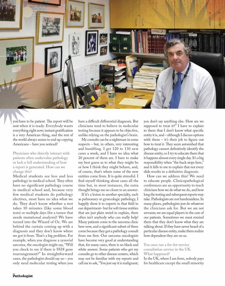
you have to be patient. The report will be
sent when it is ready. Everybody wants
everything right now; instant gratification
is a very American thing, and the rest of
the world always seems to end up copying
Americans – have you noticed?
Physicians who directly interact with
patients often undervalue pathology
or lack a full understanding of how
a report is generated. How can we
change this?
Medical students see less and less
pathology in medical school. They often
have no significant pathology course
in medical school and, because very
few medical students do pathology
electives, most have no idea what we
do. They don’t know whether a test
takes 10 minutes (like some blood
tests) or multiple days (for a tumor that
needs mutational analysis)! We have
turned into the Wizard of Oz. We are
behind the curtain coming up with a
diagnosis and they don’t know where
we got it from. That’s a big problem. For
example, when you diagnose a synovial
sarcoma, the oncologist might say, “Will
you check to see if there is SS18 gene
rearrangement?” In straightforward
cases, the pathologist should say no – you
only need molecular testing when you
have a difficult differential diagnosis. But
clinicians tend to believe in molecular
testing because it appears to be objective,
unlike relying on the pathologist’s brain.
My consults can be a nightmare in some
respects – but, in others, very interesting
and humbling. I get 120 to 130 new
cases a week, and I have no idea what
20 percent of them are. I have to make
my best guess as to what they might be
or how I think they might behave, and,
of course, that’s where some of the new
entities come from. It is quite stressful. I
find myself thinking about cases all the
time but, in most instances, the extra
thought brings me no closer to an answer.
If it’s a lesion in another specialty, such
as pulmonary or gynecologic pathology, I
happily show it to experts in that field in
our department– but for soft tissue entities
that are just plain weird to explain, there
often isn’t anybody who can really help!
Many patients come to the sarcoma clinic
here now, and a significant subset of them
come because they got a pathology consult
from me first. Our sarcoma oncologists
have become very good at understanding
that, for many cases, there is no black and
white answer. Some patients who get my
consults go to other disease centers, which
may not be familiar with my reports and
call me to ask, “You just say it is malignant;
you don’t say anything else. How are we
supposed to treat it?” I have to explain
to them that I don’t know what specific
entity it is, and – although I discuss options
with them – it’s their job to figure out
how to treat it. They seem astonished that
pathology cannot definitively identify the
disease entity, so I try to educate them that
it happens almost every single day. It’s a big
responsibility when “the buck stops here,”
and it falls to me to explain that not every
slide results in a definitive diagnosis.
How can we address this? We need
to educate people. Clinicopathological
conferences are an opportunity to teach
clinicians how we do what we do, and how
long the testing and subsequent reports may
take. Pathologists are not handmaidens. In
many places, pathologists just do whatever
the clinicians ask for. But we are not
servants; we are equal players in the care of
our patients. Sometimes we must remind
them that they don’t know what they are
talking about. If they have never heard of a
particular disease entity, make them realize
how dependent they are on us.
You once ran a fee-for-service
consultation service in the UK.
What happened?
In the UK, where I am from, nobody pays
for healthcare except the small minority
yoou uuuuu have to be patient. The report will
sent when it is ready. Everybody wan
everything right now; instant gratificati

www.thepathologist.com
who use private practice. It’s a socialized
system – but there was a time in the early
1990s when the government wanted to
make healthcare market-driven. They
wanted the referring hospital to pay
the receiving hospital for every patient
or case that went from one district to
another, even though it was all one
central budget and the hospitals were all
a part of the National Health Service. I
was unlucky because they picked me as
a pilot project for consultation services.
They instructed me to send bills for the
consults, starting with those from the
UK. Nobody in the UK or Europe had
ever billed for a pathology consult before.
As you can imagine, people were very
angry and called to say, “What is this
bill? Nobody else sends bills.” I spent
hours on the phone explaining that it
was the government who wanted to send
the bill, not me!
I had a soft tissue tumor unit with two
histotechnologists and a secretary. The
hospital administrators were instructed
to close the unit down if I didn’t bring
in enough money to cover salaries for
the technicians and secretary, as well as
lab costs (which, of course, I never did).
They put the staff on monthly renewable
contracts instead of annual ones. It was
quite stressful. European patients generally
didn’t have to pay for healthcare in other
European countries – but then the UK
Department of Health decided that I
should start billing for European consults
as well. That was funny, because cases
from Europe were sent anyway without
ever sending payments. In the UK, people
were wondering whether they could keep
sending cases or not, what was going to
happen with the bills, and whether they
were going to get into trouble. It was in
the newspapers and on television. It was
horrible – but the government scheme
failed and was eventually dropped.
There is an increasing trend in
the USA towards mergers and
corporatization, particularly in
pathology. How do mergers
affect pathology?
Medicine in the USA has become more
and more corporate, and less and less
a profession where you try to heal the
sick. Physicians are often employees
of a company that is trying to make
money. Even not-for-profit organizations
need funding to put up new buildings
or to buy the latest technology. It has
various negative consequences. For
example, many places want doctors to
do more and more work with less and
less support. Even hospitals here in
Boston cut back on support staff, such
as histotechnologists, secretaries, and
so on. They make changes in the name
of efficiency – things like asking us to
use voice recognition rather than typing
assistance. It just puts pressure on doctors
to do more with less help.
To ma x imize bi l l ing revenue,
administrators want the most complex
cases to be done in the big hospitals.
Big teaching hospitals have hundreds of
trainees, complicated equipment, and lots
of expenditures. In hospital networks, they
try to push smaller procedures to the smaller
hospitals – for example, lumpectomies,
herniorrhaphies, or varicose vein surgeries
– because their overhead isn’t so high. It
changes the pathology as well. I have been
here at Harvard for almost 25 years and
the case mix is constantly changing. I see
less and less simple material. Our trainees
rarely see a hernia sac, an appendix, or a
gallbladder. A lot of the simple things have
just disappeared because they’re now done
in the community hospitals.
How much should pathologists
interact directly with patients?
Decades ago, I established a patient
consultation practice in the UK. I had
no plans to do so, but I had started
getting quite a few consults and people
wanted treatment advice. We didn’t
have a sarcoma clinic in the hospital I
worked at in the late 1980s. I persuaded
an orthopedic surgeon to be interested
in limb tumors, a general surgeon to
take care of trunk and retroperitoneal
tumors, and an oncologist (who did both
radiation oncology and chemotherapy
back then because there wasn’t much
chemotherapy) to handle that side of
things. This group of practitioners began
seeing patients and it grew into an actual
scheduled sarcoma clinic. I would see the
patients with them, and on my own if
they were out of town. If the patients got
lung metastases, they often sent me into
the room to explain about metastases!
If patients feel they don’t understand
their disease and need to talk to somebody,
clinicians should always consider directing
them to pathologists – and pathologists
should make themselves available. These
interactions happen more by email in
the US, which is difficult because you
don’t necessarily have precise or complete
patient details (especially if you’re talking
to somebody thousands of miles away).
You have to be very careful about how
you phrase things if you don’t know the
specific circumstances. That said, patients
here sometimes want to come and look at
the slides and are generally appreciative. If
they ask, I don’t think we should ever say
no or be shy of talking to patients. I know
there is a community hospital in Lowell,
Massachusetts, where they recently had
clinicians offer patients, after a diagnosis
of cancer, the option of talking to the
pathologist. Most of the patients who
met with the pathologist loved it and felt
they knew more about their disease. The
conversation helped them visualize the
disease and feel better about it. Clinicians
like to appear to be the ones to know
whether something is benign or malignant,
when in fact the pathologist makes the
diagnosis. The patients end up appreciating
the clinician for work the pathologist has
done. Many clinicians don’t talk about
pathologists too much, so I appreciate the
ones who offer patients the opportunity
g p y p
or case that went from one district to
another, even though it was all one
central budget and the hospitals were all
a part of the National Health Service. I
was unlucky because they picked me as
a pilot project for consultation services.
They instructed me to send bills for the
consults starting with those from the
me more
nd less
a profession where you try to heal the
sick. Physicians are often employees
of a company that is trying to make
money. Even not-for-profit organizations
need funding to put up new buildings
or to buy the latest technology. It has
various negative consequences. For
example, many places want doctors to
do more and more work with less and
worked at in the late 1980s. I persuaded
an orthopedic surgeon to be interested
in limb tumors, a general surgeon to
take care of trunk and retroperitoneal
tumors, and an oncologist (who did both
radiation oncology and chemotherapy
back then because there wasn’t much
chemotherapy) to handle that side of
things. This group of practitioners began
seeing patients and it grew into an actual
scheduled sarcoma clinic. I would see the
patients with them, and on my own if
they were out of town. If the patients got
lung metastases they often sent me into
who use private pracaaa tice. It’s aaaaa socializezezezezezeed
system – but there was a time in the earlllyl
1990s when the government wanted to
make healthcare market-driven. They
wanted the referring hospital to pay
the receiving hospital for every patient
corppppoooroooooooooooooo atization, particularly in
pathology. How do mergers
affect pathology?
Medicine in the USA has becom
and more corporate, and less an
a profession where you try to h
Profession 45

to speak to us – even if not every patient
takes that offer.
One problem is that a lot of people think
of pathology as something vaguely creepy
that has to do with death. I find even now,
when I go to social events, people don’t talk
very much if I say, “I am a pathologist.” If
I say instead, “When you have a biopsy,
people like me decide whether it is benign
or malignant,” then they become interested.
When their perception is that pathology
is autopsies or “weird stuff,” they don’t
want to hear about it. We have an image
problem, and I don’t know how to change
that except to say that we shouldn’t hide.
My father, a surgeon, wasn’t very pleased
when I went into pathology. Remember,
this was in 1982; for him, pathologists
and anesthesiologists were like servants.
Fortunately, he changed his mind after
about 15 years! I find that, when I meet
residency applicants or new residents,
their families may not have much idea
about their profession, and some don’t
even understand that pathologists are
“proper” physicians!
People have a lot of ideas for improving
the image of pathology. There was a time,
about 15 years ago, when pathologists
thought it might be a good idea to make
FAQ lists for diseases that we could give
clinicians to share with patients. That
never really caught on, because clinicians
didn’t necessarily understand the answers
or how to explain them. They didn’t use
them because they felt uncomfortable. If we
make the effort to answer their questions
in person, we can avoid that discomfort
and spread awareness of our profession and
its value.
How can we encourage medical
students to apply for pathology?
The drop in applications is very troubling. I
think that, to solve the problem for the long
term, we have to restore more pathology to
the medical school curriculum. That will
be hard because, in modern education, they
shorten the curricula and pack far too much
into them. In the way that pathology used
to be taught (40 years ago!), systematically,
I had a full three-month pathology course
in my fourth year of medical school. We
learned about etiology, pathogenesis,
and microscopic appearances. There
was no immunohistochemistry or
molecular biology then, but we learned
about outcomes and complications of
disease. So much of that pathology was
invaluable in taking exams across most
other medical specialties!
Over the last 20 years, everybody has
been focused on social medicine and
training good family practitioners. In
almost every medical school, pathology
(and histology and anatomy) teaching is
much more limited than it used to be. The
Medical University of South Carolina
in Charleston does still have a three-
month pathology course just like I had
at medical school, and 10 to 15 students
go into pathology there each year because
they’ve had that exposure. But here at
Harvard, only two or three students each
year go into pathology – so the impact
is obvious. Those students who do come
into pathology residency, no matter how
smart they are, often have very little idea
what they are doing. It’s not their fault;
they have probably never seen normal
pancreas under a microscope! I think
it’s very hard to learn pathology from
that kind of starting point, and I find
it worrying that this is the new normal.
How could pathology training
be improved?
Residency programs are successful when
faculty like to teach. It’s no good when
trainees are taken for granted; they
shouldn’t just be grossing specimens with
little guidance or passively sitting on
the other side of the two-headed scope.
Modern medicine sometimes takes away
teaching time; faculty are frustrated by
the number of commitments and demands
placed on their time and teaching falls
by the wayside. Insurance companies
often demand shortened turnaround
times as a “quality measure,” so trainees
don’t always get to preview things – but
that’s how I think residents gain the
most knowledge. When I met with some
insurance company representatives 20
years ago in Massachusetts, they wanted
our turnaround times to be shorter. They
were trying to find a way to pay less if we
didn’t meet the turnaround times they
wanted to set. Somebody questioned
the need for resident preview time to be
factored in. I said, “Those are the people
who will be looking at your prostate
biopsies in about 15 years. Do you want
us to teach them or not?” They hadn’t
thought about it that way. As soon as
they realized it could be their specimens
under the microscope, they changed
their minds!
I think the most important thing is
to get good training. One of the reasons
residency training in the US was shortened
to four years was financial – to save paying
for that fifth year. But being well-trained
is critical to what we do. It doesn’t matter
how smart you are, you can’t learn without
seeing enough cases. There is a tendency for
places to use trainee logbooks to tick off, for
example, that they have seen 20 colorectal
carcinomas – but then the trainee may
wrongly imagine that they don’t need to
see any more. That’s not true, because the
more you see, the more you realize how
heterogeneous human disease is. There is
no substitute for experience. The more you
see, the better you get, and the more you
realize how much you don’t know.
People go through phases; quite a few
have little or no self-doubt in their 30s
or 40s and think that they know almost
everything. Most of them, if they are
smart, will eventually discover that they
don’t really know as much as they think.
By the time they are in their 60s, they will
hedge a little more often or admit to being
unsure. As you grow older, it becomes
okay to say, “I don’t know.” People don’t
like ambiguity when they are younger;
nt
nk
py
w,
lk
If
y,
gn
or malignant,” then they become interested.
When their perception is that pathology
is autopsies or “weird stuff,” they don’t
want to hear about it. We have an image
into them. In the way that pathology used
to be taught (40 years ago!), systematically,
I had a full three-month pathology course
in my fourth year of medical school. We
learned about etiology, pathogenesis,
and microscopic appearances. There
was no immunohistochemistry or
molecular biology then, but we learned
about outcomes and complications of
disease. So much of that pathology was
invaluable in taking exams across most
other medical specialties!
Over the last 20 years, everybody has
often demand shortene
times as a “quality measu
don’t always get to previe
that’s how I think resid
most knowledge. When I met with some
insurance company representatives 20
years ago in Massachusetts, they wanted
our turnaround times to be shorter. They
were trying to find a way to pay less if we
didn’t meet the turnaround times they
wanted to set. Somebody questioned
the need for resident preview time to be
factored in. I said, “Those are the people
totttttotttttt ssssssssssssssssssssssspppepepppepepepppppepppepepepppepepeakaakakakakakakakakaaaa tto o oo o oo ususuuuuuu – even if nototttttt eeeeeeeveveveeery patien
takekes ssssss thththhhththhhhththhththththththhhhattatatattatataaaaaaaaataaaa ooffffffeff r.
OnOnOnOne e prprprprprprprrprrrprppp obobobobobobbobobobobobobbbbbobobbooboblllllllellllelelelllll m m m mm m m m iisisisisisis ttttttthahahahahahahat t t tt t a a a a a a lololololololol t ttttt of people thin
off ppatthohohhohhhh lollllololll gyggygygygygygy aaaaaaaaas somethththththht inininiii g gg vav guely creep
thhatat hhhhhhhhhhasasasaasaasass tttttto o o o o oo do with deeeath. I find even now
whwhwhhwhwhwwwww enenenenenenenne IIIIIIII gggggggggo to social events, peoplelll don’t tal
vvvveeryryryryyryy muccccch hhhhh if I say, “I am a pathologist.” I
III IIII saay y instead, “When you have a biops
people like me decide whether it is benig
or malignant,” then they become interested
Profession46
ed turnaround
ure,” so trainees
ew things – but
dents gain the
met with some

www.thepathologist.com
they want everything in black and white.
But many things about pathology (and
life!) are ambiguous. There are always
shades of grey.
What do you foresee in pathology’s future?
Genetics and genomics will play a
significant role – but they won’t be the right
solution for everyone. I think every new
technology becomes very important for a
subset of patients. For example, finding a
new therapeutic target via next-generation
sequencing is great for 7 to 10 percent of
cancer patients, but the other 90 percent
don’t benefit at all. In most tumors, it is
currently of limited help. It is fashionable
to try to find something when there are
no options. Patients read newspapers or
leaflets in oncologists’ offices and ask to
have tests done. Physicians order the tests
because the patients (customers!) want
them – and so, medicine is changing. We
have become more responsive to what
patients want, rather than telling them
what should be done. I sometimes deal
with patients who demand tests. If the test
they want is completely irrelevant to their
disease, I try to explain that it is not useful
and that all they are doing is throwing
money away. Clinicians don’t say no very
much these days – and sometimes that can
lead to crazy tests.
When Gleevec came out, people
around the country wanted to test for
KIT (CD117) expression in tumor types
without any known KIT mutations.
American medicine is not very good at
saying, “I am sorry. There is nothing more
we can do, so let’s concentrate on making
you comfortable, reducing your pain, and
finishing your bucket list.” Instead, patients
get endless chemotherapy, complications,
and ICU stays. I don’t know if it is the
patients who refuse to give up, or society
that has created false hope. It is not good.
I wouldn’t do it. Most doctors, when
extremely sick with cancer, avoid crazy
testing, and instead focus on pain relief and
support, and know when there is nothing
more to be done. It is very telling that
most doctors don’t want these aggressive
or protracted treatment efforts – so why
do we do it for so many of our patients?
Telepathology may also become a
routine part of our work, but that depends
on the specimen type. For a small biopsy
with a single slide, it could be very useful
– but if you have 30 slides of a pancreatic
resection, for example, current technology
is still slow and challenging to use. The
technology keeps getting better, so I think
it will probably become useful, but right
now I don’t like it much. When we look
at a glass slide under the microscope, we
all scan the slide at low power – perhaps
subconsciously – before we decide what’s
worth focusing on. It’s usually much
harder when you have a digital slide to
realize what is important at low power,
because software is often slow to focus
when scanning. When people send me
weird cases as digital slides, I find I
usually can’t make a definitive diagnosis.
It takes me much longer than when using
a microscope and glass slides.
It would be great to get an opinion on a
frozen section consult from someone who
is off-campus if you have good software.
We set up a system here more than 10 years
ago but, in the end, nobody used it very
much. Why? Because the person on the
receiving end wasn’t comfortable enough
making a diagnosis that way and would
drive to the hospital to make it in person.
We older folks grew up in pathology the
old-fashioned way and it’s hard to change
brains that are wired that way. Millennials,
on the other hand, have grown up using
technology since early childhood, so they
may more easily become proficient in using
digital images! By the time they are my age,
digital pathology may be entirely routine,
and pathologists may sign out cases from
home without having to come to the
hospital. Society changes!
Christopher DM Fletcher is Professor of Pathology at Harvard Medical School, Senior Pathologist and Vice Chair of Anatomic Pathology at Brigham and Women’s Hospital, and Chief of Onco-Pathology at the Dana-Farber Cancer Institute, Boston, USA.Pallavi A. Patil is a graduate of the Brown University Pathology residency program and fellow in gastrointestinal and liver pathology at Yale University, New Haven, USA.
What do you foresee in pathology’s future?
Genetics and genomics will play a
significant role – but they won’t be the right
solution for everyone. I think every new
technology becomes very important for a
subset of patients. For example, finding a
new therapeutic target via next-generation
sequencing is great for 7 to 10 percent of
cancer patients, but the other 90 percent
don’t benefit at all. In most tumors, it is
they want everything in black and whwhwhite.
But many things about pathology (and
life!) are ambiguous. There are always
shades of grey.

Applicat ion Note 48
Minimize turnaround and maximize clinical utility. PierianDx Clinical Genomics Workspace™ and Clinical Genomics Knowledgebase, completewith ready-to-use interpretations, support for all variant types, and robust clinical sharingnetwork, provide the ideal combination of human expertise and technology to rapidly andaccurately identify clinically significant variants and produce an actionable report. www.pieriandx.com
20mm+ 6Mb+ 60+ 55,000+Number of reviewed
publicationsAmount of sequence
coverage Number of Institutionssharing de-identified
clinical interpretations
Number of unique, signedout interpretations
Clinical NGS Reports for Every Assay
Next-generation sequencing (NGS) technologies facilitate
the accurate detection of genetic variants. Yet, the process of
analyzing and classifying more complex alterations using a
standard variant lookup table remains challenging.
In our informative video, we focus on providing best
practices for the analysis of gene fusions, co-occurring variants,
copy number variants, and tumor mutational burden (TMB).
Further, we provide practical strategies for analyzing and
classifying these complex variants using a comprehensive
knowledgebase to minimize turnaround time and maximize
the clinical utility of the resulting report.
By watching this video, you will:
• Gain strategies for filtering, classifying, and interpreting
variants within a clinical context.
• Learn about bioinformatic methods for calling variants
and assessing quality.
• See example reports using de-identified samples
for several popular NGS assays, including Archer
VariantPlex® and FusionPlex®, TruSight™ Oncology
500, and AmpliSeq.
Analyzing Complex Genomic Variants
The process of interpreting genetic variants can be complex and time-consuming, made even more
so by complex variants, such as gene fusions, co-occurring variants, and tumor mutation burden.
Laboratories must develop best practices for analyzing and classifying these complex variants.

Sponsored Feature 49
Spotlight on... Technology
Precise. Simple. Fast. AccuLift Laser Capture Microdissection System
Get precise and efficient cell capture,
down to single cel ls, even from
challenging tissues. The simplified
design includes a high-precision stage,
uniquely aligned IR and UV lasers, and
a novel consumable cap for more efficient
cell capture. Increase your confidence
in preserving biomolecule integrity for
more successful downstream analysis.
Fluidigm.com/LCM
RedRick Technologies Ergonomic Workstations Alleviate the Risk of Repetitive Stress Injury
The shift to digital pathology will require
pathology departments to create flexible
and stable ergonomic workspaces that
accommodate both a digital pathology viewer
and a microscope. As other digital clinical
departments have discovered, a well-designed
workspace also facilitates collaboration and
teaching and maximizes the use of space.
https://bit.ly/2GXBUBT
MindPeak Showcases Second Product for PD-L1 After Successful Launch of BreastIHC
After the successful product launch of
MindPeak’s BreastIHC, our second pilot
product for PD-L1 stained cell quantification
is coming. First tests on real-world clinical
data show promising results concerning
accuracy and robustness. Our goal is to
relieve pathologists of the burden of PD-L1
scoring in clinical routine and research.
https://www.mindpeak.ai/
Color Reproducibility for Whole Slide Imaging Devices Through ICC Color Management FFEI’s patented Sierra color management
solution enables the standardization of
digital images produced by WSI scanners.
Sierra uses a biologically stained slide that
mimics human tissue. To measure the
spectral accuracy of a scanner, an ICC
profile is then generated – meeting FDA
color reproducibility guidelines.
https://ffei.ai/

The Pathologists’ AssistantSitting Down With… Sarah Garner, Pathologists’ Assistant and Director of the Pathologists’ Assistant Program at Tulane University, New Orleans, Louisiana, USA

www.thepathologist.com
51Sit t ing Down With
What drew you to pathology and a career
as a pathologists’ assistant?
Like many who pursue pathology, I fell
into it and realized it was perfect for
me. During my undergraduate studies,
I worked in internal medicine and saw
patients daily. One day, a man came in
with a large testicular mass and – although
I loved speaking with him and wanted to
help him – what I really wanted to know
was how and why the tumor had formed,
what it looked like, and what treatment
he would need. Now I know I wanted to
understand the epidemiology, pathogenesis,
and morphologic features. That’s the day
I realized I was meant to do something
other than see patients – but, at the time,
I had no idea what pathology really was
(and, unfortunately, none of the physicians
I worked with did either).
While working in internal medicine, I
took a gross anatomy class with cadaveric
dissection. I loved the class so much that
I stuck around as a teaching assistant and
quickly learned that I wanted my career
to involve teaching and learning about the
human body. When I stumbled across the
pathologists’ assistant (PA) profession after
Internet searches for careers in anatomy
and abnormalities, I knew I had found
something that would set my world alight!
It seemed like the perfect way to combine
all my passions: gross anatomy, dissection,
pathology, art, and teaching.
When I speak to other PAs about their
route into the career, many of them have
similar stories about stumbling upon the
profession and realizing it was a perfect
fit. It always makes me wonder how many
people who would really love this job never
find it. We shouldn’t have to stumble
upon this wonderful profession – or any
pathology specialties – and that is why, as
a practicing PA and educator, I strive to
promote pathology and the PA profession
wherever I can.
Can you describe “a day in the life” of
a PA?
That’s impossible to answer. And that’s
exactly why I love my job so much! Even
if I’m technically doing the same things,
every specimen, patient, and day is different.
Sometimes I spend the whole day in the
gross room, grossing specimens or teaching
residents, medical students, and PA students.
Equally, I may be working outside the gross
room – and the majority of my days are spent
doing a little of both. If I had to generalize a
typical day, it would go something like this:
teach gross anatomy to first-year medical
students, spend a few hours in the gross room
either grossing or training others, teach a
didactic class for PA students, and then one
for my graduate and undergraduate students.
Other days can include autopsy, presenting at
resident conferences, and sometimes teaching
for an entire day. I love that I am always
doing something different, because nothing
ever gets boring and I get to work with a lot
of different people.
Are there differences in how you
approach teaching pathology residents
and PAs?
I designed the curriculum for our PA
students and play some role in all of it, so I
always know their exact progress and can
tailor my teaching techniques to fit their
current knowledge. It’s highly personalized,
and the integrated curriculum teaches
them what they need and want to learn.
In contrast, residents typically come from a
variety of medical schools and backgrounds,
so it’s more difficult to gauge individual
needs. Some might have worked extensively
in pathology, whereas others might have
no experience at all. Luckily, our residency
program is relatively small, so I can get to
know each of them and adapt accordingly.
Most of the time, I find it more
challenging to teach a first-year pathology
resident than a PA student in the gross
room, because the PA students have
didactic coursework specifically designed
to teach them the concepts of grossing and
why each section is important. Residents
often don’t arrive with that knowledge,
because it’s something they’re expected to
learn during residency. The residents also
get limited time, or their rotations are
broken up into months spread throughout
the year, so the lack of consistency can
make it more challenging for them to learn
and familiarize themselves with grossing
concepts. I do my best to teach the “why”
behind all of the sections and measurements
and not just the “how,” because I think that
leads to better patient care, whether you’re
a PA or a resident. Our PA curriculum and
residency education overlap significantly –
and all classes and conferences are available
for everyone to attend.
If you could go back and give yourself
one piece of advice at the start of your
career, what would it be?
I would tell myself to be patient, which I
sometimes find difficult because I like to
take on big tasks and I get really excited
about what I’m doing. I’m still learning
how to be patient, especially now that I am
more involved with research. I’m also bad at
accepting help from others and even worse
at asking for it. I am getting better – but
it’s something that would have greatly
benefited me years ago. You don’t have to
do everything yourself!
“I do my best to teach
the ‘why’ behind all
of the sections and
measurements and
not just the ‘how,’
because I think that
leads to better
patient care.”

BFR
0000-7741-01
1 Test. 43 Targets. ~1 Hour.
Innovative diagnostics for evolving bugs.Now available: The BioFire® Blood Culture Identification 2 (BCID2) Panel.
As the leader in syndromic testing, BioFire knows that when bugs evolve, testing should too. Stay ahead of changing multi-drug resistant organisms with the new leading test for bloodstream infections—the BioFire BCID2 Panel. In about an hour, the BioFire BCID2 Panel tests for 43 of the most common gram-positive bacteria, gram-negative bacteria, yeast, and antimicrobial resistance genes—all in a single test.
The BioFire BCID2 Panel
99% Sensitivity. 99.8% Specifi city.*
GRAM-NEGATIVE BACTERIAAcinetobacter calcoaceticus-
baumannii complexBacteroides fragilisEnterobacterales
Enterobacter cloacae complexEscherichia coli Klebsiella aerogenesKlebsiella oxytoca Klebsiella pneumoniae groupProteus SalmonellaSerratia marcescens
Haemophilus infl uenzae Neisseria meningitidis Pseudomonas aeruginosa Stenotrophomonas maltophilia
GRAM-POSITIVE BACTERIAEnterococcus faecalisEnterococcus faeciumListeria monocytogenesStaphylococcus
Staphylococcus aureusStaphylococcus epidermidisStaphylococcus lugdunensis
Streptococcus Streptococcus agalactiae Streptococcus pneumoniaeStreptococcus pyogenes
YEASTCandida albicansCandida aurisCandida glabrataCandida kruseiCandida parapsilosisCandida tropicalisCryptococcus neoformans/gattii
ANTIMICROBIAL RESISTANCE GENESCarbapenemasesIMPKPCOXA-48-likeNDMVIM
Colistin Resistancemcr-1
ESBLCTX-M
Methicillin ResistancemecA/CmecA/C and MREJ (MRSA)
Vancomycin ResistancevanA/B
biofiredx.com
* The stated performance is the aggregate of the prospective data from the clinical study.