Embed Size (px)
Citation preview
CBIS Exam Prep Course Section 2 10/27/2016
Rainbow Rehab Centers 1
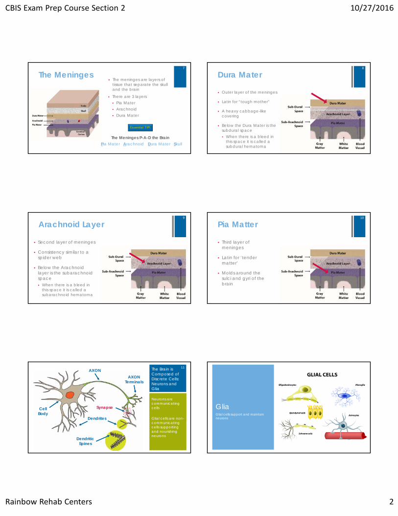
Neuroanatomy & NeuroplasticitySection 2
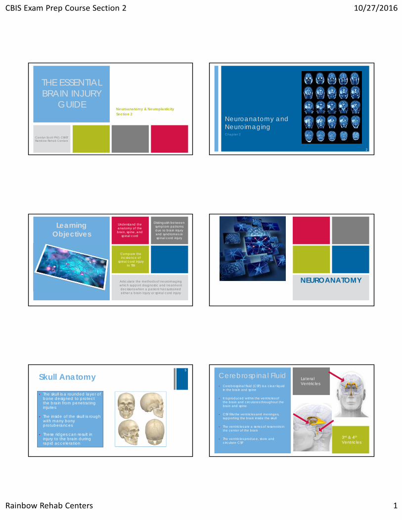
THE ESSENTIAL BRAIN INJURY
GUIDE
Carolyn Scott PhD, CBIST Rainbow Rehab Centers
Neuroanatomy and NeuroimagingChapter 2
2
Learning Objectives
Understand the anatomy of the brain, spine, and
spinal cord
Compare the incidence of
spinal cord injury to TBI
Distinguish between symptom patternsdue to brain injury and syndromes inspinal cord injury
Articulate the methods of neuroimagingwhich support diagnostic and treatmentdecisions when a patient has sustainedeither a brain injury or spinal cord injury
NEUROANATOMY
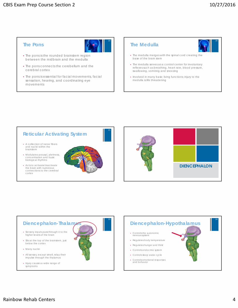
Skull Anatomy
The skull is a rounded layer of bone designed to protect the brain from penetrating injuries
The inside of the skull is rough with many bony protuberances
These ridges can result in injury to the brain during rapid acceleration
5
Cerebrospinal Fluid Cerebrospinal fluid (CSF) is a clear liquid
in the brain and spine
It is produced within the ventricles of the brain and circulates throughout the brain and spine
CSF fills the ventricles and meninges, supporting the brain inside the skull
The ventricles are a series of reservoirs in the center of the brain
The ventricles produce, store and circulate CSF
Lateral Ventricles
3rd & 4th
Ventricles
6
CBIS Exam Prep Course Section 2 10/27/2016
Rainbow Rehab Centers 2
The Meninges The meninges are layers of
tissue that separate the skull and the brain
There are 3 layers Pia Mater Arachnoid Dura Mater
Pia Mater Arachnoid Dura Mater Skull
Essential TIP!
The Meninges P-A-D the Brain
7
Dura Mater
Outer layer of the meninges
Latin for “tough mother”
A heavy cabbage-like covering
Below the Dura Mater is the subdural space When there is a bleed in
this space it is called a subdural hematoma
8
Arachnoid Layer
Second layer of meninges
Consistency similar to a spider web
Below the Arachnoid layer is the subarachnoid space When there is a bleed in
this space it is called a subarachnoid hematoma
9
Pia Matter
Third layer of meninges
Latin for ‘tender matter’
Molds around the sulci and gyri of the brain
10
The Brain is Composed of Discrete Cells: Neurons and Glia
Neurons are communicating cells
Glial cells are non-communicating cells supporting and nourishing neurons
AXON
CellBody
Dendrites
Dendritic Spines
AXONTerminals
Synapse
11
GliaGlial cells support and maintain neurons
CBIS Exam Prep Course Section 2 10/27/2016
Rainbow Rehab Centers 3
Neurons Communicate with Each Other Via Synapses Neurons have processes that
support electrochemical transmission
This allows them to communicate via an electrochemical process
Synapses are junctions between neurons where this electrochemical process takes place
The gap between an axon of one neuron and the dendrite of another neuron is the synapse
AXONTERMINALsynaptic
vesicle
SYNAPTIC CLEFTneurotransmitters
receptor
DENDRITIC SPINE
13
A signal from the axon of Neuron A travels to the dendrite of Neuron B
An action potential then generates from the cell body of Neuron B
This sends a signal down the axon and passes that signal to the dendrite of Neuron C
The process proceeds forward so long as an
action potential continues to generate
and the needed neurotransmitters and
other chemicals such as calcium and potassium,
etc., are present
Neuron B
Neuron C
Neuron A
14
15
BRAIN STEM
Brain StemComponents
MedullaPonsMidbrain
The brainstem is located at the top of
the spinal column
It is the central point for all incoming and outgoing information
and basic life functions
There are 3 components
17
Midbrain
The smallest part of the brain stem
Involved in elementary forms of vision and hearing
Plays a pivotal role in alertness and arousal
18
CBIS Exam Prep Course Section 2 10/27/2016
Rainbow Rehab Centers 4
The Pons
The pons is the rounded brainstem region between the midbrain and the medulla
The pons connects the cerebellum and the cerebral cortex
The pons is essential for facial movements, facial sensation, hearing, and coordinating eye movements
19
The Medulla
The medulla merges with the spinal cord creating the base of the brain stem
The medulla serves as a control center for involuntary reflexes such as breathing, heart rate, blood pressure, swallowing, vomiting and sneezing
Involved in many basic living functions; injury to the medulla is life threatening
20
Reticular Activating System
A collection of nerve fibers and nuclei within the brainstem
Modulates arousal, alertness, concentration and basic biological rhythms
Acts to activate/inactivate the brain with numerous connections to the cerebral cortex
21
DIENCEPHALON
Diencephalon-Thalamus Sensory inputs pass through it to the
higher levels of the brain
Sits at the top of the brainstem, just below the cortex
Many nuclei
All senses, except smell, relay their impulse through the thalamus
Injury causes a wide range of symptoms
23
Diencephalon-Hypothalamus
Controls the autonomic nervous system
Regulates body temperature
Regulates hunger and thirst
Controls endocrine system
Controls sleep wake cycle
Controls emotional responses and behavior
24
CBIS Exam Prep Course Section 2 10/27/2016
Rainbow Rehab Centers 5
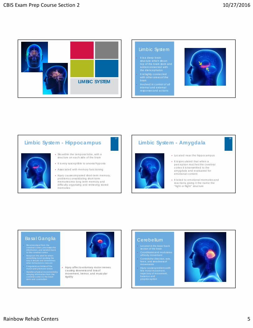
LIMBIC SYSTEM
Limbic System• It is a deep brain
structure which sits on top of the brain stem and is interconnected with the diencephalon
• It is highly connected with other areas of the brain
• Involved in control of all internal and external responses and actions
Limbic System - Hippocampus
Sits within the temporal lobe, with a structure on each side of the brain
It is very susceptible to anoxia/hypoxia
Associated with memory functioning
Injury causes impaired short-term memory, problems consolidating short-term memories into long term memory and difficulty organizing and retrieving stored memories
27
Limbic System - Amygdala
Located near the hippocampus
It is speculated that when a perception reaches the cerebral cortex it is transmitted to the amygdala and evaluated for emotional content
It is tied to emotional memories and reactions, giving it the name the “fight or flight” structure
28
Basal Ganglia Receives input from the
cerebral cortex, processes the information, and sends it back to the cerebral cortex
Keeps on the alert for when something is not working the way it should and determines what behaviors to execute
It regulates activities of the motor and premotor areas
Handles physical movements by relaying information from the cerebral cortex to the brain stem and cerebellum
Injury affects voluntary motor nerves causing slowness and loss of movement, tremor, and muscular rigidity
Cerebellum Located in the lower back
section of the brain Coordinates and modulates
all body movement Controls the direction, rate,
force, and steadiness of movements
Injury causes problems with fine motor movement, trajectory of movement, balance and proprioception
CBIS Exam Prep Course Section 2 10/27/2016
Rainbow Rehab Centers 6
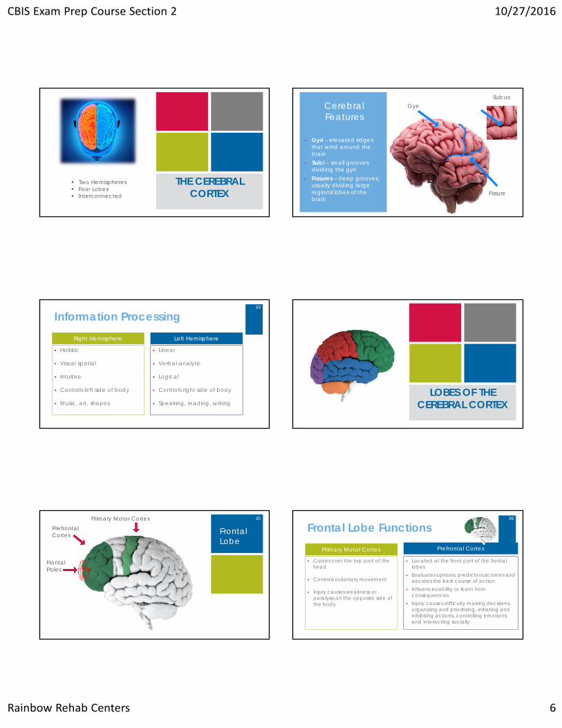
THE CEREBRAL CORTEX
Two Hemispheres Four Lobes Interconnected
Cerebral Features
• Gyri – elevated ridges that wind around the brain
• Sulci – small grooves dividing the gyri
• Fissures – deep grooves, usually dividing large regions/lobes of the brain
GyriSulcus
Fissure
Information Processing
Holistic
Visual spatial
Intuitive
Controls left side of body
Music, art, shapes
Linear
Verbal-analytic
Logical
Controls right side of body
Speaking, reading, writing
Right Hemisphere Left Hemisphere
33
LOBES OF THE CEREBRAL CORTEX
Frontal Lobe
Primary Motor CortexPrefrontal Cortex
Frontal Poles
35
Frontal Lobe Functions
Curves over the top part of the head
Controls voluntary movement
Injury causes weakness or paralysis on the opposite side of the body
Located at the front part of the frontal lobes
Evaluates options, predicts outcomes and decides the best course of action
Influences ability to learn from consequences
Injury causes difficulty making decisions, organizing and prioritizing, initiating and inhibiting actions, controlling emotions and interacting socially
Primary Motor Cortex Prefrontal Cortex
36
CBIS Exam Prep Course Section 2 10/27/2016
Rainbow Rehab Centers 7
Frontal Lobe Functions Planning
Organizing
Problem solving
Judgment
Impulse control
Decision making
Working memory
37
Frontal Lobe Damage
Changes in personality
Poor self-awareness
Reduced motivation/goal-directed behavior
Impaired attention/short-term memory
Poor judgment
Inability to plan
38
Prefrontal Cortex Injuries in Children
Brain is not fully developed Effects not immediately apparent In their teens, deficits may become
more apparent in teen May cause a wide range of poorly
controlled behaviors
39
Temporal Lobe
40
Temporal Lobe
Memory
Language
Hearing
Auditory processing
New learning
Understanding, storing, and retrieving new information
Functions Impairments
41
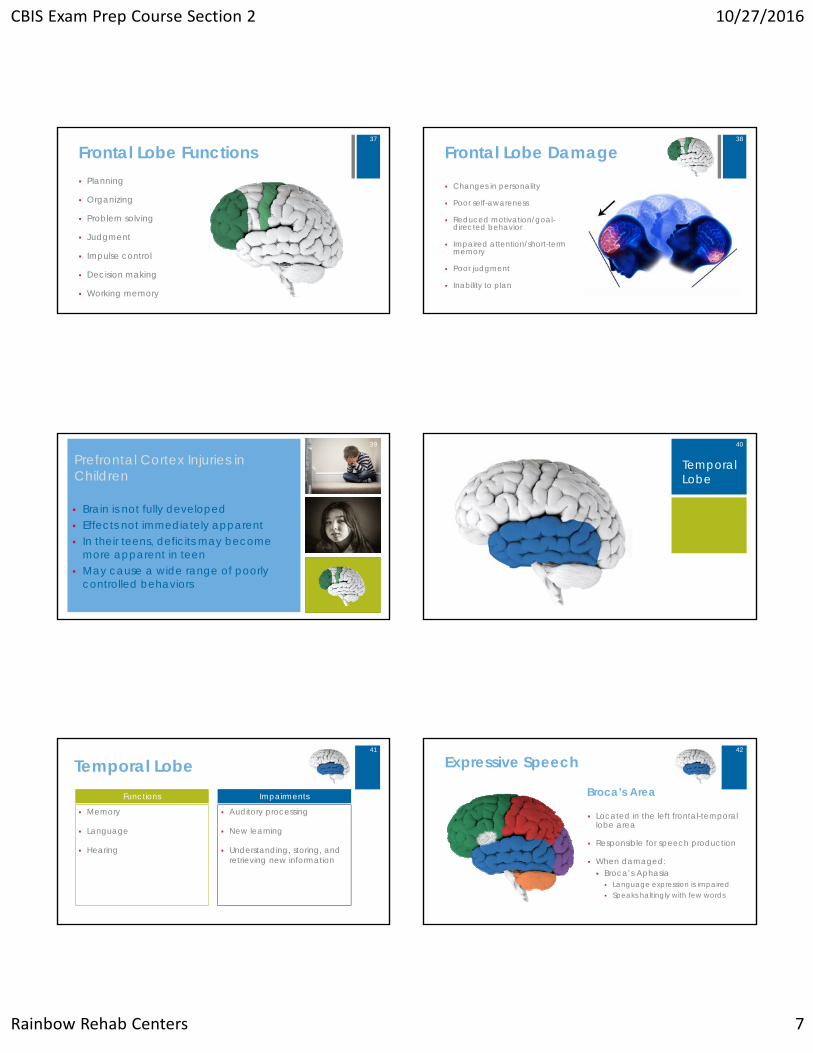
Expressive Speech
Broca’s Area
Located in the left frontal-temporal lobe area
Responsible for speech production
When damaged: Broca’s Aphasia Language expression is impaired Speaks haltingly with few words
42
CBIS Exam Prep Course Section 2 10/27/2016
Rainbow Rehab Centers 8
Receptive SpeechWernicke’s Area
Located in the left temporal-parietal lobe
Responsible for speech comprehension
When damaged: Wernicke’s Aphasia Language comprehension is
impaired Speaks fluently but does not make
sense
43 Speech Production & Understanding
Left frontal-temporal lobe
Speech Production
Language expression is impaired
Speaks haltingly with few words
Left temporal-parietal lobe
Speech Comprehension
Language comprehension is impaired
Speaks fluently but does not make sense
Broca’s Area Wernicke’s Area
44
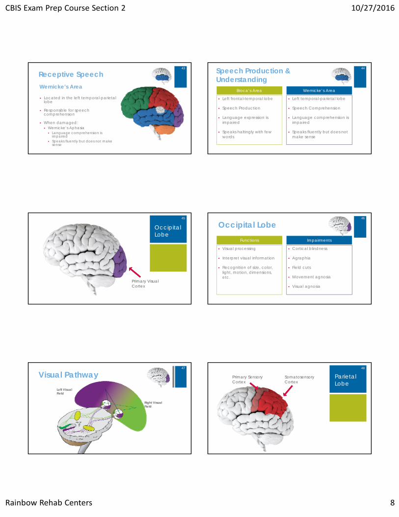
Occipital Lobe
Primary Visual Cortex
45
Occipital Lobe
Visual processing
Interpret visual information
Recognition of size, color, light, motion, dimensions, etc.
Cortical blindness
Agraphia
Field cuts
Movement agnosia
Visual agnosia
Functions Impairments
46
Visual PathwayLeft Visual Field
Right Visual Field
47
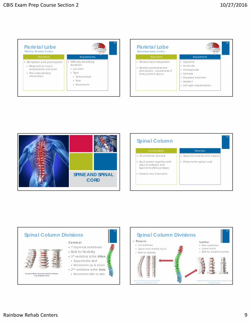
Parietal Lobe
Somatosensory Cortex
Primary Sensory Cortex
48
CBIS Exam Prep Course Section 2 10/27/2016
Rainbow Rehab Centers 9
Parietal Lobe
Sensation and perception Responds to touch,
temperature and pain Processes sensory
information
Difficulty identifying sensation Location Type Temperature Pain Movement
Functions Impairments
Primary Sensory Cortex
49
Parietal Lobe
Sensory input integration
Spatial awareness and perception - awareness of body parts in space
Agraphia Acalculia Anosognosia Aphasia Impaired attention Neglect Left-right disorientation
Functions Impairment
Somatosensory Cortex
50
SPINE AND SPINAL CORD
Spinal Column
33 vertebrae (bones)
Each joined together with discs (cartilage) and ligaments (fibrous tissue)
Divided into 5 sections
Supports muscles and organs
Protects the spinal cord
Composition Function
52
Spinal Column DivisionsCervical 7 topmost vertebrae Built for flexibility 1st vertebra is the Atlas Supports the skull Movement up & down
2nd vertebra is the Axis Movement side to sideCervical Spine showing Lateral, Anterior,
and Posterior view
53
Spinal Column DivisionsLumbar Five vertebrae Lower back Built for weight bearing
LumbarVertebraeshowingLateral,Anterior,andPosteriorviews
ThoracicVertebraeshowingLateral,Anterior,andPosteriorviews
Thoracic 12 vertebrae Upper and middle back Built for stability
54
CBIS Exam Prep Course Section 2 10/27/2016
Rainbow Rehab Centers 10
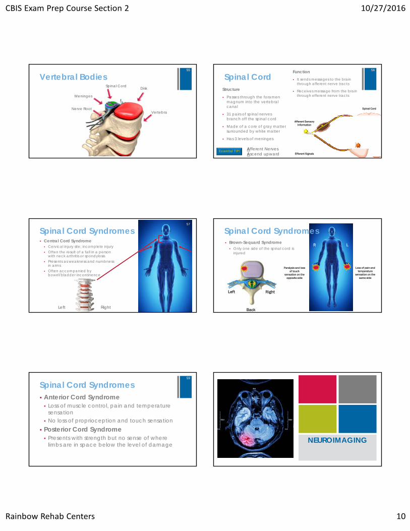
Vertebral BodiesSpinal Cord
Meninges
Vertebra
Disk
Nerve Root
55
Spinal CordStructure
Passes through the foramen magnum into the vertebral canal
31 pairs of spinal nerves branch off the spinal cord
Made of a core of gray matter surrounded by white matter
Has 3 levels of meninges
Function It sends messages to the brain
through afferent nerve tracts
Receives message from the brain through efferent nerve tracts
Afferent Sensory Information
Efferent Signals
Spinal Cord
56
Essential TIP! Afferent Nerves Ascend upward
Spinal Cord Syndromes Central Cord Syndrome Cervical injury site; incomplete injury Often the result of a fall in a person
with neck arthritis or spondylosis Presents as weakness and numbness
in arms Often accompanied by
bowel/bladder incontinence
Left Right
57
Spinal Cord Syndromes Brown-Sequard Syndrome Only one side of the spinal cord is
injured
Left Right
Paralysis and loss of touch
sensation on the opposite side
Loss of pain and temperature
sensation on the same side
Back
LR
58
Spinal Cord Syndromes Anterior Cord Syndrome Loss of muscle control, pain and temperature
sensation No loss of proprioception and touch sensation Posterior Cord Syndrome Presents with strength but no sense of where
limbs are in space below the level of damage
59
NEUROIMAGING
CBIS Exam Prep Course Section 2 10/27/2016
Rainbow Rehab Centers 11
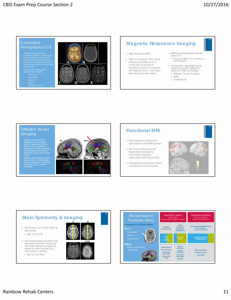
Computed Tomography (CT) An X-ray procedure that
combines multiple x-ray images with the aid of a computer to produce cross sectional views and three dimensional images of the internal organs and structures of the body
Useful for identifying gross anatomical changes Skull fracture Hemorrhage Blood clot Swelling Penetrating object
Magnetic Resonance Imaging
Also known as MRI
Uses a magnetic field, radio frequency pulses, and a computer to produce detailed pictures of organs, soft tissues, bone, and other internal body structures
MRI provides better detail than CT More sensitive to a variety of
pathologies
Computer algorithms are used to create different forms of MRI, including Diffusion Tensor Imaging fMRI T2 Weighted
62
Diffusion Tensor Imaging A type of Magnetic
Resonance Imaging which uses the rate at which water diffuses between cells to provide information about the internal structures of the body
DTI provides information about the connectivity and continuity of neural pathways (i.e., white matter)
Functional MRI
The individual performs a task while in the MRI scanner
The scan measures and maps brain activity by detecting changes associated with blood flow
Changes in blood flow follow changes in neural activity
64
Brain Symmetry & Imaging
The brain in a normal state is symmetric See T1 and T2
Non-pathological brains and the structures that make up the brain should all appear similar to one another (i.e., from brain to brain) See C and Flair
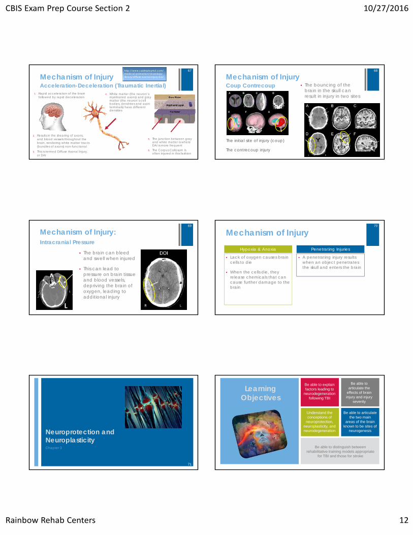
65MechanismsofTraumaticInjury
TRAUMATICIMPACTContactInjury
Headstruckbyoragainstanobject
TRAUMATICINERTIALNon‐ContactInjury
Brainmoveswithinskull
CLOSEDNon‐Penetrating
OPENPenetratingSkullFracture
MeningesBreach
Rotational/AngularForcesNon‐ContactInjury
Brainmoveswithinskull
FOCAL‐ OR‐
DIFFUSE
PRIMARILYDIFFUSEMULTIFOCAL
PRIMARILYFOCAL
BrainContusions
BrainLacerations
IntracerebralHemorrhage
DiffuseAxonalInjury
EpiduralHematomas
SubduralHematomas
IntracerebralHemorrhage
Infections
DiffuseAxonalInjury
WhiteMatterLesion
Hemorrhage
Categories of Brain Injury Focal Contusions
Lesions
Hematomas
Diffuse Diffuse Axonal Injury Hemorrhage
CBIS Exam Prep Course Section 2 10/27/2016
Rainbow Rehab Centers 12
Mechanism of Injury
1. Rapid acceleration of the brain followed by rapid deceleration
Acceleration-Deceleration (Traumatic Inertial)
5. The junction between gray and white matter is where DAI is more frequent
6. The Corpus Callosum is often injured in this fashion
http://www.calshipleymd.com/medical-animation/neurology-library/diffuse-axonal-injury-dai/
2. Results in the shearing of axons, and blood vessels throughout the brain, rendering white matter tracts (bundles of axons) non-functional
3. This is termed Diffuse Axonal Injury, or DAI
4. White matter (the neuron’s myelinated axons) and gray matter (the neuron’s cell bodies, dendrites and axon terminals) have different densities
67
Mechanism of Injury The bouncing of the
brain in the skull can result in injury in two sites
Coup Contrecoup
The initial site of injury (coup)
The contrecoup injury
68
Mechanism of Injury:
The brain can bleed and swell when injured
This can lead to pressure on brain tissue and blood vessels, depriving the brain of oxygen, leading to additional injury
Intracranial Pressure
69
Mechanism of Injury
Lack of oxygen causes brain cells to die
When the cells die, they release chemicals that can cause further damage to the brain
A penetrating injury results when an object penetrates the skull and enters the brain
Hypoxia & Anoxia Penetrating Injuries
70
Neuroprotection and NeuroplasticityChapter 3
71
Learning Objectives
Understand the conceptions of
neuroprotection, neuroplasticity, andneurodegeneration
Be able to explain factors leading to
neurodegeneration following TBI
Be able to articulate the
effects of braininjury and injury
severity
Be able to articulate the two main
areas of the brain known to be sites of
neurogenesis
Be able to distinguish betweenrehabilitative training models appropriate
for TBI and those for stroke
CBIS Exam Prep Course Section 2 10/27/2016
Rainbow Rehab Centers 13
NEUROPLASTICITY
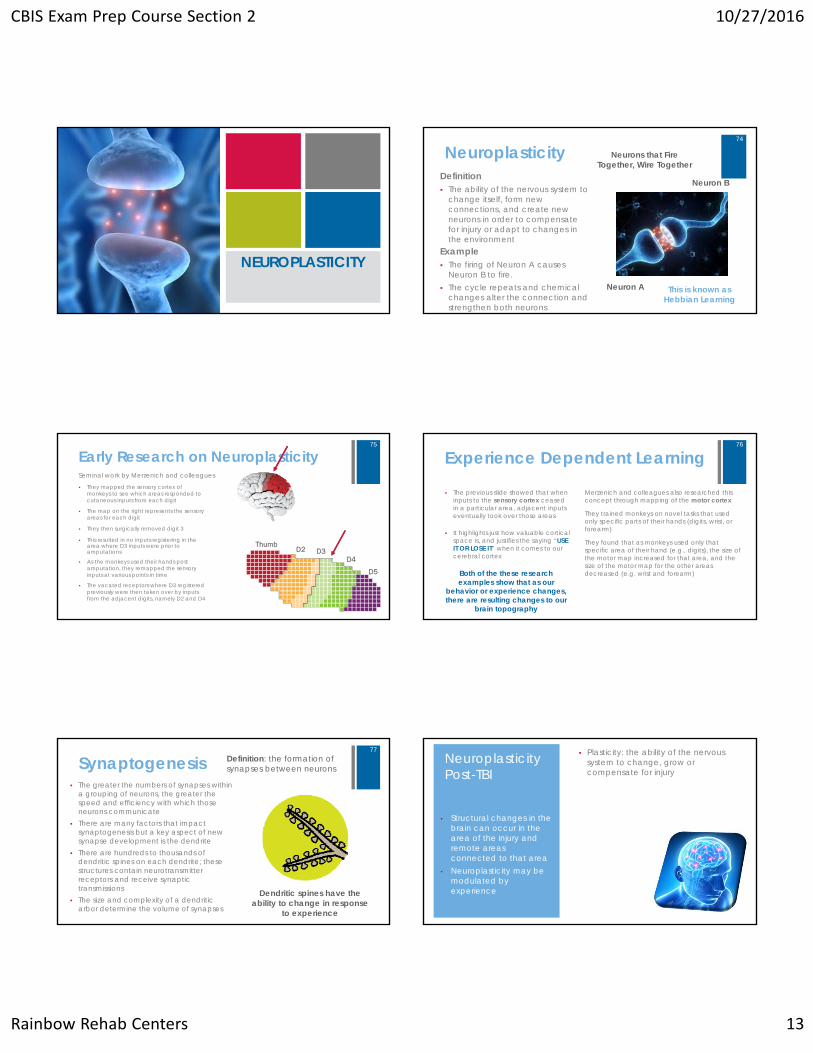
NeuroplasticityDefinition The ability of the nervous system to
change itself, form new connections, and create new neurons in order to compensate for injury or adapt to changes in the environment
Neurons that Fire Together, Wire Together
Neuron A
Neuron B
Example The firing of Neuron A causes
Neuron B to fire. The cycle repeats and chemical
changes alter the connection and strengthen both neurons
This is known as Hebbian Learning
74
Early Research on Neuroplasticity
ThumbD2 D3
D4
D5
Seminal work by Merzenich and colleagues
They mapped the sensory cortex of monkeys to see which areas responded to cutaneous inputs from each digit
The map on the right represents the sensory areas for each digit
They then surgically removed digit 3
As the monkeys used their hands post amputation, they remapped the sensory inputs at various points in time
This resulted in no inputs registering in the area where D3 inputs were prior to amputations
The vacated receptors where D3 registered previously were then taken over by inputs from the adjacent digits, namely D2 and D4
75
Experience Dependent Learning
The previous slide showed that when inputs to the sensory cortex ceased in a particular area, adjacent inputs eventually took over those areas
It highlights just how valuable cortical space is, and justifies the saying “USE IT OR LOSE IT” when it comes to our cerebral cortex
Merzenich and colleagues also researched this concept through mapping of the motor cortex
They trained monkeys on novel tasks that used only specific parts of their hands (digits, wrist, or forearm)
They found that as monkeys used only that specific area of their hand (e.g., digits), the size of the motor map increased for that area, and the size of the motor map for the other areas decreased (e.g. wrist and forearm)Both of the these research
examples show that as our behavior or experience changes, there are resulting changes to our
brain topography
76
Synaptogenesis The greater the numbers of synapses within
a grouping of neurons, the greater the speed and efficiency with which those neurons communicate
There are many factors that impact synaptogenesis but a key aspect of new synapse development is the dendrite
There are hundreds to thousands of dendritic spines on each dendrite; these structures contain neurotransmitter receptors and receive synaptic transmissions
The size and complexity of a dendritic arbor determine the volume of synapses
Definition: the formation of synapses between neurons
Dendritic spines have the ability to change in response
to experience
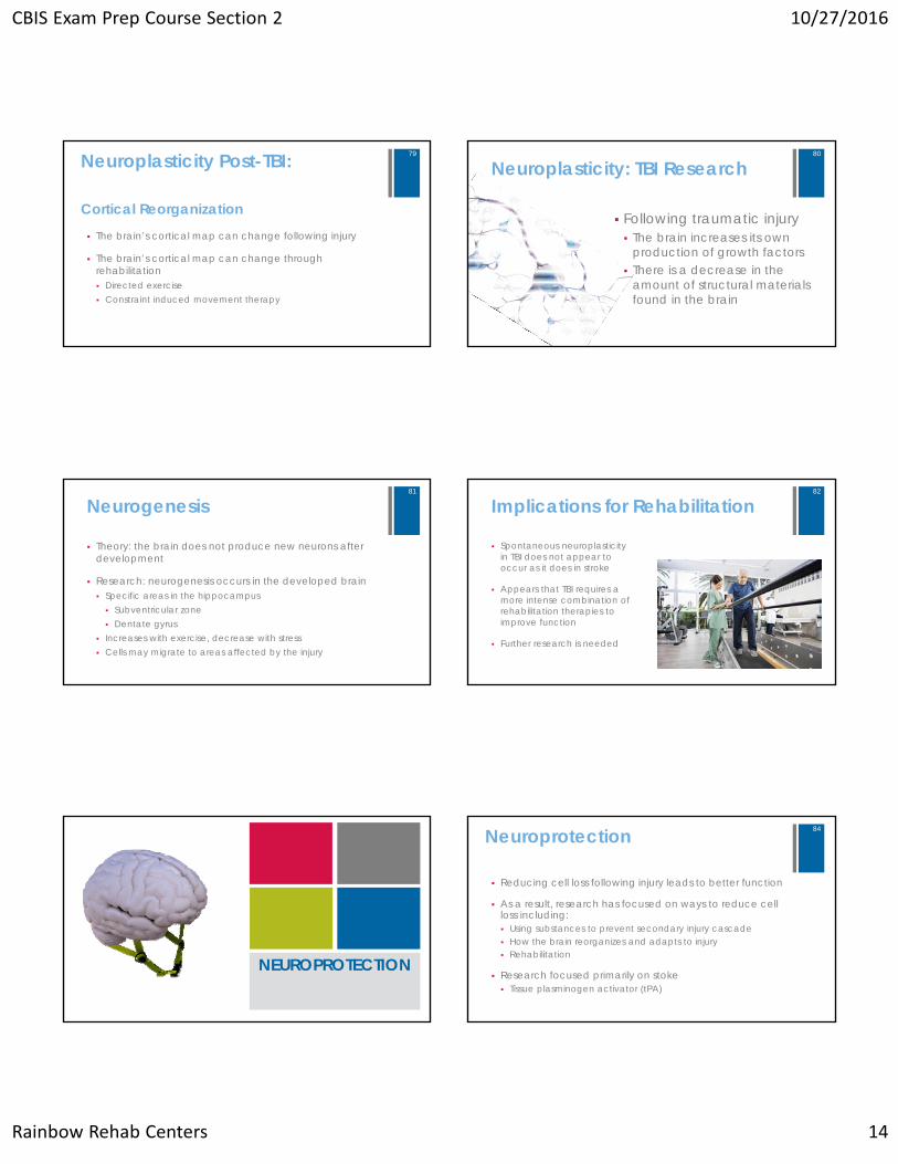
77Neuroplasticity Post-TBI
Plasticity: the ability of the nervous system to change, grow or compensate for injury
• Structural changes in the brain can occur in the area of the injury and remote areas connected to that area
• Neuroplasticity may be modulated by experience
CBIS Exam Prep Course Section 2 10/27/2016
Rainbow Rehab Centers 14
Neuroplasticity Post-TBI:
The brain’s cortical map can change following injury
The brain’s cortical map can change through rehabilitation Directed exercise Constraint induced movement therapy
Cortical Reorganization
79
Neuroplasticity: TBI Research
Following traumatic injury The brain increases its own
production of growth factors There is a decrease in the
amount of structural materials found in the brain
80
Neurogenesis
Theory: the brain does not produce new neurons after development
Research: neurogenesis occurs in the developed brain Specific areas in the hippocampus Subventricular zone Dentate gyrus
Increases with exercise, decrease with stress Cells may migrate to areas affected by the injury
81
Implications for Rehabilitation
Spontaneous neuroplasticity in TBI does not appear to occur as it does in stroke
Appears that TBI requires a more intense combination of rehabilitation therapies to improve function
Further research is needed
82
NEUROPROTECTION
Neuroprotection
Reducing cell loss following injury leads to better function
As a result, research has focused on ways to reduce cell loss including: Using substances to prevent secondary injury cascade How the brain reorganizes and adapts to injury Rehabilitation
Research focused primarily on stoke Tissue plasminogen activator (tPA)
84
CBIS Exam Prep Course Section 2 10/27/2016
Rainbow Rehab Centers 15
Biological Cascade Following TBI Primary Injury – direct damage to the
brain Secondary Injury – causes additional
damage Excitotoxicity Edema Apoptosis
85
Secondary Injury
Excitotoxicity Failure of neurons to
maintain resting state
Sodium-potassium pump failure
Excitotoxicaccumulation of sodium and calcium
Edema Swelling in cells Increased
Intracranial Pressure
Contributes to apoptosis
Apoptosis Cells surrounding
the primary injury die as a result
This expands the size of the injury
86
Potential Neuroprotective Agents for TBI Neuroprotective agents limit neuronal death following injury
and/or enhance recovery
Neuroprotective Agent Intervention Target Animal Models Showing Efficacy (Stroke)
Human Studies Showing Efficacy (TBI)
Magnesium Increase Mg2 (decreased Mg2 results in excessive
production of free radical and mild inflammation)
Failed
Progesterone Decrease cerebral edema
Initial Efficacy; Follow Up Trial
Nicotinimide Reduce injury volume; decrease glial activation;
reduce BBB breaches; reduce edema
Unknown
?
87
THEEND
88