Embed Size (px)
Citation preview
Your Horse's Health
Veterinary Medicine with
Matt Durham, DVM
Published in Bay Area Equestrian Network June 2008
The Equine Heart Part 2: Common Cardiac Disease
Horse owners have probably all experienced “heart” in a
favorite horse, that indefinable quality that makes certain
horses stand out. In the article The Equine Heart: Part 1,
we examined the remarkable abilities of the equine heart,
and its role in making horses superior athletes. In this
article, we will examine some of the more common
cardiac problems found in horses.
How the heart works (the boring part)
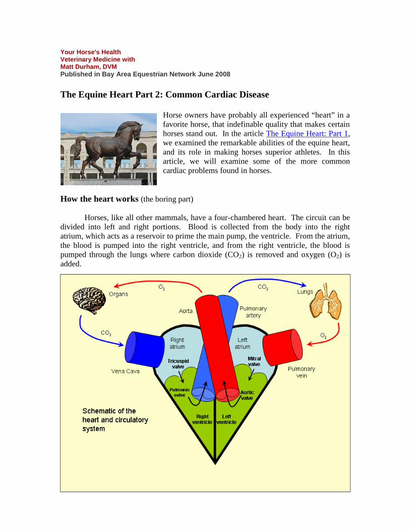
Horses, like all other mammals, have a four-chambered heart. The circuit can be
divided into left and right portions. Blood is collected from the body into the right
atrium, which acts as a reservoir to prime the main pump, the ventricle. From the atrium,
the blood is pumped into the right ventricle, and from the right ventricle, the blood is
pumped through the lungs where carbon dioxide (CO2) is removed and oxygen (O2) is
added.
This oxygenated blood then continues back to the heart into the left atrium, and
follows a similar course through the left ventricle. The left ventricle is the most muscular
portion of the heart, having to pump blood throughout the entire body via the aorta.
Blood is delivered to muscle, brain, and other organs, which then extract O2 and add the
waste product CO2 back in. The unoxygenated blood then returns to the right heart via
the vena cava, and the cycle is started again.
The atria and ventricles are composed of a type of muscle somewhat different
from skeletal muscle. Cardiac muscular contraction is initiated by electrical impulses.
The heart has a built-in pacemaker called the sino-atrial (SA) node, living at the top of
the heart, which governs heart rate and rhythm under most circumstances. The SA node
sends its impulse out in a wave across the atria, causing muscular contraction in a similar
downward-moving wave, pushing blood downward into the ventricles. Once the
electrical impulse reaches the bottom of the atria, the atrio-ventricular (AV) node is
triggered, which sends an electrical impulse through the bundle of His, which carries the
impulse down to the bottom of the heart. During this time, muscle tissue is bypassed. At
the bottom of the heart, the Purkinje fibers carry the electrical impulse in an upward
direction, causing the individual muscle fibers to contract in a coordinated upward wave
towards the openings of the “great vessels” at the top of the heart (the aorta in the left
ventricle and the pulmonary artery in the right ventricle).
The cardiac cycle is divided into two cycles: diastole, where the right and left
atria contract, and systole, where the right and left ventricles contract.
Valves are present between the atria and ventricles, and between the ventricles
and the “great vessels” to prevent significant backflow. The valves are flexible flaps of
tissue which are pushed out of the way by forward flow of blood. When back pressure
starts to develop, the valves start to push back, but are held in place by connective tissue
bands called chordae tendinae, thereby sealing the valve opening and preventing
backflow. The left atrio-ventricular valve is called the mitral valve, and the right one is
called the tricuspid valve. The valves associated with the great vessels are the aortic
valve on the left and the pulmonic valve on the right.
Evaluation of the heart (the slightly less boring part)
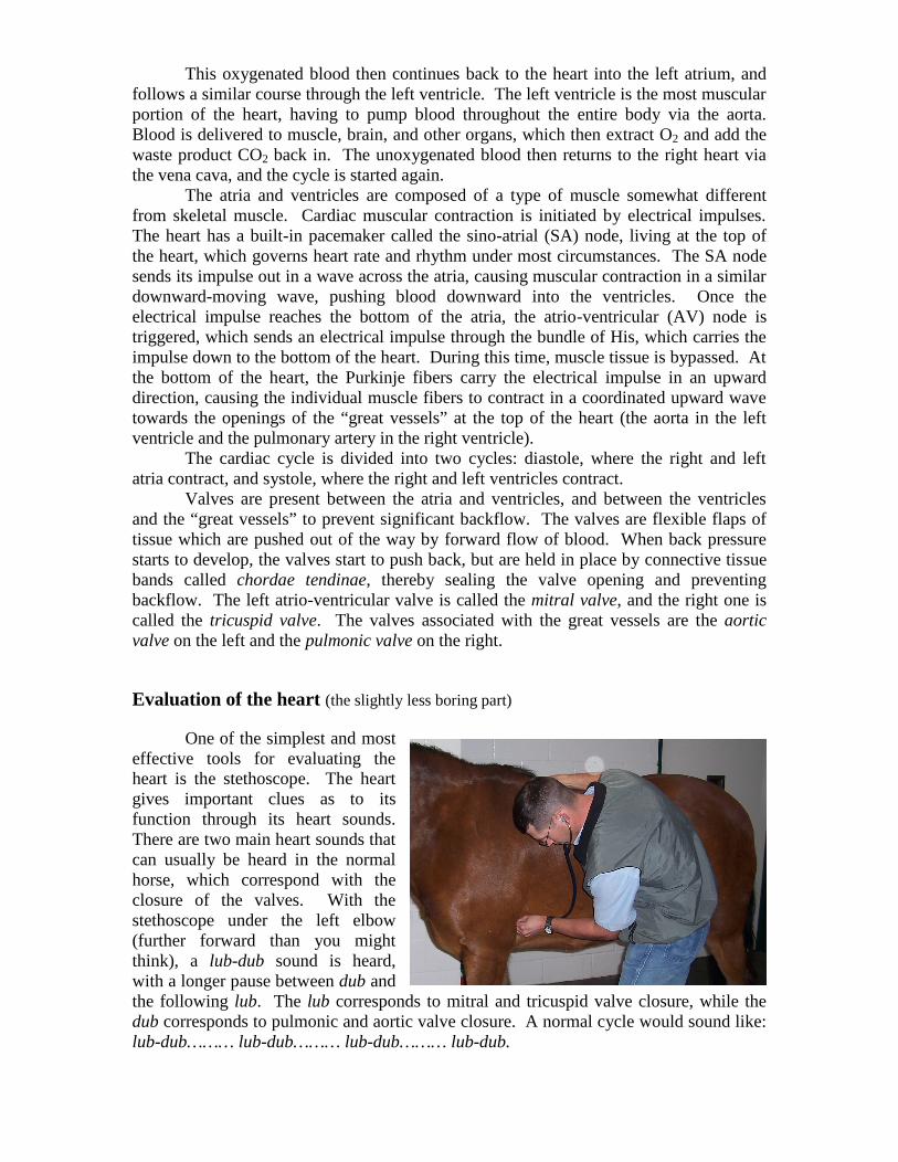
One of the simplest and most
effective tools for evaluating the
heart is the stethoscope. The heart
gives important clues as to its
function through its heart sounds.
There are two main heart sounds that
can usually be heard in the normal
horse, which correspond with the
closure of the valves. With the
stethoscope under the left elbow
(further forward than you might
think), a lub-dub sound is heard,
with a longer pause between dub and
the following lub. The lub corresponds to mitral and tricuspid valve closure, while the
dub corresponds to pulmonic and aortic valve closure. A normal cycle would sound like:
lub-dub……… lub-dub……… lub-dub……… lub-dub.
In between the lub-dub sounds is systole, the period when the ventricles are
contracting. The longer pause, between dub and the following lub, is diastole, when the
atria contract to refill the ventricles. Each lub-dub accounts for a full cardiac cycle, or
one beat. Typical heart rates in resting adult horses are between 32 and 40 beats per
minute. (Count the number of beats for 15 seconds and multiply by 4) Exercise and
anxiety are common causes for elevations in heart rate, while pain and cardiac problems
can also cause elevations. The heart rate will stay elevated in horses with pain or cardiac
disease, while the rate will come back to normal in the nervous or recently exercised
horse. The heart rhythm in horses is typically stable, meaning that each beat occurs at a
predictable interval. By tapping your foot to each beat while counting, deviations from
the expected rhythm can be detected.
A heart murmur is an abnormal sound caused by turbulent blood flow. The
murmur itself is not a disease, but is merely a physical finding. Most murmurs are
associated with turbulence created from leaking valves. Turbulent blood flow can
sometimes be created in normal highly fit horses, causing a ‘physiologic flow’ murmur.
During a cardiac examination, the lungs sounds are also evaluated, since certain heart
disease conditions can lead to a buildup of fluid in the lungs.
Palpation of pulses can be useful in counting the heart rate without a stethoscope,
and in evaluating the strength and character of ventricular contraction.
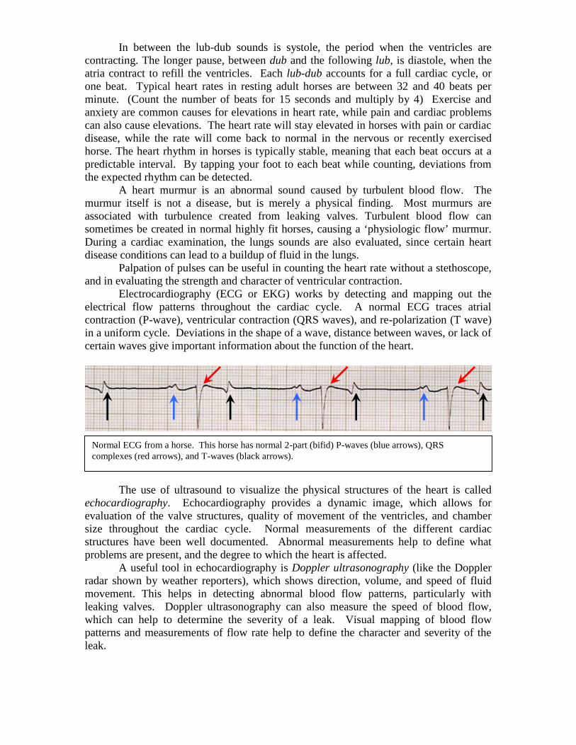
Electrocardiography (ECG or EKG) works by detecting and mapping out the
electrical flow patterns throughout the cardiac cycle. A normal ECG traces atrial
contraction (P-wave), ventricular contraction (QRS waves), and re-polarization (T wave)
in a uniform cycle. Deviations in the shape of a wave, distance between waves, or lack of
certain waves give important information about the function of the heart.
The use of ultrasound to visualize the physical structures of the heart is called
echocardiography. Echocardiography provides a dynamic image, which allows for
evaluation of the valve structures, quality of movement of the ventricles, and chamber
size throughout the cardiac cycle. Normal measurements of the different cardiac
structures have been well documented. Abnormal measurements help to define what
problems are present, and the degree to which the heart is affected.
A useful tool in echocardiography is Doppler ultrasonography (like the Doppler
radar shown by weather reporters), which shows direction, volume, and speed of fluid
movement. This helps in detecting abnormal blood flow patterns, particularly with
leaking valves. Doppler ultrasonography can also measure the speed of blood flow,
which can help to determine the severity of a leak. Visual mapping of blood flow
patterns and measurements of flow rate help to define the character and severity of the
leak.
Normal ECG from a horse. This horse has normal 2-part (bifid) P-waves (blue arrows), QRS
complexes (red arrows), and T-waves (black arrows).
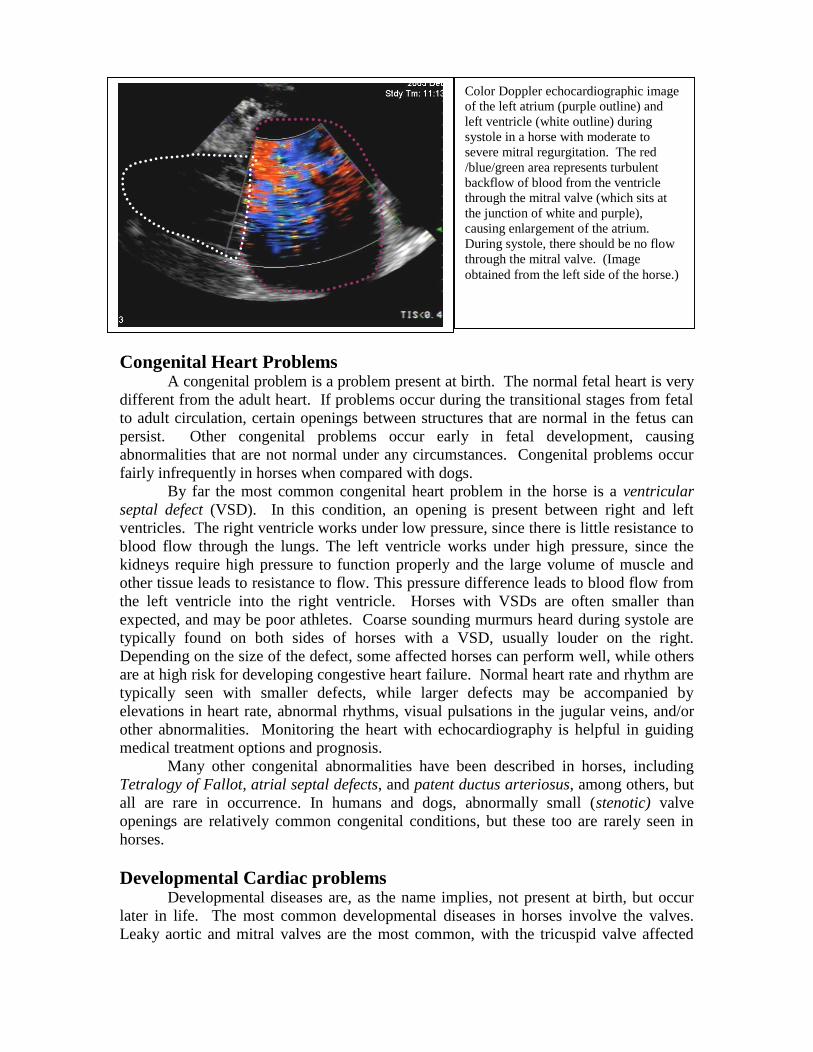
Color Doppler echocardiographic image
of the left atrium (purple outline) and
left ventricle (white outline) during
systole in a horse with moderate to
severe mitral regurgitation. The red
/blue/green area represents turbulent
backflow of blood from the ventricle
through the mitral valve (which sits at
the junction of white and purple),
causing enlargement of the atrium.
During systole, there should be no flow
through the mitral valve. (Image
obtained from the left side of the horse.)
Congenital Heart Problems
A congenital problem is a problem present at birth. The normal fetal heart is very
different from the adult heart. If problems occur during the transitional stages from fetal
to adult circulation, certain openings between structures that are normal in the fetus can
persist. Other congenital problems occur early in fetal development, causing
abnormalities that are not normal under any circumstances. Congenital problems occur
fairly infrequently in horses when compared with dogs.
By far the most common congenital heart problem in the horse is a ventricular
septal defect (VSD). In this condition, an opening is present between right and left
ventricles. The right ventricle works under low pressure, since there is little resistance to
blood flow through the lungs. The left ventricle works under high pressure, since the
kidneys require high pressure to function properly and the large volume of muscle and
other tissue leads to resistance to flow. This pressure difference leads to blood flow from
the left ventricle into the right ventricle. Horses with VSDs are often smaller than
expected, and may be poor athletes. Coarse sounding murmurs heard during systole are
typically found on both sides of horses with a VSD, usually louder on the right.
Depending on the size of the defect, some affected horses can perform well, while others
are at high risk for developing congestive heart failure. Normal heart rate and rhythm are
typically seen with smaller defects, while larger defects may be accompanied by
elevations in heart rate, abnormal rhythms, visual pulsations in the jugular veins, and/or
other abnormalities. Monitoring the heart with echocardiography is helpful in guiding
medical treatment options and prognosis.
Many other congenital abnormalities have been described in horses, including
Tetralogy of Fallot, atrial septal defects, and patent ductus arteriosus, among others, but
all are rare in occurrence. In humans and dogs, abnormally small (stenotic) valve
openings are relatively common congenital conditions, but these too are rarely seen in
horses.
Developmental Cardiac problems
Developmental diseases are, as the name implies, not present at birth, but occur
later in life. The most common developmental diseases in horses involve the valves.
Leaky aortic and mitral valves are the most common, with the tricuspid valve affected
less commonly, and the pulmonic valve rarely affected. Abnormalities in rhythm are
somewhat less common, as are aneurysms and pericarditis. Myocarditis, which is
common in humans, is uncommon in horses.
Valvular disease
Slowly progressive scarring of the valve margins is relatively common in older
horses in the aortic and mitral valves, which can lead to poorly functioning valves and
variable degrees of leaking. Tearing in the valve leaflets, buckling of the leaflets, or
ruptured chordae tendinae lead to improperly functioning valves as well. Rarely,
bacterial infections can occur on or near the valves, which can severely affect cardiac
function, and can be life threatening.
Leaking of the aortic valve (aortic regurgitation) is common in older horses, and
is often tolerated well. The backflow causes an overload in volume in the left ventricle,
but in early stages the ventricle does a good job of adapting. Murmurs are present during
diastole (after the ‘dub’), present on both sides, but louder on the left. The murmur can
sometimes have a ‘divebomber’ sound, which likely occurs from vibrations of the aortic
valve leaflets. Long term aortic regurgitation can lead to stretching of the ventricle and
the mitral valve opening, which can cause leaking in this valve as well.
Leaking of the mitral valve (mitral regurgitation) is also relatively common. The
backflow causes the left atrium to enlarge, which causes a backup of blood into the
pulmonary vein. Murmurs are during systole (between ‘lub’ and ‘dub’), and are heard on
the left side of the horse. Most mitral valve leaks can be tolerated well if the onset is
slow, allowing the lungs to compensate for the increase in blood pressure. Sudden-onset
mitral valve leaks, such as with ruptured chordae tendinae, are more difficult for the body
to handle, because sudden backflow causes a sudden pressure increase in the lungs,
which causes fluid to leak into the air sacs of the lung. In this situation, the outcome is
usually poor, while small or slowly progressing leaks can often be managed medically.
Leaking of the tricuspid valve (tricuspid regurgitation) occurs less commonly and
is typically well tolerated by horses. Backflow can sometimes be seen as prominent
jugular pulses, prominence in other blood vessels, or edema in the legs or abdomen.
Murmurs are during systole (between ‘lub’ and ‘dub’), and are heard on the right side of
the horse. Leaks in the tricuspid valve are most commonly caused by enlargement of the
inside of the right ventricle, which can happen from resistance through the lungs in
chronic respiratory conditions or mitral valve regurgitation. The right ventricle can also
be enlarged in certain athletic disciplines such as in the sustained aerobic effort of
Standardbred racehorses. Tricuspid regurgitation is more common in athletic humans as
well.
Aneurysm
Aortic root aneurysm is a very rare, but serious condition. The typical aneurysm
is an abnormal bulging of the aorta, which is at risk of rupturing. This can lead to
collapse or sudden death, usually when the horse is exercising. Aneurysms in the blood
vessels leading to the intestines can be caused by the parasite Strongylus vulgaris (large
strongyle), but this is easily prevented by proper de-worming practices.
Myocarditis
Damaged or inflamed heart muscle (myocarditis), usually occurs in horses as a
result of certain viral infections or toxins. Monensin is a feed additive found in some
cattle feeds, which is extremely toxic to horses. Oleander, Yew, and avocado branches
and leaves are among other plants that are cardiotoxic to horses. Very rarely, coronary
artery disease leading to myocarditis can be caused by infections within the heart.
Cholesterol plaques, which cause coronary artery disease in humans, are not seen in
horses. While myocarditis is rare in horses, coronary artery disease is the leading cause
of death in humans.
Arrhythmias
Abnormal cardiac rhythms, or arrhythmias, are uncommon in horses. Second-
degree AV block is common in horses, but is almost always considered normal. In this
rhythm, the horse’s heart actually skips a beat. The SA node sends its signal, but the AV
node decides that the heart is functioning efficiently, and does not send the signal through
for one beat. In the normal situation, only one beat is skipped, and the normal rhythm is
resumed. This sounds like: lub-dub……… lub-dub……… (pause)……… lub-dub………
lub-dub. If keeping rhythm by tapping your foot, you should detect a gap when you
expect a beat, but then find the following beat with one more foot tap.
Atrial fibrillation occurs when the normal wave of electrical impulses that moves across
the atria is broken up into many random waves, moving in different directions at the same
time in an uncoordinated manner. This causes the atria to contract in a random
uncoordinated manner, and does not provide the AV node with a reliable signal. The
rhythm of the ventricles must be taken over by the AV node, which usually does not
create a predictable rhythm. This typically sounds something like: lub-dub.. lub-
dub………………… lub-dub……… lub-dub… lub-dub……………… lub-dub in an
unpredictable pattern.
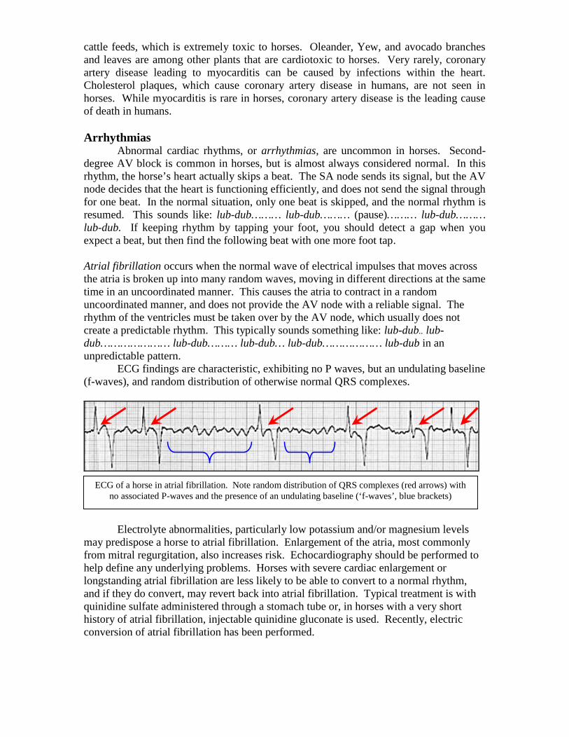
ECG findings are characteristic, exhibiting no P waves, but an undulating baseline
(f-waves), and random distribution of otherwise normal QRS complexes.
Electrolyte abnormalities, particularly low potassium and/or magnesium levels
may predispose a horse to atrial fibrillation. Enlargement of the atria, most commonly
from mitral regurgitation, also increases risk. Echocardiography should be performed to
help define any underlying problems. Horses with severe cardiac enlargement or
longstanding atrial fibrillation are less likely to be able to convert to a normal rhythm,
and if they do convert, may revert back into atrial fibrillation. Typical treatment is with
quinidine sulfate administered through a stomach tube or, in horses with a very short
history of atrial fibrillation, injectable quinidine gluconate is used. Recently, electric
conversion of atrial fibrillation has been performed.
ECG of a horse in atrial fibrillation. Note random distribution of QRS complexes (red arrows) with
no associated P-waves and the presence of an undulating baseline (‘f-waves’, blue brackets)
Atrial premature contractions are relatively common, while ventricular premature
contractions are relatively rare. Both of these conditions occur when there is localized
myocarditis. The conditions can sometimes be differentiated by carefully listening to
rhythm abnormalities, but definitive diagnosis is made with an ECG. Both conditions
require a period of rest and anti-inflammatory medications, as well as investigation into
any underlying causes of disease.
Ventricular tachycardia and ventricular fibrillation are rare and generally are associated
with serious underlying disease processes.
Management of Horses with Cardiac Disease (a little good news)
Depending on the severity and type of abnormality, most common heart
conditions in horses are manageable. In fact, most horses can compete effectively in all
but the most rigorous events with mild to moderate valvular problems. Horses with
valvular disease should be evaluated periodically with a thorough physical examination
and echocardiography to monitor progression of disease. Horses with mild to moderate
valvular disease often benefit from exercise. The amount of exercise should be tailored
to the individual horse, based on age, fitness, degree of cardiac dysfunction, and
soundness.
Depending on the type of disease present, horses may need to be maintained on
daily medications. Enalapril and furosemide (Lasix), with or without digoxin, are
common choices for treatment of ongoing conditions. The use of these medications in
some horses may delay the progression of cardiac disease, and can often alleviate some
of the clinical signs seen in moderately affected horses. Severely affected horses may
improve somewhat, but typically have a poor prognosis even with aggressive treatment.
Insignificant aortic and mitral regurgitation are commonly found cardiac
problems, typically with only a subtle murmur, and no other abnormal findings on a
physical examination. In these mildly affected horses, medical therapy is not necessary,
but the murmur should be monitored at each examination for signs of progression.
Final word
Horses are remarkably athletic creatures, in large part due to their incredible
hearts. Fortunately, significant heart disease is uncommon in these amazing creatures,
allowing us to enjoy our equine friends for many years. We are enriched by their
company, we marvel at their physical abilities, and we always remember that special
horse with lots of “heart.”
Matt Durham, DVM grew up in Reno, Nevada. During the summers growing up, Dr. Durham
worked in the Sierra Nevadas as a backcountry guide at McGee Creek and Mammoth Lakes
Pack Outfits, where he met his wife, Tiffany. He attended Cal Poly, San Luis Obispo, and
obtained a degree in Animal Science. After graduating from veterinary school at UC Davis, he
performed a one year internship at Alamo Pintado Equine Medical Center in Los Olivos,
California. After four years in practice, he performed a one year fellowship in large animal
cardiology and ultrasound at the University of Pennsylvania's New Bolton Center. Dr. Durham
has been at Steinbeck Country Equine Clinic since 2001.