Embed Size (px)
Citation preview

THE JOURNAL OF BIOLOGICAL CHEVISTRY Vol. 241, No. 21, Issue of November 10, PP. 4955-4966,1966
Printed in U.S. A.
The Enzymic Synthesis of L-Cysteine in Escherichia coli
and Salmonella typhimurium
(Received for publication, June 13, 1966)
NICHOLAS M. KREDICH AND GORDON M. TOMKINS
From the National Institute of Arthritis and Metabolic Diseases, National Institutes of Health, United States Public Health Service, Bethesda, Maryland 20014
SUMMARY
A two-step pathway from L-serine to L-cysteine is de- scribed in Escherichia coli and Salmonella iyphimurium. Serine transacetylase, the first enzyme in this pathway, catalyzes the formation of 0-acetyl-L-serine from L-serine and acetyl coenzyme A and is inhibited by L-cysteine. The enzyme has been purified approximately lOOO-fold and has a spectrum similar to that of pyridoxal phosphate-containing enzymes. 0-Acetylserine sulfhydrylase, the second enzyme in the pathway, catalyzes the formation of L-cysteine from 0-acetyl-L-serine and sulfide. It is repressed in Salmonella grown on L-cysteine and derepressed when grown on L-
djenkolic acid. Cys E mutants in S. typhimurium contain low or un-
detectable levels of serine transacetylase and variable levels of 0-acetylserine sulfhydrylase.
Although it has been established that the synthesis of cysteine in animals takes place by trans-sulfuration from homocysteine to serine, nutritional data suggest that in microorganisms a different pathway of cysteine biosynthesis de nova exists (1). Schloss- mann and Lynen (2) have purified a pyridoxal phosphate-de- pendent enzyme, serine sulfhydrase, from yeast, which catalyzes the following reaction:
L-Serine + HzS + L-cysteine + Hz0
Nakamura and Sato (3) have demonstrated the enzymic forma- tion of X-sulfocysteine from thiolsulfate and serine by extracts of Aspergillus nidulans, and on the basis of genetic and nutritional evidence they propose that this compound is an intermediate in cysteine biosynthesis.
Genetic and enzymatic studies on cysteine auxotrophs of Salmonella typhimurium have partially elucidated the pathway of reduction of sulfate to sulfide, the precursor of cysteine sulfur in this organism (4). Isotope studies in vivo in Escherichia coli (5) suggest that serine is the precursor of the carbon skeleton of cysteine. However, the mechanism of cysteine biosynthesis from serine and sulfide in bacteria is unknown.
The purpose of this paper is to describe the purification and characterization of enzymic activities in E. coli and S. typhi-
m&urn, which catalyze the conversion of L-serine to n-cysteine in two steps as follows:
L-Serine + acetyl-CoA + 0-acetyl-L-serine + CoA (1)
0-Acetyl-L-serine + H2S -+ n-cysteine + Hz0 + acetate (2)
We refer to the first enzyme as serine transacetylase and have given the second enzyme the trivial name, 0-acetylserine sulfhy- drylase. Genetic evidence in S. typhimurium suggests that cysteine biosynthesis normally proceeds via these two reactions in that organism.
EXPERIMENTAL PROCEDURE
Materials
DTNr was purchased from Aldrich. Acetyl-CoA, CoA, N- ethylmaleimide, and L-serylglycine were obtained from Mann. 0-Acetyl-L-serine and 0-acetyl-L-threonine were products of Yeda Research and Development, Ltd., Rehovoth, Israel, and were purchased from New England Nuclear. N-Acetyl-nn-ser- ine was prepared according to Narita (6). O-Succinyl-m-homo- serine was generously provided by Dr. M. Flavin, and malonyl- CoA and acetyl-acyl carrier protein were gifts of Dr. P. R. Vagelos. Formyl-CoA was made by the method of Sly and St&man (7). Uniformly labeled i4C-L-serine with a specific activity of 120 mC per mmole was purchased from New England Nuclear. Acylase I was obtained from Worthington, and bovine serum albumin was a product of Armour. Nitro blue tetrazo- lium was obtained from Borden. DEAE-cellulose was a product of Schleicher and Schuell, and 2-hydrazino-3-imidazol-4(5)-yl- propionic acid was a gift of Merck Sharp and Dohme. All other chemicals were purchased from commercial sources and were of the highest purity generally available.
Cysteine-requiring mutants of S. typhimurium were kindly provided by Dr. Philip Hartman.
Assays for Serine Transacetylase
Two assays were used. The first method was adapted from the procedure of Alpers, Appel, and Tomkins (8) for the assay of thiogalactoside transacetylase. It is based on a disulfide inter- change between the CoA liberated from acetyl-CoA during the reaction and dithiobis(2-nitrobenzoic acid), where the production of thionitrobenzoic acid is followed spectrophotometrically at 412
1 The abbreviation used is: DTN, 5,5’-dithiobis(2-nitrobenzoic acid).
4955
by guest on July 25, 2019http://w
ww
.jbc.org/D
ownloaded from

4956 Synthesis of L-Cysteine in E. coli and S. typhimurium Vol. 241, No. 21
,120 r STA 0~~0~ with DTN PI
OASS reoct~on I
0 30 60 90 120 150 0 ~‘9 PROTEIN pg PROTEIN
FIG. 1. Dependence of initial rates of the two enzymatic reac- tions on the amount of enzyme added. The crude extract was used in these determinations. Purified enzyme preparations also gave linear results. A, serine transacetylase (STA) with the DTN assay; B, 0-acetylserine sulfhydrylase (OAKS) with the assay described in “Methods.”
mp. An extinction coefficient of 13,600 at 412 rnp was used for thionitrobenzoic acid. A unit of enzyme is defined as that amount of enzyme which will catalyze the acetylation of 1 pmole of L-serine per min under the conditions of the assay. Reactions were carried out in cells of lO-mm path length at 25” in a final volume of 0.5 ml which contained Tris-HCI, pH 7.6, 25 pmoles; DTN, 0.5 pmole; sodium-EDTA, 0.5 pmole; acetyl-CoA, 0.05 pmole; L-serine, 0.5 pmole; and an appropriate amount of en- zyme. The reaction was started by the addition of enzyme, and the initial rate of reaction was followed at 412 rnp in a Gilford recording spectrophotometer, model 2000. A blank containing all materials except L-serine was run simultaneously and sub- tracted from the reaction rate obtained with L-serine. After the first two steps of purification, blanks were negligible and could be omitted. The rate of increase of absorbance at 412 rnp decreased slowly during the course of the reaction because of the falling con- centration of acetyl-CoA, which was added at a concentration near its K, for the reaction. When 0.005 unit or less of enzyme per 0.5 ml was used, this decrease in velocity was less than 10% per min and did not interfere with the estimation of initial veloc- ities. As shown in Fig. I$, the initial reaction rate was linearly proportional to the amount of enzyme added.
When the serine transacetylase reaction was studied in the presence of compounds containing free -SH groups, another method of assay was required because of the reactivity of DTN with thiols. This second assay is based on the rate of loss of ab- sorbance at 232 rnp, followed spectrophotometrically, which oc- curs upon cleavage of the thioester bond of acetyl-CoA (9). Except for the omission of DTN, the reaction mixture was identi- cal with that used for the DTN procedure. From the difference in the absorbances of the reactants and the products, the loss of absorbance at 232 rnp was calculated to be 4200. Both assays gave identical initial rates, indicating that DTN has no effect on the reaction. All serine transacetylase determinations were done by the DTN method, unless thiols were included in the reaction, in which case the second procedure was used.
Acetyl-CoA was assayed by the phosphotransacetylase method of Stadtman (10).
Assay for 0-Acetyl-L-serine Sulfhydrylase
A 1 M solution of the hydrochloride of 0-acetyl-L-serine in 1.0 M Tris base was prepared fresh daily and stored in an ice bath. The pH was carefully adjusted to between 5 and 6 with additional
Tris base, carefully avoiding a more alkaline pH, since an 0- to N-acetyl shift, giving N-acetylserine, occurs rapidly at pH values much above 7.6 (11). Sulfide solutions were prepared by bub- bling freshly generated Ha into 0.2 M Tris base in a stream of NP until the sulfide concentration was approximately 60 mM, as determined by the DTN reagent (1 mole of sulfide determined titrametrically liberates 2 moles of thionitrobenzoic acid). The pH of these solutions was approximately 8.2. Portions were sealed in glass ampules under Nz, stored at 4”, and opened just before use.
The reaction mixture for the 0-acetylserine sulfhydrylase reac- tion contained the following materials in a 0.20-ml volume with a final pH of 7.2 to 7.4; Tris-HCl, 52 pmoles; 0-acetyl-r-serine, 20 pmoles; sulfide, 0.4 to 0.7 pmole; and sodium EDTA, 0.2 pmole. The reactions were started by adding 0.03 to 0.3 unit of enzyme. 0-Acetyl-L-serine was omitted from the blank tubes and from standards to which known concentrations of L-cysteine were added. Reactions were carried out at room temperature in small, capped test tubes, and samples were taken for analysis 3 to 5 min after the addition of enzyme.
The cysteine formed was determined by a modification of the procedure of Liddell and Saville (12) for aliphatic thiols. In this procedure the thiol is treated with an excess of nitrous acid to give the stable S-nitrosothiol derivative. After excess nitrous acid is removed with ammonium sulfamate, a mixture of mercuric chlo- ride, sulfanilamide, and N-1-naphthylethylenediamine is added. In the presence of mercuric ion the S-nitrosothiol rapidly decom- poses to give nitrous acid, diazotizing the sulfanilamide, which then couples with the naphthylethylenediamine to give a stable azo dye as the chromaphore.
A 0.05-ml aliquot of the reaction mixture containing 2 to 35 mpmoles of thiol was added with vigorous mixing to 0.25 ml of 1 mM nitrous acid (freshly prepared by adding 1 part of 0.1 M NaNOz to 99 parts of 0.4 N H2S04). After 5 min, 0.10 ml of 0.5% ammonium sulfamate was added, and the solution was thor- oughly mixed. After 2 more min, 0.80 ml of the following mix- ture was added: 1 part of 1% HgClz in 0.4 N HCl; 4 parts of 3.44% sulfanilamide in 0.4 N HCl; 2 parts of 0.1% l-naphthyl- ethylenediamine dihydrochloride in 0.4 N HCl. The maximum color was developed in 5 min, was stable for 15 min, and was read at 540 mp. A sample containing 30 mpmoles of cysteine gave an absorbance of approximately 1.0. The amount of thiol formed per min was calculated by comparing with known stand- ards. One unit of enzyme is defined as an amount which cata- lyzes the formation of 1 pmole of cysteine per min under the above conditions. Neither Tris buffer nor H*S at a concentra- tion of 3 mu gave a color, and the latter produced only a 5% lowering of the color from cysteine. Sulfide concentrations higher than 5 mM strongly inhibited color development and could not be used in the assay.
The rate of the 0-acetylserine sulfhydrylase reaction was linear to within 10% for 5 min after the addition of enzyme (Fig. 2), and the amount of cysteine formed in this period was linearly proportional to the enzyme concentration (Fig. 1B).
Histochemical dssay for Serine Transacetylase
Analytical disc gel electrophoresis was carried out with 6% acrylamide gel as described previously (13). Runs were made at room temperature with a pH 8.8 buffer containing 5.16 g of Tris base and 3.48 g of glycine per liter, or at 4” with a Tris-glycine buffer (13) at pH 7.8. Proteins were detected by staining with
by guest on July 25, 2019http://w
ww
.jbc.org/D
ownloaded from

Issue of November 10, 1966 N. M. Kredich and G. M. Tomkins 4957
a 0.25 ‘% solution of Amido Schwartz in 7 To acetic acid. A histo- chemical stain was devised for serine transacetylase activity, wherein the CoA liberated during the transacetylation reaction reduced nitro blue tetrazolium to the insoluble formazan. The gel (6 x 65 mm) was placed in a test tube (10 X 75 mm) contain- ing the following materials in a volume of 1.0 ml: Tris base, 100 pmoles; n-serine, 10 pmoles; acetyl-CoA, 1.0 pmole; phenazine methosulfate, 0.1 mg; and nitro blue tetrazolium, 2.0 mg. After incubation at 56-60” for several minutes, dark bands appeared on the surface of the gel, which corresponded to protein stains believed to be serine transacetylsse. Such bands did not appear on gels incubated without n-serine. The high temperature was used to accelerate the reduction of the dye by CoA.
Preparation of N-Acetyl-W-L-se&e and 0-Acetyl-W-L-serine
N-Acetyl-14C-n-serine was prepared by incubating 10 PC of 14C-n-serine at pH 7.6 with a Z-fold molar excess of acetyl-CoA and an amount of enzyme calculated to give almost 100% trans- acetylation in 5 min. After 30 min at room temperature the pH was adjusted to 8.5, and after an additional 10 min the reaction mixture was passed over a column containing 1 ml of Dowex 5OW-X8, 100 to 200 mesh, in the hydrogen ion form. N-Acetyl- I%-n-serine was eluted with 3 ml of water, neutralized with Tris base, and stored frozen. Radioautographs of thin layer chroma- tograms of this material indicated that it was free of other 14C- containing materials, specifically serine and 0-acetylserine.
0-Acetyl-14C-n-serine was prepared by the method of Sheehan, Goodman, and Hess (14) as follows. To 10 PC of Y%serine, dried by lyophilization in a test tube, 1.0 ml of glacial acetic acid saturated with dry HCl was added. The test tube was sealed, and the mixture was allowed to stand at room temperature over- night. Aliquot portions of 0.1 ml of the reaction mixture were lyophilized in small tubes and stored with a desiccant at -20” until needed. Radioautograms of thin layer chromatograms in- dicated that approximately 90% of the radioactivity insuch prep- arations was in 0-acetylserine, with the remainder of the radio- activity present as serine and N-acetylserine.
Thin Layer Chromatography
Thin layer chromatography was performed on plates (20 x 20 cm) of silica gel 250 p thick (Mann) and developed with chloro- form-ethanol-glacial acetic acid-water, 50 :32 : 10 :8. When cys- teine was to be determined by chromatography, it was found use- ful to treat it with a 5-fold excess of N-ethylmaleimide for several minutes at a neutral pH (15) before its application to the chro- matogram. The resulting maleimide derivative was not subject to oxidation as a thiol is, and thus it gave a discrete spot, which was easily resolved from the other compounds of interest in this work. All compounds were detected by the chlorine-starch- potassium iodide procedure of Rydon and Smith (16). Com- pounds possessing a free amino group were also located with ninhydrin. RF values of compounds studied in this work are listed in Table I.
Radioautographs were made by overlaying the developed and dried chromatograms with a sheet of Kodak single coated medical x-ray film, emulsion side down, and allowing exposure to take place overnight.
Other Methods
Sedimentation velocities were determined in a Spinco model E analytical ultracentrifuge at 59,780 rpm, with schlieren optics.
A
I Purified enzyme
MINUTES
FIG. 2. Time course of the 0-acetylserine sulfhydrylase reac- tion. The incubation mixture had the nronortions of materials as described in “Methods ” but in a 0.4Iml*volume. Samples of 50 ~1 were withdrawn at the indicated times and assayed for thiol.
TABLE I
Thin layer chromatography Thin layer plates of silica gel (250 p) were spotted with 0.01 to
0.1 pmole of compound and developed in chloroform-ethanol- glacial acetic acid-water, 50:32:10:8. After drying, compounds were detected by a chlorine-starch-potassium iodide technique (16).
I RF
L-Serine 0.16 N-Acetyl-nn-serine 0.55 0-Acetyl-L-serine 0.30 L-Cysteine...................... 0.33 L-Cysteine maleimide. 0.45
Absorption spectra were done in a Cary model 15 recording spectrophotometer. Proteins were determined by the method of Lowry et al. (17) with crystalline bovine serum albumin as a standard. Sonic oscillation of bacterial suspensions was done in a Branson Sonifier, model LS-75, at 4 amp for 60 sec.
Purification of Serine Transacetylase
Approximately equal levels of serine transacetylase activity were found in S. typhimurium LT-2 and in several strains of E. coli grown with or without amino acid supplements. The en- zyme described here was prepared from frozen cells of E. coli B, grown on a high peptone medium and purchased from Grain Processing Corporation. The enzyme was kept at 4” during the first two steps of the purification.
I. Crude Extract-Twenty-five kilograms (wet weight) of frozen cells were thawed, suspended in 25 liters of 0.01 M tri-
by guest on July 25, 2019http://w
ww
.jbc.org/D
ownloaded from

4958 Synthesis of L-Cysteine in E. coli and S. typhimurium
TABLE II
PuriJication of serine transacetylase
Valnes for 0-acetylserine slllfhydrylase activity were determined from samples stored for 3 months at -20”.
Fraction and step Volume
ml
I. Crude extract’. II. Streptomycin sulfate and initial
ammonium sulfate fractionations. III. Heatat65”...................... IV. Third ammonium sulfate. V. Acid pII precipitation..
VI. Calcium phosphate gel.. VII. DEAE-cellulose..
VIII. Sephadex G-200.. IX. Preparative gel electrophoresis. X. Ammonium sulfate-precipitated
protein...........................
60,000
1,270 106 20 25,400 0.16 3,600 10.5 5.3 19,100 0.50
305 70 46 14,000 0.66 400 22 21 8,400 0.96
1,190 1.57 5.0 5,950 3.2 362 1.08 7.4 2,680 6.9
25 1.13 20 500 17.7 70 0.12 3.8 266 32
1.0
a Estimated values for this fraction. -
Protein xxentratior
w/ml
30
4.7
Units of serine transacetykise
ethanolamine-HCl, pH 7.5, containing 0.01 M P-mercaptoethanol, and disintegrated in a Manton-Gualin laboratory homogenizer (model 15M-8TA) at 10,000 psi. After centrifugation in a Sharples centrifuge, the supernatant layer was diluted with origi- nal buffer to a protein concentration of 30 mg per ml. The total volume was 60 liters.
II. Streptomycin Precipitation and Initial Ammonium Xuljate Fructionations-Thirty liters of 10 y0 streptomycin sulfate were added with stirring to the crude extract, and the resulting pre- cipitate was removed by Sharples centrifugation. The super- natant solution was brought to a final concentration of 0.1 M in triethanolamine by the addition of 1.0 M triethanolamine-HCl, pH 7.5. Then ammonium sulfate crystals were slowly added with stirring to a final concentration of 0.70 saturation (472 mg per ml of original solution). After centrifugation the precipitate was dissolved in approximately 7 liters of starting buffer. The protein precipitating between 0.33 and 0.45 ammonium sulfate saturation (196 and 277 mg per ml) was then obtained by the addition of ammonium sulfate and dissolved in 0.05 M Tris-HCl, pH 7.6. This material was kindly donated to us by Dr. P. R. Vagelos, in whose laboratory the above described purification was carried out. Samples of these steps were not available for assay, but an approximation of original activity, based on other small scale purifications, was made and appears in Table II. The early steps resulted in a 6-fold purification of the enzyme with a 57% yield.
Owing to the moderate lability of serine transacetylase activity in the cold, particularly at low salt concentrations, the following procedures were done at room temperature unless stated other- wise. Fractions could be stored at 4” overnight or longer, when the salt concentration was 0.5 M or higher. The standard buffer used was 0.05 M Tris-HCI, pH 7.6. At certain stages in the puri- fication 0.5 M NaCl, 0.01 mM CoA, or both were added to the standard buffer to enhance enzyme stability.
III. Heat Treatment-The second ammonium sulfate fraction was diluted to a protein concentration of 35 mg per ml with stand- ard buffer, and batches of 750 ml were brought to 62” in a large 70” water bath over a 5-min period and held at 62-65” for an
Per ml
0.9
122
-
-
ad .dil tional 5 min. After cooling in an ice bath the precipitated protein was removed by centrifugation at 12,000 x g for 10 min. Coil was added to the enzyme-containing supernatant layer to a concentration of 0.01 mM.
Total
54,000
122
Specific activity of
serine ransacetylase
units/mg protein
0.030
26
U
-
Vol. 241, No. 21
nits of 0.acetylserim sulfhydrylase
Per ml
108
x 108
6,480
units/mg protien
3.6 120
1,240 1,570 11.7 62 148 533 14.1 28 880 268 12.6 19 530 212 24.1 25
10 11.9 6.4 2.0 15 5.3 13.9 2.0
130 3.2 115 G.4
17 1.2 142 4.5
Specific activity of
-acetylserine xlfhydrylase
Ratio of Lacetylserine ;ulfhydrylase
to swine ransacetylase
IV. Third Ammonium Sulfate Fractionation-Ammonium sulfate was added with stirring to a final concentration of 0.46 saturation (284 mg per ml). The precipitate was collected by centrifugation and dissolved in standard buffer to a final volume of 305 ml.
V. pH 4.6 Precipitation-The enzyme solution was then de- salted by passage through a Sephadex G-50 column, 120 cm x 12 cm2, equilibrated at room temperature with standard buffer- NaCl-CoA. The flow rate was 10 ml per min, and the enzyme was collected in a volume of 375 ml. The solution was then care- fully adjusted to pH 4.6 with 0.5 N acetic acid, which resulted in the precipitation of a large amount of material. The precipitate was removed by centrifugation at 10,000 X g for 10 min and discarded, and the supernatant layer was quickly adjusted to pH 6.8 with 1.0 M Tris base.
VI. Calcium Phosphate Gel Adsorption-A volume of 450 ml of calcium phosphate gel (32 mg per ml) in water was added to the supernatant obt.ained in the preceding step. After the mix- ture had been stirred at room temperature for 10 min, the gel WBS removed by centrifugation and washed with 450 ml of standard buffer-NaCl. After centrifugation the supernatant was dis- carded, and the gel was washed with 450 ml of 0.02 M sodium phosphate, pH 7.6-0.2 M NaCl for 10 min. This resulted in the elution of very little enzyme, and after centrifugation the super- natant was discarded. The enzyme was eluted by stirring the gel for 10 min in 450 ml of 0.01 M sodium phosphate, pH 7.6, and the gel was removed by centrifugation.
VII. DEAE-cellulose Chromatography-The calcium phos- phate gel eluate was diluted with water to 1190 ml and then ap- plied to a DEAE-cellulose column, 30 cm X 10 cm2, which had been equilibrated with 0.02 M sodium phosphate, pH 7.0. After the column had been washed with 100 ml of the equilibrating buffer, elution with a 4-liter linear gradient of 0 to 0.4 M NaCl in 0.02 M sodium phosphate, pH 7.0, was carried out at a flow rate
by guest on July 25, 2019http://w
ww
.jbc.org/D
ownloaded from

Issue of November 10, 1966 N. M. Kredich and G. M. Tomkins 4959
of 2 ml per min. Twenty-milliliter fractions were collected and assayed for absorbance at 280 rnp and serine transacetylase ac- tivity. Enzyme appeared at approximately 0.15 M NaCl, and fractions containing the highest specific activities were pooled.
VIII. Sephadex G-200 Step-Ammonium sulfate was added to the DEAE-cellulose-purified enzyme to a concentration of 0.60 saturation (390 mg per ml), and after centrifugation the precipi- tated protein was dissolved in 6 ml of standard buffer-NaCl-CoA. This solution was applied to a column of Sephadex G-200, 72 cm x 5.3 cm2, equilibrated with the same buffer. The enzyme was eluted with the equilibrating buffer at a flow rate of 0.5 ml per min. The tubes containing the highest specific activities were pooled, reprecipitated with ammonium sulfate, and in the same manner applied to a Sephadex G-200 column, 210 cm x 4.2 cm2. Elution was carried out at a flow rate of 0.2 ml per min, and 5-ml fractions were collected with the elution pattern shown in Fig. 3. It can be seen that there are two distinct peaks of activity, which elute with and just after the total exclusion vol- ume. Although the nature of the differences between these two peaks has not been systematically studied, disc gel electro- phoresis of samples from both peaks was consistent with the hypothesis that the first peak contains aggregates of the enzyme. Fractions containing the highest specific activities from the second peak were pooled to constitute Fraction VIII.
IX. Preparative Polyacrylamide Gel ElectrophoresisFraction VIII was precipitated by the addition of ammonium sulfate to a concentration of 0.60 saturation. The precipitate was dissolved in 3 ml of standard buffer-NaCl and then passed through a col- umn of Sephadex G-50 equilibrated with the Tris-glycine buffer used in electrophoresis. The volume of the enzyme at this point was 6 ml.
Preparative gel electrophoresis was performed at 25” for 10 hours at 300 volts and a constant current of 50 ma in a prepara- tive electrophoresis apparatus (Buchler Instruments) (18) with a column, 12.5 cm x 15 cm2, of 6% polyacrylamide gel and a gel- buffer system (19)2 with a running pH of 9.4.
Fractions (5 ml) were collected at a flow rate of 1 ml per min in test tubes containing 1 ml of six times concentrated standard buffer-NaCl-CoA and were assayed for absorbance at 280 rnp and serine transacetylase activity. The enzyme could be seen on the gel as a thin yellow band, traveling with an RF of about 0.8, and eluted just after the tracking dye (bromphenol blue). The elution profile is seen in Fig. 4. Peak fractions were pooled and assayed as Fraction IX.
The enzyme was precipitated with ammonium sufate as above, taken up in standard buffer-NaCl-CoA, and passed through a Sephadex G-50 column equilibrated with the same buffer. This material was designated Fraction X.
Samples from this purification were assayed for 0-acetylserine sulfhydrylase activity after 3 months of storage at -2O”, at which time the existence of 0-acetylserine sulfhydrlyase activity was recognized. Both activities are tabulated in Table II.
RESULTS
Se&e Transacetylase
Purity and Physical Properties of Serine Transacetylase-The correspondence of absorbance at 280 ml* and enzymatic activity in the preparative gel electrophoresis step suggested that the
2 The urea described in this system was omitted for our purposes.
FRACTION NUMBER
FIG. 3. Elution pattern of protein and serine transacetylase (STA) activity in G-260 Sephadex step. Fractions of 5 ml were collected and assayed for absorbance at 280 rnp and for serine t,ransacetylase activity.
FRACTION NUMBER
FIG. 4. Elution pattern in preparative disc gel electrophoresis step. Fractions with a final volume of 6 ml were assayed for absorbance at 280 rnp and serine transacetylase (STA) activity.
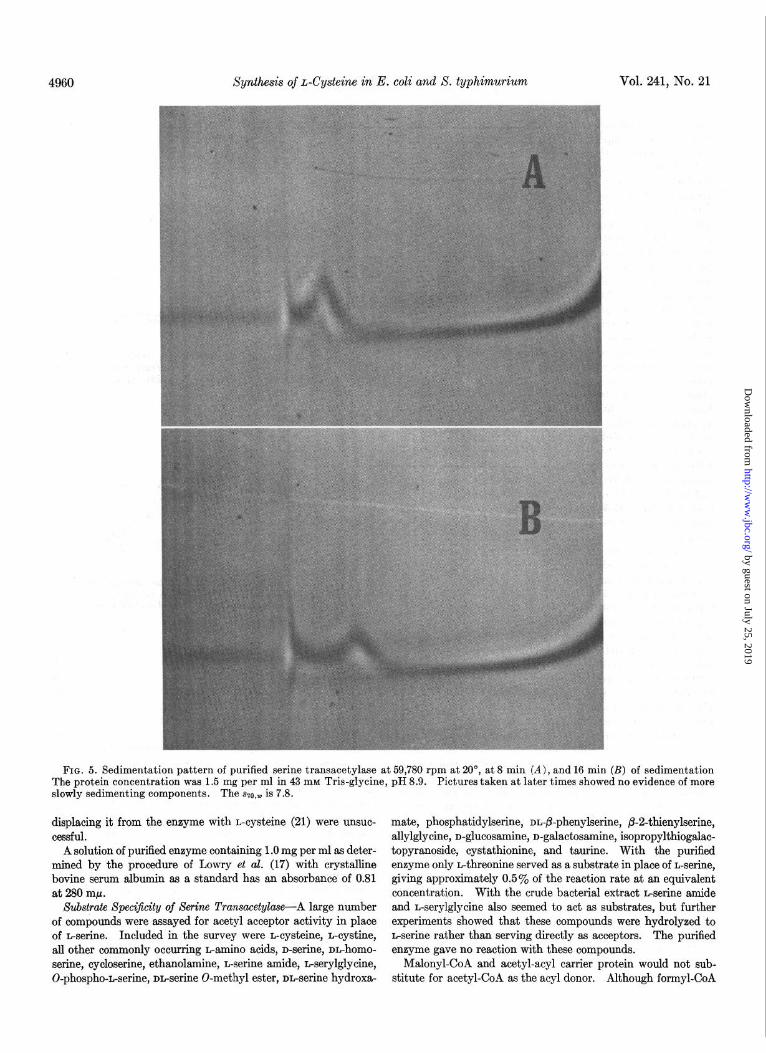
enzyme was relatively pure. When the material from Step X was initially subjected to analytical disc gel electrophoresis, four protein bands were noted, all of which showed serine transacetyl- ase activity in the histochemical assay. Three of these bands were compact with sharp edges, while the major, fastest moving component was very diffuse. At this time the enzyme prepara- tion showed three components by analytical ultracentrifugation with szo,w values of 10.2, 15.8, and 24.4. After storage at -20” for 3 months analytical ultracentrifugation revealed a single peak with an ~20,~ of 7.8 (Fig. 5). Disc gel electrophoresis now re- vealed the previously noted diffuse material, but, in place of the three more slowly moving bands, a very rapidly moving compo- nent with enzymic activity was seen. We felt that these findings represented aggregation phenomena and artifacts due to inter- actions of the enzyme with the acrylamide gel. When further information pertaining to this problem is available, it will be reported elsewhere. Contrary to disc gel electrophoresis, elec- trophoresis at several pH values on cellulose acetate strips re- vealed a single protein band. On the basis of these data we feel that the enzyme is at least 85 to 90% pure.
A solution of the purified enzyme is yellow and exhibits an absorption peak at 415 rnp (Fig. 6) which is similar to that re- ported for several pyridoxal phosphate-containing enzymes (20). Attempts to identify the chromaphore as pyridoxal phosphate by
by guest on July 25, 2019http://w
ww
.jbc.org/D
ownloaded from
4960 Synthesis of L-Cysteine in E. coli and S. typhimurium Vol. 241, No. 21
FIG. 5. Sedimentation pattern of purified serine transacetylase at 59,780 rpm at 20”, at 8 min (A), and 16 min (B) of sedimentation The protein concentration was 1.5 mg per ml in 43 mM Tris-glycine, pH 8.9. Pictures taken at later times showed no evidence of more slowly sedimenting components. The SZO,~ is 7.8.
displacing it from the enzyme with L-cysteine (21) were unsuc- mate, phosphatidylserine, nn-@-phenylserine, b-2-thienylserine, cessful. allylglycine, n-glucosamine, n-galactosamine, isopropylthiogalac-
A solution of purified enzyme containing 1 .O mg per ml as deter- topyranoside, cystathionine, and taurine. With the purified mined by the procedure of Lowry et al. (17) with crystalline enzyme only n-threonine served as a substrate in place of n-serine, bovine serum albumin as a standard has an absorbance of 0.81 giving approximately 0.5% of the reaction rate at an equivalent at 280 ml. concentration. With the crude bacterial extract n-serine amide
Substrate Spec$icity of Serine Transacetylase-A large number and L-serylglycine also seemed to act as substrates, but further of compounds were assayed for acetyl acceptor activity in place experiments showed that these compounds were hydrolyzed to of n-serine. Included in the survey were n-cysteine, L-cystine, n-serine rather than serving directly as acceptors. The purified all other commonly occurring L-amino acids, n-serine, nn-homo- enzyme gave no reaction with these compounds. serine, cycloserine, ethanolamine, n-serine amide, n-serylglycine, Malonyl-CoA and acetyl-acyl carrier protein would not sub- 0-phospho-n-serine, nn-serine O-methyl ester, nn-serine hydroxa- stitute for acetyl-CoA as the acyl donor. Although formyl-CoA
by guest on July 25, 2019http://w
ww
.jbc.org/D
ownloaded from

Issue of November 10, 1966 N. M. Kredkh and G. M. Tomkins 4961
O.OSy
I\’
i 360 300 400 420 440
WAVELENGTH, mp
FIG. 6. Absorption spectrum of purified serine transacetylase. The tracing was done on a Cary model 15 recording spectropho- tometer with a 10X scale expander with a protein concentration of 0.79 mg per ml in 43 mM Tris-glycine, pH 8.9.
hydrolyzes rapidly under the conditions of the assay, it was possi- ble to show that, with this donor, acyl transfer occurred approxi- mately 40% as fast as with acetyl-CoA at the same concentra tion.
Because of the spectral evidence for the association of pyr- idoxal with the enzyme, pyridoxal phosphate was included in incubation mixtures at concentrations of 1 to 50 pg per ml with no effect on the rate of reaction.
Characterization of Product of Se&e Transacetylase Reaction- The two most likely products of the serine transacetylase reac- tion were 0-acetyl- and N-acetylserine. The conversion of 0-acetyl- to N-acetylserine proceeds at about 1% per min at pH 7.6, and much faster at higher pH values (11). Therefore, even if 0-acetylserine were the true product of the enzymic reaction, both 0-acetyl- and N-acetylserine might be found as products. N-Acetylserine, however, is quite stable at neutral pH values, so that if it were the product of the enzymic reaction, no O-acetyl- serine should appear. In order to determine the true enzymic product, the products of several reaction mixtures carried out at different pH values with r4C-L-serine as the substrate were exam- ined by chromatography and radioautography. Fig. 7 shows the
FIG. 7. Product of the serine transacetylase reaction. Incu- silica gel and developed and radioautographed as in “Methods.” bations were done as in “Methods,” with W-L-serine, purified It can be seen that both 0-acetylserine (OAS) and N-acetylserine serine transacetylase, and Tris-HCl at the indicated pH, and with- (NAS) are formed, the latter compound being favored at the more out DTN. Aliquot portions were spotted on thin layer plates of alkaline pH.
by guest on July 25, 2019http://w
ww
.jbc.org/D
ownloaded from

4962 Synthesis of L-Cysteine in E. coli and S. typhimwium Vol. 241, No. 21
Km for L-serine in STA reaction.
r
4 8. 12 16
Y I I I , “s
1 2 3 4 5 6 7 8 mM L-SERINE
FIG. 8. Substrate concentration velocity curve for n-serine in the serine transacetylase (STA) reaction with an acetyl-CoA con- centration of 0.10 mM. The assay was done by the DTN method described in “Methods ” Moderate substrate inhibition is noted, and the K, for n-serine is 5.6 X 10e4 M.
t
Km for acetyl-CoA in STA reaction.
,100
mM ACETYL-CoA
FIG. 9. Substrate velocity curve for acetyl-CoA in the serine transacetylase (STA) reaction with an L-serine concentration of 1.0 rnM. The assay was done by the DTN method described in “Methods.” The K, for acetyl-CoA is 1.1 X lo-* M.
results of such experiments. At pH 7.1 0-acetylserine was the major product with only traces of N-acetylserine appearing with longer incubations. As the pH of the incubation was increased, more N-acetylserine was formed. When 0-acetyl-1%L-serine was incubated in place of i4C-n-serine, it was converted to N-ace- tylserine even in the absence of enzyme. When N-acetyl-14C- serine was incubated with enzyme, no serine or 0-acetylserine was formed. Therefore the enzymatic product is 0-acetylserine, which can then undergo a nonenzymatic acyl migration to give N-acetylserine.
In order to establish the optical configuration of the enzymic product, N-acetyl-14C-serine was synthesized as described in “Methods.” Chromatographic and radioautographic analysis revealed that acylase I from hog kidney quantitatively deacyl- ated this compound to serine. Since the deacylase is known to attack only N-acylated n-amino acids, the compound derived from the enzymic product (0-acetylserine) must have been N- acetylJ4C-n-serine. When authentic 0-acetyl-L-serine was con- verted chemically to N-acetylserine by incubation at pH 8.0 for
1 hour, the product was also found to be N-acetyl-n-serine by hydrolysis with acylase I, indicating that the L configuration is maintained during the 0- to N-acyl shift. We concluded, there- fore, that the product of the serine transacetylase reaction is 0-acetyl-L-serine.
Kinetics of Xerine Transacetylase Reaction-When the rate of serine acetylation was measured at different concentrations of n-serine, Michaelis-Menten kinetics was observed with moderate inhibition at serine concentrations above 3 mM (Fig. 8). The calculated K, is 5.6 X lop4 M. A plot of rate with respect to acetyl-CoA concentration also reveals Michaelis-Menten kinetics with a K, of 1.1 X 1O-4 M (Fig. 9).
The pH rate curve of the reaction is rather flat with an opti- mum at pH 7.6 to 7.8 (Fig. 10). At the same pH and ionic strength the reaction is only 70% as fast in sodium phosphate as in Tris-HCl buffer. Salt concentrations less than 0.02 M or greater than 0.2 M appreciably decrease the reaction rate.
Inhibitors of Serine Transacetylase Reaction-Each compound tested for acetyl acceptor activity in place of n-serine was also assayed for its ability to inhibit the reaction. The data in Table III show the degree of inhibition by various compounds at the concentrations indicated. The most striking finding was the ability of n-cysteine to inhibit the reaction at very low concen- trations. Fig. 11 shows this inhibition with varying concentra- tions of n-cysteine. The inhibition was noncompetitive with L-serine, but competitive with acetyl-CoA (Fig. 12). n-Cysteine had no effect, while derivatives of L-cysteine inhibited only at high concentration, where trace contaminants of L-cysteine might have been responsible for the effect.
0-Acetylserine Suljhyclrylase
Characterization and Purity of 0-Acetylserine Suljhydrylase- Until the strong inhibition of the serine transacetylase reaction by n-cysteine was discovered, we had not suspected that it was a step in the biosynthesis of cysteine. Therefore, the enzyme preparation described in this paper had been purified only for serine transacetylase activity. Yet, when sulfide was added to the usual reaction mixture containing r4C-n-serine, acetyl-CoA, and purified serine transacetylase, radioautographs revealed a
.080r pH curve for STA
t I 8 I I I I I I
6.4 6.8 7.2 7.6 8.0 8.4 PH
FIG. 10. Dependence of serine transacetylase (STA) reaction rate on nH. The assays were done bv the DTN method with either T&-HCl at a final concentration of 0.05 M or sodium phos- phate at a final phosphate concentration of 0.02 M.
by guest on July 25, 2019http://w
ww
.jbc.org/D
ownloaded from

Issue of November 10, 1966 N. M. Kredich and G. M. Tomkins 4963
labeled compound with the mobility of cysteine. The formation of this compound from labeled serine required acetyl-CoA.
When N-acetylJ4C-L-serine was used as a substrate, with or without sulfide, the compound was not formed. However, when 0-acetyl-14C-L-serine was incubated with sulfide and the purified serine transacetylase, a good yield of cysteine (chromatographi- tally identified) was obtained, even in the absence of acetyl-CoA. Therefore, the formation of cysteine was studied directly, by the procedure described in “Methods.”
The complete reaction mixture required 0-acetyl-L-serine, sulfide, and enzyme. Pyridoxal phosphate had no effect on the reaction rate at concentrations between 1 and 100 pg per ml, and no inhibition was noted with the pyridoxal phosphate inhibitors, KCN, 2 hydrazino-3-imidazol-4(5)-yl-propionic acid, or hydrox- ylamine at concentrations of 1 mM. Some nonenzymatic forma- tion of thiol occurred during long incubations, but the nature of the product was not investigated, since during the usual 5-min incubation none was detected. L-Serine, N-acetyl-DL-serine, 0-phospho-L-serine, or 0-succinyl-nL-homoserine could not sub- stitute for 0-acetyl-L-serine in the formation of thiol; neither did these compounds inhibit the reaction. 0-Acetyl-L-threonine did serve as a substrate, but only 0.7% as well as 0-acetyl-L- serine. The product of this reaction w&s not studied further. 0-Acetyl-L-threonine did not inhibit at a concentration of 24 mM.
When samples of bacterial extract at various stages of purifica- tion were assayed for both 0-acetylserine sulfhydrylase and serine transacetylase activity, it was found that the ratio of 0-acetylserine sulfhydrylase units to serine transacetylase units decreased from 120 to 4.5 between Fractions II and X (Table II). Attempts to enhance the 0-acetylserine sulfhydrylase activity in Fraction X by adding pyridoxal phosphate or metals or by mixing it with Fraction II were unsuccessful. Neither was the level of 0-acetylserine sulfhydrylase activity in Fraction II in- hibited by mixing it with more purified fractions.
The data in Table II show that the calcium phosphate gel step resulted in the greatest loss in 0-acetylserine sulfhydrylase activ- ity proportional to serine transacetylase activity but that the ratio of activities remained relatively constant after that step. Even though the two activities do not copurify well, the possi-
TABLE III Inhibition of serine lransacetylase reaction by various suljur-
containing compounds
Reaction mixtures contained Tris-HCI, pH 7.6, 25 pmoles; L- serine, 0.50 pmole; acetyl-CoA, 0.05 amole; 0.005 unit of purified enzyme in a final volume of 0.50 ml; and the compound to be tested at the indicat,ed final concentration. The reaction was followed spectrophotometrically at 232 rnp as described in “Meth- ods.”
Inhibition Concentration Inhibition
L-Cysteine L-Cysteine o-Cysteine n-Cysteine L-Cystine N-Acetyl-L-cysteine.. L-Cysteic acid. L-Homocysteine Taurine
+nM 0.01 0.1 0.1 1.0 1.0 1.0 1.0 1.0 1.0
-~
% 85 96
0 0 0
51 50
0 0
--
k-TY--+ 8 10 100 pM L-CYSTEINE
FIG. 11. Inhibition of serine transacetylase (&“A) by L-cys- teine. The assays were done with purified enzyme by following the loss of absorbance at 232 rnp as described in “Methods” with varying concentrations of L-cysteine. At 1.1 MM L-cysteine, 50yo inhibition occurred with 1 mM L-serine and 0.1 mM acetyl-CoA.
.08 .I6 10 20 30 mM ACETYL-CoA ‘4
FIG. 12. Inhibition of serine transacetylase by L-cysteine with varying concentrations of acetyl-CoA. The data suggest that the inhibition is competitive with acetyl-CoA with a Ki of 4 X 10-v M.
bility of a physical association between 0-acetylserine sulfhy- drylase and serine transacetylase was suggested by the copurifi- cation subsequent to the calcium phosphate gel step. However, more recent studies to be reported in detail later show a definite separation of the two activities by ammonium sulfate fractiona- tion and gel filtration of crude extracts.
Product of 0-Acetylserine Sulfhydrylase Reaction-To 40 ml of 0.2 M Tris-HCl, pH 7.4, containing 1 mM NaEDTA, were added 368 mg (2 mmoles) of the hydrochloride of 0-acetyl-L-serine, and the solution was adjusted to pH 6.3 with additional Tris base. Then 13 ml of Tris-sulfide, 64 mM (see “Methods”) were added, giving a final pH of 7.35. After the addition of 60 units of O- acetylserine sulfhydrylase (Fraction VIII), the reaction mixture was placed in a glass flask which was stoppered, and aliquot por- tions were assayed for thiol formation with time. After 1 hour the reaction had reached completion with the formation of 0.86 mmole of thiol.
The solution was adjusted to pH 2 with HCI, and the remaining
by guest on July 25, 2019http://w
ww
.jbc.org/D
ownloaded from

4964 Synthesis of L-Cysteine in E. coli and S. typhimurium Vol. 241, No. 21
Km for OAS in OASS reactIon.
Km.l.4xlO‘‘M
I I I I I I I 0.04 0.00
‘/s 0.12 0.16
I I I I I, I I I I I 20 40 60 80 100 t20
mM 0-ACETYL L-SERINE
FIQ. 13. Substrate velocity curve for 0-acetyl-L-serine (OAS) in the 0-acetylserine sulfhydrylase (OASS) reaction. The assay was done as described in “Methods” with varying concentrations of 0-acetyl-L-serine. The K, for 0-acetyl-L-serine is 1.4 X 10-z M.
0.2f
0.24
Km for sulfide in OASS reaction
?--
with a stream of nitrogen. The solution of thiol was then oxi- dized to the disulfide by titration with an alcoholic solution of iodine until a faint trace of iodine color remained in solution. The pH was then adjusted to 3.2 with NHIOH, and 15 ml of ethanol were added. A 6ne precipitate resulted, which was col- lected by filtration after standing at 4” overnight. This mate- rial was dissolved in a small amount of warm 1 N HCl, filtered, and recrystallized by the addition of NHhOH and ethanol as above. The final product was collected by filtration, washed with ethanol and ether, and air dried. The yield was 35 mg.
The purified product was identified as cystine by thin layer chromatography on silica gel (see “Methods”), and by paper chromatography in two solvent systems. The [ar], was found to be -210” (O.lYo in 1 N HCl), thus establishing the material as n-cystine. Since the enzymatic product is a thiol, it must be L-cysteine.
Kinetics of O-Acetylserine Xuljhydrylase Reaction-The time course of cysteine synthesis is shown in Fig. 2. The fall in reac- tion rate is probably due to loss of 0-acetyl-n-serine by conver- sion to N-acetyl-n-serine and cysteine, and to loss of sulfide by diffusion and by conversion to cysteine. The linearity of the reaction during the first few minutes shows that the product,
TABLE IV 0 Serine transacetylase and 0-acetylserine sulfhydrylase activities
0 in cys E mutants of S. lyphimurium
Mutants were grown with shaking at 37” on nutrient broth and harvested in late log phase by centrifugation. Extracts were prepared by sonic oscillation in standard buffer. Supernatants from a 37,060 X g centrifugation were used in the assays. Results are expressed as the percentage specific activity of that in wild-
0 type organisms grown in the same way. r’
I
Ea 2 Ea 6 Eb 11 Eb 17 Ea 30 Eb 396
Serine transacetylase 0-Acet lserine sulf- activity hydry ase activity P
% % 3.0 510 0 (<0.2) 290 1.8 86 0 (<0.2) 33 0 (<0.2) 25 0.7 56
TABLE V
1 , , , Serine transacetylase and 0-acetylserine sulfhydrylase activities in wild-type S. typhimurium LT-2 grown on di$erent
2 4 6 sulfur sources “S
I I I I I I I I I , I Bacteria were grown with shaking at 37” in Vogel-Bonner Me- 0.4 0.8 1.2 I .6 2.0 2.4 dium E minimal salts (23) with 0.5% glucose. The cells were
mM SULFIDE
FIG. 14. Substrate velocity curve for sulfide in the O-acetyl- serine sulfhydrylase (OASS) reaction. With the assay described in “Methods,” the sulfide concentration was varied. The K, for sulfide is 1.8 X lO+ M.
harvested in log phase, washed with fresh medium minus sulfate, and resuspended in medium in which the sulfur source was 0.5 mM as indicated. After two generations of growth the cells were harvested by centrifugation and washed with sulfur-free medium. Extracts were prepared by sonic oscillation in standard buffer, and 37,666 X g supernatants were used in the assay.
H2S was bubbled off with a stream of nitrogen. To this solution was then added 1.36 g of HgCh in 10 ml of 1 N HCl. When the pH was adjusted to 3.0 with dilute NaOH, a precipitate formed which was collected by centrifugation and dissolved in 15 ml of 1 N HCl. A stream of H&I was passed through this solution for 10 min, and the resulting precipitate was removed by centrifugation and discarded. H&S was removed from the supernatant solution
-
Sulfate. 0.031 5.3 L-Cysteine 0.057 1.6 r,-Djenkolic acid. 0.023 10.9
by guest on July 25, 2019http://w
ww
.jbc.org/D
ownloaded from

Issue of November 10, 1966 N. M. Kredich and G. M. Tom&s 4965
L-cysteine, is not inhibitory at concentrations up to 0.5 mM. The dependence of velocity on substrate concentration is shown in Figs. 13 and 14, and K, values for sulfide and 0-acetyl-L-serine are 1.8 X 1w4 M and 1.4 X low2 M, respectively.
Genetic Data
When it appeared that serine transacetylase and O-acetyl- serine sulfhydrylase were steps in cysteine biosynthesis, several cysteine-requiring mutants of S. typhimurium were examined for these enzymic activities. The serine transacetylase and O- acetylserine sulfhydrylase reactions catalyzed by highly purified preparations of serine transacetylase from wild-type S. typhi- m&urn LT-2 had the same characteristics as the enzyme de- scribed above from E. coli. Salmonella was chosen for genetic study because the genetics of cysteine metabolism had been more carefully studied in this organism, and mutants were readily available.
Mutations which produce cysteine auxotrophs unable to grow on sulfide map in the cys E region in Salmonella (22). Since these organisms presumably cannot incorporate sulfide into cysteine, it seemed likely that they would show alterations of se&e transacetylase of 0-acetylserine sulfhydrylase activity. Several cys E mutants were grown on nutrient broth and har- vested by centrifugation, and soluble extracts were prepared by sonic oscillation in standard buffer. After centrifugation at 37,000 x g for 30 min, the supernatant solutions were assayed for protein and enzymic activities. The results are shown in Table IV as percentages of the activity found in similar extracts of wild-type LT-2 organism grown in the same way. The low or undetectable activity of serine transacetylase in these mutants strongly suggests that this enzyme is involved in cysteine biosyn- thesis and indicates that the cys E region in S. typhimurium may contain the structural gene for that enzyme. Mutants in either the a or b cistrons of the cys E locus had low or absent levels of serine transacetylase. 0-Acetylserine sulfhydrylase activity was also altered in these mutants, being either elevated as much as 5-fold over the wild type or depressed. These changes in 0-acetylserine sulfhydrylase activity in cys E mutants are not presently understood.
In confirmation of the enzymatic data showing appreciable levels of 0-acetylserine sulfhydrylase, it was found that with all six cys E mutants tested a zone of growth occurred around a crystal of 0-acetyl-L-serine on minimal agar plates. In most cases, however, the amount of growth was only microscopic and considerably less than that observed with L-cysteine.
The data in Table V suggest that in S. typhimurium O-acetyl- serine sulfhydrylase is repressed by growth on L-cysteine and derepressed by growth on L-djenkolic acid in a manner similar to that reported with the sulfate to sulfide enzymes in that organism (24). Serine transacetylase activity appears to vary in the op- posite way but to a lesser degree.
DISCUSSION
The enzymatic, nutritional, and genetic data presented indi- cate that the pathway of L-cysteine biosynthesis from L-serine and sulfide in E. coli and S. typhimurium proceeds by the path- way presented in the introduction. Serine transacetylase, the
first enzyme in this short pathway, is subject to feedback inhibi- tion by very low concentrations of the end product, L-cysteine, but its synthesis is not repressed. The second enzyme, O-acetyl- serine sulfhydrylase, is repressed by L-cysteine but is not inhib- ited by L-cysteine.
Although serine transacetylase has been extensively purified, the enzyme that catalyzes the 0-acetylserine sulfhydrylase reac- tion has not yet been systematically investigated. Perhaps the serine transacetylase and 0-acetylserine sulfhydrylase activities are in some way physically associated, since the purified serine transacetylase contains an appreciable amount of 0-acetylserine sulfhydrylase activity. However, since the two activities do not copurify well in the early steps of serine transacetylase purifica- tion, are not corepressed or coderepressed, and are affected differ- ently by mutations in the cys E region in S. typhimurium, it appears that both activities do not reside in the same protein molecule. The possibility of spurious enzymatic activities capa- ble of carrying out the 0-acetylserine sulfhydrylase reaction in crude extracts makes it impossible to define the exact relation- ships between serine transacetylase, 0-acetylserine sulfhydrylase, and the cys E locus at this time. We hope that purification of the 0-acetylserine sulfhydrylase activity will resolve this problem.
Acknourledgments-The authors wish to thank Dr. R. Reisfeld, who performed the preparative gel electrophoresis; and Drs. B. Ames, R. Martin, and S. Black for their helpful discussions.
1. 2. 3. 4.
SIMMONDS, S., J. Biol. Chem., 174, 717 (1948). SCHLOSSMANN, K., AND LYNEN, F., Biochem. Z., 338,591 (1957). NAKAMURA, T., AND SATO, R., Nature, 198, 1198 (1963). DREYFUSS, J., AND MONTY, K. J., J. Biol. Chem., 238, 1019
(1963). 5. 6. 7.
ABELSON, P. H., J. Biol. Chem., 206, 335 (1954). NARITA, K., Biochim. Biophys. Acta, 30, 352 (1958). SLY, W. S., AND STADTMAN, E. R., J. Biol. Chem., 238, 2632
(1963). 8. ALPERS, D. H., APPEL, S. H., AND TOMKINS, G.M., J.BioZ.
Chem., 240, 10 (1965). 9.
10. Il. 12. 13.
STADTMAN, E. R., J. Cell. Comp. Physiol., 41, 89 (1953). STADTMAN, E. R., J. Biol. Chem., 196, 527 (1952). FLAVIN, M., AND SLAUGHTER, C., Biochemistry, 4,137O (1965). LIDDELL, H. F., AND SAVILLE, B., Analyst, 84, 188 (1959). APPEL, S. H., ALPERS, D. H., BND TOMKINS, G.M., J.MoZ.
Biol., 11, 12 (1965). 14. SHEEHAN, J.C., GOODMAN, M., ANDHESS,G.P., J.Am. Chem.
Sot., 78, 1367 (1956). 15. 16. 17.
18.
GREGORY, J. D., J. Am. Chem. Sot., 77, 3922 (1955). RYDON, H. N., AND SMITH, P. W. G., Nature, 169,922 (1952). LOWRY,~. H., ROSEBROUQH,N. J., FARR, A.L., ANDRANDALL,
R. J., J. Biol. Chem., 193, 265 (1951). JOVIN, T., CHRAMBACH, A., AND NBUGHTON, M.A.,Anal. Bio-
them., 9, 351 (1964). 19. REISFELD, R. A., AND SMALL, P. A., JR., Science, 162, 1253
(1966). 20. LABOW. R.. AND ROBINSON, W. G.. J. Biol. Chem.. 241, 1239
(1966). ’ 21. 22. 23.
SCHIRCH, L., AND MASON, M., J. Biol. Chem., 238, 1032 (1963). CLOWES. R. C., J. Gen. Microbial., 18, 154 (1958). VOGEL, ‘H. J., 'AND BONNER, D. ik, J. Baol. Chem., 218, 97
(1956). 24. DREYFUSS, J., END MONTY, K. J., J. Biol. Chem., 238, 3781
(1963).
REFERENCES
by guest on July 25, 2019http://w
ww
.jbc.org/D
ownloaded from

Nicholas M. Kredich and Gordon M. Tomkinstyphimurium
Salmonella and Escherichia coliThe Enzymic Synthesis of l-Cysteine in
1966, 241:4955-4965.J. Biol. Chem.
http://www.jbc.org/content/241/21/4955Access the most updated version of this article at
Alerts:
When a correction for this article is posted•
When this article is cited•
to choose from all of JBC's e-mail alertsClick here
http://www.jbc.org/content/241/21/4955.full.html#ref-list-1
This article cites 0 references, 0 of which can be accessed free at
by guest on July 25, 2019http://w
ww
.jbc.org/D
ownloaded from


![Mass Spectrometric Analysis of l-Cysteine Metabolism: … · tion of [U-13C3, 15N]L-cysteine to the culture, the levels of [13C3,15N]L-cysteine increased, and [13C3, 15N]L-cysteine](https://img.pdfslide.us/doc/110x75/5fe663421198753c202620ce/mass-spectrometric-analysis-of-l-cysteine-metabolism-tion-of-u-13c3-15nl-cysteine.jpg)