Embed Size (px)
Citation preview
UC IrvineCognition and Creativity
TitleThe Emotions (after Charles Darwin)
Permalinkhttps://escholarship.org/uc/item/0h35f4zs
AuthorSwack, Debra
Publication Date2009-12-12 Peer reviewed
eScholarship.org Powered by the California Digital LibraryUniversity of California
The Emotions (after Charles Darwin)
Debra Swack SUNY@Buffalo Research Foundation
40 West 116th Street, Apt. B502 New York, New York 10026
212-866-2192 [email protected]
ABSTRACT
Rapid changes in science, technology and new media will lead to
more sophisticated ideas about what it means to be human, in
thought, body, emotional response and artistic expression. New
relationships will form between humans, machines and animals
with the human functioning as a networked resource that can be
accessed globally over the internet.
This paper documents both the technical and theoretical
development of the collaborative interactive new media video
project “The Emotions (after Charles Darwin)” which explores
some of the above concepts. “The Emotions” first tries to
establish the existence of the universality of emotions at a
biological level, as empirically measured and documented by the
results of the control group (non-autistic subjects, as the goal is to
document “normal”, i.e. universal emotional response) at the
Brain Mind Institute in Switzerland. Secondly, it suggests the
potential for subsequent futuristic misuse through genetic and or
technological modification (demonstrated by the observer’s ability
to interactively modify or transform a given emotion’s video
stream at will).
Keywords
Cognitive and computational neuroscience, embodiment,
bioethics, emotions, interactivity, Plutchik, amygdala, face
perception.
1. INTRODUCTION Although Darwin was incredibly prescient in his discoveries about
what role the nervous system might play in regulating emotions,
developments in neuroscience did not begin until well over a 100
years later, partially due to the lack of sophisticated recording and
analytical tools such as neuro-imaging and computation made
easier, enhanced through software algorithms and applications
executed on computers.
This co-mingling of previously unrelated and seldom overlapping
disciplines means that new media itself, its practices, applications
and theories will continue to be in constant flux and development.
It used to be standard practice in beginning art classes to ask what
is art? But now the question is not only what is art, but who or
what makes art (i.e., sometimes art now takes on a life of its own,
extending beyond the control of its creator).
For example, the interactive new media video project “The
Emotions (after Charles Darwin)” attempts to prove the
universality of emotions by transcending cultural categorizations
such as species, race, age and gender and instead relates emotions
to their neurobiological origins and functions. It further suggests
that once empirically known, that this information can be used to
genetically or technologically alter human emotion(s) in
individuals or groups to create new beings or new emotional
interiors that better conform to culturally desirable behaviors. This
of course raises bioethical questions about the future nature of life
for humans and animals; the embodiment and containment of the
self and its symbiotic integration and enhancement with
technology and machines.
“No Longer is human existence defined by its unique temporal
and spatial coordinate; one body, one life in a specific space and
time. Instead human life is increasingly defined by the agential,
instrumental deployment of resources for bodily renewal, both its
temporal and spatial context subject to extensions or
translocations”, according to Susan Merrill Squier, in Liminal
Lives: Imagining the Human at the Frontiers of Biomedicine.
As Joanna Zylinska states in her book Bioethics in the Age of New
Media, “This is by no means to suggest that the human has been
reduced to information in the age of new media and that we can
therefore do away with embodiment; it is only to point to the
emergence of new discourses of the human which undermines its
centering around some fixed biological characteristics or moral
values.”
She adds, “The human does not disappear from the kind of
nonhumanist bioethics envisaged here: in fact, it functions as its
strategic point of entry. What we are dealing with, however, is
not so much a “human being” understood as a discrete and
disembodied moral unity but rather a “human becoming”;
relational, co-emerging with technology, materially implicated in
sociocultural networks, and kin to other life forms.”
Neil Badmington in Alien Chic talks about how recent trends in
techno-science have unsettled post humanist critics. For example
he talks about how Donna Haraway’s “Cyborg Manifesto (1991)”
first deconstructed humanist relationships such as
organism/machine, reality/fiction/human/animal, physical/non-
physical and self/other and replaced them with chimeras;
cyborgian fabrications of machine and organisms. He goes on to
say that the latest trend in post-humanism seems to involve
merging with animals, which ironically was not a concept alien to
Darwin 140 years ago when he studied, documented and sought to
define similarities with animals’ emotions and our own.
Badmington quotes numerous television and news reportage from
Newsweek to Nature, who discovered that reason, tool use, tool
making, altruism and language are not unique to humans, neither I
might add, is making or performing music (last year I presented
© Digital Arts and Culture, 2009. Permission to make digital or hard copies of all or part of this work for personal or classroom use is granted without fee provided that copies are not made or distributed for profit or commercial advantage and that copies bear this notice and the full citation on the first page. To copy otherwise, or republish, to post on servers or to redistribute to lists, requires prior specific permission from the author. Digital Arts and Culture, December 12–15, 2009, Irvine, California, USA.
“Birdsongs; the Language Gene”, in the “Sonic Fragments
Soundart Festival” at Princeton University which digitally
reconfigures bird songs into human music).
2. DARWIN AND NEUROSCIENCE Over a hundred years ago, Charles Darwin theorized that the
universality of emotions existed in humans and animals at a
biological level. He posed questions such as can we feel happy,
sad or fearful when we are alone or are emotions a unique result
of being with others in a social situation? He suggested that the
reason for the universality of emotions was due to an underlying
biological basis that communicated our needs to others. We
experience an emotion and specific areas of the brain send signals
to specialized muscle groups that respond to communicate our
feelings.
Darwin believed that the following principles were responsible for
most of the expressions and gestures involuntarily exhibited by
humans and animals while experiencing emotions: habitual
actions initiated by certain states of mind in order to relieve or
gratify certain sensations, habitual inverse actions initiated by the
exact opposite states of mind and actions initiated by the nervous
system mostly independent from both will and habit.
In post Darwin times, scientists study what regions and chemicals
in the brain control different emotions and if these regulators can
be modified to elicit alternative results. For example, emotions are
studied to determine their affect on the immune, cardiovascular
and endocrine systems. There is also the possibility for misuse,
what if we could invoke certain emotions in people at will through
a drug or by permanently or temporarily altering structures in their
brain? Perhaps at the same time we could remove their ability to
feel remorse or guilt. Could this form of genetic intervention be
used randomly against individuals or during war-time to induce
people to commit violent acts?
The neuroscientist Joseph Ledoux says the brain has not evolved
to the point where connectivity exists for cognitive systems to
control our emotions. But even so, he says that wouldn’t
necessarily be good, because Mr. Spock (a character lacking in
human emotions from the 60’s TV show Star Trek) may not be an
ideal kind of human that we'd like to become. Additionally,
Ledoux talks about futuristically controlling undesirable emotions
such as fear through drug regulation, stating that once we can
identify the neurotransmitters that are involved in producing fear,
we could create a chemical profile of fear in the amygdala and
then develop a drug to attack it.
The amygdala is an almond-shaped structure in the frontal portion
of the temporal lobe near the hippocampus in the brain that allows
us to both feel and perceive negative emotions. It regulates our
reactions to events that are important for survival such as the
presence of danger, sexual partners, enemies, food and those in
need. The amygdala works as a system with other related
structures because unique sets of regions in the brain are
connected to each other and work together to control different
emotions. It also plays an important role in emotional regulation
and studies have shown that emotional disorders can manifest
themselves both functionally and structurally (it can become
asymmetrically enlarged in depressed individuals). Patients who
have had their amygdala destroyed due to stroke are able to
recognize all emotions expressed by facial expressions except for
fear.
The amygdala’s connectivity with the neo-cortex is also not
symmetrical; the amygdala’s connection to the neo-cortex is much
stronger than the neo-cortex’s connection to it (as shown in David
Amaral’s studies of primate brains), which in part explains,
according to neuroscientist Joseph Ledoux, why emotions are
often hard to turn off once initiated. The body also releases
hormones and long acting substances at the exact time that we
experience strong emotions. Additionally, there is a relationship
between the visual system and emotions. In The Expressions of
the Emotions in Man and Animals, Darwin talks about the
importance of visual cues when seeking mates, prey and avoiding
danger, therefore it’s not surprising that studies show that the
visual cortex is more activated in response to visual emotional
stimuli than visual non-emotional stimuli.
Darwin acknowledged individual variance in emotional reactivity
due to differences in development (for example he noticed that
insane persons had strong passions which they openly expressed).
But he never addressed the idea of emotion regulation which
didn’t come into being until the development of neuroscience a
hundred years later.
Davidson defines the study of individual differences in emotional
reactivity and emotion regulation as affective style consisting of
the threshold to respond, the magnitude of the response, the rise
time to the peak of the response, the recovery function of the
response and the duration of the response. The duration of
emotional responding is important in understanding individual
differences and can also indicate psychopathology since some
mood disorders are associated with either an abnormally early
onset or inability to turn off a response quickly enough.
3. THE EMOTIONS “The Emotions” is a multi- channel interactive video where each
of four panels will display close-up graphic, moving images of
men, women and children of all ages and races, expressing a
specific emotion such as happiness, sadness, fear or anger
(categorized as such by the results of the control group). Each
panel’s images will morph/blend to form a continuous stream of
soundless images whose emotion will not be identified so as to
allow the viewer the ability to form their own conclusion as to
what emotion they feel is being expressed (which will also test the
universality of emotions).
A fifth panel will record live audience reaction/ participation at
the actual site of the installation in order to test mirroring behavior
of the emotions displayed in the other four panels. Additionally
the observer will have the ability to interactively modify, convert
or morph emotions; demonstrating a futuristic ability to alter
emotions genetically and or technologically at will. "The
Emotions" is a collaboration with the Brain Mind Institute in
Switzerland whose experiments done using my photographs
validates their universality as images of specific emotions and
forms the basis for the video.
Shortly after “The Emotions” was accepted into the New Media
Collection (Rhizome) at the New Museum, I was contacted by
Britt Russo, a neuroscientist who had seen the project posted on
their web-site. She asked me if I would be interested in
collaborating with her lab at the Brain Mind Institute in
Switzerland and would allow them to use my photographs for
emotion perception research in autistic subjects. The lab had never
used photographs from life before, only those of staged actors. In
return they would present my work at international meetings and
publish it in scientific journals. Although the lab wanted to use my
photographs for research in autism; a neurodevelopmental
disorder that impairs social functioning, I knew I would be
primarily interested in the results of the control group as I wanted
to document what was perceived as “normal” or “neurotypical”
response and therefore universal, not the responses evidenced
solely in autistic patients. However I thought that I might learn
more about emotional response in general; its measurement and
analysis by including the observation of autistic patients since I
had the opportunity.
At the first meeting I had with Britt in Manhattan in the third
week of December 2007, she informed me about the institute and
its practices. The Brain Mind Institute was considered a world-
class research facility for neuroscience whose goal was to
synthesize and create a knowledge base by advocating a
multidisciplinary approach across disciplines and by linking
different research laboratories.
As taken from their web-site: “The mission of the Brain Mind
Institute is to understand the fundamental principles of brain
function in health and disease, by using and developing unique
experimental, theoretical, technological and computational
approaches. The scientific challenge addressed by the BMI
consists in connecting different levels of analysis of brain activity,
such that cognitive functions can be understood as a manifestation
of specific brain processes; specific brain processes as emerging
from the collective activity of thousands of cells and synapses;
synaptic and neuronal activity in turn as emerging properties of
the biophysical and molecular mechanisms of cellular
compartments.” The group that I would be working with was
headed by Dr. Nouchine Hadjikhani; a specialist in neuroimaging.
3.1 Testing at the BMI Lab In the lab, functional magnetic resonance imaging (fMRI),
Electroencephalography (EEG) and magnetoencephalography
MEG) were used to visualize brain activity and
electromyography (EMG) was used to measure facial muscle
activity of autistic subjects while they viewed images of human
emotional facial expressions (autistic people display different
brain activity patterns and facial muscles reactions than normal or
“neurotypical” people). A Tobii eye tracker was used to trace the
path of the subject’s eyes, while they viewed images.
According to Dr Hadjikhani’s research, autism was thought to be
related to the dysfunction of the mirror neuron system that plays a
critical role in the perception of other people’s intentions
including empathy. Autism Spectrum Disorder (ASD) is a
behaviorally defined neurodevelopmental disorder of early onset
whose subjects suffer from a social disability that profoundly
affects their ability to understand other people’s feeling and to
establish reciprocal rewarding relationships. The disorder
manifests itself by exhibiting restrictive and or repetitive
interests and behaviors. Persons suffering with ASD typically fail
to engage in social interactions because of an inability to correctly
interpret facial expressions and their meanings. Abnormalities in
face perception (crucial to social-communicative competence) and
the accurate identification of the deficient components of the face
processing system are essential to the understanding of ASD.
The lab’s primary area of study was the functional and structural
integrity of the social cognition network as it relates to autism and
also the amygdalas’s connectivity to the mirror neuron system
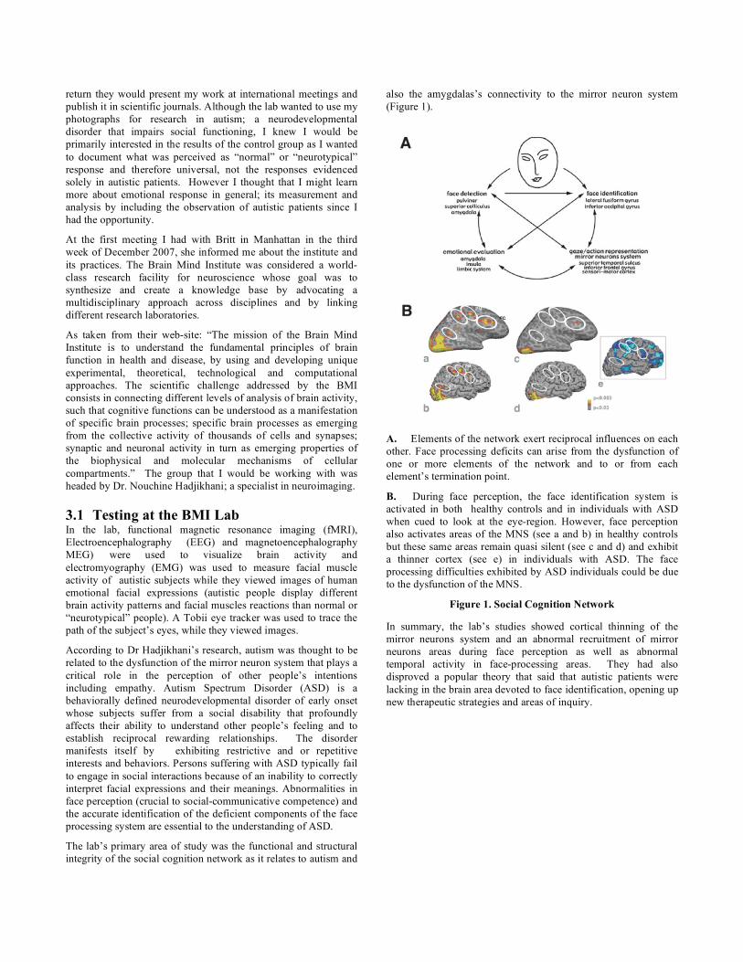
(Figure 1).
A. Elements of the network exert reciprocal influences on each
other. Face processing deficits can arise from the dysfunction of
one or more elements of the network and to or from each
element’s termination point.
B. During face perception, the face identification system is
activated in both healthy controls and in individuals with ASD
when cued to look at the eye-region. However, face perception
also activates areas of the MNS (see a and b) in healthy controls
but these same areas remain quasi silent (see c and d) and exhibit
a thinner cortex (see e) in individuals with ASD. The face
processing difficulties exhibited by ASD individuals could be due
to the dysfunction of the MNS.
Figure 1. Social Cognition Network
In summary, the lab’s studies showed cortical thinning of the
mirror neurons system and an abnormal recruitment of mirror
neurons areas during face perception as well as abnormal
temporal activity in face-processing areas. They had also
disproved a popular theory that said that autistic patients were
lacking in the brain area devoted to face identification, opening up
new therapeutic strategies and areas of inquiry.
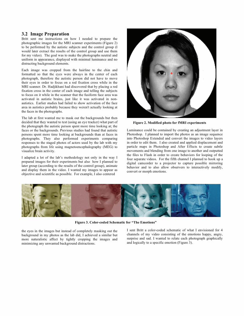
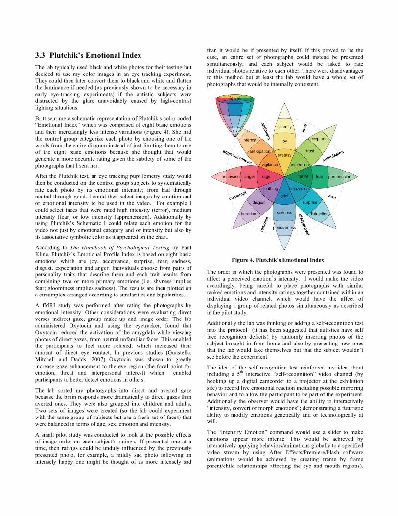
Figure 3. Color-coded Schematic for “The Emotions”
3.2 Image Preparation Britt sent me instructions on how I needed to prepare the
photographic images for the MRI scanner experiments (Figure 2)
to be performed by the autistic subjects and the control group (I
would later extract the results of the control group and use them
for my video). The goal was to make the photographs neutral and
uniform in appearance, displayed with minimal luminance and no
distracting background elements.
Each image was cropped from the hairline to the chin and
formatted so that the eyes were always in the center of each
photograph, therefore the autistic person did not have to move
their eyes in order to focus on a red fixation cross while in the
MRI scanner. Dr. Hadjikhani had discovered that by placing a red
fixation cross in the center of each image and telling the subjects
to focus on it while in the scanner that the fusiform face area was
activated in autistic brains, just like it was activated in non-
autistics. Earlier studies had failed to show activation of the face
area in autistics probably because they weren't actually looking at
the faces in the photographs.
The lab at first wanted me to mask out the backgrounds but then
decided that they wanted to test (using an eye tracker) what part of
the photograph the autistic person spent more time looking at; the
faces or the backgrounds. Previous studies had found that autistic
persons spent more time looking at backgrounds than at faces in
photographs. They also performed experiments comparing
responses to the staged photos of actors used by the lab with my
photographs from life using magnetoencephalography (MEG) to
visualize brain activity.
I adapted a lot of the lab’s methodology not only in the way I
prepared images for their experiments but also how I planned to
later group (according to the results of the control group), animate
and display them in the video. I wanted my images to appear as
objective and scientific as possible. For example, I also centered
the eyes in the images but instead of completely masking out the
background in my photos as the lab did, I achieved a similar but
more naturalistic affect by tightly cropping the images and
minimizing any unwanted background distractions.
Figure 2. Modified photo for fMRI experiments
Luminance could be contained by creating an adjustment layer in
Photoshop. I planned to import the photos as an image sequence
into Photoshop Extended and convert the images to video layers
in order to edit them. I also created and applied displacement and
particle maps in Photoshop and After Effects to create subtle
movements and blending from one image to another and outputted
the files to Flash in order to create behaviors for looping of the
four separate videos. For the fifth channel I planned to hook up a
digital camcorder to a projector to capture possible mirroring
behavior and to also allow observers to interactively modify,
convert or morph emotions.
I sent Britt a color-coded schematic of what I envisioned for 4
channels of my video consisting of the emotions happy, angry,
surprise and sad. I wanted to relate each photograph graphically
and logically to a specific emotion (Figure 3).
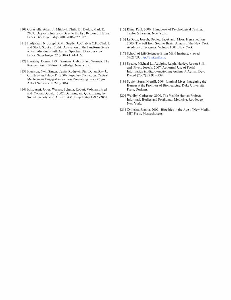
3.3 Plutchik’s Emotional Index
The lab typically used black and white photos for their testing but
decided to use my color images in an eye tracking experiment.
They could then later convert them to black and white and flatten
the luminance if needed (as previously shown to be necessary in
early eye-tracking experiments) if the autistic subjects were
distracted by the glare unavoidably caused by high-contrast
lighting situations.
Britt sent me a schematic representation of Plutchik's color-coded
“Emotional Index” which was comprised of eight basic emotions
and their increasingly less intense variations (Figure 4). She had
the control group categorize each photo by choosing one of the
words from the entire diagram instead of just limiting them to one
of the eight basic emotions because she thought that would
generate a more accurate rating given the subtlety of some of the
photographs that I sent her.
After the Plutchik test, an eye tracking pupillometry study would
then be conducted on the control group subjects to systematically
rate each photo by its emotional intensity; from bad through
neutral through good. I could then select images by emotion and
or emotional intensity to be used in the video. For example I
could select faces that were rated high intensity (terror), medium
intensity (fear) or low intensity (apprehension). Additionally by
using Plutchik’s Schematic I could relate each emotion for the
video not just by emotional category and or intensity but also by
its associative symbolic color as it appeared on the chart.
According to The Handbook of Psychological Testing by Paul
Kline, Plutchik’s Emotional Profile Index is based on eight basic
emotions which are joy, acceptance, surprise, fear, sadness,
disgust, expectation and anger. Individuals choose from pairs of
personality traits that describe them and each trait results from
combining two or more primary emotions (i.e, shyness implies
fear; gloominess implies sadness). The results are then plotted on
a circumplex arranged according to similarities and bipolarities.
A fMRI study was performed after rating the photographs by
emotional intensity. Other considerations were evaluating direct
verses indirect gaze, group make up and image order. The lab
administered Oxytocin and using the eyetracker, found that
Oxytocin reduced the activation of the amygdala while viewing
photos of direct gazes, from neutral unfamiliar faces. This enabled
the participants to feel more relaxed; which increased their
amount of direct eye contact. In previous studies (Guastella,
Mitchell and Dadds, 2007) Oxytocin was shown to greatly
increase gaze enhancement to the eye region (the focal point for
emotion, threat and interpersonal interest) which enabled
participants to better detect emotions in others.
The lab sorted my photographs into direct and averted gaze
because the brain responds more dramatically to direct gazes than
averted ones. They were also grouped into children and adults.
Two sets of images were created (so the lab could experiment
with the same group of subjects but use a fresh set of faces) that
were balanced in terms of age, sex, emotion and intensity.
A small pilot study was conducted to look at the possible effects
of image order on each subject’s ratings. If presented one at a
time, then ratings could be unduly influenced by the previously
presented photo, for example, a mildly sad photo following an
intensely happy one might be thought of as more intensely sad
than it would be if presented by itself. If this proved to be the
case, an entire set of photographs could instead be presented
simultaneously, and each subject would be asked to rate
individual photos relative to each other. There were disadvantages
to this method but at least the lab would have a whole set of
photographs that would be internally consistent.
Figure 4. Plutchik's Emotional Index
The order in which the photographs were presented was found to
affect a perceived emotion’s intensity. I would make the video
accordingly, being careful to place photographs with similar
ranked emotions and intensity ratings together contained within an
individual video channel, which would have the affect of
displaying a group of related photos simultaneously as described
in the pilot study.
Additionally the lab was thinking of adding a self-recognition test
into the protocol (it has been suggested that autistics have self
face recognition deficits) by randomly inserting photos of the
subject brought in from home and also by presenting new ones
that the lab would take themselves but that the subject wouldn’t
see before the experiment.
The idea of the self recognition test reinforced my idea about
including a 5th
interactive “self-recognition” video channel (by
hooking up a digital camcorder to a projector at the exhibition
site) to record live emotional reaction including possible mirroring
behavior and to allow the participant to be part of the experiment.
Additionally the observer would have the ability to interactively
“intensity, convert or morph emotions”; demonstrating a futuristic
ability to modify emotions genetically and or technologically at
will.
The “Intensify Emotion” command would use a slider to make
emotions appear more intense. This would be achieved by
interactively applying behaviors/animations globally to a specified
video stream by using After Effects/Premiere/Flash software
(animations would be achieved by creating frame by frame
parent/child relationships affecting the eye and mouth regions).

“Morph Emotions” would utilize a program/behaviors that would
scramble all channels simultaneously by selecting and replacing
video content from each of the four channels at random. “Convert
Emotion” would allow the user to morph any stream of emotions
into another by utilizing a program/behaviors that would select
and replace video content from one video stream to another. The
original color filter associated with Plutchik’s color coded
schematic would be applied to the new video stream, maintaining
its original Emotional Index categorization reference point.
The lab decided to organize an open-house of talks and
presentations for the public to celebrate the first World Autism
Day on April 2, 2008, as instituted by the U.N. They teamed up
with two other autism labs, one that worked with rats and other
with robots. They hoped that it would generate more research
subjects and also enlighten the public about autism. The lab’s
areas of research (including the brain areas studied) and my
collaborative role are graphically summarized in Figure 5.
Figure 5. Hadjikhani Autism Lab
4. CONCLUSION AND FUTURE WORK We finished corresponding in the summer of 2008, as the research
was completed and my photographic images were categorized and
documented by the control group. Throughout our
correspondence, I had Britt send me any relevant documentation
on what her group under Dr. Hadjikhani was researching; the
technological and computational tools used to both measure and
record experiments and their theoretical methods, applications and
implications. The photographs that I submitted to Britt were
spontaneous photos from life, never posed and taken well before I
had ever thought of doing the project (so I never associated any of
them with a particular emotion). They were pretty objective, the
only issue being that the person being photographed was
sometimes briefly aware of my presence (the lab previously used
only staged photographs by actors for their testing).
In conclusion the interactive new media project “The Emotions
(after Charles Darwin)”; a multi-channel interactive video
consisting of multiple panels displaying close-up graphic, moving
images of men, women and children of all ages and races, each
expressing a specific emotion such as happiness, sadness, fear or
anger (as categorized by the results of the control group)
supported Darwin’s ideas about the universality of emotions on a
biological level.
A strong relationship was shown to exist between the control
group’s rating and ranking of each image’s emotion (as
determined by Plutchik’s Emotional Index) and emotional
intensity as determined by the battery of tests including
pupillometry eyetracking after Oxytocin administration,
functional magnetic resonance imaging (fMRI),
Electroencephalography (EEG) and magnetoencephalography
(MEG) to visualize brain activity and electromyography (EMG) to
measure facial muscle activity.
So far emotions appear to be universal at a biological level which
futuristically suggests that now that we know that, how can we
modify them to elicit more desirable behaviors? Does the
intensification, conversion and morphing (induced by the
application of random software behaviors) of universal
scientifically determined emotions used in this project bring up
suggestive ideas about genetic and technological modifications of
emotion regulation of the future?
Ongoing work would include the exploration and visual
interactive representation of emotion regulation and control
through the implementation of one or a combination of drugs,
genetics or technological enhancements.
5. REFERENCES [1] Amaral, David G. 2003. The Amygdala, Social Behavior,
and Danger Detection. Center for Neuroscience. University
of California-Davis, Davis.
[2] Badminton, Neil. 2004. Alien Chic. Posthumanism and the
Other Within. Routledge, New York.
[3] Bradley, Margaret M., Miccoli, Laura, Escrig, Miguel A. and
Lang, Peter J. 2008. The Pupil as a Measure of Emotional
Arousal and Automatic Activation. Pychophysiology. 45
(2008).
[4] Brockman, John. 1997. Parallel Memories: Putting Emotions
Back Into the Brain. Joseph LeDoux interviewed
http://www.edge.org/3rd_culture/ledoux/ledoux_p1.html.
[5] Corden, Ben, Chilvers, Rebecca, Skuse, David. 2008.
Avoidance of Emotionally Arousing Stimuli Predicts Social-
perceptual Impairment in Asperger’s Syndrome.
Neuropsychologia 46 (2008) 137-147.
[6] Darwin, Charles. 1998. The Expression of the Emotions in
Man and Animals. Third Edition, Oxford University Press,
New York (First Edition 1872, Murray, John, Great Britain).
[7] Davidson, Richard J. 2003. Darwin and the Neural Bases of
Emotion and Affective Style. Laboratory for Affective
Neuroscience. University of Wisconsin, Madison.
[8] Eckman, Paul, Campos, Joseph J., Davidson, Richard J. and
de Waal, Frans B.M., editors. 2003. Emotions Inside and
Out. 130 Years after Darwin’s the Expression of the
Emotions in Man and Animals. Annals of the New York
Academy of Sciences. Volume 1000. New York.
[9] Ekman, Paul. 2003. Emotions Revealed. Recognizing Faces
and Feelings to Improve Communication and Emotional
Life. Second Edition, Henry Holt and Company, New York.
[10] Gusastella, Adam J., Mitchell, Philip B., Dadds, Mark R.
2007. Oxytocin Increases Gaze to the Eye Region of Human
Faces. Biol Psychiatry (2007) 006-3223/07.
[11] Hadjikhani N, Joseph R.M., Snyder J., Chabris C.F., Clark J.
and Steele S., et al. 2004. Activation of the Fusiform Gyrus
when Individuals with Autism Spectrum Disorder view
Faces. NeuroImage 22 (2004) 1141-1150.
[12] Haraway, Donna. 1991. Simians, Cyborgs and Women: The
Reinvention of Nature. Routledge, New York.
[13] Harrison, Neil, Singer, Tania, Rothstein Pia, Dolan, Ray J.,
Critchley and Hugo D. 2006. Pupillary Contagion: Central
Mechanisms Engaged in Sadness Processing. Soc2 Cogn
Affect Neurosci. PCM (2006).
[14] Klin, Ami, Jones, Warren, Schultz, Robert, Volkmar, Fred
and Cohen, Donald. 2002. Defining and Quantifying the
Social Phenotype in Autism. AM J Psychiatry 159:6 (2002).
[15] Kline, Paul. 2000. Handbook of Psychological Testing.
Taylor & Francis, New York.
[16] LeDoux, Joseph, Debiec, Jacek and Moss, Henry, editors.
2003. The Self from Soul to Brain. Annals of the New York
Academy of Sciences. Volume 1001, New York.
[17] School of Life Sciences-Brain Mind Institute, viewed
09/21/09. http://bmi.epfl.ch/.
[18] Spezio, Michael L., Adolphs, Ralph, Hurley, Robert S. E.
and Piven, Joseph. 2007. Abnormal Use of Facial
Information in High-Functioning Autism. J. Autism Dev.
Disord (2007) 37:929-939.
[19] Squier, Susan Merrill. 2004. Liminal Lives: Imagining the
Human at the Frontiers of Biomedicine. Duke University
Press, Durham.
[20] Waldby, Catherine. 2000. The Visible Human Project:
Informatic Bodies and Posthuman Medicine. Routledge ,
New York.
[21] Zylinska, Joanna. 2009. Bioethics in the Age of New Media.
MIT Press, Massachusetts.