Embed Size (px)
Citation preview
The EMG Signal
H-reflexes
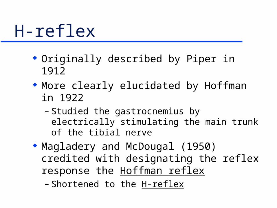
H-reflex
Originally described by Piper in 1912 More clearly elucidated by Hoffman in
1922– Studied the gastrocnemius by electrically
stimulating the main trunk of the tibial nerve Magladery and McDougal (1950) credited
with designating the reflex response the Hoffman reflex – Shortened to the H-reflex

H-reflex Theory.1 The electrical equivalent of the deep tendon
reflex for testing a monosynaptic loop
Group Ia fiber
Spindle
Gross muscle
Reflex hammerAlpha motoneuron
Spinal cord

H-reflex Theory.2 The electrical equivalent of the deep tendon
reflex for testing a monosynaptic loop1
Group Ia fiber
Spindle
Gross muscle
Alpha motoneuron
Spinal cord
EMG
RecordingElectrodes
GalvanicStimulator
StimulatingElectrodes
1Hugon, 1971

H-reflex Theory.3
Confirms the integrity of the afferent - efferent nerve connections
Amplitudes (mV) provide an index of alpha motoneuron excitability at the spinal cord level

H-reflex Uses.1
First used as a clinical/diagnostic tool– Most frequently used to study the tibial nerve in
the posterior compartment of the thigh» Gastrocnemius usually studied
» Compare latencies between M-waves and H-reflex bilaterally
Correlates with leg length - range = 22.64 - 40.14 msec (tibial nerve)
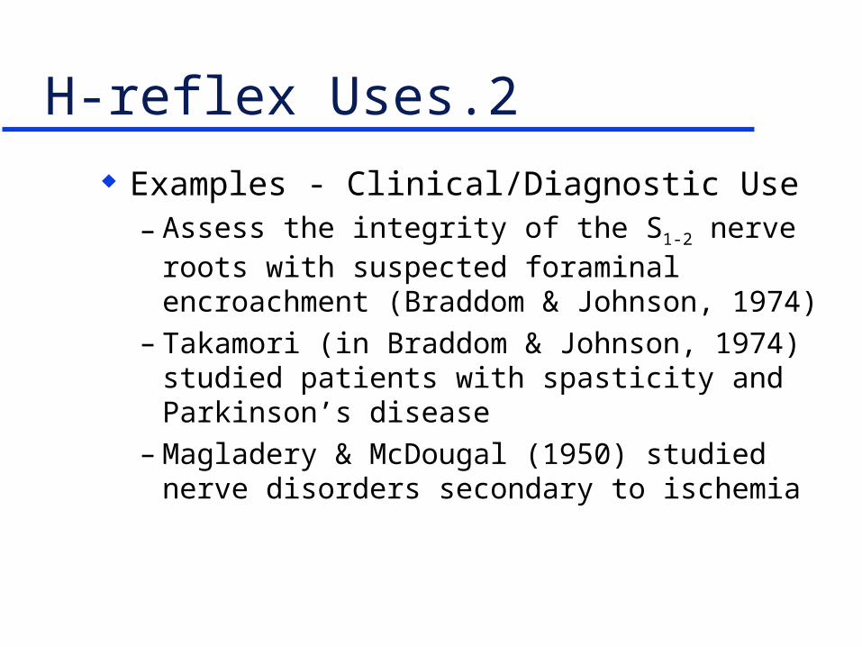
H-reflex Uses.2
Examples - Clinical/Diagnostic Use– Assess the integrity of the S1-2 nerve roots with
suspected foraminal encroachment (Braddom & Johnson, 1974)
– Takamori (in Braddom & Johnson, 1974) studied patients with spasticity and Parkinson’s disease
– Magladery & McDougal (1950) studied nerve disorders secondary to ischemia
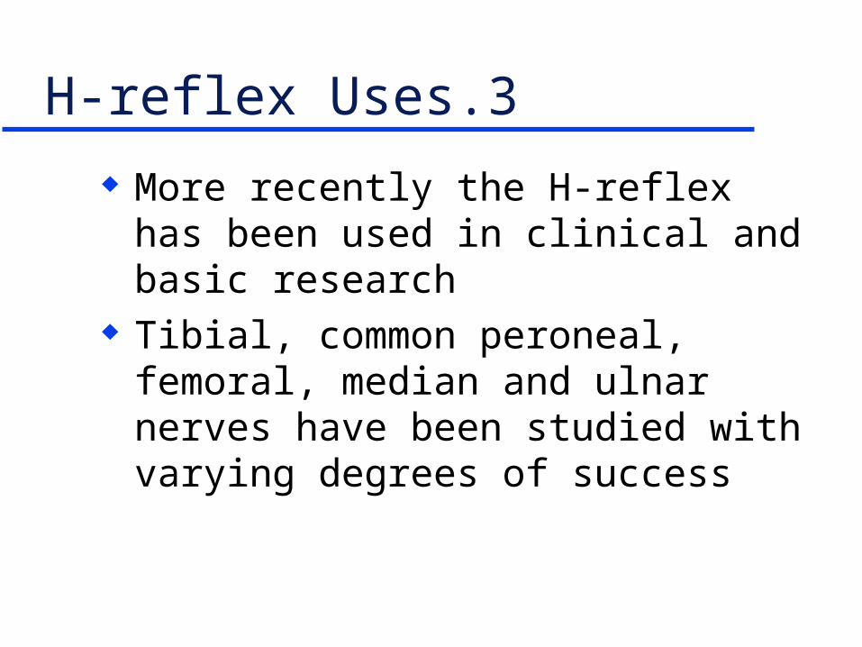
H-reflex Uses.3
More recently the H-reflex has been used in clinical and basic research
Tibial, common peroneal, femoral, median and ulnar nerves have been studied with varying degrees of success

H-reflex - Uses.4
Mongia (1972) studied the femoral nerve and quadriceps
Bulbulian & Bowles (1992) studied eccentric contractions in downhill running
Kennedy et al. (1982); Spencer et al. (1984); and McDonough & Weir (1996) studied reflex inhibition in the quadriceps secondary to knee joint capsular swelling
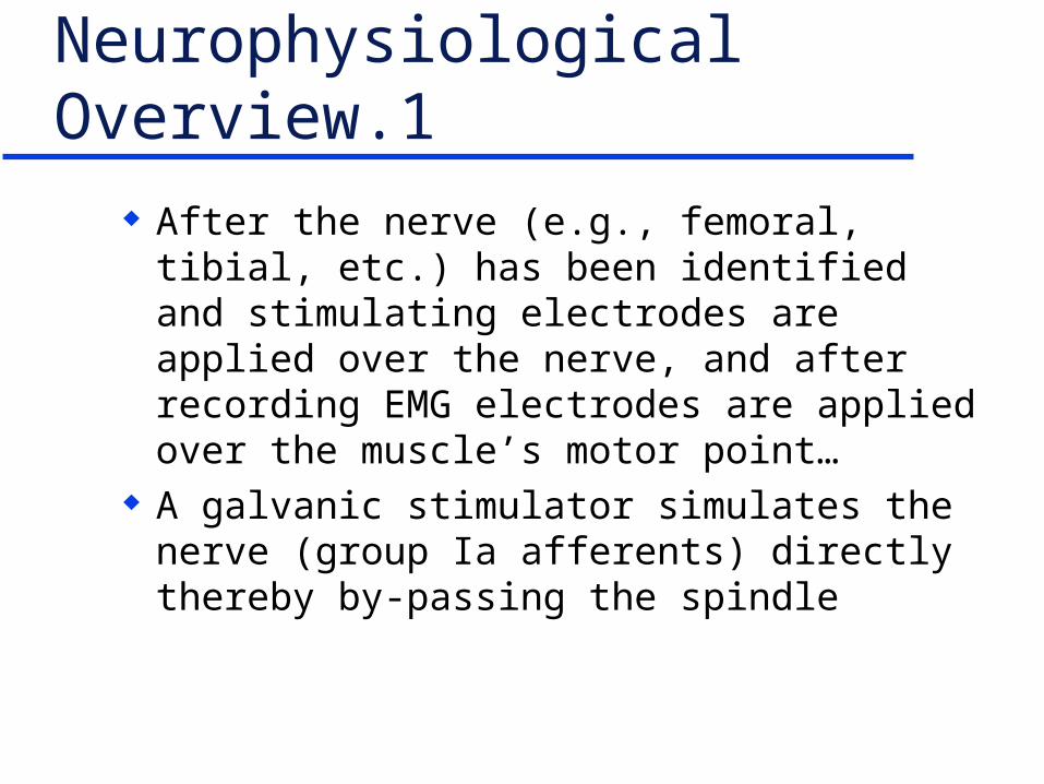
Neurophysiological Overview.1
After the nerve (e.g., femoral, tibial, etc.) has been identified and stimulating electrodes are applied over the nerve, and after recording EMG electrodes are applied over the muscle’s motor point…
A galvanic stimulator simulates the nerve (group Ia afferents) directly thereby by-passing the spindle

Neurophysiological Overview.2
Group Ia fibers monosynaptically connect with alpha motoneurons in the anterior horn of the spinal cord
The alpha motoneurons activate extrafusal (somatic) fibers in the homonymous muscle– Recording EMG electrodes pick-up electrical
activity in the muscle A twitch contraction is elicited
– Similar is appearance to a DTR response

EMG Response - Vastus Medialis
Stimulus artifact
M-wave H-reflex
Volts
Latency
msec

Potential Confounding Influences
Head/neck position Mental/emotional/alertness state Cognitive state Ambient temperature The specific nerve being studied Stimulating electrode conditions

Procedure.1 - Femoral Nerve
Using a galvanic stimulator with a probe electrode identify the motor points of VM and VL– Apply EMG electrodes in the standard way with
standard skin prep» Reference electrode over ASIS
– Confirm signal using Scope1.vi Palpate the femoral artery pulse in the femoral
triangle - mark the skin

Procedure.2 - Femoral Nerve
Using a galvanic stimulator with a probe electrode locate the femoral nerve 1-2 cm lateral to the femoral artery - mark skin– Confirm with Scope1.vi
Apply a pre-gelled, self-adhesive electrode over the femoral nerve– Large dispersal electrode on back of thigh

Procedure.3 - Femoral Nerve
Stimulator settings– Rate: 0.30 - 0.40 Hz– Duration: 0.50 - 1.0 msec– Intensity: 40 - 110 volts
Collect data using Bincolct.vi– Saves data in binary format
~10 seconds of rest between trials

Procedure.4 - Femoral Nerve
Using Scope1a.vi bring intensity (voltage) up gradually until an M-wave and H-reflex are visualized
Decrease intensity in ~5 volt increments until the M-wave decreases in amplitude but the H-reflex remains constant

Procedure.5 - Femoral Nerve
Run Bincolct.vi– Will continue to collect data until Stop button is
pushed– Every 10 seconds press stimulator switch “On”
to elicit H-reflexes

Bincolct.vi

Procedure.6 - Femoral Nerve
Analyze data using Hread.vi (reads binary format data)– Run the VI– Each time the SHOW NEXT button is pushed
1000 data points will be advanced– Approximately every 10 seconds a triplex of
signals will appear» Stimulus artifact
» M-wave
» H-reflex

Hread.vi

EMG Response - Vastus Medialis
Stimulus artifact
M-wave H-reflex
Volts
Latency
msec
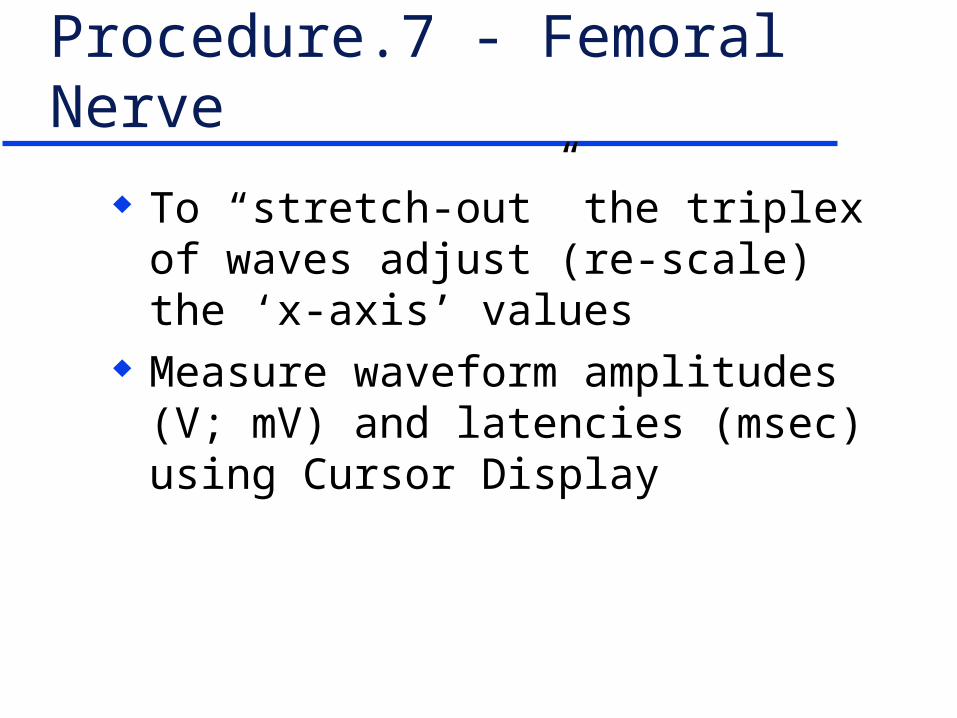
Procedure.7 - Femoral Nerve
To “stretch-out” the triplex of waves adjust (re-scale) the ‘x-axis’ values
Measure waveform amplitudes (V; mV) and latencies (msec) using Cursor Display

References
Braddom, R.L., & Johnson, E.W. (1974) H-reflex: Review and classification with clinical uses. Archives of Physical Medicine and Rehabilitation, 55, 412
Enoka, R.M. (1994). Neuromechanical basis of kinesiology (2nd ed.). Champaign, IL: Human Kinetics, pp. 177-179.
Enoka, R.M., Hutton, R.S., & Eldred, E. (1980). Changes in excitability of tendon tap and Hoffman reflexes following voluntary contractions. Electroencephalography and Clinical Neurophysiology, 18, 664-672.
References
Garland, S., Gerilovsky, L, & Enoka, R.M. (1994). Association between muscle architecture and quadriceps femoris H-reflex. Muscle & Nerve, 17, 581-592.
Hugon, M. (1973). Methodology of the Hoffman reflex in man. In J.E. Desmedt (Ed.). New developments in electromyography and clinical neurophysiology. Basel: Karger, pp. 277-293.
Kennedy, J.C., Alexander, I.J., & Hayes, K.C. (1982). Nerve supply o f the human knee and its functional importance. The American Journal of Sports Medicine, 10, 187-194.
References
Magladery, J.W., & McDougal, D.B. (1950). Electrophysiological studies of nerve and reflex activity in normal man: Identification of certain reflexes in electromyogram and conduction velocity of peripheral nerves. Bulletin of Johns Hopkins Hospital, 86, 265-290.
McDonough, A.L., & Weir, J.P. (1996). The effect of post-surgical edema of the knee on reflex inhibition of the quadriceps femoris - a case study. Journal of Sport Rehabilitation, 5, 172-181.
References
Mongia, S.K. (1972). H-reflex from quadriceps and gastrocnemius muscle. Electromyography and Clinical Neurophysiology, 12, 179-190.
Spencer, J.D., Hayes, K.C., & Alexander, I.J. (1984). Knee joint effusion and quadricepsreflex inhibition in man. Archives of Physical Medicine and Rehabilitation, 65, 171-177.