Embed Size (px)
Citation preview
Florida International UniversityFIU Digital CommonsNicole Wertheim College of Nursing and HealthSciences
Nicole Wertheim College of Nursing and HealthSciences
10-21-2014
The Elusive Role of the Left Temporal Pole (BA38)in Language: A Preliminary Meta-AnalyticConnectivity StudyAlfredo ArdillaDepartment of Communication Sciences and Disorders, Florida International University, [email protected]
Byron BernalMiami Children's Hospital
Monica RosselliFlorida Atlantic University
Follow this and additional works at: https://digitalcommons.fiu.edu/cnhs_fac
Part of the Medicine and Health Sciences Commons
This work is brought to you for free and open access by the Nicole Wertheim College of Nursing and Health Sciences at FIU Digital Commons. It hasbeen accepted for inclusion in Nicole Wertheim College of Nursing and Health Sciences by an authorized administrator of FIU Digital Commons. Formore information, please contact [email protected].
Recommended CitationAlfredo Ardila, Byron Bernal, and Monica Rosselli, “The Elusive Role of the Left Temporal Pole (BA38) in Language: A PreliminaryMeta-Analytic Connectivity Study,” International Journal of Brain Science, vol. 2014, Article ID 946039, 7 pages, 2014. doi:10.1155/2014/946039
Research ArticleThe Elusive Role of the Left Temporal Pole (BA38) in Language:A Preliminary Meta-Analytic Connectivity Study
Alfredo Ardila,1 Byron Bernal,2 and Monica Rosselli3
1 Department of Communication Sciences and Disorders, Florida International University,11200 SW 8th Street, AHC3-431B, Miami, FL 33199, USA
2Department of Radiology/Research Institute, Miami Children’s Hospital, Miami, FL, USA3Department of Psychology, Florida Atlantic University, Davie, FL, USA
Correspondence should be addressed to Alfredo Ardila; [email protected]
Received 26 August 2014; Revised 4 October 2014; Accepted 7 October 2014; Published 21 October 2014
Academic Editor: Joao Quevedo
Copyright © 2014 Alfredo Ardila et al. This is an open access article distributed under the Creative Commons Attribution License,which permits unrestricted use, distribution, and reproduction in any medium, provided the original work is properly cited.
It has been suggested that the left temporal pole (Brodmann area 38 (BA38)) participates in diverse language functions, includingsemantic processing, speech comprehension, and naming. Utilizing the activation likelihood estimation (ALE), a meta-analyticconnectivity study was conducted to further our understanding on the role of BA38 in language. Departing from the BrainMapfunctional database, 11 papers corresponding to 12 paradigms including 201 participants were selected. Initially, 𝑃 < 0.01 wasemployed as the significance level, resulting in the presence of four different activation clusters. However, when the significance levelwas lowered to𝑃 < 0.05, sixteen activation clusters appeared, including classical language areas such as Broca’s andWernicke’s areas.It was concluded that (1) this meta-analytic connectivity study suggests the presence of two major connection circuits involvingBA38; one is related to language, while the othermay be involved in visuospatial and integrative audiovisual functions. Furthermore,(2) BA38 also contributes to various brain networks supporting linguistic processes related not only to language comprehensionbut also to language production.
1. Introduction
The temporal lobe is a particularly complex brain areainvolved in a diversity of functions, some of which includeauditory, olfactory, memory, vestibular, visual, and linguisticprocessing [1, 2]. With regard to other primates, the temporallobe is the largest in humans representing about 17% ofthe cerebral cortex [3]. The temporal pole (Brodmann area38; BA38) represents no more than 10% of total temporallobe volume and about 1.7% of the cerebral cortex. Dueto its sizeable nature, several subareas have been distin-guished in the temporal pole. Pascual et al. [4] proposedseparation possibilities between different cytoarchitectonicsubfields: (1) dorsal, with predominant connectivity to audi-tory/somatosensory and language networks; (2) ventrome-dial, predominantly connected to visual networks; (3)medial,connected to paralimbic structures; and (4) anterolateral,connected to the default-semantic network. Consequently,significant language roles of BA38 are assumed.
The specific linguistic role of the left temporal poleremains elusive. Regardless of its evident participation inthe brain language system, the left BA38 is not usuallyincluded in Wernicke’s area. Nevertheless, various linguisticfunctions have been related to this brain area. Contemporaryneuroimaging studies suggest that the left BA38 is involvedin the following language-related functions: semantic pro-cessing [5–8], speech comprehension, responsive naming [9],naming of items learned in early life, word retrieval for spe-cific entities [10], lexicosemantic ambiguity processing [11],processing negative sentences [12], narrative comprehension[13–15], and the processing of abstract information [16].
Clinical observations have significantly advanced theunderstanding of left temporal pole function. Analyses ofindividuals with left-hemisphere strokes have revealed theleft anterior temporal cortices role in syntactic processing.Patients with brain damage to this area are more likely topresent with receptive agrammatism than patients in which
Hindawi Publishing CorporationInternational Journal of Brain ScienceVolume 2014, Article ID 946039, 7 pageshttp://dx.doi.org/10.1155/2014/946039
2 International Journal of Brain Science
anterior temporal cortex remains spared [17]. It has beenfurther proposed that the temporal pole represents a semantichub of the brain, or the neural substrates that subserveparticipating in the processing of “unique entities” (i.e.,proper names of people and places) [18–21]. Tsapkini et al.[22] compared a group of 20 patients with acute anteriortemporal pole stroke damage to a group of 28 patientswithout anterior temporal pole damage matched by infarctvolume. The authors calculated the average percent errorin two language tasks, auditory comprehension and namingtasks, as a function of infarct volume using a nonparametricregressionmethod.They attributed infarct volume as the solepredictive variable in the production of semantic errors inboth auditory comprehension and object naming tasks.Thesefindings were interpreted as support to the hypothesis thatleft unilateral anterior temporal pole lesions, even acutely, areunlikely to cause significant deficits in mapping meaning tosound by themselves although they contribute to networksunderlying both naming and comprehension of objects.Theyconcluded that the anterior temporal lobe may be a semantichub for object meaning, but its role must be representedbilaterally.
Other authors (e.g., [23]) have reported that namingperformance in a typical confrontation naming test (i.e.,Boston Naming Test) is related to metabolite levels in theanterior left temporal pole. Nonetheless, some authors haveargued that the role of the temporal lobe in naming is limitedto the naming of unique entities, such as naming people[24]. However, some contradictory evidence has also beenpresented [25].
As reported in the semantic subtype of primary pro-gressive aphasia—which is characterized by the progressivedeterioration of word comprehension, particularly nounswhile other cognitive abilities remain spared—marked tem-poropolar atrophy and pronounced impairments of odornaming and matching are observed [26]. These authorspredicted that the temporal pole would play a key role inlinking odor object representations to transmodal networks,given its anatomical proximity to olfactory and visual objectprocessing areas.
Interestingly, the temporal poles are among brain regionsoften considered as the brain network sustaining our ability tounderstand other people’s mental states or “Theory of Mind”(ToM) (e.g., [27, 28]). However, it has also been reported thatthe left temporal lobe is not necessary for ToM reasoning, atleast in nonverbal conditions, as long as its right counterpartis preserved [29].
Pinpointing the connectivity of the anterior temporallobe represents a crucial question to understand its rolein language-like function. Using high angular resolutiondiffusion imaging (HARDI) MRI, Makris et al. [30, 31]described two major fiber connections of the human middlelongitudinal fascicle. They did so by examining morphol-ogy, topography, cortical connections, biophysical measures,volume, and length in seventy-four brains. These two fiberconnections course together through the dorsal tempo-ral pole and the superior temporal gyrus maintaining acharacteristic topographic relationship in the mediolateraland ventrodorsal dimensions. The authors suggested that
the superior temporal-angular connection of the middlelongitudinal fascicle plays a role in language and attention,while the superior temporal-superior parietal connection isinvolved in visuospatial and integrative audiovisual func-tions. Menjot De Champfleu et al. [32] reported that themiddle longitudinal fasciculus is clearly delineated fromthe other fascicles that constitute the language pathways,especially the ventral pathway. It runs within the superiortemporal gyrus whitematter from the temporal pole and thenextends caudally in the upper part of the sagittal stratum andthe posterior part of the corona radiata to reach the inferiorparietal lobule (angular gyrus). Augustine [33] emphasizedthe relationship between the temporal pole and the insula.Although the insula is recognized for its language responsibil-ities, vagueness remains regarding its participation in variouslanguage functions [34–37]. The uncinate fasciculus, on theother hand, connects the anterior temporal and inferiorfrontal lobes. The uncinate fasciculus connects the poleof the temporal lobe (BA38) uncus and parahippocampalgyrus with the ipsilateral orbital and lateral frontal cortices.Propositions have been made involving its association withsemantic memory retrieval and processing [38].
Recently, a new alternative to study brain connectivityhas been proposed by Robinson et al. [39] known as meta-analytic connectivity modeling (MACM). MACM is basedin automatic meta-analysis executed by pooling coactivationpatterns. The technique takes advantage of the BrainMaprepository of functional MRI studies and a software program(Sleuth) provided by the same group to find, filter, organize,plot, and export peak coordinates for further statisticalanalysis. Sleuth provides a list of foci, in Talairach, or MNIcoordinates, each one representing the center of mass ofan activation cluster. This method acquires the region ofinterest (i.e., the temporal pole), deems it the independentvariable, and interrogates the database for studies indicatingcomparable activation of the chosen target. The query isconveniently filtered for different conditions such as age,normal versus patients, type of paradigm, and domain ofcognition. By pooling the data with these conditions, thetool is able to provide a universe of coactivations that can bestatistically analyzed for significant commonality. As a finalstep, activation likelihood estimation (ALE) [40, 41] can beperformed utilizing GingerALE, another software providedby BrainMap, generating probability of an event occurringat voxel level across the studies. Areas of coactivation willdisplay a network related to the function and domainsselected as filter criteria.
Considering the diverse language functions linked toBA38, a meta-analytic connectivity was developed utilizingMACM on the participation of this brain area in language.Deviating from previous clinical and neuroimaging studies,it was hypothesized that the left temporal pole participates indifferent brain language circuits.
Our research question is what are the areas that coac-tivate when BA38 activates in language tasks? With thisapproach, the commonality criteria are quite stringent, as itonly requires BA38 activation occurrence during a languagetask. Notice that we were not interested in ascertainingthe activation related to a specific task. The area may be
International Journal of Brain Science 3
activated in “nonverbal” tasks, but that was not explored. Asa result, this concentrates the results on language domains.It is important to observe that BA38 may reveal restricted orample connectivity (coactivation network) depending uponthe spectrum of studies accepted to enter the pool of ALEanalysis, that is, for example, only expressive, only receptive,only lexical, lexical and semantics, and so forth, but thatdecision depends upon the scope of the study.
2. Materials and Methods
The database of BrainMap [42] was accessed utilizing Sleuth2.2 on December 10, 2013. Sleuth is software providedby BrainMap to query its database. Sleuth software wasimplemented in aWindows platform.This meta-analysis wasintended to assess the network of coactivations in which theBA38 is involved.
The search conditions were as follows: (1) studies report-ing BA38 activation; (2) studies using fMRI; (3) context:normal subjects; (4) activation: activation only; (5) handed-ness: right-handed subjects; (6) age: 20–60 years; (7) domain:cognition, subtype: language.
ALE meta-analysis was then performed utilizing Gin-gerALE. ALE maps were initially thresholded at 𝑃 <0.01 corrected for multiple comparisons and false discov-ery rates. We used the predefined values found in Gin-gerALE [40] and only clusters of 200 or more cubic mmwere accepted as valid clusters. ALE results were over-laid onto an anatomical template suitable for MNI coordi-nates, also provided by http://brainmap.org/. For this pur-pose, we utilized the Multi-Image Analysis GUI (Mango)(http://ric.uthscsa.edu/mango/). Mosaics of 5 × 7 insets oftransversal fusioned images were generated utilizing a plug-in of the same tool, selecting every other image, starting onimage number 10, and exported to a 2D-jpg image. Because ofthe limited activation observed when employing the𝑃 < 0.01threshold, a second analysis was performed with a loweredsignificance level of 𝑃 < 0.05.
3. Results
3.1. First Analysis. Eleven papers corresponding to 12 exper-imental conditions with a total of 201 subjects were finallyselected (subjects participating in two different experimentswere counted as two subjects) (Table 1).
Table 2 presents the main loci of brain connectivity ofBA38 by meta-analytic connectivity modeling (MACM).Four different clusters of activity were found, two in the leftand two in the right hemisphere.
As indicated in Table 2, the first cluster includes BA38,while the second cluster contains the left insula and superiortemporal lobe, indicating its involvement in language. Thelast two clusters include the right parietal and temporal lobes.Seemingly, this area has twomajor connection pathways: onewithin the left hemisphere and the second involving the righthemisphere.
3.2. Second Analysis. Table 3 presents the main loci of brainconnectivity of BA38 bymeta-analytic connectivitymodeling
(MACM) when significance level 𝑃 < 0.05 was selected.Sixteen different clusters of activity were found.
The first clusters include basically the same areas found inthe first analysis.Thus, there is one connection pathway in theleft including the insula and the temporal lobe and anotherconnection pathway to the right including the parietal andtemporal lobes. Some increased activation is also found atthe level of the claustrum, probably as an extension of someinsula activation. Other additional clusters of activation arelocated within the left occipital area (BA17), frontal languageareas (BA44 and BA47), subcortical areas such as the caudatenucleus and even brainstem areas (red nucleus), Wernicke’sarea (left BA22), and homologous right area (right BA22).
4. Discussion
It iswell known that BA38has someparticipation in language,although pinpointing its specific function has been not easy.It has been suggested that one of its major language functionsincludes naming, particularly the naming of unique entities(i.e., names of people and places) [18–21]. Interestingly, nam-ing using fingerspelling and signing by native ASL signershave also shown that naming famous persons activates theleft temporal pole, whereas naming animals (whether fin-gerspelled or signed) activates the left inferotemporal cortex[54]. This observation demonstrates that left BA38 functionin naming is not exclusively dependent on the auditoryinformation of language but is a more general and conceptualone.
Functional studies have advanced the understanding ofthe unexpected complexity of BA38 functions (see [55]).Because of its location in the brain (somehow in betweenBroca’s and Wernicke’s areas), it is understandable that BA38participates in language. Our first meta-analytic study iscongruent with Makris et al. [30, 31] observation that twofiber connections course through the temporal pole; one isinvolved in language (superior temporal-angular connectionof the middle longitudinal fascicle), while the other isinvolved in visuospatial and integrative audiovisual functions(superior temporal-superior parietal connection).
It was unexpected to find so few activation clusters inthe first meta-analysis and so many activation clusters in thesecond meta-analysis. This observation may suggest that theleft temporal pole has two fundamental connections; thus, itparticipates in the following two major brain networks: onerelated to language and the other related to more visuospatialand integrative audiovisual functions, as it has been suggestedby Makris et al. [30, 31]. Moreover, BA38 may also havea secondary contribution to several networks supportinglinguistic processes and it may be related not only to languageunderstanding but also to language production. Indeed, bothWernicke and Broca’s areas appeared interconnected to leftBA38.
Interestingly, two different pathways in language process-ing have been recognized. Electrophysiological analysis of theauditory system in primates and functional neuroimagingstudies in human subjects have suggested that there aretwo pathways arising from the primary auditory cortex;a “ventral” pathway is thought to project anteriorly from
4 International Journal of Brain Science
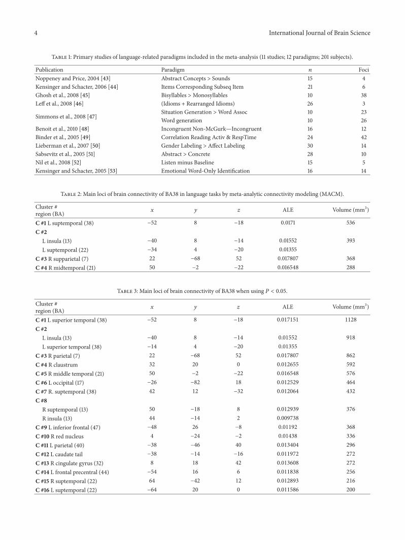
Table 1: Primary studies of language-related paradigms included in the meta-analysis (11 studies; 12 paradigms; 201 subjects).
Publication Paradigm 𝑛 FociNoppeney and Price, 2004 [43] Abstract Concepts > Sounds 15 4Kensinger and Schacter, 2006 [44] Items Corresponding Subseq Item 21 6Ghosh et al., 2008 [45] Bisyllables >Monosyllables 10 38Leff et al., 2008 [46] (Idioms + Rearranged Idioms) 26 3
Simmons et al., 2008 [47]Situation Generation >Word Assoc 10 23Word generation 10 26
Benoit et al., 2010 [48] Incongruent Non-McGurk—Incongruent 16 12Binder et al., 2005 [49] Correlation Reading Activ & RespTime 24 42Lieberman et al., 2007 [50] Gender Labeling > Affect Labeling 30 14Sabsevitz et al., 2005 [51] Abstract > Concrete 28 10Nil et al., 2008 [52] Listen minus Baseline 15 5Kensinger and Schacter, 2005 [53] Emotional Word-Only Identification 16 14
Table 2: Main loci of brain connectivity of BA38 in language tasks by meta-analytic connectivity modeling (MACM).
Cluster #region (BA) 𝑥 𝑦 𝑧 ALE Volume (mm3)
C #1 L suptemporal (38) −52 8 −18 0.0171 536C #2
L insula (13) −40 8 −14 0.01552 393L suptemporal (22) −34 4 −20 0.01355
C #3 R supparietal (7) 22 −68 52 0.017807 368C #4 R midtemporal (21) 50 −2 −22 0.016548 288
Table 3: Main loci of brain connectivity of BA38 when using 𝑃 < 0.05.
Cluster #region (BA) 𝑥 𝑦 𝑧 ALE Volume (mm3)
C #1 L superior temporal (38) −52 8 −18 0.017151 1128
C #2L insula (13) −40 8 −14 0.01552 918
L superior temporal (38) −14 4 −20 0.01355
C #3 R parietal (7) 22 −68 52 0.017807 862
C #4 R claustrum 32 20 0 0.012655 592
C #5 R middle temporal (21) 50 −2 −22 0.016548 576
C #6 L occipital (17) −26 −82 18 0.012529 464
C #7 R. suptemporal (38) 42 12 −32 0.012064 432
C #8R suptemporal (13) 50 −18 8 0.012939 376
R insula (13) 44 −14 2 0.009738
C #9 L inferior frontal (47) −48 26 −8 0.01192 368
C #10 R red nucleus 4 −24 −2 0.01438 336
C #11 L parietal (40) −38 −46 40 0.013404 296
C #12 L caudate tail −38 −14 −16 0.011972 272
C #13 R cingulate gyrus (32) 8 18 42 0.013608 272
C #14 L frontal precentral (44) −54 16 6 0.011838 256
C #15 R suptemporal (22) 64 −42 12 0.012893 216
C #16 L suptemporal (22) −64 20 0 0.011586 200
International Journal of Brain Science 5
the primary auditory cortex to prefrontal areas along thesuperior temporal gyrus, while a separate “dorsal” routeconnects these areas posteriorly via the inferior parietal lobe.A similar pattern of pathways has been found in humans: adorsal pathway fromWernicke’s area to Broca’s area includingthe arcuate fasciculus, the supramarginal gyrus, the lateralsuperior temporal gyrus, and the lateral middle temporalgyrus. Additionally, a ventral route between Wernicke’s areaand Broca’s area has been exhibited connecting these twoareas through the external capsule/uncinate fasciculus andthe medial superior temporal gyrus. As expected, theseconnections are stronger in the left hemisphere [56, 57].BA38 language connections would correspond to the ventralpathway.
The connections found between BA38 and the associationauditory areas of the right hemisphere (BA21/22) intriguinglysuggest that the left temporal pole has an assortment offunctions beyond the purely linguistic functions. Markedly,the right temporal pole has been included in a systeminvolved in emotion processing, with the insula and theamygdala [58]. Pizzamiglio et al. [59] analyzed whethersounds referring to actions have a different representationin the brain from other types of sounds. By method of ERP,they found the left posterior superior temporal and premotorareas to be selectively modulated by action-related sounds.Contrastingly, the temporal pole is bilaterally modulatedby nonaction-related sounds. Thus, the left temporal poleseemingly participates in a broad brain system involved inemotion and sound processing.
A significant involvement of the right hemisphere tem-poral lobe in emotional language is commonly accepted[60]. However, brain processing of emotional prosody seemsparticularly complex and probably includes the right tempo-ral pole. The presence of three successive processing stagesduring recognition of emotional prosody has been postu-lated: (1) extraction of suprasegmental acoustic informationpredominantly subserved by right-sided primary and higherorder acoustic regions, (2) representation of meaningfulsuprasegmental acoustic sequences within posterior aspectsof the right superior temporal sulcus, and (3) explicit eval-uation of emotional prosody at the level of the bilateralinferior frontal cortex. Explicit evaluation of linguistic aspectsof prosody appears to be linked to left-hemisphere languageareas. Specifically, the bilateral orbitofrontal cortex was foundto be involved in explicit evaluation of emotional prosody[61]. Furthermore, it has been proposed that evaluation ofaffective prosody requires prior analysis of acoustic featureswithin temporal regions and that transfer of informationfrom the temporal cortex to the frontal lobes occurs viaparallel pathways [62].
Many limitations could be mentioned regarding thepresent study. The major critique of meta-analysis studiescommonly refers to the lack of homogeneity of the pooledtasks, methods, and individuals. Furthermore, MACM isstill new requiring performance of future validation studies.We have used BA38 as the independent variable and aspectrum of coactivated areas as the dependent variable,which may be unusual. However, the current results are quite
consistent with clinical observations, positively supportingthe structural connectivity findings.
Conflict of Interests
The authors declare that there is no conflict of interestsregarding the publication of this paper.
Acknowledgment
The authors express their most sincere gratitude to CarleyKeim for her editorial support.
References
[1] K. Kolb and I. Q. Whishaw, Fundamentals of Human Neuropsy-chology, Worth Publishers, New York, NY, USA, 6th edition,2009.
[2] L. R. Squire, C. E. L. Stark, and R. E. Clark, “The medialtemporal lobe,”Annual Review of Neuroscience, vol. 27, pp. 279–306, 2004.
[3] J. A. Kiernan, “Anatomy of the temporal lobe,” Epilepsy Researchand Treatment, vol. 2012, Article ID 176157, 12 pages, 2012.
[4] B. Pascual, J. C. Masdeu, M. Hollenbeck et al., “Large-scalebrain networks of the human left temporal pole: a functionalconnectivity MRI study,” Cerebral Cortex, 2013.
[5] U. Noppeney and C. J. Price, “Retrieval of visual, auditory, andabstract semantics,” NeuroImage, vol. 15, no. 4, pp. 917–926,2002.
[6] U. Noppeney and C. J. Price, “A PET study of stimulus- andtask-induced semantic processing,” NeuroImage, vol. 15, no. 4,pp. 927–935, 2002.
[7] C. Palliera, A.-D. Devauchellea, and S. Dehaenea, “Corticalrepresentation of the constituent structure of sentences,” Pro-ceedings of the National Academy of Sciences of the United Statesof America, vol. 108, no. 6, pp. 2522–2527, 2011.
[8] R. Vandenberghe, A. C. Nobre, and C. J. Price, “The responseof left temporal cortex to sentences,” Journal of CognitiveNeuroscience, vol. 14, no. 4, pp. 550–560, 2002.
[9] L. Tomaszewki, S. Farias, G. Harrington, C. Broomand, andM. Seyal, “Differences in functional MR imaging activationpatterns associated with confrontation naming and responsivenaming,”American Journal of Neuroradiology, vol. 26, no. 10, pp.2492–2499, 2005.
[10] T. J. Grabowski, H. Damasio, D. Tranel, L. L. Boles Ponto, R. D.Hichwa, and A. R. Damasio, “A role for left temporal pole in theretrieval of words for unique entities,” Human Brain Mapping,vol. 13, no. 4, pp. 199–212, 2001.
[11] K. Hoenig and L. Scheef, “Mediotemporal contributions tosemantic processing: fMRI evidence from ambiguity processingduring semantic context verification,”Hippocampus, vol. 15, no.5, pp. 597–609, 2005.
[12] U. Kumar, P. Padakannaya, R. K. Mishra, and C. L. Khetrapal,“Distinctive neural signatures for negative sentences in Hindi:an fMRI study,” Brain Imaging and Behavior, vol. 7, no. 2, pp.91–101, 2013.
[13] J. T. Crinion, E. A. Warburton, M. A. Lambon-Ralph, D.Howard, and R. J. S. Wise, “Listening to narrative speech afteraphasic stroke: the role of the left anterior temporal lobe,”Cerebral Cortex, vol. 16, no. 8, pp. 1116–1125, 2006.
6 International Journal of Brain Science
[14] E. A. Maguire, C. D. Frith, and R. G. Morris, “The functionalneuroanatomy of comprehension andmemory: the importanceof prior knowledge,” Brain, vol. 122, no. 10, pp. 1839–1850, 1999.
[15] N. Tzourio, B. Nkanga-Ngila, and B. Mazoyer, “Left planumtemporale surface correlates with functional dominance duringstory listening,” NeuroReport, vol. 9, no. 5, pp. 829–833, 1998.
[16] B. Straube, Y. He, M. Steines et al., “Supramodal neural process-ing of abstract information conveyed by speech and gesture,”Frontiers in Behavioral Neuroscience, vol. 7, article 120, 2013.
[17] S. Magnusdottir, P. Fillmore, D. B. den Ouden et al., “Damageto left anterior temporal cortex predicts impairment of complexsyntactic processing: a lesion-symptommapping study,”HumanBrain Mapping, vol. 34, no. 10, pp. 2715–2723, 2013.
[18] K. Patterson, P. J. Nestor, and T. T. Rogers, “Where do you knowwhat you know? The representation of semantic knowledge inthe human brain,” Nature Reviews Neuroscience, vol. 8, no. 12,pp. 976–987, 2007.
[19] W. K. Simmons and A. Martin, “The anterior temporal lobesand the functional architecture of semantic memory,” Journal ofthe International Neuropsychological Society, vol. 15, no. 5, pp.645–649, 2009.
[20] D. Tranel, H. Damasio, and A. R. Damasio, “A neural basis forthe retrieval of conceptual knowledge,” Neuropsychologia, vol.35, no. 10, pp. 1319–1327, 1997.
[21] D. Tranel, “Impaired naming of unique landmarks is associatedwith left temporal polar damage,” Neuropsychology, vol. 20, no.1, pp. 1–10, 2006.
[22] K. Tsapkini, C. E. Frangakis, and A. E. Hillis, “The function ofthe left anterior temporal pole: evidence from acute stroke andinfarct volume,” Brain, vol. 134, no. 10, pp. 3094–3105, 2011.
[23] L. Rami, C. Caprile, B. Gomez-Anson et al., “Naming is associ-ated with left temporal pole metabolite levels in neurodegener-ative diseases,”Dementia and Geriatric Cognitive Disorders, vol.25, no. 3, pp. 212–217, 2008.
[24] C. Papagno, “Naming and the role of the uncinate fasciculusin language function,” Current Neurology and NeuroscienceReports, vol. 11, no. 6, pp. 553–559, 2011.
[25] I. P. Martins and L. Farrajota, “Proper and common names: adouble dissociation,” Neuropsychologia, vol. 45, no. 8, pp. 1744–1756, 2007.
[26] J. K. Olofsson, E. Rogalski, T. Harrison, M.-M. Mesulam, and J.A. Gottfried, “A cortical pathway to olfactory naming: evidencefrom primary progressive aphasia,” Brain, vol. 136, no. 4, pp.1245–1259, 2013.
[27] S. J. Carrington and A. J. Bailey, “Are there theory of mindregions in the brain? A review of the neuroimaging literature,”Human Brain Mapping, vol. 30, no. 8, pp. 2313–2335, 2009.
[28] F. Van Overwalle, “Social cognition and the brain: a meta-analysis,” Human Brain Mapping, vol. 30, no. 3, pp. 829–858,2009.
[29] C.Michel, L. Dricot, R. Lhommel et al., “Extensive left temporalpole damage does not impact on theory of mind abilities,”Journal of Cognitive Neuroscience, vol. 25, no. 12, pp. 2025–2046,2013.
[30] N. Makris, M. G. Preti, D. Wassermann et al., “Human middlelongitudinal fascicle: segregation and behavioral-clinical impli-cations of two distinct fiber connections linking temporal poleand superior temporal gyrus with the angular gyrus or superiorparietal lobule using multi-tensor tractography,” Brain Imagingand Behavior, vol. 7, no. 3, pp. 335–352, 2013.
[31] N. Makris, M. G. Preti, T. Asami et al., “Humanmiddle longitu-dinal fascicle: variations in patterns of anatomical connections,”Brain Structure and Function, vol. 218, no. 4, pp. 951–968, 2013.
[32] N. Menjot De Champfleur, I. Lima Maldonado, S. Moritz-Gasser et al., “Middle longitudinal fasciculus delineation withinlanguage pathways: a diffusion tensor imaging study in human,”European Journal of Radiology, vol. 82, no. 1, pp. 151–157, 2013.
[33] J. R. Augustine, “Circuitry and functional aspects of the insularlobe in primates including humans,” Brain Research Reviews,vol. 22, no. 3, pp. 229–244, 1996.
[34] H. Ackermann and A. Riecker, “The contribution of the insulato motor aspects of speech production: a review and a hypoth-esis,” Brain and Language, vol. 89, no. 2, pp. 320–328, 2004.
[35] A. Ardila, “The role of insula in language: an unsettled ques-tion,” Aphasiology, vol. 13, no. 1, pp. 79–87, 1999.
[36] A. Ardila, D. F. Benson, and F. G. Flynn, “Participation of theinsula in language,” Aphasiology, vol. 11, no. 12, pp. 1159–1169,1997.
[37] A. Ardila, B. Bernal, and M. Rosselli, “Participation of theinsula in language revisited: ameta-analytic connectivity study,”Journal of Neurolinguistics, vol. 29, no. 1, pp. 31–41, 2014.
[38] A. S. Dick, B. Bernal, and P. Tremblay, “The language connec-tome: new pathways, new concepts,” Neuroscientist, vol. 20, no.5, pp. 453–467, 2014.
[39] J. L. Robinson, A. R. Laird, D. C. Glahn, W. R. Lovallo, andP. T. Fox, “Metaanalytic connectivity modeling: delineating thefunctional connectivity of the human amygdala,” Human BrainMapping, vol. 31, no. 2, pp. 173–184, 2010.
[40] A. R. Laird, P. M. Fox, C. J. Price et al., “ALE meta-analysis:controlling the false discovery rate and performing statisticalcontrasts,” Human Brain Mapping, vol. 25, no. 1, pp. 155–164,2005.
[41] P. E. Turkeltaub, G. F. Eden, K. M. Jones, and T. A. Zeffiro,“Meta-analysis of the functional neuroanatomy of single-wordreading:method and validation,”NeuroImage, vol. 16, no. 3, part1, pp. 765–780, 2002.
[42] http://brainmap.org/.[43] U. Noppeney and C. J. Price, “Retrieval of abstract semantics,”
NeuroImage, vol. 22, no. 1, pp. 164–170, 2004.[44] E. A. Kensinger and D. L. Schacter, “Amygdala activity is
associated with the successful encoding of item, but not source,information for positive and negative stimuli,” Journal of Neu-roscience, vol. 26, no. 9, pp. 2564–2570, 2006.
[45] S. S. Ghosh, J. A. Tourville, and F.H.Guenther, “Aneuroimagingstudy of premotor lateralization and cerebellar involvement inthe production of phonemes and syllables,” Journal of Speech,Language, and Hearing Research, vol. 51, no. 5, pp. 1183–1202,2008.
[46] A. P. Leff, T. M. Schofield, K. E. Stephan, J. T. Crinion, K. J.Friston, and C. J. Price, “The cortical dynamics of intelligiblespeech,” Journal of Neuroscience, vol. 28, no. 49, pp. 13209–13215,2008.
[47] W. K. Simmons, S. B. Hamann, C. L. Harenski, X. P. Hu, and L.W. Barsalou, “fMRI evidence for word association and situatedsimulation in conceptual processing,” Journal of Physiology, vol.102, no. 1–3, pp. 106–119, 2008.
[48] M. M. Benoit, T. Raij, F.-H. Lin, I. P. Jaaskelainen, and S.Stufflebeam, “Primary and multisensory cortical activity iscorrelated with audiovisual percepts,” Human Brain Mapping,vol. 31, no. 4, pp. 526–538, 2010.
International Journal of Brain Science 7
[49] J. R. Binder, D. A. Medler, R. Desai, L. L. Conant, and E.Liebenthal, “Some neurophysiological constraints on models ofword naming,” NeuroImage, vol. 27, no. 3, pp. 677–693, 2005.
[50] M. D. Lieberman, N. I. Eisenberger, M. J. Crockett, S. M. Tom,J. H. Pfeifer, and B. M. Way, “Putting feelings into words:affect labeling disrupts amygdala activity in response to affectivestimuli,” Psychological Science, vol. 18, no. 5, pp. 421–428, 2007.
[51] D. S. Sabsevitz, D. A. Medler, M. Seidenberg, and J. R. Binder,“Modulation of the semantic system by word imageability,”NeuroImage, vol. 27, no. 1, pp. 188–200, 2005.
[52] L. F. De Nil, D. S. Beal, S. J. Lafaille, R. M. Kroll, A. P. Crawley,and V. L. Gracco, “The effects of simulated stuttering andprolonged speech on the neural activation patterns of stutteringand nonstuttering adults,” Brain and Language, vol. 107, no. 2,pp. 114–123, 2008.
[53] E. A. Kensinger and D. L. Schacter, “Retrieving accurateand distorted memories: neuroimaging evidence for effects ofemotion,” NeuroImage, vol. 27, no. 1, pp. 167–177, 2005.
[54] K. Emmorey, T. Grabowski, S. McCullough et al., “Neuralsystems underlying lexical retrieval for sign language,” Neu-ropsychologia, vol. 41, no. 1, pp. 85–95, 2003.
[55] “Brodmann’s Interactive Atlas,” http://www.fmriconsulting.com/brodmann/BA5.html.
[56] G. J. Parker, S. Luzzi, D. C. Alexander, C. A.Wheeler-Kingshott,O. Ciccarelli, and M. Lambon Ralph, “Lateralization of ventraland dorsal auditory-language pathways in the human brain,”NeuroImage, vol. 24, no. 3, pp. 656–666, 2005.
[57] J. P. Rauschecker and S. K. Scott, “Maps and streams in theauditory cortex: nonhuman primates illuminate human speechprocessing,” Nature Neuroscience, vol. 12, no. 6, pp. 718–724,2009.
[58] S. Hsieh, M. Hornberger, O. Piguet, and J. R. Hodges, “Braincorrelates of musical and facial emotion recognition: evidencefrom the dementias,” Neuropsychologia, vol. 50, no. 8, pp. 1814–1822, 2012.
[59] L. Pizzamiglio, T. Aprile, G. Spitoni et al., “Separate neuralsystems for processing action- or non-action-related sounds,”NeuroImage, vol. 24, no. 3, pp. 852–861, 2005.
[60] R. D. Lane and L. Nadel, Cognitive Neuroscience of Emotion,Oxford University Press, New York, NY, USA, 2000.
[61] D. Wildgruber, H. Ackermann, B. Kreifelts, and T. Ethofer,“Chapter 13 Cerebral processing of linguistic and emotionalprosody: fMRI studies,” Progress in Brain Research, vol. 156, pp.249–268, 2006.
[62] T. Ethofer, S. Anders, M. Erb et al., “Cerebral pathways inprocessing of affective prosody: a dynamic causal modelingstudy,” NeuroImage, vol. 30, no. 2, pp. 580–587, 2006.

Submit your manuscripts athttp://www.hindawi.com
Neurology Research International
Hindawi Publishing Corporationhttp://www.hindawi.com Volume 2014
Alzheimer’s DiseaseHindawi Publishing Corporationhttp://www.hindawi.com Volume 2014
International Journal of
ScientificaHindawi Publishing Corporationhttp://www.hindawi.com Volume 2014
Hindawi Publishing Corporationhttp://www.hindawi.com Volume 2014
BioMed Research International
Hindawi Publishing Corporationhttp://www.hindawi.com Volume 2014
Research and TreatmentSchizophrenia
The Scientific World JournalHindawi Publishing Corporation http://www.hindawi.com Volume 2014
Hindawi Publishing Corporationhttp://www.hindawi.com Volume 2014
Neural Plasticity
Hindawi Publishing Corporationhttp://www.hindawi.com Volume 2014
Parkinson’s Disease
Hindawi Publishing Corporationhttp://www.hindawi.com Volume 2014
Research and TreatmentAutism
Sleep DisordersHindawi Publishing Corporationhttp://www.hindawi.com Volume 2014
Hindawi Publishing Corporationhttp://www.hindawi.com Volume 2014
Neuroscience Journal
Epilepsy Research and TreatmentHindawi Publishing Corporationhttp://www.hindawi.com Volume 2014
Hindawi Publishing Corporationhttp://www.hindawi.com Volume 2014
Psychiatry Journal
Hindawi Publishing Corporationhttp://www.hindawi.com Volume 2014
Computational and Mathematical Methods in Medicine
Depression Research and TreatmentHindawi Publishing Corporationhttp://www.hindawi.com Volume 2014
Hindawi Publishing Corporationhttp://www.hindawi.com Volume 2014
Brain ScienceInternational Journal of
StrokeResearch and TreatmentHindawi Publishing Corporationhttp://www.hindawi.com Volume 2014
Neurodegenerative Diseases
Hindawi Publishing Corporationhttp://www.hindawi.com Volume 2014
Journal of
Cardiovascular Psychiatry and NeurologyHindawi Publishing Corporationhttp://www.hindawi.com Volume 2014