Embed Size (px)
Citation preview
CASE REPORT Open Access
The elusive diagnosis and emergentmanagement of a late-presenting rupturedinterstitial pregnancy: a case reportLauren M. Ahlschlager1*, David Mysona2 and A. Jenna Beckham3
Abstract
Background: Interstitial pregnancies are rare and often difficult to diagnose given their proximal position to theuterine cavity, however most are identified by 12 weeks gestation. Delayed or missed diagnosis contributes toheightened incidence of poor outcomes including hemorrhage and death.
Case presentation: A 35-year-old woman at 15 weeks gestation with confirmed intrauterine pregnancy on firsttrimester ultrasound and prior negative MRI presented in hemorrhagic shock and was found to have a rupturedinterstitial pregnancy. Exploratory laparotomy revealed the fetus to be in the abdomen as well as a large cornualdefect and abnormal placentation that resulted in supracervical hysterectomy.
Conclusions: Interstitial pregnancy should be considered in a patient presenting with symptoms consistent withectopic rupture, especially in the setting of equivocal or suboptimal prior imaging. Earlier diagnosis may allow forfertility-sparing intervention and decreased risk of morbidity and mortality.
Keywords: Interstitial pregnancy, Ectopic, Ultrasound, Obstetrics, Case report
Teaching points
1) First trimester dating ultrasounds and evenadvanced imaging such as MRI have difficultyvisualizing and definitively diagnosing interstitialectopic pregnancies.
2) Clinical suspicion for an interstitial ectopicpregnancy should remain high if a patient haspersistent symptoms consistent with an ectopicpregnancy despite prior imaging indicating anintrauterine pregnancy.
3) While fertility-preserving and conservative surgicalapproaches are the ideal management for aninterstitial ectopic pregnancy, in certain cases -hysterectomy may be necessary.
BackgroundEctopic pregnancies account for 2 % of all pregnanciesbut are the cause of nearly 3 % of maternal deaths in theUnited States [1]. While interstitial pregnancies make up2 to 4 % of ectopic pregnancies, they are associated withsignificant mortality, accounting for 20 % of all deathsassociated with ectopic pregnancies [2, 3].Interstitial, cornual, and angular pregnancy are terms
that are often used interchangeably, likely secondary toclose anatomic proximity. However, each of these has aspecific definition. A cornual pregnancy is most accur-ately defined as one occurring “in the rudimentary hornof a uterus with a Mullerian anomaly,” though manypractitioners use this term to describe any pregnancy oc-curring near the cornua [3]. Angular pregnancies areclassified as occurring within the uterine cavity but justmedial to the uterotubal junction. Interstitial pregnan-cies, by contrast, are gestations occurring in the mostproximal portion of the fallopian tube, embedded in
© The Author(s). 2021 Open Access This article is licensed under a Creative Commons Attribution 4.0 International License,which permits use, sharing, adaptation, distribution and reproduction in any medium or format, as long as you giveappropriate credit to the original author(s) and the source, provide a link to the Creative Commons licence, and indicate ifchanges were made. The images or other third party material in this article are included in the article's Creative Commonslicence, unless indicated otherwise in a credit line to the material. If material is not included in the article's Creative Commonslicence and your intended use is not permitted by statutory regulation or exceeds the permitted use, you will need to obtainpermission directly from the copyright holder. To view a copy of this licence, visit http://creativecommons.org/licenses/by/4.0/.The Creative Commons Public Domain Dedication waiver (http://creativecommons.org/publicdomain/zero/1.0/) applies to thedata made available in this article, unless otherwise stated in a credit line to the data.
* Correspondence: [email protected] of North Carolina at Chapel Hill School of Medicine, 321 SColumbia St, Chapel Hill, NC 27516, USAFull list of author information is available at the end of the article
Ahlschlager et al. BMC Pregnancy and Childbirth (2021) 21:553 https://doi.org/10.1186/s12884-021-04026-7
myometrium, and thus are considered extrauterine be-cause they are not within the uterine cavity [3].Differentiation between an interstitial ectopic versus
an eccentrically located intrauterine pregnancy (cornualor angular) is challenging and often leads to delayeddiagnosis of interstitial ectopic pregnancies, contributingto a higher mortality rate for these pregnancies [4]. Suchdistinctions are exceedingly important as managementof these conditions varies greatly. Whereas eccentricallylocated intrauterine pregnancies often result in healthyterm pregnancies, interstitial ectopic pregnancies are rarelyviable, with a rupture rate of approximately 15 % [3].Here we present the unique case of a woman in
hemorrhagic shock who was found to have an undiag-nosed interstitial ectopic pregnancy at 15 weeks despiteprior first trimester ultrasound and recent magneticresonance imaging (MRI) which were interpreted asindicating a normal intrauterine pregnancy. This caseunderscores the importance of both early detection andhigh suspicion for interstitial ectopic pregnancies evenin the setting of negative prior imaging.
Case presentationA 35-year-old G3P2002 with two prior uncomplicatedvaginal deliveries and no significant medical history pre-sented at 15 3/7 weeks gestation by last menstrualperiod. She was brought in by emergency medical ser-vices due to a syncopal episode at home. Her historywas notable for one-month of progressive abdominalpain and associated nausea, vomiting and diarrhea. Shereported having regular follow-up with her obstetricianand multiple recent emergency department visits. Shehad no prior surgeries. Her vitals showed a heart rate inthe 130s and her blood pressure was 76/43. On physicalexam her abdomen was diffusely tender with peritoneal
signs. Her labs were significant for a leukocytosis to28,700, hemoglobin of 7.6, creatinine of 1.07 mg/dL andlactate of 5.7 mmol/L. Ultrasonography showed freefluid in the abdomen. Both the obstetrics and traumasurgery teams were immediately consulted as the sourceof her hemorrhagic shock was unknown. The patientbecame hemodynamically stable with resuscitation; so,the decision was made to proceed with a computedtomography (CT) scan of the abdomen and pelvis withcontrast.Concurrently, outside hospital records were made
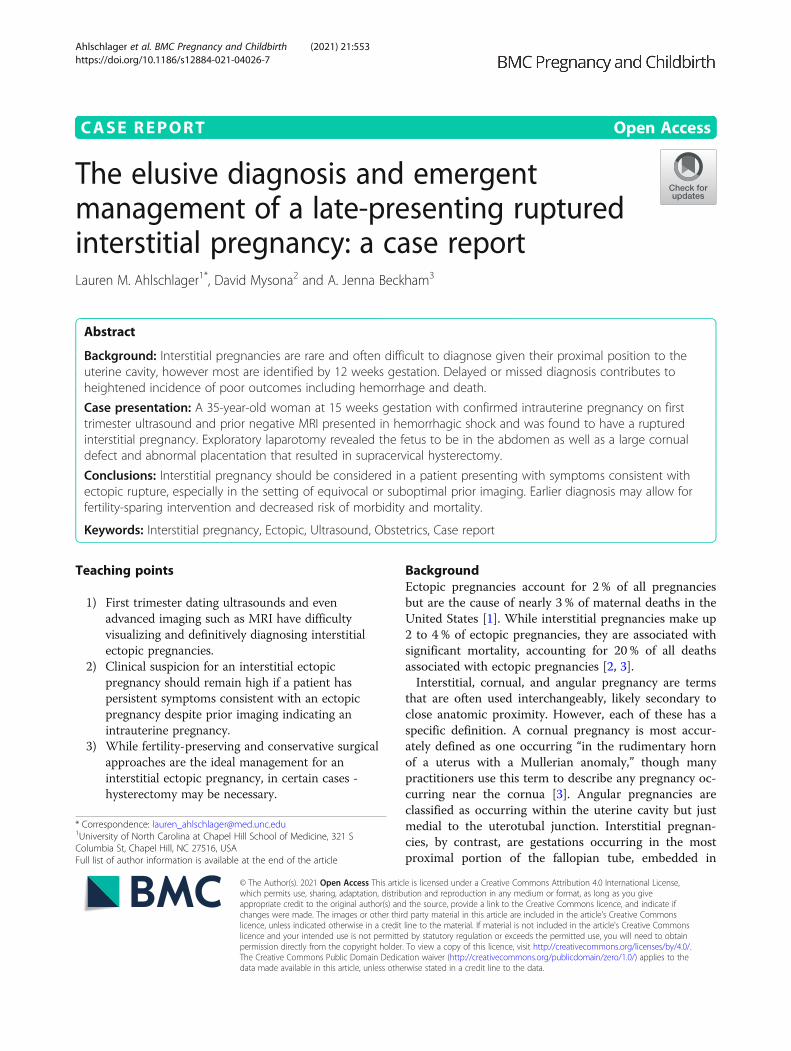
available and reviewed by both teams. They revealed thatthe patient had ultrasounds performed at 8 5/7, 12 4/7,and 13 0/7. Her ultrasound at 12 4/7 noted a possiblearcuate uterus with the pregnancy located more on thepatient’s right. An additional scan at 13 0/7 reportedsuboptimal visualization of the pregnancy on both trans-abdominal scan and transvaginal scan. These recordsalso showed that one week prior to presentation at ourfacility, the patient had gone to an outside emergencydepartment with right lower quadrant pain. There shehad an elevated white blood cell count to 18,000, atransabdominal ultrasound and abdominal MRI. Neitherthe ultrasound nor the MRI were interpreted as showingany acute abnormalities. Both specifically commented onthere being an intrauterine pregnancy. The patient wasdischarged home from the outside emergency depart-ment with a presumed viral illness. A timeline of eventsis presented in Fig. 1.At our facility the patient’s CT scan demonstrated a
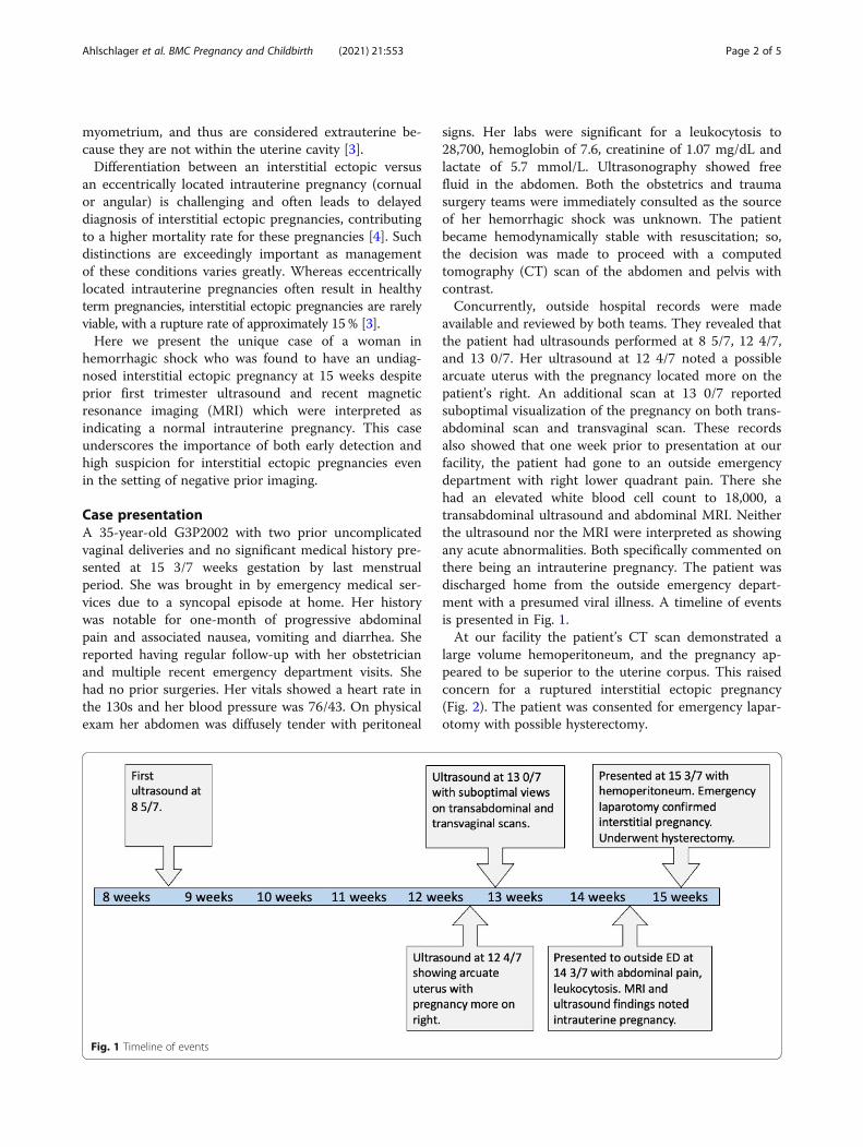
large volume hemoperitoneum, and the pregnancy ap-peared to be superior to the uterine corpus. This raisedconcern for a ruptured interstitial ectopic pregnancy(Fig. 2). The patient was consented for emergency lapar-otomy with possible hysterectomy.
Fig. 1 Timeline of events
Ahlschlager et al. BMC Pregnancy and Childbirth (2021) 21:553 Page 2 of 5
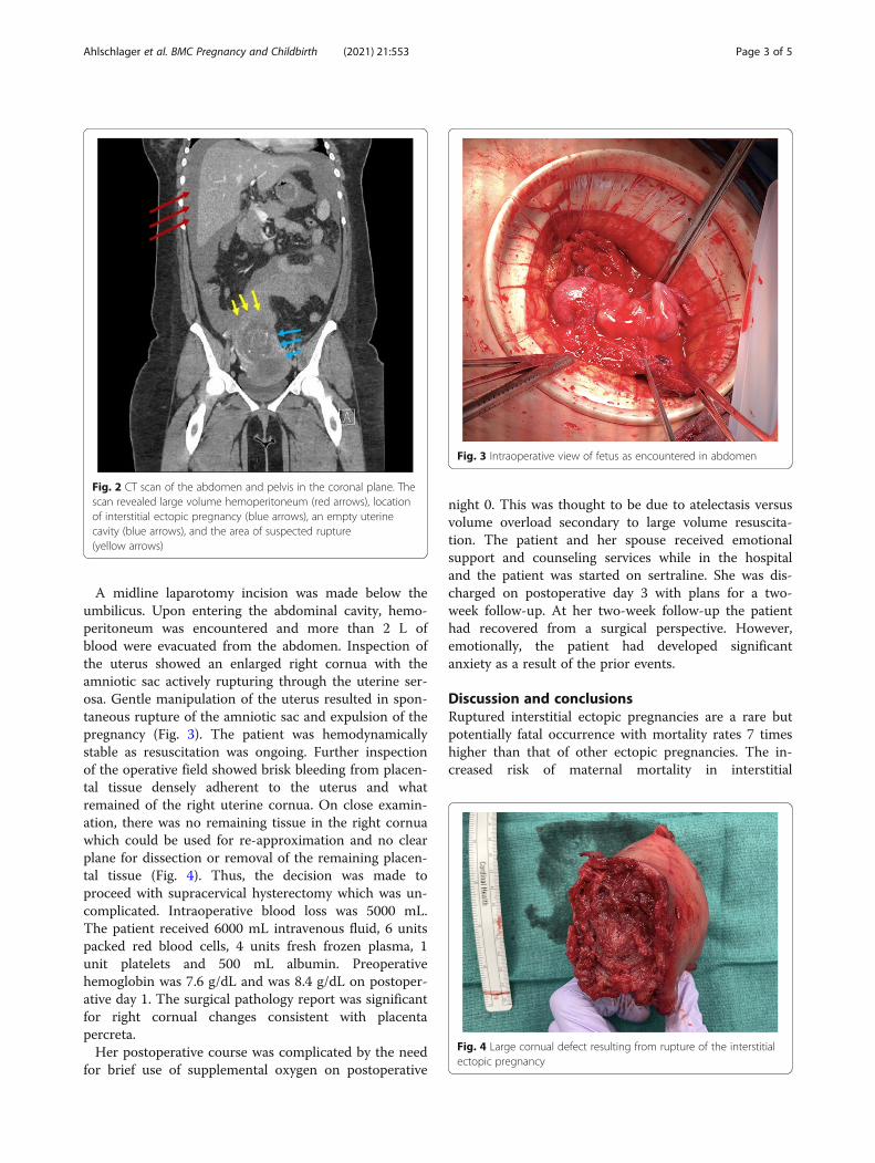
A midline laparotomy incision was made below theumbilicus. Upon entering the abdominal cavity, hemo-peritoneum was encountered and more than 2 L ofblood were evacuated from the abdomen. Inspection ofthe uterus showed an enlarged right cornua with theamniotic sac actively rupturing through the uterine ser-osa. Gentle manipulation of the uterus resulted in spon-taneous rupture of the amniotic sac and expulsion of thepregnancy (Fig. 3). The patient was hemodynamicallystable as resuscitation was ongoing. Further inspectionof the operative field showed brisk bleeding from placen-tal tissue densely adherent to the uterus and whatremained of the right uterine cornua. On close examin-ation, there was no remaining tissue in the right cornuawhich could be used for re-approximation and no clearplane for dissection or removal of the remaining placen-tal tissue (Fig. 4). Thus, the decision was made toproceed with supracervical hysterectomy which was un-complicated. Intraoperative blood loss was 5000 mL.The patient received 6000 mL intravenous fluid, 6 unitspacked red blood cells, 4 units fresh frozen plasma, 1unit platelets and 500 mL albumin. Preoperativehemoglobin was 7.6 g/dL and was 8.4 g/dL on postoper-ative day 1. The surgical pathology report was significantfor right cornual changes consistent with placentapercreta.Her postoperative course was complicated by the need
for brief use of supplemental oxygen on postoperative
night 0. This was thought to be due to atelectasis versusvolume overload secondary to large volume resuscita-tion. The patient and her spouse received emotionalsupport and counseling services while in the hospitaland the patient was started on sertraline. She was dis-charged on postoperative day 3 with plans for a two-week follow-up. At her two-week follow-up the patienthad recovered from a surgical perspective. However,emotionally, the patient had developed significantanxiety as a result of the prior events.
Discussion and conclusionsRuptured interstitial ectopic pregnancies are a rare butpotentially fatal occurrence with mortality rates 7 timeshigher than that of other ectopic pregnancies. The in-creased risk of maternal mortality in interstitial
Fig. 2 CT scan of the abdomen and pelvis in the coronal plane. Thescan revealed large volume hemoperitoneum (red arrows), locationof interstitial ectopic pregnancy (blue arrows), an empty uterinecavity (blue arrows), and the area of suspected rupture(yellow arrows)
Fig. 3 Intraoperative view of fetus as encountered in abdomen
Fig. 4 Large cornual defect resulting from rupture of the interstitialectopic pregnancy
Ahlschlager et al. BMC Pregnancy and Childbirth (2021) 21:553 Page 3 of 5
pregnancies can be partially attributed to difficulty indiagnosis which often results in missed or delayed iden-tification [5]. Diagnostic criteria for interstitial ectopicpregnancy on ultrasound include (1) an empty uterinecavity (2) a gestational sac located at least 1 cm from lat-eral uterine wall, and (3) a thin (< 5mm) myometriallayer surrounding the gestational sac. Presence of an“interstitial line sign” has also been posed as an add-itional diagnostic criterion and refers to visualization ofa thin, echogenic line on ultrasound representing theinterstitial region of the fallopian tube just lateral to thegestational sac and endometrial cavity [3, 6]. Notably,these criteria are only valid in the first trimester beforethe gestational sac enlarges. In the case of our patient,her first trimester ultrasound at 8 5/7 weeks reportedlydid not support the diagnosis [7].Compared to ultrasound, considerably less has been
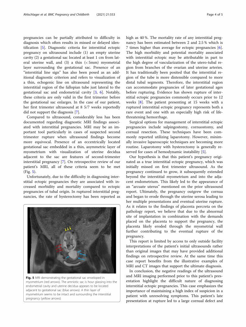
documented regarding diagnostic MRI findings associ-ated with interstitial pregnancies. MRI may be an im-portant tool particularly in cases of suspected secondtrimester rupture when ultrasound findings becomemore equivocal. Presence of an eccentrically locatedgestational sac embedded in a thin, asymmetric layer ofmyometrium with visualization of uterine deciduaadjacent to the sac are features of second-trimesterinterstitial pregnancy [7]. On retrospective review of ourpatient’s MRI, all of these criteria seem to be met(Fig. 5).Unfortunately, due to the difficulty in diagnosing inter-
stitial ectopic pregnancies they are associated with in-creased morbidity and mortality compared to ectopicpregnancies of tubal origin. In ruptured interstitial preg-nancies, the rate of hysterectomy has been reported as
high as 40 %. The mortality rate of any interstitial preg-nancy has been estimated between 2 and 2.5 % which is7 times higher than average for ectopic pregnancies [6].The high morbidity and potential mortality associatedwith interstitial ectopic may be attributable in part tothe high degree of vascularization of the utero-tubal re-gion from branches of the ovarian and uterine arteries.It has traditionally been posited that the interstitial re-gion of the tube is more distensible compared to moredistal tubal segments. Therefore, the interstitial regioncan accommodate pregnancies of later gestational agesbefore rupturing. Evidence has shown rupture of inter-stitial ectopic pregnancies commonly occurs prior to 12weeks [8]. The patient presenting at 15 weeks with aruptured interstitial ectopic pregnancy represents both arare event and one with an especially high risk of life-threatening hemorrhage.Surgical options for management of interstitial ectopic
pregnancies include salpingostomy, cornuostomy, andcornual resection. These techniques have been com-monly reported utilizing laparotomy. However, minim-ally invasive laparoscopic techniques are becoming moreroutine. Laparotomy with hysterectomy is generally re-served for cases of hemodynamic instability [5].Our hypothesis is that this patient’s pregnancy origi-
nated as a true interstitial ectopic pregnancy, which wasinitially missed on first trimester ultrasound. As thepregnancy continued to grow, it subsequently extendedbeyond the interstitial myometrium and into the adja-cent endometrium. This likely led to the appearance ofan “arcuate uterus” mentioned on the prior ultrasoundreport. Ultimately, the pregnancy outgrew the cornuaand began to erode through the uterine serosa leading toher multiple presentations and eventual uterine rupture.As it relates to the findings of placenta percreta on thepathology report, we believe that due to the abnormalsite of implantation in combination with the demandsplaced on the placenta to support the pregnancy, theplacenta likely eroded through the myometrial wallfurther contributing to the eventual rupture of thepregnancy.This report is limited by access to only outside facility
interpretations of the patient’s initial ultrasounds ratherthan original images that may have provided additionalfindings on retrospective review. At the same time thiscase report benefits from the illustrative examples ofMRI and CT images that support the ultimate diagnosis.In conclusion, the negative readings of the ultrasound
and MRI imaging performed prior to this patient’s pres-entation highlight the difficult nature of diagnosinginterstitial ectopic pregnancies. This case emphasizes theimportance of maintaining a high index of suspicion in apatient with unresolving symptoms. This patient’s latepresentation at rupture led to a large cornual defect and
Fig. 5 MRI demonstrating the gestational sac enveloped inmyometrium (red arrows). The amniotic sac is hour glassing into theendometrial cavity and uterine decidua appears to be locatedadjacent to gestational sac (blue arrows). A thin layer ofmyometrium seems to be intact and surrounding the interstitialpregnancy (yellow arrows)
Ahlschlager et al. BMC Pregnancy and Childbirth (2021) 21:553 Page 4 of 5
profound hemorrhage which fortunately did not end inmaternal mortality. Earlier diagnosis may have allowedfor a more conservative, fertility-preserving interventionsuch as methotrexate therapy or uterine arteryembolization. We report this case in hopes that it willencourage consideration of this rare but potentiallydeadly condition in patients who present with symptomssuggestive of ectopic pregnancy in the setting of subopti-mal or equivocal prior imaging, as keeping a broad dif-ferential diagnosis may reduce mortality and improvepatient outcomes.
AbbreviationsCT: Computed tomography; MRI: Magnetic resonance imaging
AcknowledgementsWe are grateful to the patient for her willingness to contribute to this report.
Authors’ contributionsLA collected the clinical data and drafted the manuscript. DM and LAconceptualized and designed the report. AJB supervised the study. Allauthors made substantive revisions to the text and approved the finalmanuscript for submission.
FundingNo funding was received for this report.
Availability of data and materialsAll data analyzed during this study are included herein.
Declarations
Ethics approval and consent to participateNot applicable.
Consent for publicationThe authors attest that written consent to publish the report along withaccompanying clinical data and images was obtained from the patient priorto composition of this manuscript. Copies of this form are available to thepublisher upon request.
Competing interestsThe authors declare that they have no competing interests.
Author details1University of North Carolina at Chapel Hill School of Medicine, 321 SColumbia St, Chapel Hill, NC 27516, USA. 2Department of Obstetrics andGynecology, University of North Carolina School of Medicine, Chapel Hill,USA. 3Department of Obstetrics and Gynecology, WakeMed Health andHospitals, 3024 New Bern Ave Suite 309, Raleigh, NC 27610, USA.
Received: 20 March 2021 Accepted: 28 July 2021
References1. Creanga AA, Syverson C, Seed K, Callaghan WM. Pregnancy-Related
Mortality in the United States, 2011–2013. Obstet Gynecol. 2017;130(2):366–73.
2. Dagar M, Srivastava M, Ganguli I, Bhardwaj P, Sharma N, Chawla D.Interstitial and cornual ectopic pregnancy: conservative surgical andmedical management. J Obstet Gynaecol India. 2018;68(6):471–6.
3. Finlinson AR, Bollig KJ, Schust DJ. Differentiating pregnancies near theuterotubal junction (angular, cornual, and interstitial): a review andrecommendations. Fertil Res Pract. 2020;6:8.
4. Jurkovic D, Mavrelos D. Catch me if you scan: ultrasound diagnosis ofectopic pregnancy. Ultrasound Obstet Gynecol. 2007;30(1):1–7.
5. Panelli DM, Phillips CH, Brady PC. Incidence, diagnosis and management oftubal and nontubal ectopic pregnancies: a review. Fertil Res Pract. 2015;1:15.
6. Brincat M, Bryant-Smith A, Holland TK. The diagnosis and management ofinterstitial ectopic pregnancies: a review. Gynecol Surg. 2019;16(1):2.
7. Hamouda ES, Littooij AS, Thia EW, Ong CL. Ruptured interstitial ectopicpregnancy at 18 weeks gestation diagnosed by MRI: a case report. J RadiolCase Rep. 2013;7(10):34–42.
8. Tulandi T, Al-Jaroudi D. Interstitial pregnancy: results generated from thesociety of reproductive surgeons registry. Obstet Gynecol. 2004;103(1):47–50.
Publisher’s NoteSpringer Nature remains neutral with regard to jurisdictional claims inpublished maps and institutional affiliations.
Ahlschlager et al. BMC Pregnancy and Childbirth (2021) 21:553 Page 5 of 5