Embed Size (px)
Citation preview
www.elsevier.com/locate/ajem
Diagnostics
The electrocardiogram in the patient with syncope
Jacqueline Dovgalyuka, Christopher Holstege MDa,Amal Mattu MDb, William J. Brady MDa,*
aDepartment of Emergency Medicine, University of Virginia, Charlottesville, VA, USAbDepartment of Emergency Medicine, University of Maryland, Baltimore, MD, USA
Received 2 December 2006; accepted 4 December 2006
Abstract Syncope is a common and challenging presentation for the emergency physician. Various
investigators have developed clinical risk score and clinical decision rules which are designed to identify
the population at highest risk for adverse events. In each of these clinical decision tools, the
electrocardiogram (ECG) is one of the key clinical variables used to evaluate the patient. Certain
electrocardiographic presentations in the patient with syncope will not only provide a reason for the loss
of consciousness but also guide early therapy and disposition in this individual. Bradycardia,
atrioventricular block, intraventricular conduction abnormality, and tachydysrhythmia in the appropriate
clinical setting provide an answer to the clinician for the sync opal event. Morphologic findings
suggesting the range of cardiovascular malady are also encountered; these entities are far ranging,
including the various ST-segment and T-wave abnormalities of acute coronary syndrome, ventricular
preexcitation as seen in the Wolff-Parkinson-White syndrome, Brugada syndrome with the associated
tendency for sudden death, prolonged QT interval common in the diverse long QT interval
presentations, and right ventricular hypertrophy suggestive of hypertrophic cardiomyopathy. This
review discusses the ECG in the patient with syncope. The general use of the 12-lead ECG in this
patient population is discussed. Furthermore, specific electrocardiographic presentations seen in the
patient with syncope are also reviewed.
D 2007 Elsevier Inc. All rights reserved.
1. Introduction
Syncope is a common and challenging presentation for
the emergency physician. It is estimated that 1% to 3% of
visits to the ED and 6% of hospital admissions are related to
syncope; furthermore, 20% to 50% of adults will experience
a syncopal episode at least once in their lifetime [1].
Although most causes of syncope are benign and require no
further evaluation, there is a small subset of patients for
0735-6757/$ – see front matter D 2007 Elsevier Inc. All rights reserved.
doi:10.1016/j.ajem.2006.12.016
* Corresponding author. Tel.: +1 4344651816.
E-mail address: [email protected] (W.J. Brady).
whom a syncopal episode may herald a potentially life-
threatening condition [2-4]. Investigators have developed a
clinical risk score [4] and clinical decision rules [2,3] which
are designed to identify the population at highest risk for
adverse events. In each of these clinical decision tools, an
abnormal electrocardiogram (ECG) is one of the key clinical
variables used to evaluate the patient.
Four basic diagnostic categories of syncope are encoun-
tered, including reflex-mediated, orthostatic, cerebrovascu-
lar, and cardiac. Of these 4 generally accepted categories of
syncope, cardiac causes represent 10% to 30% of cases [1].
The ECG may be helpful in all 4 categories, yet the cardiac
subtype is likely to yield the highest rate of electrocardio-
American Journal of Emergency Medicine (2007) 25, 688–701
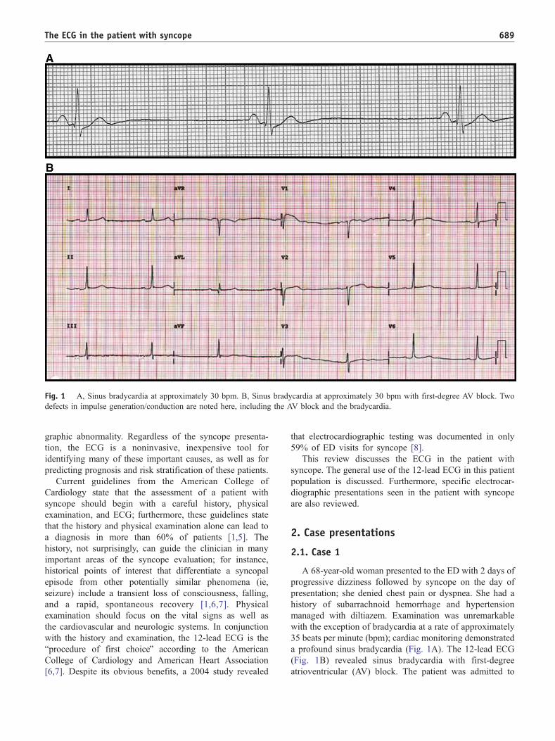
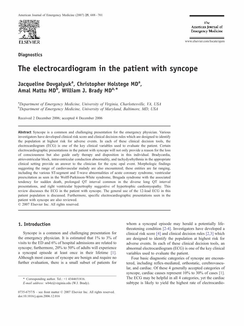
Fig. 1 A, Sinus bradycardia at approximately 30 bpm. B, Sinus bradycardia at approximately 30 bpm with first-degree AV block. Two
defects in impulse generation/conduction are noted here, including the AV block and the bradycardia.
The ECG in the patient with syncope 689
graphic abnormality. Regardless of the syncope presenta-
tion, the ECG is a noninvasive, inexpensive tool for
identifying many of these important causes, as well as for
predicting prognosis and risk stratification of these patients.
Current guidelines from the American College of
Cardiology state that the assessment of a patient with
syncope should begin with a careful history, physical
examination, and ECG; furthermore, these guidelines state
that the history and physical examination alone can lead to
a diagnosis in more than 60% of patients [1,5]. The
history, not surprisingly, can guide the clinician in many
important areas of the syncope evaluation; for instance,
historical points of interest that differentiate a syncopal
episode from other potentially similar phenomena (ie,
seizure) include a transient loss of consciousness, falling,
and a rapid, spontaneous recovery [1,6,7]. Physical
examination should focus on the vital signs as well as
the cardiovascular and neurologic systems. In conjunction
with the history and examination, the 12-lead ECG is the
bprocedure of first choiceQ according to the American
College of Cardiology and American Heart Association
[6,7]. Despite its obvious benefits, a 2004 study revealed
that electrocardiographic testing was documented in only
59% of ED visits for syncope [8].
This review discusses the ECG in the patient with
syncope. The general use of the 12-lead ECG in this patient
population is discussed. Furthermore, specific electrocar-
diographic presentations seen in the patient with syncope
are also reviewed.
2. Case presentations
2.1. Case 1
A 68-year-old woman presented to the ED with 2 days of
progressive dizziness followed by syncope on the day of
presentation; she denied chest pain or dyspnea. She had a
history of subarrachnoid hemorrhage and hypertension
managed with diltiazem. Examination was unremarkable
with the exception of bradycardia at a rate of approximately
35 beats per minute (bpm); cardiac monitoring demonstrated
a profound sinus bradycardia (Fig. 1A). The 12-lead ECG
(Fig. 1B) revealed sinus bradycardia with first-degree
atrioventricular (AV) block. The patient was admitted to
Fig. 2 Wide complex tachycardia in a young patient. The ventricular rate is very rapid, in excess of 240 bpm.
J. Dovgalyuk et al.690
the hospital to a critical care unit with placement of
transcutaneous pacer pads. Further monitoring demonstrated
continued bradycardia. Serum cardiac markers were nega-
tive for myocardial infarction. The patient underwent
placement of a permanent right ventricular pacemaker
without complication. She was discharged from the hospital
with bradycardia likely related to chronic conduction system
disease exacerbated by the calcium channel blocking agent.
She was well at follow-up.
2.2. Case 2
A 24-year-old man with no known medical history was
evaluated by emergency medical service for acute weakness.
Vital signs were significant for a blood pressure of 70 mm
Hg, a pulse of 200 bpm, and a respiratory rate of 32 per
minute. The cardiac monitor revealed a wide complex
tachycardia at a rate of 200 bpm (Fig. 2). Immediate direct
current cardioversion was administered with an initial
energy of 100 J which successfully converted the rhythm
to sinus tachycardia. He was transported uneventfully to the
Fig. 3 Wolff-Parkinson-White syndrome. Note the classic triad, includin
Pseudo-ischemic findings, such as Q waves and T-wave inversion, are als
ED. Upon ED arrival, the patient lost consciousness; the
cardiac monitor revealed a recurrent wide complex tachy-
cardia. Immediate electrical cardioversion was performed
with the return of sinus tachycardia. Twelve-lead ECG
(Fig. 3) demonstrated normal sinus rhythm with shortened
PR interval, widened QRS complex, and delta wave—all
findings consistent with Wolff-Parkinson-White (WPW)
syndrome. The patient received intravenous procainamide
and was admitted to the hospital. Electrophysiologic study
revealed an accessory pathway (AP) with anterograde
conduction properties. Because of the patient’s initial
presentation and electrophysiologic study result, he under-
went ablative therapy (transcatheter radiofrequency tech-
nique) with good results; no further rhythm disturbances
were noted during 2 years of follow-up.
2.3. Case 3
A 38-year-old man presented to the ED complaining of
3 brief episodes of severe lightheadedness; approximately
30 minutes before presentation, he noted a full syncopal
g the shortened PR interval, delta wave, and widened QRS complex.
o seen and do not represent sequelae of coronary artery disease.
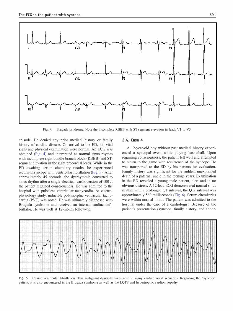
Fig. 4 Brugada syndrome. Note the incomplete RBBB with ST-segment elevation in leads V1 to V3.
The ECG in the patient with syncope 691
episode. He denied any prior medical history or family
history of cardiac disease. On arrival to the ED, his vital
signs and physical examination were normal. An ECG was
obtained (Fig. 4) and interpreted as normal sinus rhythm
with incomplete right bundle branch block (RBBB) and ST-
segment elevation in the right precordial leads. While in the
ED awaiting serum chemistry results, he experienced
recurrent syncope with ventricular fibrillation (Fig. 5). After
approximately 45 seconds, the dysrhythmia converted to
sinus rhythm after a single electrical cardioversion of 100 J;
the patient regained consciousness. He was admitted to the
hospital with pulseless ventricular tachycardia. At electro-
physiology study, inducible polymorphic ventricular tachy-
cardia (PVT) was noted. He was ultimately diagnosed with
Brugada syndrome and received an internal cardiac defi-
brillator. He was well at 12-month follow-up.
Fig. 5 Coarse ventricular fibrillation. This malignant dysrhythmia is
patient, it is also encountered in the Brugada syndrome as well as the L
2.4. Case 4
A 12-year-old boy without past medical history experi-
enced a syncopal event while playing basketball. Upon
regaining consciousness, the patient felt well and attempted
to return to the game with recurrence of the syncope. He
was transported to the ED by his parents for evaluation.
Family history was significant for the sudden, unexplained
death of a paternal uncle in the teenage years. Examination
in the ED revealed a young male patient, alert and in no
obvious distress. A 12-lead ECG demonstrated normal sinus
rhythm with a prolonged QT interval; the QTc interval was
approximately 560 milliseconds (Fig. 6). Serum chemistries
were within normal limits. The patient was admitted to the
hospital under the care of a cardiologist. Because of the
patient’s presentation (syncope, family history, and abnor-
seen in many cardiac arrest scenarios. Regarding the bsyncopeQQTS and hypertrophic cardiomyopathy.
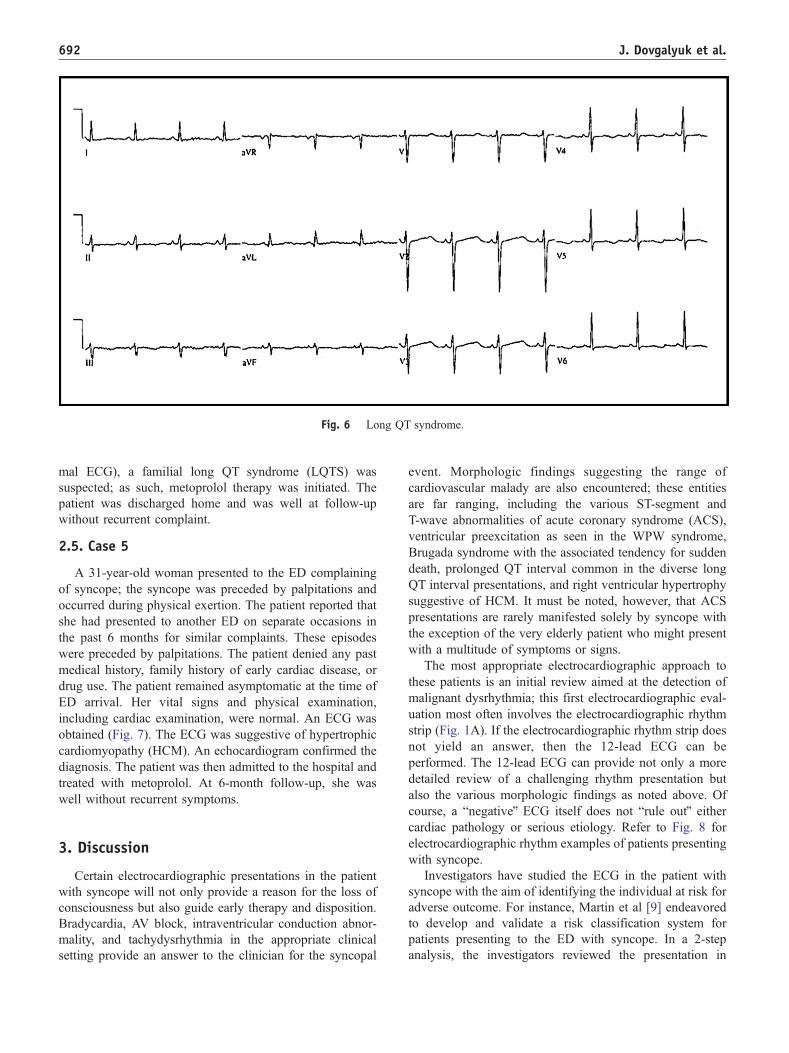
Fig. 6 Long QT syndrome.
J. Dovgalyuk et al.692
mal ECG), a familial long QT syndrome (LQTS) was
suspected; as such, metoprolol therapy was initiated. The
patient was discharged home and was well at follow-up
without recurrent complaint.
2.5. Case 5
A 31-year-old woman presented to the ED complaining
of syncope; the syncope was preceded by palpitations and
occurred during physical exertion. The patient reported that
she had presented to another ED on separate occasions in
the past 6 months for similar complaints. These episodes
were preceded by palpitations. The patient denied any past
medical history, family history of early cardiac disease, or
drug use. The patient remained asymptomatic at the time of
ED arrival. Her vital signs and physical examination,
including cardiac examination, were normal. An ECG was
obtained (Fig. 7). The ECG was suggestive of hypertrophic
cardiomyopathy (HCM). An echocardiogram confirmed the
diagnosis. The patient was then admitted to the hospital and
treated with metoprolol. At 6-month follow-up, she was
well without recurrent symptoms.
3. Discussion
Certain electrocardiographic presentations in the patient
with syncope will not only provide a reason for the loss of
consciousness but also guide early therapy and disposition.
Bradycardia, AV block, intraventricular conduction abnor-
mality, and tachydysrhythmia in the appropriate clinical
setting provide an answer to the clinician for the syncopal
event. Morphologic findings suggesting the range of
cardiovascular malady are also encountered; these entities
are far ranging, including the various ST-segment and
T-wave abnormalities of acute coronary syndrome (ACS),
ventricular preexcitation as seen in the WPW syndrome,
Brugada syndrome with the associated tendency for sudden
death, prolonged QT interval common in the diverse long
QT interval presentations, and right ventricular hypertrophy
suggestive of HCM. It must be noted, however, that ACS
presentations are rarely manifested solely by syncope with
the exception of the very elderly patient who might present
with a multitude of symptoms or signs.
The most appropriate electrocardiographic approach to
these patients is an initial review aimed at the detection of
malignant dysrhythmia; this first electrocardiographic eval-
uation most often involves the electrocardiographic rhythm
strip (Fig. 1A). If the electrocardiographic rhythm strip does
not yield an answer, then the 12-lead ECG can be
performed. The 12-lead ECG can provide not only a more
detailed review of a challenging rhythm presentation but
also the various morphologic findings as noted above. Of
course, a bnegativeQ ECG itself does not brule outQ eithercardiac pathology or serious etiology. Refer to Fig. 8 for
electrocardiographic rhythm examples of patients presenting
with syncope.
Investigators have studied the ECG in the patient with
syncope with the aim of identifying the individual at risk for
adverse outcome. For instance, Martin et al [9] endeavored
to develop and validate a risk classification system for
patients presenting to the ED with syncope. In a 2-step
analysis, the investigators reviewed the presentation in
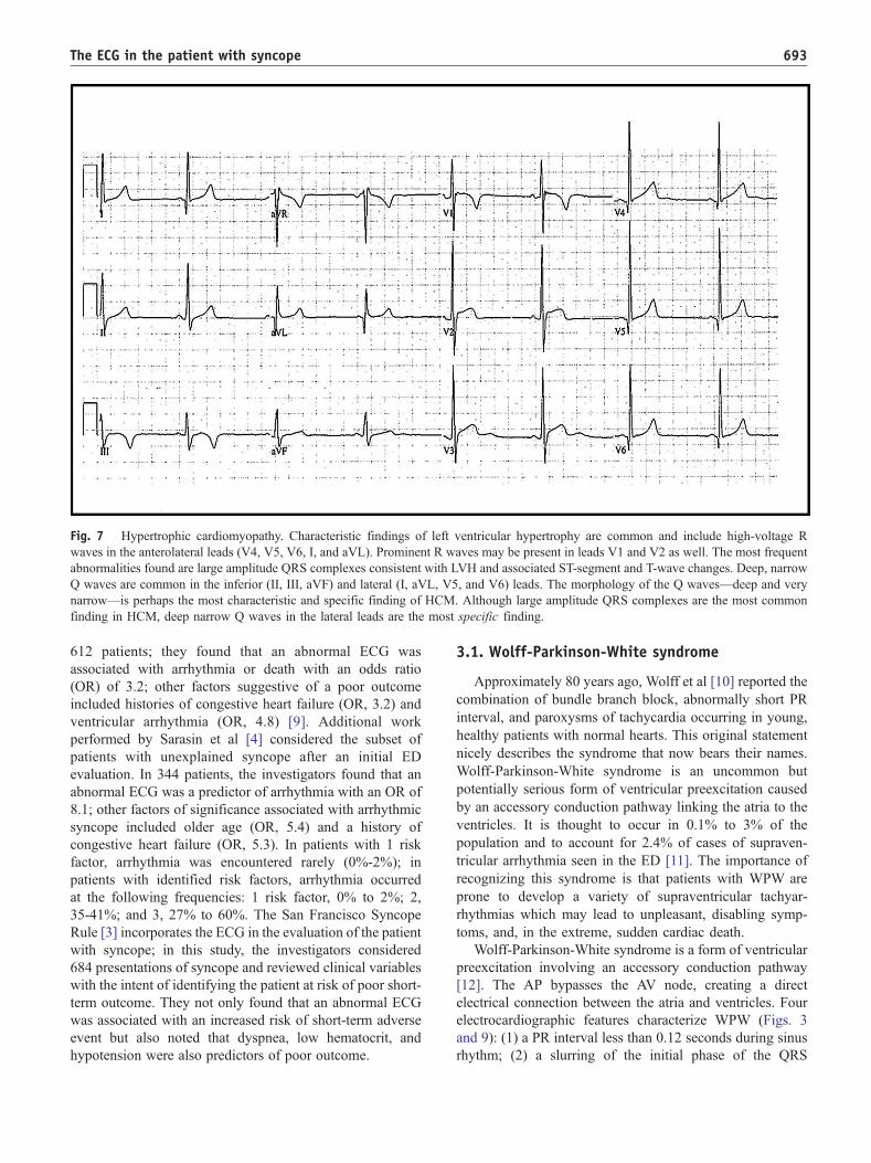
Fig. 7 Hypertrophic cardiomyopathy. Characteristic findings of left ventricular hypertrophy are common and include high-voltage R
waves in the anterolateral leads (V4, V5, V6, I, and aVL). Prominent R waves may be present in leads V1 and V2 as well. The most frequent
abnormalities found are large amplitude QRS complexes consistent with LVH and associated ST-segment and T-wave changes. Deep, narrow
Q waves are common in the inferior (II, III, aVF) and lateral (I, aVL, V5, and V6) leads. The morphology of the Q waves—deep and very
narrow—is perhaps the most characteristic and specific finding of HCM. Although large amplitude QRS complexes are the most common
finding in HCM, deep narrow Q waves in the lateral leads are the most specific finding.
The ECG in the patient with syncope 693
612 patients; they found that an abnormal ECG was
associated with arrhythmia or death with an odds ratio
(OR) of 3.2; other factors suggestive of a poor outcome
included histories of congestive heart failure (OR, 3.2) and
ventricular arrhythmia (OR, 4.8) [9]. Additional work
performed by Sarasin et al [4] considered the subset of
patients with unexplained syncope after an initial ED
evaluation. In 344 patients, the investigators found that an
abnormal ECG was a predictor of arrhythmia with an OR of
8.1; other factors of significance associated with arrhythmic
syncope included older age (OR, 5.4) and a history of
congestive heart failure (OR, 5.3). In patients with 1 risk
factor, arrhythmia was encountered rarely (0%-2%); in
patients with identified risk factors, arrhythmia occurred
at the following frequencies: 1 risk factor, 0% to 2%; 2,
35-41%; and 3, 27% to 60%. The San Francisco Syncope
Rule [3] incorporates the ECG in the evaluation of the patient
with syncope; in this study, the investigators considered
684 presentations of syncope and reviewed clinical variables
with the intent of identifying the patient at risk of poor short-
term outcome. They not only found that an abnormal ECG
was associated with an increased risk of short-term adverse
event but also noted that dyspnea, low hematocrit, and
hypotension were also predictors of poor outcome.
3.1. Wolff-Parkinson-White syndrome
Approximately 80 years ago, Wolff et al [10] reported the
combination of bundle branch block, abnormally short PR
interval, and paroxysms of tachycardia occurring in young,
healthy patients with normal hearts. This original statement
nicely describes the syndrome that now bears their names.
Wolff-Parkinson-White syndrome is an uncommon but
potentially serious form of ventricular preexcitation caused
by an accessory conduction pathway linking the atria to the
ventricles. It is thought to occur in 0.1% to 3% of the
population and to account for 2.4% of cases of supraven-
tricular arrhythmia seen in the ED [11]. The importance of
recognizing this syndrome is that patients with WPW are
prone to develop a variety of supraventricular tachyar-
rhythmias which may lead to unpleasant, disabling symp-
toms, and, in the extreme, sudden cardiac death.
Wolff-Parkinson-White syndrome is a form of ventricular
preexcitation involving an accessory conduction pathway
[12]. The AP bypasses the AV node, creating a direct
electrical connection between the atria and ventricles. Four
electrocardiographic features characterize WPW (Figs. 3
and 9): (1) a PR interval less than 0.12 seconds during sinus
rhythm; (2) a slurring of the initial phase of the QRS
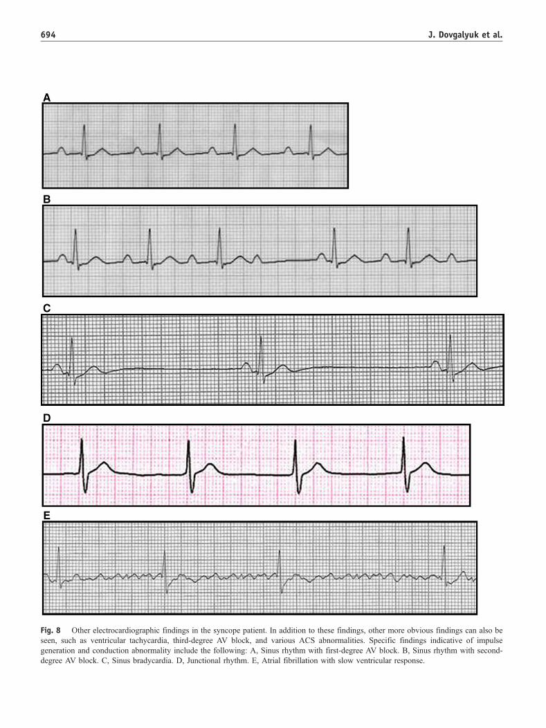
Fig. 8 Other electrocardiographic findings in the syncope patient. In addition to these findings, other more obvious findings can also be
seen, such as ventricular tachycardia, third-degree AV block, and various ACS abnormalities. Specific findings indicative of impulse
generation and conduction abnormality include the following: A, Sinus rhythm with first-degree AV block. B, Sinus rhythm with second-
degree AV block. C, Sinus bradycardia. D, Junctional rhythm. E, Atrial fibrillation with slow ventricular response.
J. Dovgalyuk et al.694
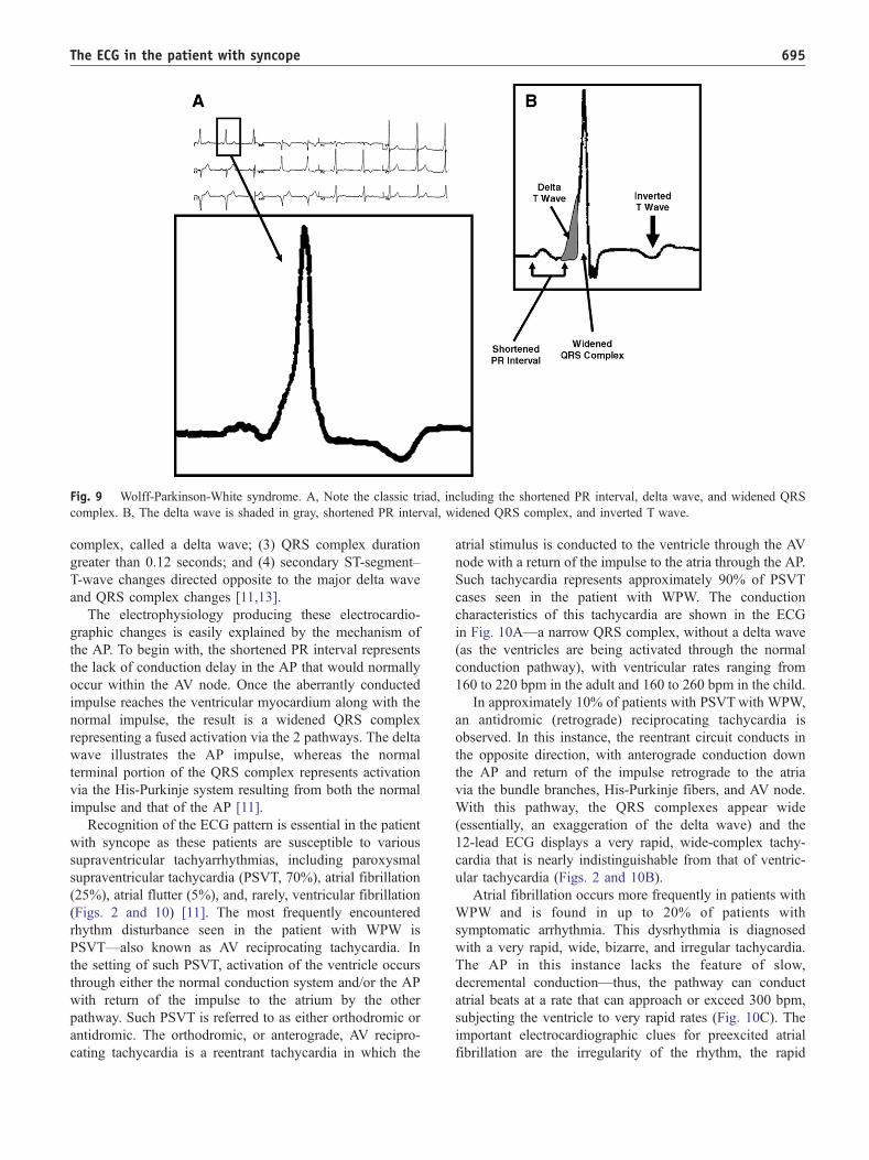
Fig. 9 Wolff-Parkinson-White syndrome. A, Note the classic triad, including the shortened PR interval, delta wave, and widened QRS
complex. B, The delta wave is shaded in gray, shortened PR interval, widened QRS complex, and inverted T wave.
The ECG in the patient with syncope 695
complex, called a delta wave; (3) QRS complex duration
greater than 0.12 seconds; and (4) secondary ST-segment–
T-wave changes directed opposite to the major delta wave
and QRS complex changes [11,13].
The electrophysiology producing these electrocardio-
graphic changes is easily explained by the mechanism of
the AP. To begin with, the shortened PR interval represents
the lack of conduction delay in the AP that would normally
occur within the AV node. Once the aberrantly conducted
impulse reaches the ventricular myocardium along with the
normal impulse, the result is a widened QRS complex
representing a fused activation via the 2 pathways. The delta
wave illustrates the AP impulse, whereas the normal
terminal portion of the QRS complex represents activation
via the His-Purkinje system resulting from both the normal
impulse and that of the AP [11].
Recognition of the ECG pattern is essential in the patient
with syncope as these patients are susceptible to various
supraventricular tachyarrhythmias, including paroxysmal
supraventricular tachycardia (PSVT, 70%), atrial fibrillation
(25%), atrial flutter (5%), and, rarely, ventricular fibrillation
(Figs. 2 and 10) [11]. The most frequently encountered
rhythm disturbance seen in the patient with WPW is
PSVT—also known as AV reciprocating tachycardia. In
the setting of such PSVT, activation of the ventricle occurs
through either the normal conduction system and/or the AP
with return of the impulse to the atrium by the other
pathway. Such PSVT is referred to as either orthodromic or
antidromic. The orthodromic, or anterograde, AV recipro-
cating tachycardia is a reentrant tachycardia in which the
atrial stimulus is conducted to the ventricle through the AV
node with a return of the impulse to the atria through the AP.
Such tachycardia represents approximately 90% of PSVT
cases seen in the patient with WPW. The conduction
characteristics of this tachycardia are shown in the ECG
in Fig. 10A—a narrow QRS complex, without a delta wave
(as the ventricles are being activated through the normal
conduction pathway), with ventricular rates ranging from
160 to 220 bpm in the adult and 160 to 260 bpm in the child.
In approximately 10% of patients with PSVTwith WPW,
an antidromic (retrograde) reciprocating tachycardia is
observed. In this instance, the reentrant circuit conducts in
the opposite direction, with anterograde conduction down
the AP and return of the impulse retrograde to the atria
via the bundle branches, His-Purkinje fibers, and AV node.
With this pathway, the QRS complexes appear wide
(essentially, an exaggeration of the delta wave) and the
12-lead ECG displays a very rapid, wide-complex tachy-
cardia that is nearly indistinguishable from that of ventric-
ular tachycardia (Figs. 2 and 10B).
Atrial fibrillation occurs more frequently in patients with
WPW and is found in up to 20% of patients with
symptomatic arrhythmia. This dysrhythmia is diagnosed
with a very rapid, wide, bizarre, and irregular tachycardia.
The AP in this instance lacks the feature of slow,
decremental conduction—thus, the pathway can conduct
atrial beats at a rate that can approach or exceed 300 bpm,
subjecting the ventricle to very rapid rates (Fig. 10C). The
important electrocardiographic clues for preexcited atrial
fibrillation are the irregularity of the rhythm, the rapid
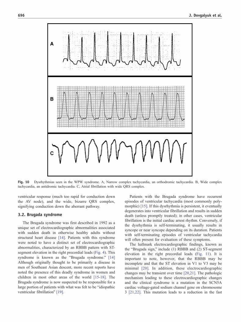
Fig. 10 Dysrhythmias seen in the WPW syndrome. A, Narrow complex tachycardia, an orthodromic tachycardia. B, Wide complex
tachycardia, an antidromic tachycardia. C, Atrial fibrillation with wide QRS complex.
J. Dovgalyuk et al.696
ventricular response (much too rapid for conduction down
the AV node), and the wide, bizarre QRS complex,
signifying conduction down the aberrant pathway.
3.2. Brugada syndrome
The Brugada syndrome was first described in 1992 as a
unique set of electrocardiographic abnormalities associated
with sudden death in otherwise healthy adults without
structural heart disease [14]. Patients with this syndrome
were noted to have a distinct set of electrocardiographic
abnormalities, characterized by an RBBB pattern with ST-
segment elevation in the right precordial leads (Fig. 4). This
syndrome is known as the bBrugada syndrome.Q [14]
Although originally thought to be primarily a disease in
men of Southeast Asian descent, more recent reports have
noted the presence of this deadly syndrome in women and
children in most other areas of the world [15-18]. The
Brugada syndrome is now suspected to be responsible for a
large portion of patients with what was felt to be bidiopathicventricular fibrillationQ [19].
Patients with the Brugada syndrome have recurrent
episodes of ventricular tachycardia (most commonly poly-
morphic) [15]. If this dysrhythmia is persistent, it eventually
degenerates into ventricular fibrillation and results in sudden
death (unless promptly treated); in other cases, ventricular
fibrillation is the initial cardiac arrest rhythm. Conversely, if
the dysrhythmia is self-terminating, it usually results in
syncope or near syncope depending on its duration. Patients
with self-terminating episodes of ventricular tachycardia
will often present for evaluation of these symptoms.
The hallmark electrocardiographic findings, known as
the bBrugada sign,Q include (1) RBBB and (2) ST-segment
elevation in the right precordial leads (Fig. 11). It is
important to note, however, that the RBBB may be
incomplete and that the ST elevation in V1 to V3 may be
minimal [20]. In addition, these electrocardiographic
changes may be transient over time [20,21]. The pathologic
mechanism leading to these electrocardiographic changes
and the clinical syndrome is a mutation in the SCN5A
cardiac voltage-gated sodium channel gene on chromosome
3 [21,22]. This mutation leads to a reduction in the fast
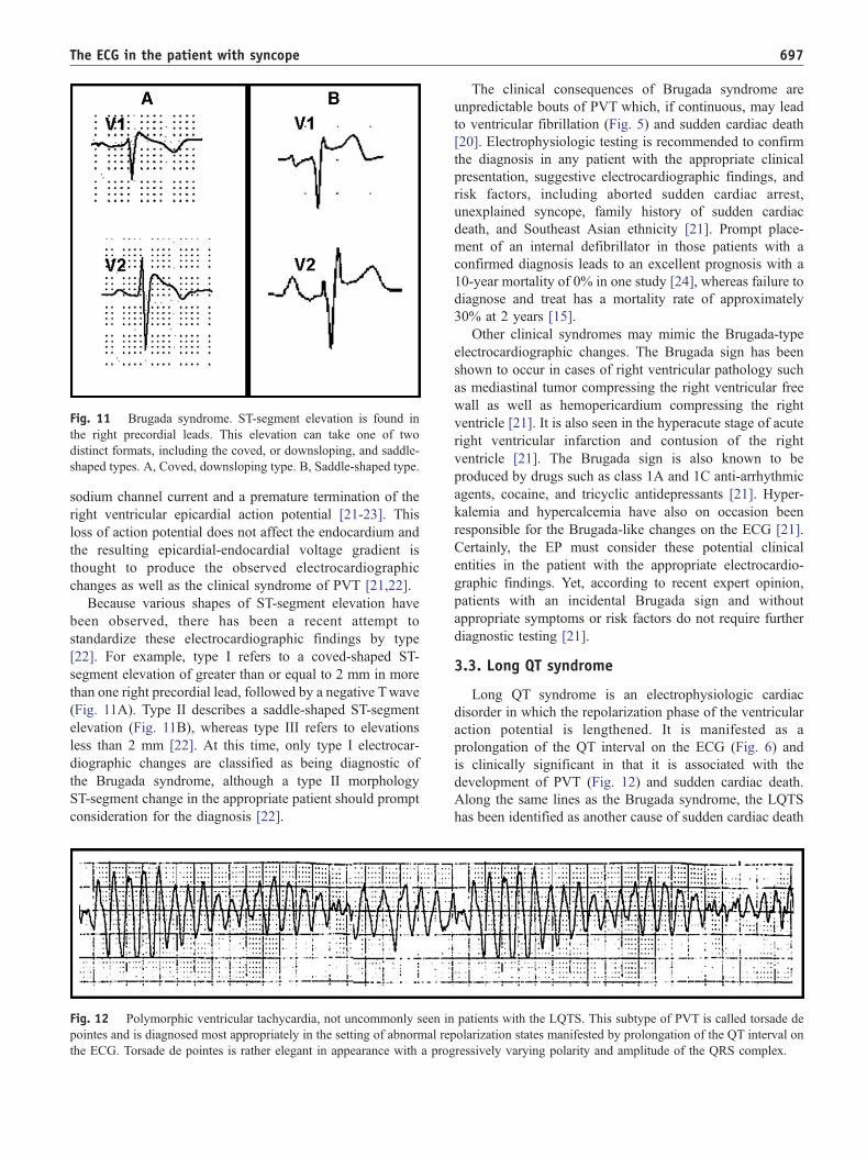
Fig. 11 Brugada syndrome. ST-segment elevation is found in
the right precordial leads. This elevation can take one of two
distinct formats, including the coved, or downsloping, and saddle-
shaped types. A, Coved, downsloping type. B, Saddle-shaped type.
The ECG in the patient with syncope 697
sodium channel current and a premature termination of the
right ventricular epicardial action potential [21-23]. This
loss of action potential does not affect the endocardium and
the resulting epicardial-endocardial voltage gradient is
thought to produce the observed electrocardiographic
changes as well as the clinical syndrome of PVT [21,22].
Because various shapes of ST-segment elevation have
been observed, there has been a recent attempt to
standardize these electrocardiographic findings by type
[22]. For example, type I refers to a coved-shaped ST-
segment elevation of greater than or equal to 2 mm in more
than one right precordial lead, followed by a negative Twave
(Fig. 11A). Type II describes a saddle-shaped ST-segment
elevation (Fig. 11B), whereas type III refers to elevations
less than 2 mm [22]. At this time, only type I electrocar-
diographic changes are classified as being diagnostic of
the Brugada syndrome, although a type II morphology
ST-segment change in the appropriate patient should prompt
consideration for the diagnosis [22].
Fig. 12 Polymorphic ventricular tachycardia, not uncommonly seen in
pointes and is diagnosed most appropriately in the setting of abnormal rep
the ECG. Torsade de pointes is rather elegant in appearance with a prog
The clinical consequences of Brugada syndrome are
unpredictable bouts of PVT which, if continuous, may lead
to ventricular fibrillation (Fig. 5) and sudden cardiac death
[20]. Electrophysiologic testing is recommended to confirm
the diagnosis in any patient with the appropriate clinical
presentation, suggestive electrocardiographic findings, and
risk factors, including aborted sudden cardiac arrest,
unexplained syncope, family history of sudden cardiac
death, and Southeast Asian ethnicity [21]. Prompt place-
ment of an internal defibrillator in those patients with a
confirmed diagnosis leads to an excellent prognosis with a
10-year mortality of 0% in one study [24], whereas failure to
diagnose and treat has a mortality rate of approximately
30% at 2 years [15].
Other clinical syndromes may mimic the Brugada-type
electrocardiographic changes. The Brugada sign has been
shown to occur in cases of right ventricular pathology such
as mediastinal tumor compressing the right ventricular free
wall as well as hemopericardium compressing the right
ventricle [21]. It is also seen in the hyperacute stage of acute
right ventricular infarction and contusion of the right
ventricle [21]. The Brugada sign is also known to be
produced by drugs such as class 1A and 1C anti-arrhythmic
agents, cocaine, and tricyclic antidepressants [21]. Hyper-
kalemia and hypercalcemia have also on occasion been
responsible for the Brugada-like changes on the ECG [21].
Certainly, the EP must consider these potential clinical
entities in the patient with the appropriate electrocardio-
graphic findings. Yet, according to recent expert opinion,
patients with an incidental Brugada sign and without
appropriate symptoms or risk factors do not require further
diagnostic testing [21].
3.3. Long QT syndrome
Long QT syndrome is an electrophysiologic cardiac
disorder in which the repolarization phase of the ventricular
action potential is lengthened. It is manifested as a
prolongation of the QT interval on the ECG (Fig. 6) and
is clinically significant in that it is associated with the
development of PVT (Fig. 12) and sudden cardiac death.
Along the same lines as the Brugada syndrome, the LQTS
has been identified as another cause of sudden cardiac death
patients with the LQTS. This subtype of PVT is called torsade de
olarization states manifested by prolongation of the QT interval on
ressively varying polarity and amplitude of the QRS complex.
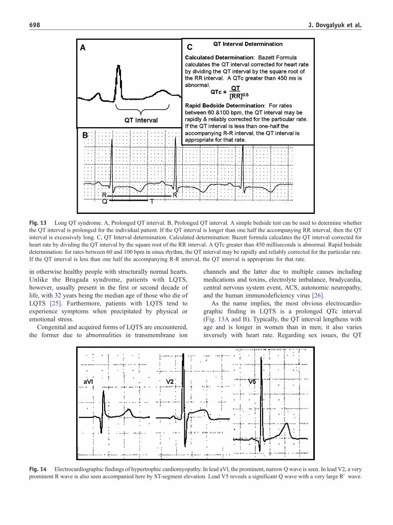
Fig. 13 Long QT syndrome. A, Prolonged QT interval. B, Prolonged QT interval. A simple bedside test can be used to determine whether
the QT interval is prolonged for the individual patient. If the QT interval is longer than one half the accompanying RR interval, then the QT
interval is excessively long. C, QT Interval determination. Calculated determination: Bazett formula calculates the QT interval corrected for
heart rate by dividing the QT interval by the square root of the RR interval. A QTc greater than 450 milliseconds is abnormal. Rapid bedside
determination: for rates between 60 and 100 bpm in sinus rhythm, the QT interval may be rapidly and reliably corrected for the particular rate.
If the QT interval is less than one half the accompanying R-R interval, the QT interval is appropriate for that rate.
J. Dovgalyuk et al.698
in otherwise healthy people with structurally normal hearts.
Unlike the Brugada syndrome, patients with LQTS,
however, usually present in the first or second decade of
life, with 32 years being the median age of those who die of
LQTS [25]. Furthermore, patients with LQTS tend to
experience symptoms when precipitated by physical or
emotional stress.
Congenital and acquired forms of LQTS are encountered,
the former due to abnormalities in transmembrane ion
Fig. 14 Electrocardiographic findings of hypertrophic cardiomyopathy.
prominent R wave is also seen accompanied here by ST-segment elevatio
channels and the latter due to multiple causes including
medications and toxins, electrolyte imbalance, bradycardia,
central nervous system event, ACS, autonomic neuropathy,
and the human immunodeficiency virus [26].
As the name implies, the most obvious electrocardio-
graphic finding in LQTS is a prolonged QTc interval
(Fig. 13A and B). Typically, the QT interval lengthens with
age and is longer in women than in men; it also varies
inversely with heart rate. Regarding sex issues, the QT
In lead aVl, the prominent, narrowQwave is seen. In lead V2, a very
n. Lead V5 reveals a significant Q wave with a very large RV wave.
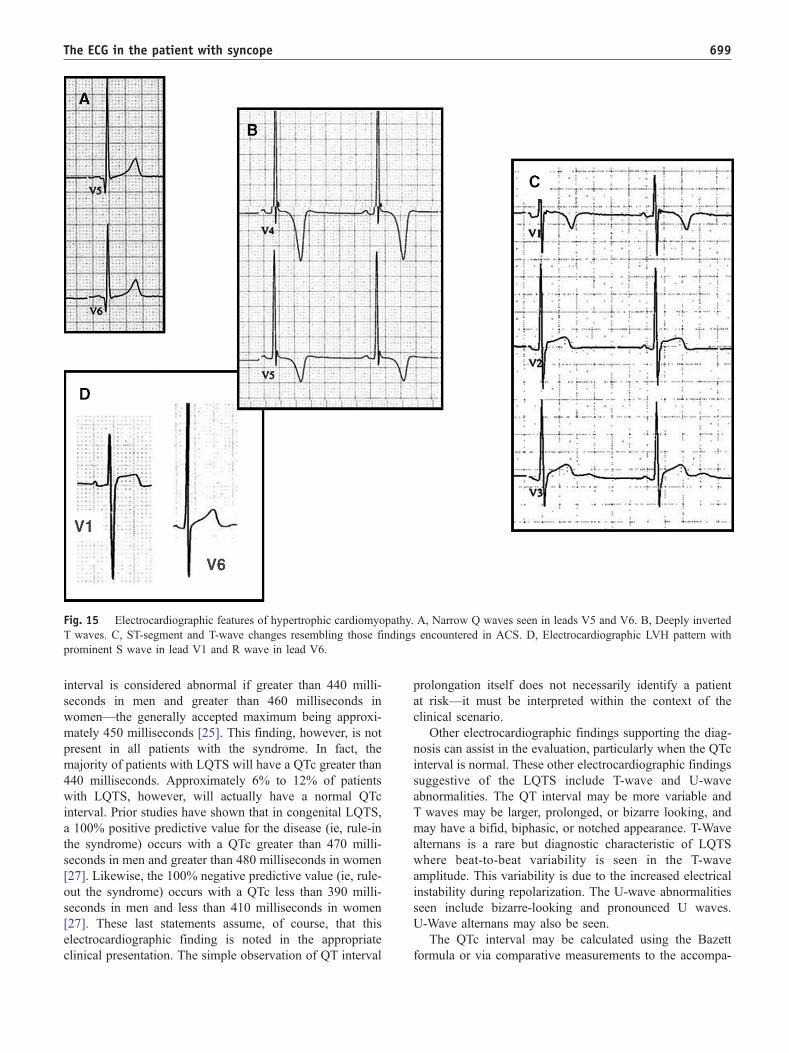
Fig. 15 Electrocardiographic features of hypertrophic cardiomyopathy. A, Narrow Q waves seen in leads V5 and V6. B, Deeply inverted
T waves. C, ST-segment and T-wave changes resembling those findings encountered in ACS. D, Electrocardiographic LVH pattern with
prominent S wave in lead V1 and R wave in lead V6.
The ECG in the patient with syncope 699
interval is considered abnormal if greater than 440 milli-
seconds in men and greater than 460 milliseconds in
women—the generally accepted maximum being approxi-
mately 450 milliseconds [25]. This finding, however, is not
present in all patients with the syndrome. In fact, the
majority of patients with LQTS will have a QTc greater than
440 milliseconds. Approximately 6% to 12% of patients
with LQTS, however, will actually have a normal QTc
interval. Prior studies have shown that in congenital LQTS,
a 100% positive predictive value for the disease (ie, rule-in
the syndrome) occurs with a QTc greater than 470 milli-
seconds in men and greater than 480 milliseconds in women
[27]. Likewise, the 100% negative predictive value (ie, rule-
out the syndrome) occurs with a QTc less than 390 milli-
seconds in men and less than 410 milliseconds in women
[27]. These last statements assume, of course, that this
electrocardiographic finding is noted in the appropriate
clinical presentation. The simple observation of QT interval
prolongation itself does not necessarily identify a patient
at risk—it must be interpreted within the context of the
clinical scenario.
Other electrocardiographic findings supporting the diag-
nosis can assist in the evaluation, particularly when the QTc
interval is normal. These other electrocardiographic findings
suggestive of the LQTS include T-wave and U-wave
abnormalities. The QT interval may be more variable and
T waves may be larger, prolonged, or bizarre looking, and
may have a bifid, biphasic, or notched appearance. T-Wave
alternans is a rare but diagnostic characteristic of LQTS
where beat-to-beat variability is seen in the T-wave
amplitude. This variability is due to the increased electrical
instability during repolarization. The U-wave abnormalities
seen include bizarre-looking and pronounced U waves.
U-Wave alternans may also be seen.
The QTc interval may be calculated using the Bazett
formula or via comparative measurements to the accompa-
J. Dovgalyuk et al.700
nying R-R interval. These techniques are depicted in
Fig. 13B and C.
3.4. Hypertrophic cardiomyopathy
Another cause of syncope with potentially lethal con-
sequences is HCM. Hypertrophic cardiomyopathy is a
heterogeneous genetic disorder that affects proteins of the
cardiac sarcomere and is seen in 1 of every 500 adults in the
general population [28]. Many patients remain asymptom-
atic throughout their lives; there is potential, however, for
dyspnea, exercise intolerance, angina, syncope, and sudden
death due to the mechanical effects of this disorder [28,29].
The classic pattern of hypertrophy occurs in an asymmetric
manner along the ventricular septum, although apical, mid-
ventricular, and concentric hypertrophy have also been
observed [29]. Symptoms occur when the hypertrophied
basal septum partially blocks the outflow tract of the left
ventricle creating a functional obstruction [28,29]. In
addition, patients with severe obstruction may experience
anterior motion of the mitral valve into the left ventricle
during systole, further reducing outflow and leading to
mitral regurgitation [28,29]. Tragically, sudden cardiac
death may be the first presenting symptom for patients with
this disease; in fact, HCM is known to be the most common
cause of sudden death in young athletes [28].
Key elements of the history in patients with HCM at risk
for sudden cardiac death may include a history of
unexplained syncope and a family history of sudden cardiac
death, although frequently patients will report neither [30].
Other factors which have been related through various
studies to an increased risk of sudden cardiac death in
patients with HCM include severe left ventricular hypertro-
phy (LVH; maximum wall thickness N30 mm), resting peak
instantaneous outflow tract gradient greater than 30 mm Hg,
abnormal blood pressure response during exercise, and the
presence of nonsustained ventricular tachycardia during
48-hour ambulatory ECG monitoring [30].
The ECG is abnormal in approximately 90% of patients
with HCM [29]. Yet, a majority of the electrocardiographic
abnormalities that are encountered in the patient with HCM
are not specific for the diagnosis. In all patients with HCM,
the most common electrocardiographic changes (Figs. 7, 14,
and 15) include increased QRS complex voltage, QRS
complex widening, Q waves, and ST-segment/T-wave
changes consistent with ventricular hypertrophy [29]. In
younger patients, these electrocardiographic findings are
more specific for the diagnosis and should certainly be
cause for concern in the appropriate setting [29]. In the
apical form of HCM, giant T-wave inversions are typical
and may be seen in other forms as well [29]. In the older
patient in whom HCM is being considered, these electro-
cardiographic findings must be distinguished from those
occurring in conditions such as chronic hypertension, ACS,
ischemic heart disease, and various conduction abnor-
malties [29].
Perhaps the most specific electrocardiographic finding
(Figs. 14 and 15), if present, in young patients with HCM is
the appearance of Q waves in leads II, III, aVF, V5, and V6
in the early teenage years. A recent study suggests that this
finding is the earliest electrocardiographic manifestation of
certain patients with HCM, preceding both wall hypertrophy
and other echocardiographic abnormalities [31]. In a large
population of patients with HCM studied, ST-segment/
T-wave changes were found in greater frequency after age
20 years; furthermore, the electrocardiographic LVH pattern
increased with age, whereas conduction disturbances were
primarily seen after age 40 years [31]. The appearance of
these abnormal Q waves in the teens studied had a reported
sensitivity of 67%, specificity of 100%, positive predictive
value of 100%, and negative predictive value of 78% for the
diagnosis of HCM [31]. Of course, these electrocardio-
graphic findings, if considered of significance in the patient,
must be noted in the appropriate clinical presentation.
References
[1] Miller TH, Kruse JE. Evaluation of syncope. Am Fam Physician
2005;72:1492-500.
[2] Quinn J, McDermott D, Stiell I, et al. Prospective validation of the
San Francisco syncope rule to predict patients with serious outcomes.
Ann Emerg Med 2006;47:448 -54.
[3] Quinn J, Stiell I, McDermott D, et al. Derivation of the San Francisco
Syncope Rule to predict patients with short-term serious outcomes.
Ann Emerg Med 2004;43:224 -32.
[4] Sarasin FP, Hanusa BH, Perneger T, et al. A risk score to predict
arrhythmias in patients with unexplained syncope. Acad Emerg Med
2003;10:1312-7.
[5] Strickberger SA, et al. AHA/ACCF Scientific statement on the
evaluation of syncope. J Am Coll Cardiol 2006;47:473-84.
[6] AHA/ACC guidelines summary ACC/AHA Cardiology Guideline
Summaries, vol 101, 2001. p. 126-7.
[7] Lee TH. Braunwald’s Heart Disease: a textbook of cardiovascular
medicine. 6th ed. Saunders.
[8] Sun BC, Edmond JA, Camargo Jr CA. Inconsistent electrocardio-
graphic testing for syncope in United States emergency departments.
Am J Cardiol 2004;93:1306-8.
[9] Martin TP, Hanusa BH, Kapoor WN. Risk stratification of patients
with syncope. Ann Emerg Med 1997;29:459-66.
[10] Wolff L, Parkinson J, White PD. Bundle-branch block with short PR
interval in healthy young people prone to paroxysmal tachycardia.
Am Heart J 1930;5:685 -704.
[11] Rosner MH, Brady Jr WJ, Kefer MP, Martin ML. Electrocardiography
in the patient with the Wolff-Parkinson-White syndrome: diagnostic
and initial therapeutic issues. Am J Emerg Med 1999;17:705-14.
[12] Kent AFS. Researches on the structure and function of the mammalian
heart. J Physiol 1893;14:233.
[13] Zipes D. Braunwald’s heart disease: a textbook of cardiovascular
medicine. 7th ed Saunders.
[14] Brugada P, Brugada J. Right bundle branch block persistent ST
segment elevation and sudden death: a distinct clinical and electro-
cardiographic syndrome. A multicenter report. J Am Coll Cardiol
1992;20:1391-6.
[15] Brugada P, Brugada R, Brugada J. The Brugada syndrome. Curr
Cardiol Reports 2000;2:507 -14.
[16] Monroe MH, Littmann L. Two-year case collection of the Brugada
syndrome electrocardiogram pattern at a large teaching hospital. Clin
Cardiol 2000;23:849 -51.
The ECG in the patient with syncope 701
[17] Hermida J, Lemoine J, Aoun FB, et al. Prevalence of the Brugada
syndrome in an apparently healthy population. Am J Cardiol 2000;
86:91-4.
[18] Priori SG, Napolitano C, Giordana U, et al. Brugada syndrome and
sudden cardiac death in children. Lancet 2000;355:808 -9.
[19] Alings M, Wilde A. bBrugadaQ syndrome—clinical data and
suggested pathophysiological mechanism. Circulation 1999;99:
666 -73.
[20] Mattu A, Rogers RL, Kim H, Perron AD, Brady WJ. The Brugada
syndrome. Am J Emerg Med 2003;21:146-51.
[21] Littman L, Monroe MH, Kerns II WP, Svenson RH, Gallagher JJ.
Brugada syndrome and bBrugada signQ: clinical spectrum with a guide
for the clinician. Am Heart J 2003;145(5):768 -78.
[22] Ott P, Marcus FI. Electrocardiographic markers of sudden death.
Cardiol Clin 2006;24:453 -69.
[23] Poelzing S, Forleo C, Samodell M, et al. SCN5A polymorphism
restores trafficking of a Brugada syndrome mutation on a separate
gene. Circulation 2006;114:360-2.
[24] Brugada J, Brugada P, Brugada R. The syndrome of right bundle
branch block ST segment elevation in V1 to V3 and sudden
death—the Brugada syndrome. Cardiovasc Drugs Ther 2002;16:
25 -7.
[25] Meyer JS, Mehdirad A, Salem BI, et al. Sudden arrhythmia death
syndrome: importance of the long QT syndrome. Am Fam Physician
2003;68:483 -8.
[26] Khan IA. Clinical and therapeutic aspects of congenital and acquired
long QT syndrome. Am J Med 2002;112:58 -66.
[27] Vincent GM, Timothy KW, Leppert M, Keating M. The spectrum of
symptoms and QT intervals in carriers of the gene for the long-QT
syndrome. N Engl J Med 1992;327:846-52.
[28] Nishimura RA, Holmes Jr DR. Hypertrophic obstructive cardiomy-
opathy. N Engl J Med 2004;350:1320 -7.
[29] Popjes ED, Sutton MSJ. Hypertrophic cardiomyopathy: pathophysiol-
ogy, diagnosis, and treatment (The heart). Geriatrics 2003;58(3):41-50.
[30] Frenneaux MP. Assessing the risk of sudden cardiac death in a patient
with hypertrophic cardiomyopathy. Heart 2004;90:570-5.
[31] Shimizu M, Ino H, Yamaguchi M, et al. Chronologic electrocardio-
graphic changes in patients with hypertrophic cardiomyopathy
associated with cardiac troponin I mutation. Am Heart J 2002;
143(2):289-93.