Embed Size (px)
Citation preview

University of ConnecticutOpenCommons@UConn
Master's Theses University of Connecticut Graduate School
6-30-2014
The Effects of Various Frequencies of Low-LevelMechanical Vibration on Retention in anOrthodontic Relapse Model in MiceAmir H. AssefniaMasters of Dental Science, [email protected]
This work is brought to you for free and open access by the University of Connecticut Graduate School at OpenCommons@UConn. It has beenaccepted for inclusion in Master's Theses by an authorized administrator of OpenCommons@UConn. For more information, please [email protected].
Recommended CitationAssefnia, Amir H., "The Effects of Various Frequencies of Low-Level Mechanical Vibration on Retention in an Orthodontic RelapseModel in Mice" (2014). Master's Theses. 625.https://opencommons.uconn.edu/gs_theses/625
i
The Effects of Various Frequencies of Low-Level Mechanical Vibration
on Retention in an Orthodontic Relapse Model in Mice
Amir H. Assefnia, D.D.S., M.S.
D.D.S., University of California, Los Angeles School of Dental Medicine, 2011
M.S., University of California, Los Angeles School of Dental Medicine, 2011
A Thesis
Submitted in Partial Fulfillment of the
Requirements for the Degree of
Master of Dental Science
At the
University of Connecticut
2014

ii
APPROVAL PAGE
Masters of Dental Science Thesis
The Effects of Various Frequencies of Low-Level Mechanical Vibration
on Retention in an Orthodontic Relapse Model in Mice
Presented by
Amir H. Assefnia, D.D.S., M.S.
Major Advisor_____________________________________________________
Ravindra Nanda, B.D.S., M.D.S., Ph.D.
Associate Advisor__________________________________________________
Flavio Uribe, D.D.S., M.D.S.
Associate Advisor_____________________________________________________
Sumit Yadav, B.D.S., M.D.S., Ph.D.
Associate Advisor__________________________________________________
Ivo Kalajzic, M.D., Ph.D.
University of Connecticut
2014

iii
TABLE OF CONTENTS Page
TITLE PAGE i
APPROVAL PAGE ii
TABLE OF CONTENTS iii
ABSTRACT v
BACKGROUND 1
Introduction 1
Nature of Relapse 2
Review of Retention Studies 4
Vibration Research 8
OTM Models and Retention Model 11
RATIONALE 16
HYPOTHESES 17
SPECIFIC AIMS 18
MATERIALS AND METHODS 18
Study Design 18
Method for Orthodontic Force Application 19
Application of Mechanical Vibration 20
Wellness Monitoring and Euthanasia 21
Micro-CT Analysis and Tooth Movement Measurements 21
STATISTICAL ANALYSIS 22
RESULTS 23
DISCUSSION 24
CONCLUSIONS 29

iv
TABLES AND FIGURES 31
REFERENCES 50
v
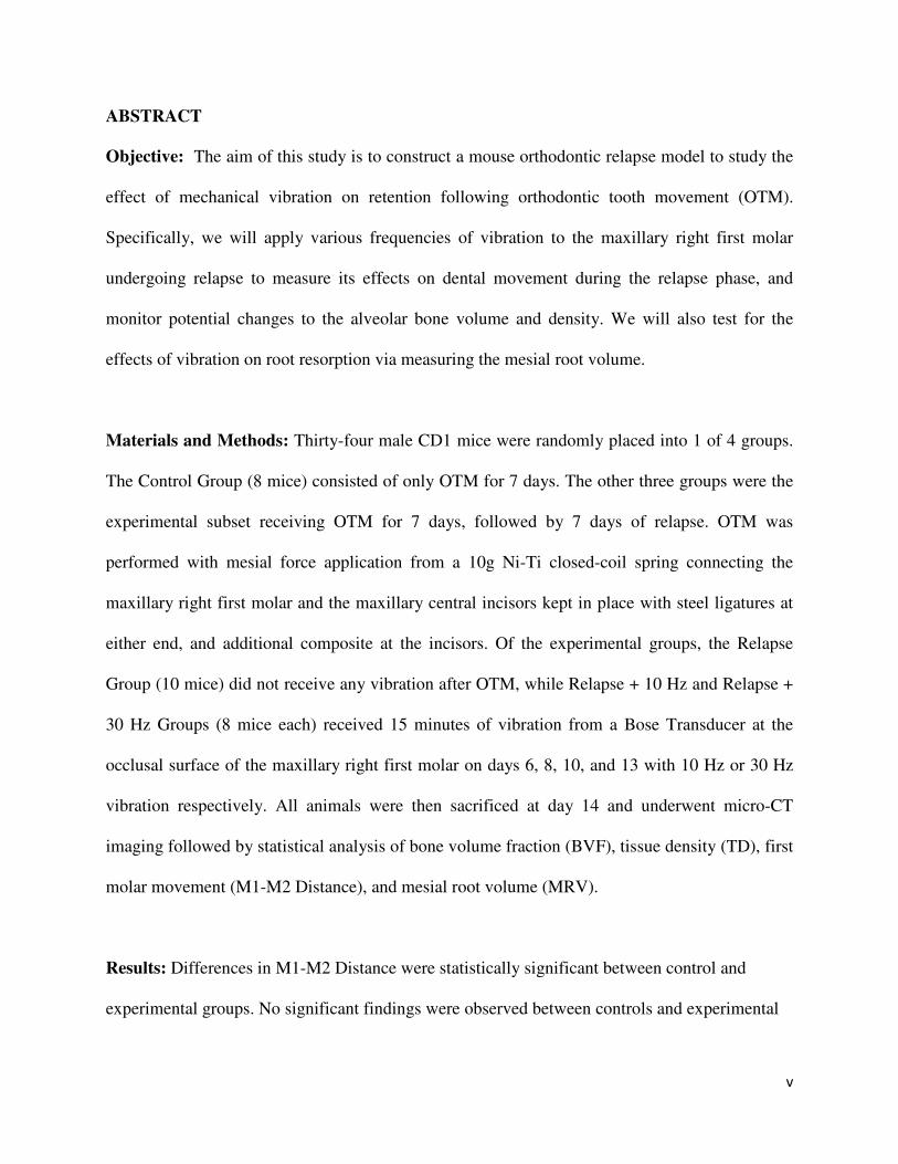
ABSTRACT
Objective: The aim of this study is to construct a mouse orthodontic relapse model to study the
effect of mechanical vibration on retention following orthodontic tooth movement (OTM).
Specifically, we will apply various frequencies of vibration to the maxillary right first molar
undergoing relapse to measure its effects on dental movement during the relapse phase, and
monitor potential changes to the alveolar bone volume and density. We will also test for the
effects of vibration on root resorption via measuring the mesial root volume.
Materials and Methods: Thirty-four male CD1 mice were randomly placed into 1 of 4 groups.
The Control Group (8 mice) consisted of only OTM for 7 days. The other three groups were the
experimental subset receiving OTM for 7 days, followed by 7 days of relapse. OTM was
performed with mesial force application from a 10g Ni-Ti closed-coil spring connecting the
maxillary right first molar and the maxillary central incisors kept in place with steel ligatures at
either end, and additional composite at the incisors. Of the experimental groups, the Relapse
Group (10 mice) did not receive any vibration after OTM, while Relapse + 10 Hz and Relapse +
30 Hz Groups (8 mice each) received 15 minutes of vibration from a Bose Transducer at the
occlusal surface of the maxillary right first molar on days 6, 8, 10, and 13 with 10 Hz or 30 Hz
vibration respectively. All animals were then sacrificed at day 14 and underwent micro-CT
imaging followed by statistical analysis of bone volume fraction (BVF), tissue density (TD), first
molar movement (M1-M2 Distance), and mesial root volume (MRV).
Results: Differences in M1-M2 Distance were statistically significant between control and
experimental groups. No significant findings were observed between controls and experimental
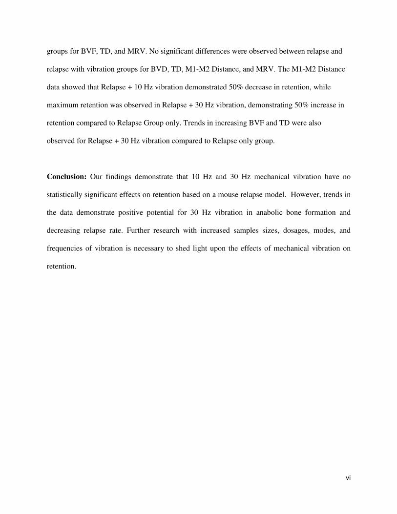
vi
groups for BVF, TD, and MRV. No significant differences were observed between relapse and
relapse with vibration groups for BVD, TD, M1-M2 Distance, and MRV. The M1-M2 Distance
data showed that Relapse + 10 Hz vibration demonstrated 50% decrease in retention, while
maximum retention was observed in Relapse + 30 Hz vibration, demonstrating 50% increase in
retention compared to Relapse Group only. Trends in increasing BVF and TD were also
observed for Relapse + 30 Hz vibration compared to Relapse only group.
Conclusion: Our findings demonstrate that 10 Hz and 30 Hz mechanical vibration have no
statistically significant effects on retention based on a mouse relapse model. However, trends in
the data demonstrate positive potential for 30 Hz vibration in anabolic bone formation and
decreasing relapse rate. Further research with increased samples sizes, dosages, modes, and
frequencies of vibration is necessary to shed light upon the effects of mechanical vibration on
retention.
1
BACKGROUND
Introduction
Retention is a phase of orthodontic treatment that serves to maintain teeth in their final positions
after active tooth movement [1]. Usually, teeth have a tendency to relapse back to their original
positions following active movement [2]. Some of the different types of relapse observed
following fixed orthodontic therapy include decreases in arch length and intercanine width, and
increase in mandibular crowding [3, 4]. Interestingly, Little et al. performed analysis of
longitudinal changes in anterior alignment of the mandibular dentition for 10-20 years,
demonstrating that the maximum relapse occurred during the first 10 years post-retention, and
continued into 20 years, with only 10% of treatments remaining clinically satisfactory [3].
Relapse has been thought to be multifactorial in nature, often involving the origin of
malocclusion, bone turnover, periodontal factors, soft tissue forces, growth, and function [5, 6].
Due to the diverse elements involved, relapse remains incompletely understood, giving rise to
wide variations in retention protocols amongst clinicians [6].
Retention has been a controversial topic since the early stages of the specialty; Angle in 1907
stated that “the problem involved in retention is so great…greater than the difficulties being
encountered in the treatment [7].” Calvin Case later added in 1920: “the very cases which create
in us the greatest pride, are going back to their former malpositions and disharmonies, in spite of
everything we have been able to do with retaining appliances [8].” Later, McCauley debated that
the transverse width of the canines and molars determine stability in 1944, while Tweed reported
the inclination of the incisors was the important factor in retention [9, 10]. In a landmark article
in 1988, Little et al., mentioned above, declared that “the only way to ensure continued
satisfactory alignment post-treatment probably is by use of fixed or removable retention for life.”
Later in 1999, Little further retrospectively reviewed a collection of over 800 patients from

2
University of Washington for relapse, and concluded that (1) Arch length decreases after
orthodontic treatment. (2) Arch width measured across the mandibular canine teeth typically
reduces post-treatment, whether or not the case was expanded during treatment. (3) Mandibular
anterior crowding during the post-treatment phase is a continuing phenomenon well into the 20-
to-40 years age bracket and likely beyond. (4) Third molar absence or presence, impacted or
fully erupted, seems to have little effect on the occurrence or degree of relapse. (5) The degree of
post-retention anterior crowding is both unpredictable and variable and no pretreatment variables
either from clinical findings, casts, or cephalometric radiographs seem to be useful predictors
either before or after treatment [11]. Finally, Littlewood et al. concluded in their systematic
review in 2006 that “There is currently insufficient evidence on which to base the clinical
practice of orthodontic retention.” [5]
Nature of Relapse
There are multiple theories regarding the causes of relapse. Proffit mentions according to the
Equilibrium theory that muscle and soft tissue pressures on the occlusion may be driving causes
of relapse [12]. Another school of thought concentrates on the periodontal ligament (PDL),
suggesting that forces exerted by stretched periodontal and gingival connective tissue fibers may
place tensile forces on moved teeth or that collagen fibers of the PDL are leading causes of
relapse [13, 14]. The role of the supracrestal fibers of the PDL was later questioned and Edwards
demonstrated that circumferential supracrestal fibrotomy (CSF) may also aid in retention [15].
On a separate note, Tweed explained that changes in tooth inclination, such as proclination of the
incisors, are important for stability in 1944 [10]. Gianelly later argued that intercanine distance,
especially in the mandible is important in preventing relapse [16]. The amount of continuous

3
bone turnover has also been associated with relapse, as well as continuous growth, especially in
the vertical dimension [17, 18]. Other theories include continuous, interproximal force,
originating in the periodontium and acting on adjacent teeth at their contact points, as proposed
by Southard et al. [19]. In a review of literature performed in 1998, six major criteria for the
stability of finished orthodontic cases were identified: 1) pretreatment lower arch form should be
maintained, 2) Original lower intercanine width should be maintained; expansion of this is the
most predictable of all relapses, 3) mandibular arch length decreases with time, 4) the most
stable position of the lower incisors is their pretreatment position, 5) Fiberotomy is an effective
means of reducing rotational relapse, and 6) Lower incisor reproximation can improve long-term
post-treatment stability [20].
The application of orthodontic force for tooth movement causes compression sides and tension
sides of the affected teeth. This strain in turn induces an inflammatory response in the
surrounding tissues, producing a cellular response of macrophages, osteoclasts, osteoblasts, and
fibroblasts to the periodontium and bone to induce remodeling [21, 22]. The compression side of
tooth movement involves osteoclastic activity resulting in bone resorption while osteoblastic
activity occurs at the opposite tension side where new woven bone is deposited [21]. After
removal of fixed orthodontic treatment, the new woven bone is remodeled and replaced with
mature lamellar bone. Lamellar bone has characteristics of being more organized and having
higher mineral content and strength than woven bone thus making it less susceptible to
resorption [23]. Remodeling may initiate relapse of teeth due to a temporary void and absence of
lamellar bone adjacent to the PDL. This in conjunction with rebound forces of the PDL can
promote relapse [24, 25]. Furthermore, other tissues remodel around the teeth; the PDL
reorganizes over 3-4 months, gingival collagen fiber networks remodel in 4-6 months, and
4
supracrestal fibers exceed even 7 months of remodeling [26]. A retention period of over 12
months allows ample time for remodeling of each type of tissue described above [27].
Review of Retention Studies
Orthodontists utilize various appliances to maintain dentition in their final positions following
OTM. The most common appliances are Hawley retainers, Vacuum-formed retainers, and
permanent fixed retainer. Pratt et al. performed a survey of the American Association of
Orthodontics members in 2011 which revealed that in the maxillary arch, the Hawley retainer is
used most by 47% of their sample, followed by the vaccum-formed retainer 41%, and permanent
fixed retainer at 11%. In the mandibular arch, these devices were utilized 29%, 29% and 42%
respectively [28]. The Hawley retainer consists of an acrylic base and labial bow of stainless
steel wire which can be adjusted according to provider preference [29]. Its advantage is that it
allows settling of the posterior teeth, if no occlusal impediments are provided such as an Adam’s
clasp and teeth are allowed to erupt freely. A disadvantage of the Hawley retainer is its
extensive palatal coverage [30]. The vacuum-formed retainer, commonly known as the Essix
retainer, is a removable retainer that is constructed out of various thicknesses of plastic and
covers all surfaces of the teeth. Its advantage is that it is nearly invisible and has no palatal
coverage. Its disadvantage is that it does not allow for occlusal settling and that it is less durable
[30]. Despite the mechanical design of these removable appliances, the limitation to their
function is patient compliance, although the Hawley has been reported to be more successful in
compliance with patients over two years in retention [30, 31]. Due to majority of orthodontic
patients falling within the adolescent and teenage years, variable rates of compliance are
achieved and can be very discouraging for clinicians [32]. Also, patient compliance with
retainers decreases over time, with fewer than half of the patients wearing them as instructed

5
following completion of active treatment [33]. Interestingly, more than 50% of patients admitted
that they did not wear retainers as instructed, with the most common reasons being discomfort
and forgetfulness [34].
Fixed retainers have been utilized to manage compliance problems in patients. Fixed retainers
are often placed lingual to the mandibular anterior dentition, and are often bonded from canine to
canine to prevent relapse of the mandibular incisors [30, 35]. Although compliance problems are
eliminated, the technique has some disadvantages [28]. The wire has to be passive, and oral
hygiene is a concern [30]. Pandis et al. have shown higher calculus accumulation, greater
marginal recession and increased probing depths in a group of patients with mandibular lingual
fixed retention. This study emphasized careful selection of retention protocols after a thorough
consideration of anatomic, hygiene, social, and cultural factors followed by close monitoring of
patients [36].
Less popular retention strategies include surgical-based interventions such as frenectomies or
circumferential supracrestal fibrotomy (CSF). For example, patients with a large maxillary
anterior diastema and labial frenum attachment to the alveolar ridge penetrating from buccal to
lingual benefit from a frenectomy to relieve the fibers inhibiting stability of diastema closure
[27]. Often frenectomies are recommended to be performed after closure of the diastema to
prevent scar tissue from hindering closure [23]. CSF targets the supracrestal fibers that contribute
to the tensile forces that cause relapse and take a considerable amount of time to remodel [27].
This procedure consists of the surgical transsection of supracrestal free gingival fibers
surrounding the tooth, and has been shown to decrease the relapse of teeth [13]. Fiberotomy has
been shown to be effective in retention, especially in preventing pure rotational relapse rather

6
than reduction of labiolingual relapse over the long term, and its results are better in the
maxillary anterior region than in the mandibular anterior region [15].
Other views regarding relapse include theories that compressive interproximal force, originating
continuously from the periodontium and acting on adjacent teeth at their contact points may be
responsible for some long term arch constriction and decreased stability [19]. Furthermore,
Southard et al. found significant correlation between mandibular anterior alignment and
interproximal force. This is potentially due to the narrower contacts of the lower incisors, and
broadening them can resist contact slippage and increase stability [30]. Another suggested mode
of treatment to relieve this force was prophylactic extraction of third molars, which has currently
been rejected as a mode of treatment [37, 38]. In addition, based on the type of relapse,
overcorrection may help in retention. Anterior-posterior overcorrection of the occlusal
relationship is recommended in Class II patients, as is overextrusion of anterior teeth in open bite
cases, and overintrusion in deep bite cases [23, 39].
Kim et al. investigated the effectiveness of pharmacological agents in preventing orthodontic
relapse in rats, showing that systemic administration of the bisphosphonate, pamidronate
significantly inhibits initial relapse of mesialized molars by inhibiting osteoclastic activity. They
attempted to create a relapse model by placing elastic bands between the first and second molar
of rats, and applied injections of bisphosphonates at the site after band removal to observe this
trend [40]. Hassan et al. created another relapse model in sheep in 2010 where they extracted
central incisors and tipped lateral incisors and used Bone Morphogenetic Proteins (BMPs) to
evaluate the post-orthodontic stability. They discovered less relapse when injecting BMPs into
the PDL of tipped incisors as compared to the controls. Active bone remodeling and
hypercementosis were also observed [41]. Statins have also been shown to stimulate osteoblastic
7
activity which would theoretically strengthen the bony housing of teeth and prevent their relapse.
In 2010, Han et al., explored the effects of simvastatin on a rat relapse model, where springs
were placed at the molars and incisors bilaterally to induce movement of the molar, and the drug
was injected systemically. They observed increased retention potential and increased
osteoprotegerin (OPG) levels [42]. OPG is an osteoclast inhibitor that binds to receptor activator
of nuclear factor κβ Ligand (RANKL) and functions as a competitive inhibitor of the RANK
receptor necessary for osteoclast activation, thus inhibiting osteoclastogenesis and bone
resportion [25]. Hudson et al. utilized the same rat relapse model as Han et al., but administered
OPG adjacent to the molars, observing over close to 50% decrease in relapse in low dosage OPG
groups and over 50% decrease in relapse in high dosage OPG groups compared to controls [25].
Limitations to pharmacological agents include local delivery and prevention of systemic side-
effects, as well as pain and discomfort. Zhao et al. addressed controlling local delivery of drugs
in 2010, utilizing a rat model with a spring force between the right maxillary first molar and
incisors. They utilized local OPG gene transfer with inactivated hemagglutinating virus and
OPG expression plasmid to periodontal tissues at the molar. The OPG was injected into the
palatal mucosa on the distal surface of moved tooth and the percentage of relapse in the
experimental groups was significantly less than in the control group (35.7±8.9% versus
96.3±7.0%). They also tested for systemic effects via monitoring inflammation at the tibia, and
saw no changes. Kanzaki et al. performed a similar OPG gene transfer in a relapse model in rats,
where a compressive 17 gram spring was placed between first molars palatally to tip them, and
injection at the site was made to test retention. This group reported almost half the amount of
relapse observed compared to controls [43]. While this method delivers promising results, gene
transfer may cause severe immunologic reactions to the inactivated virus as well as accidental

8
activation of oncogenes, forming neoplasms, or accidental diffusion from palatal to buccal
surfaces during injections [43, 44].
Pharmacological methods of tooth stability, though promising in theory, have several limitations.
The majority of methods under research are in animals models, have systemic effects, require
questionable methods of delivery, and lack of long term data. Currently, mechanical retention
(i.e. retainers) is the suggested and most popular mode of retention clinically. Development of an
adjunct to retainers would minimize the time needed to wear retainers and increase the overall
stability of OTM. Studies relating retention with vibration are limited. Vibration would serve as
a potential minimally invasive adjunct to mechanical retention to minimize orthodontic relapse.
Vibration Research
Studies of whole-body vibration have been performed in both animal and humans models.
Christiansen and Silva studied the effect of this type of vibratory stimuli on 40 adult mice using a
frequency of 45 Hz with varying magnitudes of force for 15 minutes per day in a 5 week
interval. They found an increase in trabecular bone volume in the experimental vibration group,
independent of dosage [45]. Rubin et al. performed a 1-year prospective, randomized, double-
blinded, placebo-controlled clinical trial on seventy post-menopausal women. In these subjects
they administered whole-body vibration at a frequency of 30 Hz with 0.2 grams of magnitude for
twenty minutes per day. They found an inhibition of bone loss in both the spine and the femur
with pronounced findings associated with lower body mass [46]. These studies promoted that
low-magnitude, high frequency vibration for relatively short durations has an anabolic potential
for bone, with findings mostly demonstrating increased numbers and sizes of trabeculae, with
improved stiffness and strength of cancellous bone [46]. Since the molecular mechanisms

9
involving bone turnover, specifically modeling and remodeling, are similar to those required for
OTM, applying vibratory stimuli might have an effect on the rate of tooth movement.
Other studies have looked at applying a pulsed electromagnetic field (PEMF) in order to create a
vibratory stimulus. Stark and Sinclair looked at applying PEMF in 40 male Hartley guinea pigs,
where they measured the effects of 25 Hz PEMF with 12 grams of orthodontic force for ten days.
They observed an overall increase in the rate and amount of tooth movement along with greater
bone matrix deposition and numbers of osteoclasts [47]. Darendeliler, Sinclair and Kusy in 1995
also studied the effects of PEMF along with a samarium-cobolt magnet, applying 15 Hz
vibration with 15 grams of orthodontic force for 10 days. They concluded that the amount of
OTM in the magnet and PEMF groups was greater than the group with orthodontic force alone
[48]. They proposed that the change in the rate of OTM was due to a reduction of the initial lag
phase which follows force application [48]. Darendeliler et al.then further investigated the
effects of PEMF and neodymium-iron-boron magnets in 45 Wistar rats. They applied 25 grams
of orthodontic force with a frequency of 30 Hz and demonstrated significantly greater OTM in
the group exposed to PEMF vibration [49].
Other types of vibration studied also include resonance vibration (with continuously changing
frequency) and ultrasonic vibration. Nishimura et al.tested the effect of resonance vibration on
OTM in 42 Wistar rats divided in two groups over 21 days. A 0.012 nickel-titanium (Ni-Ti)
expansion spring with 12.8 grams of orthodontic force was applied with and without weekly 8
minute resonance vibration (60 ± 8 Hz) session to the occlusal surface of 1st molars. They
concluded that there was 15% greater OTM rate with combined resonance vibration and force,
and histologically noted greater receptor activator of nuclear factor kappa-B ligand (RANKL)
expression by osteoclasts and fibroblasts on day 3, with increased numbers of osteoclasts present

10
(1.7x control) on day 8 [50]. Similarly, Ohmae et al. researched ultrasonic vibration in a split-
mouth model on 5 adult male beagle dogs with bilaterally extracted maxillary first premolars.
An eighty gram force using a sectional archwire between the canine and first premolar was
applied to close the extraction space, with one side exposed to homo-directional ultrasonic
vibration (2 minute interval, two times per week for a total of 8 – 10 weeks) while the other side
served as a control. They also identified a greater amount of tooth movement in the teeth
exposed to vibration [51].
Throughout the last few years, the AcceleDentTM
company has produced a device that can be
used in humans in order to apply a vibratory force of 30 Hz to the dentition with two
corresponding studies. The first study was a non-controlled experiment in 14 subjects for 20
minutes of appliance use per day over a total of 6 months. While no controls were used, they
postulated that the observed 3mm per month of tooth movement in the maxilla and the 2.1mm
per month in the mandible were greater results compared to current clinically accepted norms
(approximately 1mm tooth movement per month) [52]. Following these findings, they
conducted a prospective, randomized, blinded, sham-controlled clinical trial on 45 human
subjects at the University of Texas at San Antonio, with promising results pending publication.
They found significantly greater tooth movement during the aligning phase (106%) and
significantly greater tooth movement during space closure (38%). On the contrary, data
regarding the effect of mechanical vibration on OTM in an animal model at the University of
Connecticut Health Center have demonstrated different results [53]. Twenty six female Sprague
Dawley rats were divided into four groups: Control un-loaded, Vibration, OTM of 25 grams
mesial load, and OTM with vibration with 0.4 N and 30 Hz twice per week for 10 minutes.
Rather than increases in OTM, cyclical forces inhibited the amount of OTM with histological
11
analysis showing disorganization of collagen fibril structure of the PDL, and increased osteoclast
parameters with significant decrease in bone volume fraction in the molar region.
OTM Models and Retention Model
Several animal models have been designed to study tissue responses to mechanical loading
during orthodontic tooth movement. Primate, dog and cat models have been reported in initial
histological studies using light microscopy [54, 55] and electron microscopy [54, 56]. The rat
model proposed by Waldo in 1954 [57] had increased levels of experimental control over other
animal models and has become the investigative approach for researching the processes of
mechanotransduction and alveolar bone remodeling in OTM [58]. Currently, rats are most
commonly used, accounting for over half of all orthodontic tooth movement animal studies [58].
Compared with most other animals, the use of the rat model has several advantages: it is
relatively inexpensive, which allows using large sample sizes, longer housing periods allow for
longer duration of experiments, histological preparation of the rat is easier than other animal
models, there is greater availability of antibodies required for cellular and molecular biological
techniques, and their sizes are larger than mice, allowing for easier placement of orthodontic
appliances. Yet, the rat model has some limitations: denser alveolar bone as compared to
humans, lack of osteons and less abundant osteoid tissue, structural dissimilarities in the
arrangement of PDL fibers and the supporting structures, and faster tissue development during
root formation and changes incident to orthodontic treatment than in humans, while maintaining
relatively the same principle mechanisms [58].
Rat models provide for a diverse scope of orthodontic research ranging from measuring
proliferation rates of PDL cells under load to assessing the effects of prostaglandins,

12
bisphosphonates and leukotrienes on tooth movement [59-61]. Ren et al.’s systematic review of
the 153 (57% of the total tooth movement models) studies performed on rats in the past twenty
years determined that the majority of the experimental models designed poor force systems that
lacked controls throughout the duration of tooth movement [58]. Only three methods met Ren’s
inclusion criteria for a good model [58]: a force magnitude of less than 20cN; mesial movement
of molars; an experimental duration greater than 2 weeks without other experimental conditions,
such as drug intervention. Most of the studies did not consider physiology of the rat (i.e. natural
distal drift of the molars and continual eruption of the incisors), nor faulty appliance design. The
distal drift of the molars underestimates the amount of mesial movement of the molars with
continual eruption of the incisors leading to minimized control of force direction. The appliance
design is poor when the 50 fold decrease in rat molar root surface area is not considered
compared to humans, or if there is a lack of constant and continual force [58].
Pavlin et al. (2000) first developed a mouse model for testing the load conditions necessary to
generate an optimal biological response of paradental tissues [62, 63]. They used an elastomeric
“o-ring” tied between maxillary incisors and the first molar, and a red elgiloy (alloy of nickel and
cobalt) open coil spring (0.0056 x 0.022 inches, Rocky Mountain Orthodontics, Denver, CO) tied
and bonded to the same teeth, respectively. It was found that the coil spring has considerable
advantages over the “o-ring.” Firstly, bonding of a coil spring to the molar and the incisors
eliminates contact of the appliance with gingival tissues, greatly reducing the risk of tissue
irritation [62, 63]. This correlated with the criticisms of Charles Waldo, whom in 1954, was
among the first pioneers responsible for the advent of the rat model. The Waldo method utilized
an orthodontic intermaxillary elastic, which was stretched and inserted into the interproximal
space just cervical to the contact between the molars of rats [57]. This method has been

13
criticized due to the unknown force decay of the elastic. Springs have proven to be more reliable
due to delivery of a reproducible force of 10±2 cN over a range of 3-15 mm of activation [58].
Secondly, the spring has a lower force/deflection rate (F/∆). This allows for a more precise and
reproducible application of a low level force, which also remains more constant compared with
that delivered by an elastomeric “o-ring.”
In 1990’s, King [64], Keeling [65], and Nixon [66] met al.l of Ren’s criteria for an ideal rat
model [58]. Forces of 20, 40, and 60 cN were used in all 3 articles. These studies were
criticized for having an initial constant force without proper reactivation, as well as forces of 40
and 60 cN as too heavy. The appliance consisted of a 9 mm length of closed coil spring (0.006
inch NiTi; arbor diameter: 0.022 inch, Unitek, Monrovia, Calif.) suspended between a cleat
bonded to the occlusal surface of the maxillary first molars and the lateral surface of the
maxillary incisors. Initial force values were measured by suspending known weights from the
anterior end of these coils prior to fixation to the incisors. Tooth movement was based on
enlarged cephalograms, and was measured from the position of a reproducible landmark on the
molar cleat with respect to either zygomatic amalgam implants, or a barbed broach placed
submucosally on the palate. Palatally placed barbed broaches represented a more reliable, less
traumatic, and more easily executed superpositional landmark than zygomatic amalgams. They
only had a 79% appliance success rate, the animals lost weight, and they extracted mandibular
first and second molars. All of these factors contributed to poor overall animal care [58, 64-66].
Finally in 2004, Ren’s model was fabricated due to the shortcomings of previous rat models, and
used a spilt-mouth design. This design compensated for the physiological distal drift of the
molars, growth of the snout and forward movement of the incisors, and the continuous eruption
with possible distal tipping of the incisors. Stainless steel ligature wires with a diameter of 0.2

14
mm were bent to enclose all three maxillary molars as one unit. To this ligature wire a
Sentalloy® closed coil spring (Ni Ti, 10 cN, wire diameter 0.22 mm, eyelet diameter 0.56 mm,
GAC, New York, USA) was attached to deliver a reproducible force of 10 ± 2 cN over a range of
3-15 mm activation. A transverse hole was drilled through the alveolar bone and both maxillary
incisors at the mid-root level using a drilling bur (D0205, Dentsply). A stainless steel ligature
wire (diameter 0.3 mm, Dentaurum) was inserted through the hole and bonding was applied until
the buccal and palatal wires were fully embedded in the bonding material prior to light curing.
The coil was activated and attached to the ligature wire through the snout and the incisors [58].
Recently in 2006, Yoshimatsu et al. used a variation of the Ren model with Ni-Ti closed coil
springs [67] in order to further develop the mouse model for OTM. Their mouse model included
a Ni-Ti closed coil spring with wire diameter of 0.15mm and coil diameter 0.9mm. The
appliance was inserted between the maxillary incisor and the first molar on the left side. It was
fixed with a 0.1mm wire around each tooth using a dental adhesive agent (Superbond;
Sunmedical Shiga, Japan). To prevent detachment of the maxillary incisors during the
experiment, a shallow groove, 0.5mm from the gingiva, was made on the maxillary incisor every
4 days, and the wire was reattached at the new groove with 10 grams of force after activation.
The maxillary left molar was used as the experimental side, and the right as the control, taking
into account the distal molar drift that would naturally occur [67]. Our experimental models will
utilize the above mentioned advances in OTM in mice to construct a reproducible model for
OTM.
We decided to perform our experiments in mice due to several reasons. Mice are commonly
used for studies of skeletal biology due to their similarity to humans when investigating genetic
or molecular factors, and the National Human Genome Research Institute has confirmed that

15
overall, mice and humans share almost every gene in a closely related form. Of the
approximately 4,000 genes that have been studied, less than 10 are found in one species but not
in the other (5). Also, murine strains allow for proteomics studies, which help elucidate
functions of different cells, signaling pathways, secondary mediators, and transcription factors at
a molecular level, for example, in retention and OTM (94). Furthermore, mice reproduce
quickly, and are cheaper to house, grow, and maintain. Finally, utilizing a certain strain of cloned
mice (CD-1 for example), we have a genetically and phenotypically homogenous sample.
As for an orthodontic retention model, several models exist that were mentioned in the retention
studies section above [14, 25, 40-44]. The Waldo method has been utilized where a rubber band
elastic is placed between the first and second molar of rats, and after a set period of time, is
removed and relapse is noted [14, 40]. Sheep maxillary central incisors have also been extracted
and lateral incisors were tipped mesially, followed by a period monitoring their relapse [41]. Rat
molars have also been tipped palatally using a palatal spring appliance, and then studied for
relapse [43]. Mesial movement of rat molars with a spring has been studied unilaterally and
bilaterally as well [25, 42, 44]. For example, Hudson et al. utilized a Sprague-Dawley rats
relapse model divided into two phases: initial tooth movement (days 1–28), during which springs
were placed between the incisors and maxillary first molars bilaterally, and the tooth relapse
phase (days 28–52), during which injections of OPG or PBS were administered throughout the
molar relapsing phase. Zhao et al. performed a gene transfer study where they separated the right
palatal rat first molar mesially and unilaterally with a spring, and performed OPG gene transfer
to measure relapse results [44]. Our model is a modified version of the Zhao et al. model, where
we attach our spring at the incisors and the right maxillary first molar of mice unilaterally, and
provide vibration at different frequencies with the same dosage interval during the relapse phase
16
in a shorter experimental timespan. Our experimental model is the first relapse model utilizing
mice for retention in conjunction with vibration.
RATIONALE
One of major limitations to orthodontic treatment is relapse after active OTM. Relapse occurs
often, and retention strategies require patient compliance for effectiveness. Introduction of a
simple protocol for prevention of relapse would have large implications in clinical orthodontics,
and help to maintain the final outcomes of orthodontic treatment. Due to the multifactorial nature
of relapse, research to decrease relapse rate is difficult and scarce. Currently, orthodontic animal
retention models have been primarily used for pharmacological studies on relapse [14, 25, 40-
44]. While pharmacological studies present promising results on animal models, their drawbacks
include systemic effects and uncertainties in drug delivery vectors that limit their application
clinically on humans. Therefore, less invasive procedures should be developed to limit
orthodontic relapse and the need for long-term wear of retainers. Cyclical loading, especially at
a frequency of 30 Hz, has been studied in the past several decades in bone turnover, and has
demonstrated anabolic bone formation with confirmed results. Recently, new vibration devices,
including a popular unit that uses 30 Hz frequency vibration, have been released for use
clinically in orthodontics, and advertise faster tooth movement. Nevertheless, research has
demonstrated conflicting data regarding the effects of such appliances, with decreases in the rate
of OTM observed in some studies. Since decreasing OTM would promote retention of teeth, and
previous studies have confirmed anabolic activity for vibration, we decided to examine changes
in orthodontic relapse with different dosages of vibration. We will be the first to launch a relapse
study utilizing vibration in a mouse model. The objective of our controlled study is to evaluate

17
the effect of various frequencies of cyclical loading on the rate of retention, bone quality, and
root resorption in a mouse dental relapse model following OTM.
HYPOTHESES
Hypothesis 1: We hypothesize that the application of vibration shortly prior to finishing OTM
and continued after removal of force will decrease the rate of relapse (tooth movement) of the
teeth.
Hypothesis 2: We hypothesize that the application of vibration shortly prior to finishing OTM
and continued after removal of force will induce notable changes in bone density around the
teeth to aid in increased retention.
Hyposthesis 3: We hypothesize that the application of vibration shortly prior to finishing OTM
and continue after removal of force will prevent root resorption of the tooth undergoing relapse.
Null Hypothesis 1: There will be no difference in retention (movement) of teeth after OTM in
our relapse model with vibration groups compared to the non-vibration relapse group.
Null Hypothesis 2: There will be no difference in the bone quality at the tooth undergoing
relapse in the vibration groups compared to the non-vibration relapse group.
Null Hypothesis 3: There will be no difference in root resorption of the tooth undergoing relapse
in the vibration groups compared to the non-vibration groups.

18
SPECIFIC AIMS
Specific Aim 1: To utilize an in vivo mouse model to measure the effects of two different
frequencies of vibration on retention (movement) of a tooth undergoing relapse.
Specific Aim 2: To determine the effects of two different frequencies of vibration on bone
quality at the site of relapse of a tooth relapsing after OTM.
Specific Aim 3: To determine the effects of two different frequencies of vibration on root
resorption of a tooth undergoing relapse.
MATERIALS AND METHODS
Study Design
All experimental procedures were performed at the University of Connecticut Health Center
under the strict guidelines of an approved protocol (ACC# 100340-0115) for animal
experimentation. The study consisted of 34 male CD1 mice (12 weeks old), which were
randomly placed into 1 of 4 groups (1 control/ 3 experimental). In each group, the procedure
was applied to the right side of the maxilla. OTM of the maxillary first molar was performed via
an orthodontic force from a spring for 7 days. Relapse groups required removal of the spring
after 7 days, allowing for 7 days of additional relapse of the right maxillary first molar.
Additional mechanical vibration of the maxillary first molar, if applied, was performed at the end
of the OTM phase, and throughout the relapse phase of the experiment.
The following is the control group:

19
(1) OTM (Control group-8 mice)
The following are the 3 experimental groups:
(1) OTM + relapse (Relapse group-10 mice)
(2) OTM + relapse + 10 Hz Vibration (Relapse + 10 Hz group-8 mice)
(3) OTM + relapse + 30 Hz Vibration (Relapse + 30 Hz group-8 mice)
Method for Orthodontic Force Application
Animals were anesthetized with an intraperitoneal injection of ketamine and xylazine (6µL/g
body-weight). A custom mouth-prop was fabricated from 0.032 mm SS wire and was placed
between the maxillary and mandibular incisors in order to hold the mouth open.
OTM required subjecting the mice to an orthodontic force via a Nickel-Titanium (Ni-Ti) coil-
spring placed between the central incisors and the maxillary right first molar. Specifically, a low
force/deflection rate Ni-Ti closed coil-spring (G&H wires, Indianapolis, IN) was placed and
activated 1.5mm delivering a continual force of approximately 10g (Figure 1). The
force/deflection rate (F/∆) for the spring was determined in order to calibrate the amount of force
produced by activation of the spring.
Prior to appliance delivery, Ni-Ti coil spring appliances were pre-fabricated consisting of two
separate segments of 0.004 inch stainless-steel (SS) 304 V annealed ligature wire (Xylem
Company, Fort Wayne, IN), one connected to either end of the Ni-Ti coil spring (wrapped
around two coils for stability).
In order to connect the spring appliances, one end of the spring was connected to the molar and
the other end of the spring was connected to the incisors utilizing the 0.004 inch SS ligature wire.
At the molar, 0.004 inch SS ligature wire was threaded through the contact between the first and

20
second right maxillary molars from buccal to palatal, wrapped and tightened around the first
molar, and cinched below its height of contour on the palatal side. The spring was then activated
to the incisors with the 0.004 inch SS ligature wire wrapped tightly around both maxillary central
incisors. The maxillary incisors were notched disto-gingivally. To prevent any dislodging of the
ligature and spring, the ligature wire around the incisors was secured into the disto-gingival
notches using composite resin (Transbond XT Light Cure Adhesive Paste, 3M Unitek,
Monrovia, CA), which was cured using a commercial LEDemetron-1 unit (Dentsply, York, PA)
following a round of etching (Reliance Ortho Prod Inc, Itasca, IL), washing, drying, and
application of Assure Bonding Agent (Reliance Ortho Prod Inc, Itasca, IL). Finally, the
mandibular incisors were reduced 2mm in length incisally to decrease appliance breakage and
failure when the mice were masticating [67].
After appliance insertion, the mice were allowed to recover in the presence of an incandescent
light for warmth, and then returned to their cages once full ambulation, function, and self-
cleansing had returned. The appliance was checked every day to ensure optimal force delivery
for OTM, and additional bonding material was added if necessary. After completion of day 7, all
intraoral appliances (ligatures and spring) were removed. The mice then continued the final 7
days of the experiment without any intraoral appliances, allowing for relapse of OTM. The
duration of the experiment was 14 days.
Application of Mechanical Vibration
Following adequate induction of general anesthesia using a mixture of ketamine and xylazine
(described above), a custom mouth-prop fabricated from 0.017” x 0.025” Titanium Molybdenum
Alloy (TMA) wire was placed between the maxillary and mandibular incisors in order to hold
21
the mouth open. At this point, a feedback-loop controlled electromechanical actuator (Model
3230, Bose/EnduraTec, Minnetonka, MN) was utilized in order to apply unilateral mechanical
vibration to the occlusal surface of the maxillary right first molar along the long axis of the tooth,
with a loading force of 1g (Figure 2). Loading protocols for individual animals consisted of 15
minutes of mechanical vibration at 10 or 30 Hertz (cycles/second) depending on the
experimental relapse group. Mechanical vibration was applied at days 6, 8, 10, and 13 (Figure 3).
Wellness Monitoring and Euthanasia
Depending on the group assignment, mice were exposed to orthodontic force, mechanical
vibration, or the combination of both. Prior to any experimentation, all mice were acclimated to
a 12-hour light/dark cycle for at least 1 week.
All animals were housed under normal laboratory conditions and were fed a soft powder diet
(Bio-Serve Frenchtown, NJ) and water ad libitum. In order to monitor the food intake during the
experiment, all mice were weighed every 3 days. Any mouse that lost more than 20% body-
weight was sacrificed and excluded from the study.
Upon completion of the experiment (day 14), all mice were euthanized by CO2 inhalation. All
animal experimental procedures were in compliance with the guidelines set forth in the Guide for
Care and Use of Laboratory Animals [68].
Micro-CT Analysis and Tooth Movement Measurements
Following euthanasia, at day 14, the mice were decapitated and cleansed of soft tissues. The
skulls were then placed in 10% neutral buffered Formalin for seven days at +4°C with constant
agitation, upon which time they were sent for radiographic imaging. Specifically, three-
22
dimensional images were obtained using a micro-focus X-ray computed tomography (micro-CT)
machine. All micro-CT imaging and subsequent analysis was performed by the Micro-CT
facility, located in The Medical Arts and Research Building (MARB) at the University of
Connecticut Health Center.
Scanning was performed at 55 kV and 145 amps, collecting 1,000 projections per rotation at 300
millisecond integration times. Three-dimensional images were then constructed using standard
convolution and back projection algorithms with Shepp and Logan filtering and rendered within
a 12.3 mm field of view at a discrete density of 578,704 voxels/mm³ (isometric 12 mm voxels).
The images obtained were then utilized to determine the amount of orthodontic tooth movement
by measuring the distance between the right maxillary first and second molars. The two points
that were used were the most distal point of the first molar (M1) and the most mesial point of the
second molar (M2), with the difference (M1-M2 distance) being the total distance the tooth has
moved as in the control group, or the total distance left between the molars after relapse
following removal of orthodontic force seen in the experimental groups. These measurements
were made in the sagittal plane along the path of the tooth movement, which was located by
determining which image plane showed the most root structure.
The region of interest for the analysis of bone volume fraction (BVF) and tissue density (TD)
consisted of a square region that extended 200 µm from the mesial surface of the disto-lingual
root to the distal surface of the mesio-buccal root of the right maxillary first molar (Figure 4).
The mesial root volume (MRV) was also measured to check for resorption due to OTM.
STATISTICAL ANALYSIS
Descriptive statistics were used to examine the distribution of BVF, tissue density, first molar
movement, and mesial root volume. A One-Sample Kolmogorov-Smirnov test was used to
23
examine the normality of data distribution. Outcomes were compared between control, relapse,
relapse + 30Hz, and relapse + 10 Hz groups using One-way Analysis of Variance (ANOVA) and
Kruskal Wallis test where applicable. Multiple pair-wise comparisons were conducted to
examine differences in outcomes between the control group and the treatment groups, and
amongst treatment groups themselves. In order to minimize the possibility of Type 1 errors due
to multiple pair-wise comparisons, Bonferroni adjustments were conducted. For each outcome, a
total of six pair-wise comparisons were conducted. The p-value was set at 0.008 to be
statistically significant. All statistical tests were two-sided. SPSS Version 22.0 (IBM Corp, NC)
software was used to conduct the data analysis.
RESULTS
All 34 mice included in the study remained healthy and had a slight increase in body weight by
the end of the experiment. There was no loss of the spring or breakage of the ligature wire
throughout the entire experiment.
One-Step Kolmogorov-Smirnov Test to analyze samples for normality indicated that BVF and
MRV were distributed normally (parametric) while tissue density and first molar movement were
not normally distributed (non-parametric) as seen in Table 1. The overall distribution of BVF (at
region of interest), tissue density, M1-M2 distance (distance between the right maxillary first and
second molar), and MRV by treatment groups are summarized in Table 2. Direct comparisons
between control and experimental groups for BVF, TD, and M1-M2 distances, and MRV are
summarized in Table 3. There were no significant differences in BVF, TD, and MRV amongst
the control and treatment groups, while M1-M2 Distances demonstrated significant differences
between the distribution of data across control and experimental groups (Table 3). Multiple pair-
wise comparisons for the four outcome measures are summarized in Tables 4 to 7 for all data.

24
The pair-wise comparisons showed that there were significant differences in M1-M2 Distance
between Control and Relapse group (p=<0.0001), Control and Relapse + 10 Hz group
(p<0.0001), and Control and Relapse + 30 Hz group (p=0.002) even after Bonferroni corrections.
No other statistically significant pair-wise comparisons were observed between the control and
experimental groups. Comparisons of data for controls with experimental groups are shown in
Figures 5 to 8. Comparisons of data for relapse versus relapse with vibration groups are shown in
Figures 9 to 12. Overall, the mean first molar movement was significantly higher in the control
group compared to the other three groups as expected. Nevertheless, when comparing relapse
group to relapse with vibration groups, no statistical significance was detected.
DISCUSSION
The aim of our study was to determine whether there is a difference between the amount of
relapse after OTM and that observed when the first molar is subject to low-magnitude
mechanical vibration at different frequencies in a mouse model. We chose this investigation
since there has been a great disparity in the reported findings regarding the effects of vibration
both on OTM and relapse. Research on decreasing the rate of relapse via non-fixed retention
appliances is scarce, with few studies available [14, 25, 40-44]. Furthermore, since relapse
requires OTM, even confounding results have been seen both on a macroscopic and microscopic
level in OTM models. Some of the reasons such discrepancies exist are the vast differences in
research protocols applied, frequencies or methods of vibration utilized, differing or even un-
reported force levels applied in each scenario, and major differences in the various animal
models tested in each study. Nevertheless, our objective was to pursue evidence for biological
25
trends towards an increase in anabolic bone formation, decrease in the rate of tooth movement,
decrease in the resorptive rate of osteoclasts, and decrease in root resorption.
In our relapse model, after orthodontic tooth movement, the molar is then free of any orthodontic
force and receive low magnitude vibration periodically in daily intervals. The quality and
characteristics of bone at our region of interest helps to determine if cyclical loading force may
impede the rate of relapse exerted by natural periodontal and soft tissue factors. The ability of
bone to adapt to loading forces was described originally by Wilhelm Roux in 1885 and has been
known as Wolff’s Law [69]. According to this law, when bone is subject to loading forces, the
bone will adapt and increase in strength to resist that load. Further studies in jumping rats have
also confirmed that there is an anabolic effect observed when increasing the number of jumps per
day, which plateaus after a set amount of loading [70]. Compressive and intermittent loads were
also applied in avian models, and an increase in bone formation was observed [71]. Rubin et al.
have additionally confirmed that bone mineral content and trabecular pattern increase in sheep
after 20-50 Hz daily cyclical loading, as well as in humans after specifically 30 Hz of
intermittent daily cyclical loading. [46, 72]. Therefore, we also expected to see an anabolic effect
in bone formation in our groups.
As our results indicate, there is an increasing trend in TD when comparing the relapse group to
the relapse with vibration groups (Figure 10); the TD increases slightly from Relapse to Relapse
+ 10 Hz, and again from Relapse + 10 Hz to Relapse + 30 Hz. Although this was statistically not
significant and very small in percentage (close to 1% gain in TD overall in Relapse + 30 Hz
group), the trend suggests anabolic character and follows previous trends in research. While
Rubin et al. saw increases in the bone mineral density, which includes a combined density of soft
tissue and bone in the region of interest often representative of trabecular bone, our results are

26
more specific to calcified bone. TD is a density measurement restricted to calcified bone
representative of cortical bone excluding soft tissue [46, 72, 73]. In addition, BVF represents the
amount of mineralized bone within the volume of the region of interest. Although BVF followed
normalized distribution in our models, the results did not demonstrate a significant difference
between the relapse groups (Figure 9). Our results further indicate an interesting trend in BVF;
the BVF decreased very slightly (0.16%) from Relapse to Relapse + 10 Hz group, and then
notably increased from the Relapse to Relapse + 30 Hz model (over 4.8%). The Relapse + 10 Hz
group follows trends in BVF of OTM with spring force load and cyclical loading in mouse
vibration groups (Dobie and Assefnia, pending publication), showing decreases in mineral
density that would help in accelerating OTM. However, in our sample, the difference between
BVF of the Relapse and Relapse + 10 Hz groups is negligible. The Relapse + 30 Hz group,
though not statistically significant, strongly suggests that the 30 Hz cyclical load may have
anabolic effects in increasing mineral density at the region of interest. As mentioned previously,
the Relapse + 30 Hz has the most amount of both TD and BVF in our experimental sample.
Similar findings were reported in an OTM model with 30 Hz vibration by Kalajzic et al.,
although not statistically significant, with slight increases in TD and BVF in the 30 Hz vibration
groups [53]. Therefore, if the quality of bone is the determinant factor in decreasing the rate of
relapse, 30 Hz vibration at alternate daily intervals demonstrates the most potential for increasing
cortical bone and decreasing relapse, and requires further investigation within larger samples to
obtain statistically significant results.
While our study with relapse and vibration postulates an anabolic response in bone, contradicting
results have been reported via OTM models subjected to mechanical vibration in different animal
models. Studies in guinea pigs with spring coil and samarium cobalt magnets placed in a pulse

27
electromagnetic field that provides vibration have demonstrated increases in the rate of OTM
[48]. The same groups that demonstrated the above have also demonstrated increased OTM with
Neodymium-Iron-Boron magnets and sentalloy closed coil springs with rats [74]. Other sources
have also suggested similar findings utilizing different loading techniques and vibration
protocols in rats [50], which suggest a catabolic response to bone formation when OTM is
combined with constant spring loading as well as intermittent vibration. The AcceleDentTM
device with 30 Hz vibration was used in human studies without controls and a 3mm per month
OTM in the maxilla and a 2.1mm OTM per month in the mandible was reported when compared
with the accepted norm of approximately 1mm per month often seen clinically [52]. However,
these studies did not follow the same protocol, and the frequencies of mechanical vibration were
different throughout each experiment. The major difference between these studies and our model
is that they actively studied OTM while we are considering relapse rates. Our relapse study is
one of the first reported dental relapse models in mice, and bears resemblance to another relapse
model in rats [25]. In order to relate our model to OTM vibration studies available, the trend in
M1-M2 distance needs to be considered, although deemed not statistically significant in our
experimental model. In rat spring-loaded studies, the M1-M2 distance increased since OTM was
the primary measure. In our sample, since relapse is the primary measure, M1-M2 distance needs
to decrease if OTM has been increased, representative of catabolic bone activity. While
observing the M1-M2 distance, we noted a clear (over 50%) decrease in M1-M2 distance
between the Relapse and Relapse + 10 Hz group, suggestive of increasing catabolic activity. This
increase in catabolic activity is also confirmed with the slight decrease in MRV observed in our
results for Relapse + 10 Hz when compared to Relapse only group. However, the Relapse + 30
Hz group demonstrates a contradictory increase in M1-M2 distance of 50% compared to the

28
Relapse group, which is highly suggestive of anabolic bone activity, and is similar to other
research findings of Rubin et al. and Kalajzic et al. for this frequency of vibration [46, 53, 72].
Interestingly, when comparing our Relapse + 30 Hz to the Relapse group, we see a slight
decrease in MRV, though the MRV for Relpase + 30 Hz is still larger than Relapse + 10 Hz.
However, when comparing the MRV to the Controls, we see that the differences are minimal,
and the Relapse group has even a slight increase contrary to expectation. Assuming anabolic
character of bone with vibration, differences may be due to having more bone turnover at the site
due to bone remodeling after OTM, which would recruit osteoclasts leading to increased root
resorption. Studies on root resorption have shown that with different types of vibration, root
resorption may decrease or remain the same without significant changes [50, 75]. Studies on
osteoclasts with vibration have shown that vibration can cause an increase, a decrease, or even
have no effect on osteoclast numbers [50, 76, 77]. Changes in MRV in our sample demonstrated
negligible changes in root resorption. However, evaluation of changes in osteoclast numbers was
not performed as part of this thesis, and will be covered in future research on our saved sample
specimens.
While our results seem to correlate with some studies, they also contradict many others. Since
the differences between the Relapse group and Relapse with vibration groups are not statistically
significant, we cannot draw major implications from the trends observed in our data.
Nevertheless, this study has demonstrated important outcomes that help to drive further research
in Orthodontics. After the emergence of vibration devices in the market without clear scientific
evidence to support industry claims, our research requires further notice. In order to observe
statistical significance, our sample sizes need to increase to find conclusive results. The further
role of osteoclasts needs to be elucidated in resorption and remodeling rate at the region of

29
interest. Currently, researchers at the University of Connecticut are analyzing our samples for
osteoclast labeling. Also, other mediators up-regulated or down-regulated due to vibration need
to be evaluated in the relapse model, which would require detailed studies involving microarrays
after induction of vibration at different timepoints. After reviewing that 30 Hz may induce trends
towards anabolic bone formation and decrease of relapse in our model, the dosage of vibration
and duration needs to be experimentally determined to find the ideal dosage and duration
necessary for best outcomes. Another factor is also the onset of release of secondary mediators
after vibration. Since our vibration protocol required vibration only one day prior to release of
the molar from constant spring loading, other vibration onset times need to be investigated to
pinpoint the best response in anabolic activity. Following completion of more experiments, we
may be able to define better protocols in reducing orthodontic relapse in animal models, and later
in humans. This will brighten the future of orthodontics and help to sustain optimal occlusion
and esthetics after orthodontic treatment.
CONCLUSIONS
Our research design is the first relapse study performed on a mouse model to date. We were able
to determine statistically significant differences between first molar distances amongst our
control and experimental samples, while other outcomes such as bone volume fraction, tissue
density, and mesial root volume did not show any significant changes. However, we did not find
any statistical significance between the relapse and relapse with vibration groups, and were
unable to indicate a positive statistically significant effect of vibration on retention. This may
have been due to our small sample sizes or limited frequencies and dosages of vibration tested.
Nonetheless, there was a clear trend toward 30 Hz vibration suggesting anabolic bone formation
during relapse, as well as a decrease in tooth movement following 30 Hz intermittent cyclical
30
loading. Our results correspond to results in previous tooth movement publications [53]. Further
study of molecular mechanisms involved in OTM and relapse combined with larger sample sizes
and different dosages, modes, and frequencies of vibration are necessary to shed light on
providing better means of orthodontic retention in the future.

31
TABLES AND FIGURES
Figure 1. Application of Orthodontic Force: Ni-Ti spring appliance in the mouth consisting of
a Ni-Ti coil spring attached to the maxillary right first molar (left yellow arrow) and both central
incisors (right yellow arrow) via two separate segments of 0.004” annealed stainless-steel (SS)
ligature wire. To prevent any dislodging, the wire around the incisors is secured using a
composite resin. Mouth is being held open with college pliers. Lips are retracted with a custom
mouth-prop fabricated from 0.017” x 0.025” TMA wire utilized during application of vibration
(see below).

32
Figure 2. Bose Electromechanical Actuator: Bose model of the feedback-loop controlled,
electromechanical actuator (Model 3230, Bose/EnduraTec, Minnetonka, MN) utilized to apply
unilateral mechanical vibration to the occlusal surface of the mouse maxillary right first molar
along the long axis of the tooth.

33
Figure 3. Application of Mechanical Vibration: tip of electromechanical actuator (Model
3230, Bose/EnduraTec, Minnetonka, MN) is touching the occlusal surface of the maxillary right
first molar (yellow arrow). Mouth is being held open with a custom mouth-prop fabricated from
0.017” x 0.025” TMA wire utilized during application of vibration.

34
Figure 4. Saggital view of Region of Interest (ROI) for micro-CT measurements of BVF
and TD. The mouse first molar is the large molar on the left side. Mesial is oriented toward left
side, and distal is oriented toward right side of this image.
35
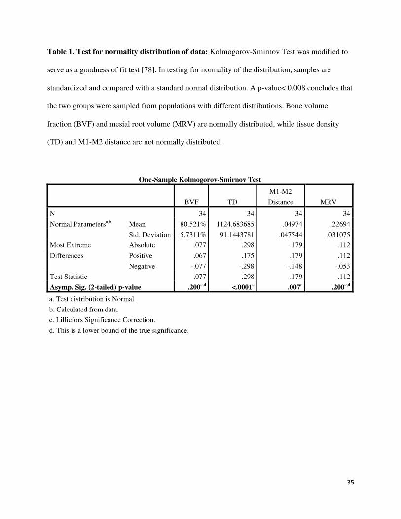
Table 1. Test for normality distribution of data: Kolmogorov-Smirnov Test was modified to
serve as a goodness of fit test [78]. In testing for normality of the distribution, samples are
standardized and compared with a standard normal distribution. A p-value< 0.008 concludes that
the two groups were sampled from populations with different distributions. Bone volume
fraction (BVF) and mesial root volume (MRV) are normally distributed, while tissue density
(TD) and M1-M2 distance are not normally distributed.
One-Sample Kolmogorov-Smirnov Test
BVF TD
M1-M2
Distance MRV
N 34 34 34 34
Normal Parametersa,b
Mean 80.521% 1124.683685 .04974 .22694
Std. Deviation 5.7311% 91.1443781 .047544 .031075
Most Extreme
Differences
Absolute .077 .298 .179 .112
Positive .067 .175 .179 .112
Negative -.077 -.298 -.148 -.053
Test Statistic .077 .298 .179 .112
Asymp. Sig. (2-tailed) p-value .200c,d
<.0001c .007
c .200
c,d
a. Test distribution is Normal.
b. Calculated from data.
c. Lilliefors Significance Correction.
d. This is a lower bound of the true significance.

36
Table 2. Outcomes by group.
Measure BVF TD M1-M2
Distance (MRV)
Control Group
Mean 80.63% 1159.85926 0.1155 0.22048
Std. Deviation 6.47% 32.541784 0.039896 0.033088
Minimum 66.70% 1108.9318 0.054 0.181
Maximum 86.50% 1209.9741 0.174 0.278
Percentiles
25 77.52% 1135.7072 0.075 0.1925
50 82.57% 1156.0503 0.126 0.21765
75 85.41% 1187.0206 0.138 0.2483
Relapse Group
Mean 79.04% 1037.65408 0.0308 0.23815
Std. Deviation 6.34% 129.54363 0.028974 0.030496
Minimum 68.40% 856.0641 0 0.187
Maximum 86.10% 1179.4683 0.09 0.279
Percentiles
25 73.11% 914.383175 0 0.21373
50 82.12% 1048.23735 0.0275 0.24305
75 83.94% 1166.3521 0.04975 0.2647
Relapse Group + 10 Hz
Mean 78.88% 1158.72884 0.012 0.21925
Std. Deviation 4.66% 20.8430233 0.02222 0.040238
Minimum 73.70% 1132.7018 0 0.153
Maximum 89.10% 1201.0795 0.048 0.294
Percentiles
25 75.44% 1146.70393 0 0.19873
50 78.66% 1153.45735 0 0.2209
75 79.63% 1170.9036 0.036 0.2384
Relapse Group + 30 Hz
Mean 83.92% 1164.24996 0.04538 0.22708
Std. Deviation 4.48% 27.2985222 0.019683 0.018649
Minimum 78.20% 1122.6259 0 0.209
Maximum 88.60% 1207.055 0.06 0.254
Percentiles
25 80.04% 1147.5552 0.0415 0.21157
50 84.06% 1159.8722 0.052 0.2204
75 88.18% 1189.75265 0.05775 0.24688

37
Table 3. Difference amongst control and experimental groups per outcome measurement.
Measurement Test p-Value
Bone volume fraction One-way ANOVA 0.252
Tissue density Kruskal-Wallis Test 0.243
M1-M2 Distance Kruskal-Wallis Test <0.0001*
Mesial root volume One-way ANOVA 0.564
*Statistically Significant (p<0.008)
38
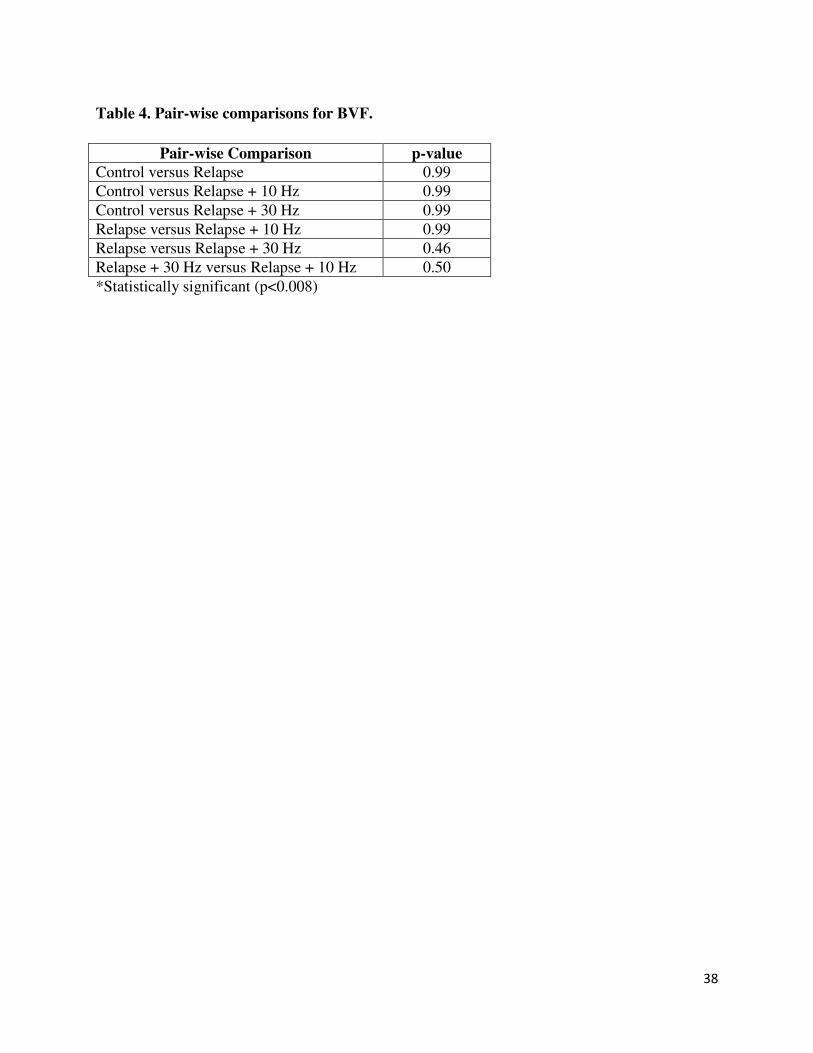
Table 4. Pair-wise comparisons for BVF.
Pair-wise Comparison p-value
Control versus Relapse 0.99
Control versus Relapse + 10 Hz 0.99
Control versus Relapse + 30 Hz 0.99
Relapse versus Relapse + 10 Hz 0.99
Relapse versus Relapse + 30 Hz 0.46
Relapse + 30 Hz versus Relapse + 10 Hz 0.50
*Statistically significant (p<0.008)
39
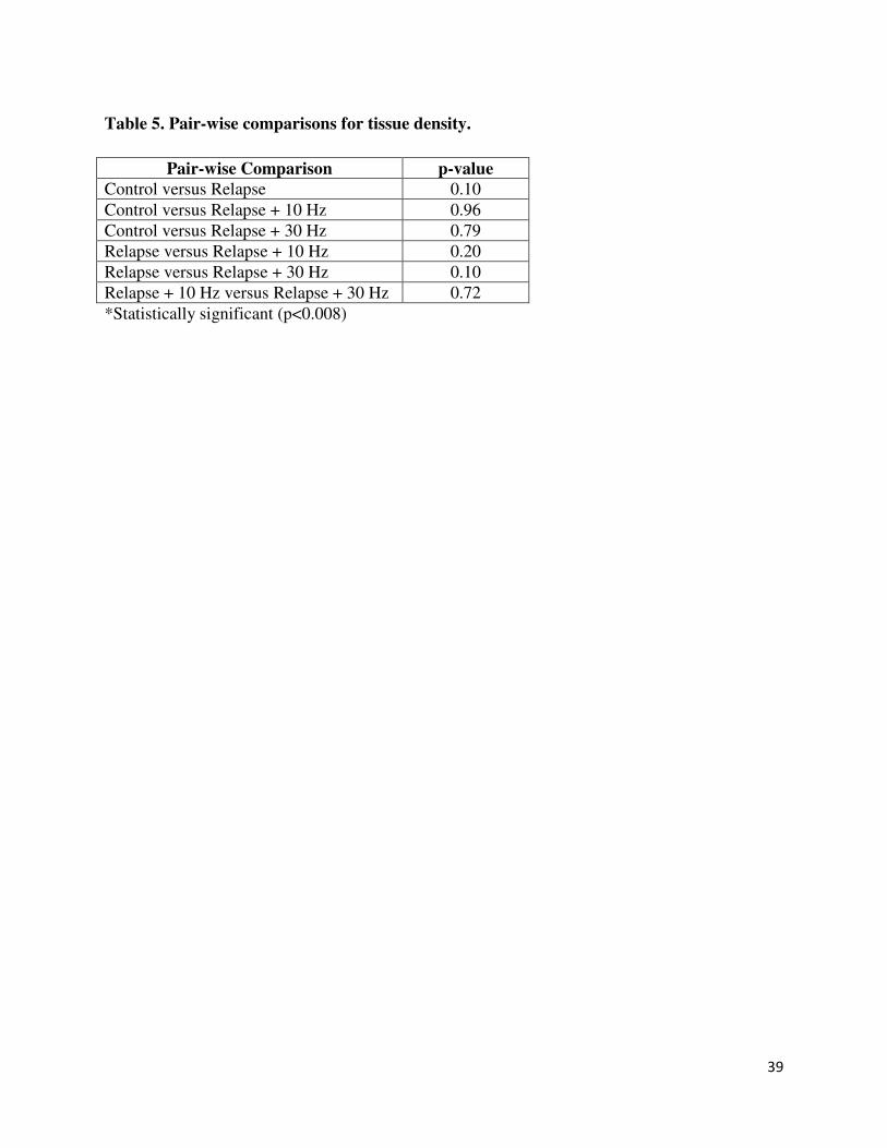
Table 5. Pair-wise comparisons for tissue density.
Pair-wise Comparison p-value
Control versus Relapse 0.10
Control versus Relapse + 10 Hz 0.96
Control versus Relapse + 30 Hz 0.79
Relapse versus Relapse + 10 Hz 0.20
Relapse versus Relapse + 30 Hz 0.10
Relapse + 10 Hz versus Relapse + 30 Hz 0.72
*Statistically significant (p<0.008)

40
Table 6. Pair-wise comparisons for M1-M2 Distance.
Pair-wise Comparison p-value
Control versus Relapse <0.0001*
Control versus Relapse + 10 Hz <0.0001*
Control versus Relapse + 30 Hz 0.002*
Relapse versus Relapse + 10 Hz 0.17
Relapse versus Relapse + 30 Hz 0.12
Relapse + 10 Hz versus Relapse + 30 Hz 0.02
*Statistically significant (p<0.008)

41
Table 7. Pair-wise Comparisons for mesial root volume.
Pair-wise Comparison p-value
Control versus Relapse 0.99
Control versus Relapse + 10 Hz 0.99
Control versus Relapse + 30 Hz 0.99
Relapse versus Relapse + 10 Hz 0.99
Relapse versus Relapse + 30 Hz 0.99
Relapse + 10 Hz versus Relapse + 30 Hz 0.99
*Statistically significant (p<0.008)

42
Figure 5. Bone volume fraction data across all groups.
0
10
20
30
40
50
60
70
80
90
Bo
ne
Vo
lum
e F
ract
ion
(%
)
All Groups
Bone Volume Fraction (BVF)
Control
Relapse
Relapse + 10 Hz
Relapse + 30 Hz
*Statistically significant (p<0.008)

43
Figure 6. Tissue density data across all groups.
0
200
400
600
800
1000
1200
Tis
sue
De
nsi
ty (
mg
/cm
³)
All Groups
Tissue Density (TD)
Control
Relapse
Relapse + 10 Hz
Relapse + 30 Hz
*Statistically significant (p<0.008)

44
Figure 7. First molar movement (M1-M2 Distance) data across all groups. Statistical
significant movement was observed between controls versus all experimental groups.
0
0.02
0.04
0.06
0.08
0.1
0.12
0.14
0.16
M1
-M2
Dis
tan
ce (
mm
)
All Groups
First Molar Movement (M1-M2 Distance)
Control
Relapse
Relapse + 10 Hz
Relapse + 30 Hz
*Statistically significant (p<0.008)
*
*
*

45
Figure 8. Mesial root volume data across all groups.
0
0.05
0.1
0.15
0.2
0.25
Me
sia
l R
oo
t
Vo
lum
e (
mm
³)
All Groups
Mesial Root Volume (MRV)
Control
Relapse
Relapse + 10 Hz
Relapse + 30 Hz
*Statistically significant (p<0.008)

46
Figure 9. Bone volume fraction across experimental groups.
0
10
20
30
40
50
60
70
80
90
Bo
ne
Vo
lum
e F
ract
ion
(%
)
Experimental Groups
Bone Volume Fraction (BVF)
Relapse
Relapse + 10 Hz
Relapse + 30 Hz
*Statistically significant (p<0.008)

47
Figure 10. Tissue density data across experimental groups.
0
200
400
600
800
1000
1200
Tis
sue
De
nsi
ty
(mg
/cm
³)
Experimental Groups
Tissue Density (TD)
Relapse
Relapse + 10 Hz
Relapse + 30 Hz
*Statistically significant (p<0.008)

48
Figure 11. First molar movement (M1-M2 Distance) data across experimental groups.
0
0.01
0.02
0.03
0.04
0.05
0.06
M1
-M2
Dis
tan
ce (
mm
)
Experimental Groups
First Molar Movement (M1-M2 Distance)
Relapse
Relapse + 10 Hz
Relapse + 30 Hz
*Statistically significant (p<0.008)

49
Figure 12. Mesial root volume data across experimental groups.
0
0.05
0.1
0.15
0.2
0.25
Me
sia
l ro
ot
vo
lum
e (
mm
³)
Experimental Groups
Mesial Root Volume (MRV)
Relapse
Relapse + 10 Hz
Relapse + 30 Hz
*Statistically significant (p<0.008)

50
REFERENCES
1. Moyers, R., Handbook of orthodontics for the student and general practitioner. Chicago, London,
Boca Raton, YearBook Publishers Inc., 1973. 3rd Ed.
2. Riedel, R.A., A review of the retention problem. The Angle orthodontist, 1960. 30: p. 179-99.
3. Little, R.M., R.A. Riedel, and J. Artun, An evaluation of changes in mandibular anterior alignment
from 10 to 20 years postretention. American journal of orthodontics and dentofacial orthopedics
: official publication of the American Association of Orthodontists, its constituent societies, and
the American Board of Orthodontics, 1988. 93(5): p. 423-8.
4. Shah, A.A., Postretention changes in mandibular crowding: a review of the literature. American
journal of orthodontics and dentofacial orthopedics : official publication of the American
Association of Orthodontists, its constituent societies, and the American Board of Orthodontics,
2003. 124(3): p. 298-308.
5. Littlewood, S.J., et al., Orthodontic retention: a systematic review. Journal of orthodontics, 2006.
33(3): p. 205-12.
6. Melrose, C. and D.T. Millett, Toward a perspective on orthodontic retention? American journal of
orthodontics and dentofacial orthopedics : official publication of the American Association of
Orthodontists, its constituent societies, and the American Board of Orthodontics, 1998. 113(5):
p. 507-14.
7. Angle, E., Malocclusion of Teeth. Anonymous Philadelphia, PA: The SS White Dental
Manufacturing Company, 1907. 7th ed.
8. Case, C., Principles in retention in orthodontia. Int J Ortod Oral Srug., 1920. 6: p. 33-51.
9. McCauley, D., The cuspid and it function in retention. AJO, 1944. 30: p. 196.
10. Tweed, C.H., Indications for the extraction of teeth in orthodontic procedure. American journal of
orthodontics and oral surgery, 1944. 42: p. 22-45.
11. Little, R.M., Stability and relapse of mandibular anterior alignment: University of Washington
studies. Seminars in orthodontics, 1999. 5(3): p. 191-204.
12. Proffit, W.R., Equilibrium theory revisited: factors influencing position of the teeth. The Angle
orthodontist, 1978. 48(3): p. 175-86.
13. Boese, L.R., Increased stability of orthodontically rotated teeth following gingivectomy in
Macaca nemestrina. American journal of orthodontics, 1969. 56(3): p. 273-90.
14. Yoshida, Y., et al., Cellular roles in relapse processes of experimentally-moved rat molars. Journal
of electron microscopy, 1999. 48(2): p. 147-57.
15. Edwards, J.G., A long-term prospective evaluation of the circumferential supracrestal fiberotomy
in alleviating orthodontic relapse. American journal of orthodontics and dentofacial orthopedics
: official publication of the American Association of Orthodontists, its constituent societies, and
the American Board of Orthodontics, 1988. 93(5): p. 380-7.
16. Gianelly, A., Evidence-based therapy: an orthodontic dilemma. American journal of orthodontics
and dentofacial orthopedics : official publication of the American Association of Orthodontists,
its constituent societies, and the American Board of Orthodontics, 2006. 129(5): p. 596-8;
discussion 598.
17. Driscoll-Gilliland, J., P.H. Buschang, and R.G. Behrents, An evaluation of growth and stability in
untreated and treated subjects. American journal of orthodontics and dentofacial orthopedics :
official publication of the American Association of Orthodontists, its constituent societies, and
the American Board of Orthodontics, 2001. 120(6): p. 588-97.
18. King, G.J., et al., Alveolar bone turnover and tooth movement in male rats after removal of
orthodontic appliances. American journal of orthodontics and dentofacial orthopedics : official
51
publication of the American Association of Orthodontists, its constituent societies, and the
American Board of Orthodontics, 1997. 111(3): p. 266-75.
19. Southard, T.E., R.G. Behrents, and E.A. Tolley, The anterior component of occlusal force. Part 2.
Relationship with dental malalignment. American journal of orthodontics and dentofacial
orthopedics : official publication of the American Association of Orthodontists, its constituent
societies, and the American Board of Orthodontics, 1990. 97(1): p. 41-4.
20. Blake, M. and K. Bibby, Retention and stability: a review of the literature. American journal of
orthodontics and dentofacial orthopedics : official publication of the American Association of
Orthodontists, its constituent societies, and the American Board of Orthodontics, 1998. 114(3):
p. 299-306.
21. Krishnan, V. and Z. Davidovitch, Cellular, molecular, and tissue-level reactions to orthodontic
force. American journal of orthodontics and dentofacial orthopedics : official publication of the
American Association of Orthodontists, its constituent societies, and the American Board of
Orthodontics, 2006. 129(4): p. 469 e1-32.
22. Wise, G.E. and G.J. King, Mechanisms of tooth eruption and orthodontic tooth movement.
Journal of dental research, 2008. 87(5): p. 414-34.
23. Proffit, W., Fields, H, Sarver, D., ed. Contemporary Orthodontics. Elsevier Science Health Science
div. Vol. 4th Ed. 2007. Chapter 18, 19.
24. Storey, E., The nature of tooth movement. American journal of orthodontics, 1973. 63(3): p. 292-
314.
25. Hudson, J.B., et al., Local delivery of recombinant osteoprotegerin enhances postorthodontic
tooth stability. Calcified tissue international, 2012. 90(4): p. 330-42.
26. Reitan, K., Clinical and histologic observations on tooth movement during and after orthodontic
treatment. American journal of orthodontics, 1967. 53(10): p. 721-45.
27. Gkantidis, N., P. Christou, and N. Topouzelis, The orthodontic-periodontic interrelationship in
integrated treatment challenges: a systematic review. Journal of oral rehabilitation, 2010. 37(5):
p. 377-90.
28. Pratt, M.C., et al., Evaluation of retention protocols among members of the American Association
of Orthodontists in the United States. American journal of orthodontics and dentofacial
orthopedics : official publication of the American Association of Orthodontists, its constituent
societies, and the American Board of Orthodontics, 2011. 140(4): p. 520-6.
29. Hawley, C., A removable retainer. Int J Ortod Oral Surg, 1919. 5: p. 219-305.
30. Rinchuse, D.J., P.G. Miles, and J.J. Sheridan, Orthodontic retention and stability: a clinical
perspective. Journal of clinical orthodontics : JCO, 2007. 41(3): p. 125-32.
31. Pratt, M.C., G.T. Kluemper, and A.F. Lindstrom, Patient compliance with orthodontic retainers in
the postretention phase. American journal of orthodontics and dentofacial orthopedics : official
publication of the American Association of Orthodontists, its constituent societies, and the
American Board of Orthodontics, 2011. 140(2): p. 196-201.
32. Mollov, N.D., et al., Patient attitudes toward retention and perceptions of treatment success. The
Angle orthodontist, 2010. 80(4): p. 468-73.
33. Kacer, K.A., et al., Retainer wear and compliance in the first 2 years after active orthodontic
treatment. American journal of orthodontics and dentofacial orthopedics : official publication of
the American Association of Orthodontists, its constituent societies, and the American Board of
Orthodontics, 2010. 138(5): p. 592-8.
34. Wong, P. and T.J. Freer, Patients' attitudes towards compliance with retainer wear. Australian
orthodontic journal, 2005. 21(1): p. 45-53.

52
35. Bearn, D.R., Bonded orthodontic retainers: a review. American journal of orthodontics and
dentofacial orthopedics : official publication of the American Association of Orthodontists, its
constituent societies, and the American Board of Orthodontics, 1995. 108(2): p. 207-13.
36. Pandis, N., et al., Long-term periodontal status of patients with mandibular lingual fixed
retention. European journal of orthodontics, 2007. 29(5): p. 471-6.
37. Mettes, T.D., et al., Surgical removal versus retention for the management of asymptomatic
impacted wisdom teeth. The Cochrane database of systematic reviews, 2012. 6: p. CD003879.
38. Southard, T.E., Third molars and incisor crowding: when removal is unwarranted. Journal of the
American Dental Association, 1992. 123(8): p. 75-9.
39. Roth, R.H., Functional occlusion for the orthodontist. Journal of clinical orthodontics : JCO, 1981.
15(1): p. 32-40, 44-51 contd.
40. Kim, T.W., et al., An ultrastructural study of the effects of bisphosphonate administration on
osteoclastic bone resorption during relapse of experimentally moved rat molars. American
journal of orthodontics and dentofacial orthopedics : official publication of the American
Association of Orthodontists, its constituent societies, and the American Board of Orthodontics,
1999. 115(6): p. 645-53.
41. Hassan, A.H., A. Al-Hubail, and A.A. Al-Fraidi, Bone inductive proteins to enhance postorthodontic
stability. The Angle orthodontist, 2010. 80(6): p. 1051-60.
42. Han, G., et al., Effects of simvastatin on relapse and remodeling of periodontal tissues after tooth
movement in rats. American journal of orthodontics and dentofacial orthopedics : official
publication of the American Association of Orthodontists, its constituent societies, and the
American Board of Orthodontics, 2010. 138(5): p. 550 e1-7; discussion 550-1.
43. Kanzaki, H., et al., Local OPG gene transfer to periodontal tissue inhibits orthodontic tooth
movement. Journal of dental research, 2004. 83(12): p. 920-5.
44. Zhao, N., et al., Local osteoprotegerin gene transfer inhibits relapse of orthodontic tooth
movement. American journal of orthodontics and dentofacial orthopedics : official publication of
the American Association of Orthodontists, its constituent societies, and the American Board of
Orthodontics, 2012. 141(1): p. 30-40.
45. Christiansen, B.A. and M.J. Silva, The effect of varying magnitudes of whole-body vibration on
several skeletal sites in mice. Ann Biomed Eng, 2006. 34(7): p. 1149-56.
46. Rubin, C., et al., Prevention of postmenopausal bone loss by a low-magnitude, high-frequency
mechanical stimuli: a clinical trial assessing compliance, efficacy, and safety. J Bone Miner Res,
2004. 19(3): p. 343-51.
47. Stark, T.M. and P.M. Sinclair, Effect of pulsed electromagnetic fields on orthodontic tooth
movement. Am J Orthod Dentofacial Orthop, 1987. 91(2): p. 91-104.
48. Darendeliler, M.A., P.M. Sinclair, and R.P. Kusy, The effects of samarium-cobalt magnets and
pulsed electromagnetic fields on tooth movement. Am J Orthod Dentofacial Orthop, 1995.
107(6): p. 578-88.
49. Darendeliler, M.A., et al., Effects of pulsed electromagnetic field vibration on tooth movement
induced by magnetic and mechanical forces: a preliminary study. Aust Dent J, 2007. 52(4): p.
282-7.
50. Nishimura, M., et al., Periodontal tissue activation by vibration: intermittent stimulation by
resonance vibration accelerates experimental tooth movement in rats. Am J Orthod Dentofacial
Orthop, 2008. 133(4): p. 572-83.
51. Ohmae M, S.S., Morohashi T, Qu H, Seki K Kurabayashi H, Biomechanical acceleration of
experimental tooth movement by ultrasonic vibration in vivo: Part 1. Homo-directional
application of ultrasonication to orthodontic force. Orthod Waves, 2001. 60: p. 201 - 212.
53
52. Kau HC, N.J., English JD, The Clinical Evaluation of a Novel Cyclical Force Generating device in
Orthodontics Orthodontic Practice, 2011. 1(1).
53. Kalajzic, Z., et al., Effect of cyclical forces on the periodontal ligament and alveolar bone
remodeling during orthodontic tooth movement. The Angle orthodontist, 2014. 84(2): p. 297-
303.
54. Rygh, P., Ultrastructural changes in tension zones of rat molar periodontium incident to
orthodontic tooth movement. Am J Orthod, 1976. 70(3): p. 269-81.
55. Storey, E., The nature of tooth movement. Am J Orthod, 1973. 63(3): p. 292-314.
56. Roberts, W.E. and J.G. Chamberlain, Scanning electron microscopy of the cellular elements of rat
periodontal ligament. Arch Oral Biol, 1978. 23(7): p. 587-9.
57. Waldo, C.M. and J.M. Rothblatt, Histologic response to tooth movement in the laboratory rat;
procedure and preliminary observations. J Dent Res, 1954. 33(4): p. 481-6.
58. Ren, Y., J.C. Maltha, and A.M. Kuijpers-Jagtman, The rat as a model for orthodontic tooth
movement--a critical review and a proposed solution. Eur J Orthod, 2004. 26(5): p. 483-90.
59. Yamasaki, K., Y. Shibata, and T. Fukuhara, The effect of prostaglandins on experimental tooth
movement in monkeys (Macaca fuscata). J Dent Res, 1982. 61(12): p. 1444-6.
60. Igarashi, K., et al., Anchorage and retentive effects of a bisphosphonate (AHBuBP) on tooth
movements in rats. Am J Orthod Dentofacial Orthop, 1994. 106(3): p. 279-89.
61. Mohammed, A.H., D.N. Tatakis, and R. Dziak, Leukotrienes in orthodontic tooth movement. Am J
Orthod Dentofacial Orthop, 1989. 95(3): p. 231-7.
62. Pavlin, D., et al., Orthodontically stressed periodontium of transgenic mouse as a model for
studying mechanical response in bone: The effect on the number of osteoblasts. Clin Orthod Res,
2000. 3(3): p. 55-66.
63. Pavlin, D., et al., Orthodontically stressed periodontium of transgenic mouse as a model for
studying mechanical response in bone: The effect on the number of osteoblasts. Clin Orthod Res,
2000. 3(2): p. 55-66.
64. King, G.J., et al., Measuring dental drift and orthodontic tooth movement in response to various
initial forces in adult rats. Am J Orthod Dentofacial Orthop, 1991. 99(5): p. 456-65.
65. Keeling, S.D., et al., Serum and alveolar bone phosphatase changes reflect bone turnover during
orthodontic tooth movement. Am J Orthod Dentofacial Orthop, 1993. 103(4): p. 320-6.
66. Nixon, C.E., et al., Histomorphometric study of dental pulp during orthodontic tooth movement. J
Endod, 1993. 19(1): p. 13-6.
67. Yoshimatsu, M., et al., Experimental model of tooth movement by orthodontic force in mice and
its application to tumor necrosis factor receptor-deficient mice. J Bone Miner Metab, 2006.
24(1): p. 20-7.
68. Alvarez, L.L. and H.G. Pardo, Guide for the care and use of laboratory animals - Natl-Res-Council.
Psicothema, 1997. 9(1): p. 232-234.
69. Lee, T.C. and D. Taylor, Bone remodelling: should we cry Wolff? Irish journal of medical science,
1999. 168(2): p. 102-5.
70. Umemura, Y., et al., Five jumps per day increase bone mass and breaking force in rats. Journal of
bone and mineral research : the official journal of the American Society for Bone and Mineral
Research, 1997. 12(9): p. 1480-5.
71. Lanyon, L.E. and C.T. Rubin, Static vs dynamic loads as an influence on bone remodelling. Journal
of biomechanics, 1984. 17(12): p. 897-905.
72. Rubin, C., et al., Quantity and quality of trabecular bone in the femur are enhanced by a strongly
anabolic, noninvasive mechanical intervention. Journal of bone and mineral research : the
official journal of the American Society for Bone and Mineral Research, 2002. 17(2): p. 349-57.
54
73. Bouxsein, M.L., et al., Guidelines for assessment of bone microstructure in rodents using micro-
computed tomography. Journal of bone and mineral research : the official journal of the
American Society for Bone and Mineral Research, 2010. 25(7): p. 1468-86.
74. Darendeliler, M.A., et al., Effects of pulsed electromagnetic field vibration on tooth movement
induced by magnetic and mechanical forces: a preliminary study. Australian dental journal, 2007.
52(4): p. 282-7.
75. Al-Daghreer, S., et al., Effect of low-intensity pulsed ultrasound on orthodontically induced root
resorption in beagle dogs. Ultrasound in medicine & biology, 2014. 40(6): p. 1187-96.
76. Lau, E., et al., Effect of low-magnitude, high-frequency vibration on osteocytes in the regulation
of osteoclasts. Bone, 2010. 46(6): p. 1508-15.
77. Lynch, M.A., M.D. Brodt, and M.J. Silva, Skeletal effects of whole-body vibration in adult and
aged mice. J Orthop Res, 2010. 28(2): p. 241-7.
78. Press, W.H., Teukolsky, S.Vetterling, W., and Flannery, B .P., Numerical Recipes: The Art of
Scientific Computing. Cambridge University Press, 2007. 3.


![CONTROL ACTIVO DE VIBRACIONES EN … · attenuate the mechanical vibrations with excitation frequencies, ... [2,7,8]. Los modelos ... globales de masa,](https://img.pdfslide.us/doc/110x75/5b96bb3209d3f2816c8bc7bc/control-activo-de-vibraciones-en-attenuate-the-mechanical-vibrations-with-excitation.jpg)