Embed Size (px)
Citation preview
Z. Zellforsch. 119, 179-187 (1971) �9 by Springer-Verlag 197I
The Effects of Steroids on the Granulosa Cells in the Domestic Fowl
ERIK DAHL
Department of Anatomy, Dental Faculty, University of Oslo, Blindern, Oslo, Norway
Received April 5, 1971
Summary. The fine structure of granulosa cells of the domestic fowl as seen after ad- ministration of steroids is described. Diaethylstilboestrol, estradiol and hydroxy-progesterone were given as intramuscular injections for a 28-days period. The main cytoplasmic changes of the granulosa cells were an increase in the number of mitochondria and dense bodies. The Golgi apparatus became enlarged, and occupied a large portion of the cell. The nucleus was found located adjacent to the oocyte, and there was an increase in number of the annular desmosomes. The investigation has demonstrated that even if steroids in high dosages induce atrophic alterations of the ovary, they stimulated proliferation of the granulosa cells of the small follicles. Apparently the small follicles with the granulosa cells retain the ability to regain development, which may be to some importance when steroids are used therapeutically (gynecologic disorders, contra-ception).
Key-Words: Granulosa cells - - Fowl - - Influence of steroids - - Alterations - - Ultra- structure.
The act ion of estrogen on the development and periodic changes of the female accessory sexual organs of mammals is well known, and i t appears probable t h a t this secretion influences the growth of the various parts of the ovary itself (Bul- lough, 1943 ; Rennels, 1951 ; Payne and Runser , 1958).
As a detail in a series of invest igat ions on the ovar ian fine s t ructure (Dahl, 1970a-c, 1971a-f) the present s tudy is concerned with the u l t ras t ruc tura l aspects of steroid influence on the granulosa cells. Previous reports from this labora tory have defined the normal u l t ras t ruc ture of the granulosa cells (Dahl, 1971d) and the steroid-producing cells of the theca in te rna (Dahi, 1970a, b, 1971a) and also the effects of steroids on the steroid-producing cells (Dahl, 1971b).
By comparing the series of experiments of these two cell types which were taken together from the same follicles under exactly the same exper imental conditions, it was the aim to achieve new informat ion about the morphology and the funct ion of the different ovar ian cell types.
Materials and Methods
Twenty White Leghorn hens, 18-24 months old, with an average body weight of 1824 g, and six 3 month-old chickens of an average weight of 900 g were used. The animals were housed in individual cages in a well-ventilated, constant-climate room (17~ controlled illumination, light on 7 a.m., off 7 p.m., relative humidity 60% ). The diet consisted of com- mercial chicken fodder, cabbage, sand grits and water ad lib. The main constituents of the
13 Z. Zellforsch., Bd. 119
180 E. Dahl :
fodder were proteins (17-19%), fat (2-4%), calcium (1.1-1.2%), phosphorus (0.7% and sodium cloride (0.5 %)). The hens werekept for at least 10days to get adapted to their environ- ment before the experiment started.
The hormone preparations used were: Estradioli valerianas (Primogyn-Depot, Schering, A-G, Berlin, Germany). This will be referred to as estradiol. Diaethylstilbestroli (Stilboestrol, Nyco@, Oslo, Norway). This will be referred to as stilbestrol. Hydroxyprogesteroni caproas (Primolut-Depot, Schering). This will be referred to as oxyprogesterone. Of the 20 hens and 6 chickens employed in this experiment, 5 hens and 3 chickens served as controls.
The steroids were administrated as follows: Three chickens received 3 mg estradiol as an intramuscular injection 7 days before sacrifice. Five hens received 1 mg estradiol as daily intramuscular injections for 28 days. Five hens received 1 mg stilbestrol as daily intramuscular injections for 28 days. Five hens received 25 mg oxyprogesterone as daily intramuscular injections for 28 days. The hens were sacrificed 24 hr after the last injection. The controls were kept under identical conditions except for the intramuscular injections.
Fixation was performed as an intracardial perfusion of dextran under nembutal anesthesia (Nembutal sodium, Abbot, 5%, followed by 1.7 % glutaraldehyde in 0.1 M phosphate buffer at pH 7.3. The perfusion lasted for a minimum 10 rain. The ovary was then excised and, while kept in a drop of fixative, cut into thin slices under the dissecting microscope. Samples from follicles of different sizes were then fixed separately in glutaraldehyde for an additional period of 2 hr at 4~ Subsequently the tissue blocks were rinsed for 10 min in 0.15 M phos- phate buffer at pH 7.3 and fixed in 1% osmium tetroxide at 4 ~ C for 2 hr. The blocks were rapidly dehydrated in a graded series of acetone and embedded in Vestopal W (Ryter and Kellenberger, 1958). Ultrathin sections were cut on an LKB Ultrobome and treated with uranyl acetate for 30 min followed by lead citrate (Reynolds, 1963) for 5 rain to increase contrast. The sections were examined in Siemens Elmiskop Ia electron microscope, equipped with 50 microns platinum objective apertures. Accelerating voltage was 80 kV. One micron thick sections were also cut for light microscopy and stained for 30 sec on a heating stage with 0.1% toluidine blue adjusted to pH 8.5 with M/15 Na~HPO 4.
Observations
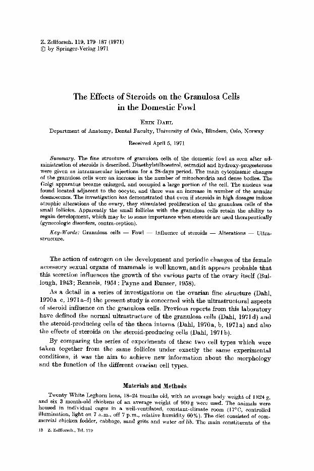
Daily inject ions of the 3 different steroids impaired the general condi t ion of the animals. After some days the egg-laying ceased, the comb became decreased in size and quite pale, and at the end of the exper imental period they s tar ted to moult , especially the progesterone t reated animals. Their appet i te decreased and their v i ta l i ty seemed to be generally reduced as compared to the control animals. The ovaries were smaller t han normal , with numerous, small follicles, all of the same size, giving the ovary an appearenee similar to cod roe. Elec t ron microscopic examina t ion of the steroid-treated mater ia l revealed the granulosa cells of the p r imary follicles to be both cuboidal (Fig. 1) and columnar (Fig. 4) in shape. The nuclei were irregular with identat ions , and were often found in the area adjacent to the oocyte (Figs. 1, 4). They were no t as large in pro- por t ion to the cytoplasm of the cell as found in the normals indicat ing a hyper- th ropy of the whole granulosa cell. The contact between the oocyte and the granulosa cells was a surface-to-surface mutua l contact with an increased n u m b e r of desmosomes (Figs. 7-9) as well as cytoplasmic processes (Fig. 1). The mito- ehondi'ia were evenly dis t r ibuted in the cytoplasm and seemed to increase in number s (Fig. 3). They had a dense, dark mat r ix (Fig. 3) and the n u m b e r of cristae seemed to increase (Fig. 2). There also seemed to be an increase in the size of the mitochondr ia as well as the n u m b e r of mierobody-like mitochondria as
compared to the controls (Figs. 1, 3).
Effects of Steroids on the Granulosa Cells 181
Fig. 1. Survey electron micrograph of granulosa cells after administrations of steroids for 28 days. There is an increase of the size of the cells, increase in number of dense bodies (Db), cytoplasmic processes towards the oocyte (arrows). Some of the nuclei (upper left and right corner) are found towards the oocyte. N Nucleus, G Golgi area, Bm Basement membrane,
M Mitochondria, L Lipid. x 7 875
The Golgi appara tus was well developed, in some cells of a ra ther enormous size and i t then occupied a large port ion of the cell (Fig. 5). I t was most often found located towards the basement membrane , cont rary to what was observed in the
13"
182 E. Dahl :
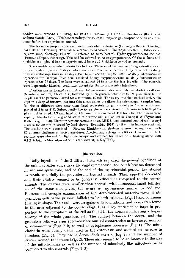
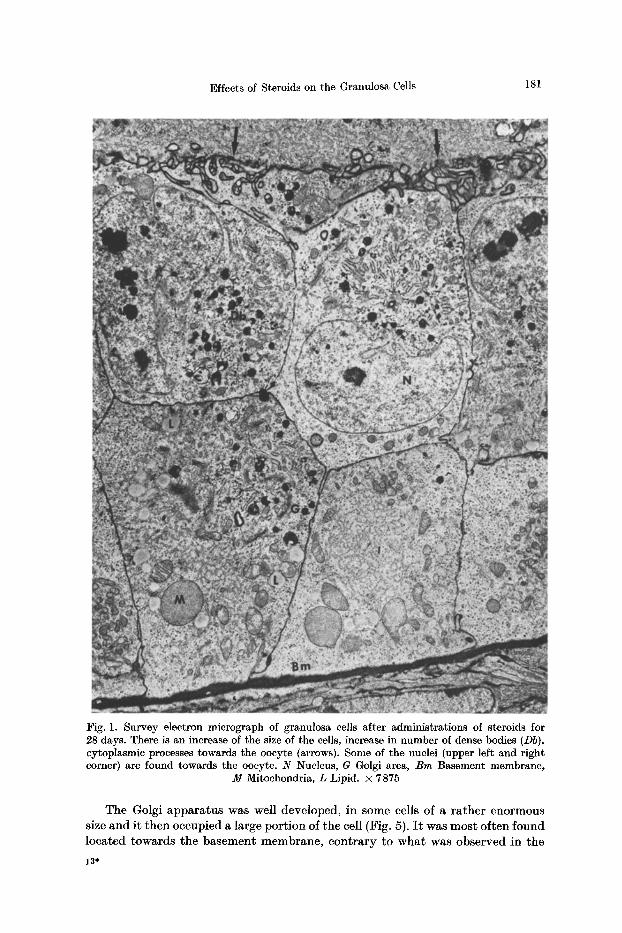
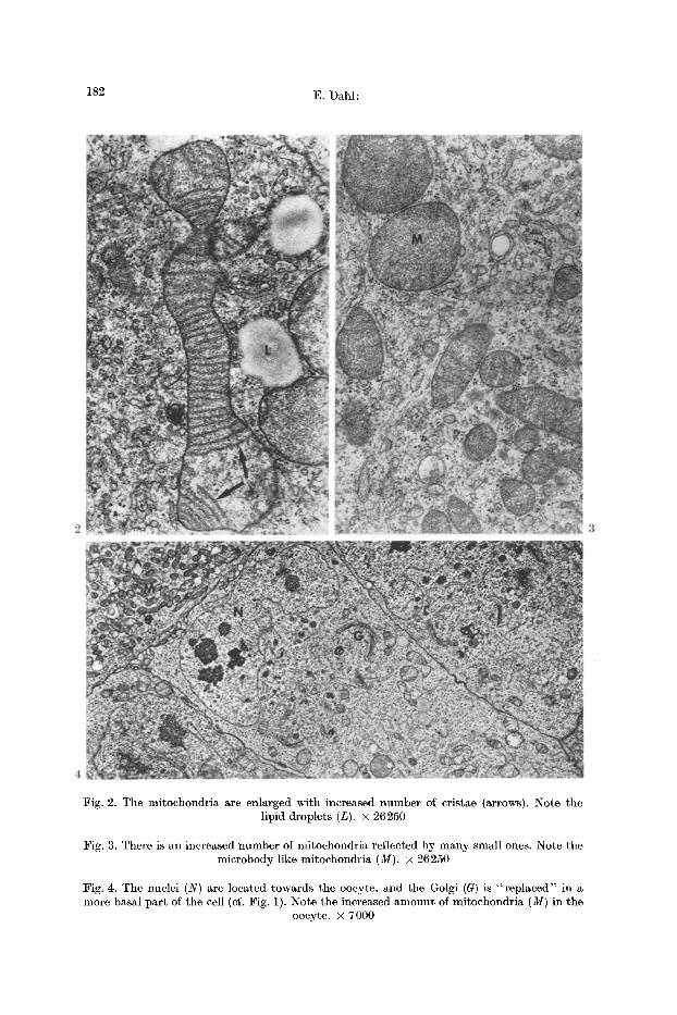
Fig. 2. The mitochondria are enlarged with increased number of cristae (arrows). Note the lipid droplets (L). • 26250
:Fig. 3. There is an increased number of mitochondria reflected by many small ones. Note the microbody-like mitochondria (M). • 26250
Fig. 4. The nuclei (N) are located towards the oocyte, and the Golgi (G) is " rep laced" in a more basal par t of the cell (cf. Fig. 1). Note the increased amount of mitochondria (M) in the
oocyte. • 7000
Effects of Steroids on the Granulosa Cells 183
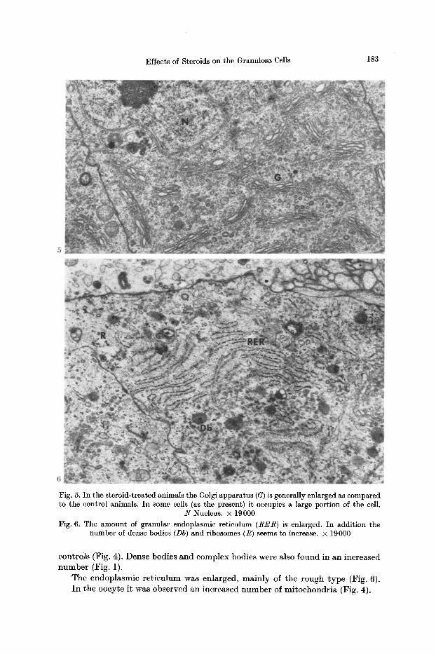
Fig. 5. In the steroid-treated animals the Golgi apparatus (G) is generally enlarged as compared to the control animals. In some cells (as the present) it occupies a large portion of the cell.
N Nucleus. • 19000 Fig. 6. The amount of granular endoplasmic reticulum (RER) is enlarged. In addition the
number of dense bodies (Db) and ribosomes (R) seems to increase. • 19000
controls (Fig. 4). Dense bodies and complex bodies were also found in an increased n u m b e r (Fig. 1).
The endoplasmic re t iculum was enlarged, main ly of the rough type (Fig. 6). I n the oocyte i t was observed an increased n u m b e r of mi toehondr ia (Fig. 4).
184 E. Dahl:
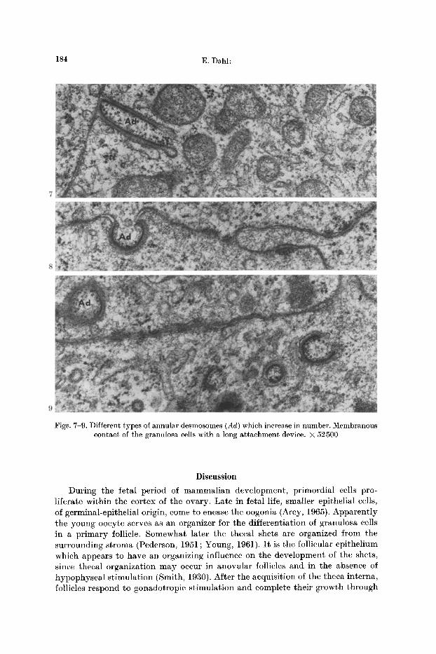
Figs. 7-9. Different types of annular desmosomes (Ad) which increase in number. Membranous contact of the granulosa cells with a long attachment device, x 52500
Discussion
During the fetal period of mammalian development, primordial cells pro- liferate within the cortex of the ovary. Late in fetal life, smaller epithelial cells, of germinal-epithelial origin, come to encase the oogonia (Arey, 1965). Apparently the young ooeyte serves as an organizer for the differentiation of granulosa cells in a primary follicle. Somewhat later the theeal shets are organized from the surrounding stroma (Pederson, 1951; Young, 1961). I t is the follicular epithelium which appears to have an organizing influence on the development of the shets, since thecal organization may occur in anovnlar follicles and in the absence of hypophyseal stimulation (Smith, 1930). After the acquisition of the theea interna, follicles respond to gonadotropie stimulation and complete their growth through
Effects of Steroids on the Granulosa Cells 185
the stage of preovulatory swelling, the maturat ion divisions and ovulation (Pederson, 1951).
Bullough (1943) found tha t large doses of oestrone given to adult female mice in dioestrus cause a reduction of the normal rapid growth of the large Graafian follicles, while the small pr imary follicles remain unaffected. No significant changes could be traced in the corpora lutea or in the interstitial and connective tissue cells. Rennels (1951) found that administration of estrogen results in a marked inhibition of ovarian growth and prevents the development of pr imary interstitial tissue. Desoxycortieosterone acetate caused some ovarian inhibition, while progesterone was without effect at the dosage employed.
Chronic administrations of estrogen to the intact animal eventually produces ovarian a t rophy by suppression of gonadotropin secretion (Everett, 1961 ; Dahl, 1971b). Among the known inhibitors of pituitary gonadotropin secretion, the estrogens are the most effective. There is complete agreement that estrogen in moderate to high dosage inhibits FSH synthesis and liberation (Greep, 1961).
Effects of steroid administration on the fine structure of the granulosa cells have not previously been demonstrated. In the present study, there was a marked reduction of the size of the ovary, with reduction of the large follicles. Ultra- structurally there were also marked alterations of the fine structure of the theca interna (Dahl, 1971 b) indicating an a t rophy of this tissue, especially the steroid- producing cells. However, the fine structure of the granulosa cells of the same follicles revealed an increase in the size of the Golgi apparatus, number of mito- chondria, granular endoplasmic reticulum, dense bodies and annular desmosomcs. Furthermore, the cells seemed enlarged, obtained a more elongated form, and the nuclei were found in the proximal part of the cells and now with the Golgi appa- ratus located towards the basal portion which was different from the location in the normals where it generally was located towards the apical part of the cell (Dahl, 1971 c). All the ultrastructural alterations observed indicate a stimulating effect on the granulosa cells of these small follicles, contrary to the generally atrophic changes observed in other ovarian structures (Dahl, 1971 b). The present s tudy therefore seems to indicate tha t steroids exert a growth-promoting influence on the pr imary follicles with a stimulating effect on the granulosa cells. Apparently these effects have not been elicited by physiologic doses. However, the possibility exist that in the neighborhood of cells that produce it, the estrogen concentration is probably far above that which would be considered physiologic for the re- mainder of the body (Everett, 1961). The high dosage of steroids used, certainly have inhibited F S H synthesis and liberation. The s tudy therefore also seems to indicate that gonadotropic hormones of the pituitary per se are not essential for the early growth of ovarian follicles.
Regarding the function of the granulosa cells, there has been some discrepancy as to their capability to produce steroid (Bjersing, 1967; Dahl, 1971 d). As pointed out (Dahl, 1971d) there seems to be little evidence for the granulosa cells to produce steroids. The present study also seems to support this view. The fact tha t steroids stimulate the granulosa cells of the small follicles, while the steroid- producing cells in the theca interna of the same follicles undergo atrophic altera- tions also indicates that granulosa cells might not be a significant source of steroids.
186 E. Dahl :
I n conclusion, the present s tudy seems to have confirmed t h a t steroids
(estrogen) have an s t imulat ing effect on the granulosa cells of the small follicles
resul t ing in proliferat ion. I n addi t ion the possibil i ty also exists t ha t steroids
render the follicles more responsive to exogenous gonadotropins (Williams, 1945 b; Dahl , 1971 b, c). The present s tudy is the first demons t ra t ion of the u l t ras t ruc tura l
changes of the granulosa cells in the fowl following adminis t ra t ion of hormones.
The s imilar i ty of the fine s t ructural changes following the t r e a t m e n t of the three
different hormones supports the concept t h a t the mechanism may be ident ical in
all three cases (Dahl, 1971 b). So far, no explanat ion can be given to the s t imulat ing
effect of progesterone on the granulosa cells. As poin ted out (Dahl, 1971b) steroid
used therapeut ica l ly in various gynecologic disorders and also for cont racept ion
m a y induce morphological a l tera t ion of the ovary. The present s tudy seems to
indicate t ha t even though the ovary becomes atrophic, the small follicles wi th
the granulosa cells re ta in the capabi l i ty to fur ther development .
R e f e r e n c e s
Arey, L. B. : The reproductive organs and sex cells. In: Developmental anatomy (L. B. Arey, ed.), 7th ed., p. 28-61. Philadelphia: W. B. Saunders Co. 1965.
Bjersing, L. : On the morphology and endocrine function of granulosa cells in ovarian follicles and corpora lutea. Acta endocr. (Kbh.), Suppl. 125, 1-23 (1967).
Bullough, W. S-: The effects of oestrone on the ovary of the mouse. J. Endocr. 3, 235-243 (1943).
Dahl, E. : Studies of the fine structure of ovarian interstitial tissue. 2. The ultrastructure of the thecal gland of the domestic fowl. Z. Zellforsch. 109, 195-211 (t970a).
- - Studies of the fine structure of ovarian interstitial tissue. 3. The innervation of the thecal gland of the domestic fowl. Z. Zellforsch. 109, 212-226 (1970b).
- - Studies of the fine structure of ovarian interstitial tissue. 6. Effects of clomiphene on the thecal gland of the domestic fowl. Z. Zellforseh. 109, 227-244 (1970c).
- - Studies of the fine structure of ovarian interstitial tissue. 1. A comparative study of the fine structure of the ovarian interstitial tissue in the rat and the domestic fowl. J. Anat. (Loud.) 108, 275-290 (1971 a).
- - Studies of the fine structure of ovarian interstitial tissue. 4. Effects of steroids on the theeal gland of the domestic fowl. Z. Zellforsch. 113, 111-132 (1971b).
- - Studies of the fine structure of ovarian interstitial tissue. 5. Effects of gonadotropins on the thecal gland of the domestic fowl. Z. Zellforsch. I18, 133-156 (1971 c).
- - The fine structure of the granulosa cells in the domestic fowl and the rat. Z. Zell- forseh. 119, 58 (1971d).
- - The effects of gonadotropins on the granulosa cells of the domestic fowl. Acta endo- crin. (Kbh.) (in press) (1971e).
- - The effects of clomiphene on the granulosa cells of the domestic fowl. Z. Zellforsch. 1 1 9 (1971 f).
Everett, J. W. : The mammalian female reproductive cycle and its controlling mechanisms. In: Sex and internal secretions, 3rd ed., vol. I (W. C. Young, ed.), p. 497-555. Baltimore: Williams & Wilkins Co. 1961.
Greep, R. O. : Physiology of the anterior hypophysis in relation to reproduction. In : Sex and internal secretions, 3rd. ed., vol. I (W. C. Young, ed.), p. 240-301. :Baltimore: Williams & Wilkins Co. 1961.
Payne, R. W., Runser, 1%. H. : The influence of estrogen and androgen on the ovarian response of hypophysectomized immature rats to gonadotropins. Endocrinology 62, 313-321 (1958).
Pederson, E. S.: Histogenesis of lutein tissue of the albino rat. Amer. J. Anat. 88, 397-416
(1951). Rennels, E. G. : Influence of hormones on the histochemistry of ovarian interstitial tissue in
the immature rat. Amer. J. Anat. 88, 63-100 (1951).
Effects of Steroids on the Granulosa Cells 187
Reynolds, E. S. : The use of lead citrate at high pH as an electron-opaque stain in electron microscopy. J . Cell Biol. 17, 208-212 (1963).
Ryter, A., Kellenberger, E. : L'inclusion au polyester pour l 'ultramicrotomie. J . Ultrastruct . Res. 2, 200-214 (1958).
Smith, P. E. : Hypophysectomy and a replacement therapy in the rat. Amer. J. Anat. 45, 205-272 (1930).
Williams, P. C. : Studies of the biological action of serum gonadotrophin. 2. Ovarian response after hypophysectomy and oestrogen t reatment . J. Endocr. 4, 131-142 (1945).
Young, W. C.: The mammalian ovary. In : Sex and internal secretions (W. C. Young, ed.), 3rd ed., vol. I, p. 449-496. Baltimore: Williams & Wilkins Co. 1961.
Dr. Erik Dahl Depar tment of Anatomy Dental Facul ty Universi ty of Oslo Blindern, Oslo 3, Norway