Embed Size (px)
Citation preview
Interventional Medicine & Applied Science, Vol. 5 (3), pp. 122–130 (2013)
DOI: 10.1556/IMAS.5.2013.3.5 ISSN 2061-1617 © 2013 Akadémiai Kiadó, Budapest 122
O R I G I N A L PA P E R
The eff ects of gamma irradiation
on diclofenac sodium, liposome and niosome
ingredients for rheumatoid arthritis
A. YEKTA ÖZER1,*, SELCAN TURKER1, SEYDA ÇOLAK2, MUSTAFA KORKMAZ2,
EKREM KILIÇ3, MERAL ÖZALP3
1Department of Radiopharmacy, Faculty of Pharmacy, Hacettepe University, Ankara, Turkey2Department of Physics Engineering, Faculty of Engineering, Hacettepe University, Ankara, Turkey
3Department of Pharmaceutical Microbiology, Faculty of Pharmacy, Hacettepe University, Ankara, Turkey
*Corresponding author: Prof. Dr. A. Yekta Özer; Department of Radiopharmacy, Faculty of Pharmacy, Hacettepe University,
06100-Sıhhiye-Ankara, Turkey; Phone: +90-312-305-2196/305 2152; Fax: +90-312-311-4777;
E-mails: [email protected], [email protected]
(Received: May 3, 2013; Revised manuscript received: June 20, 2013; Accepted: June 20, 2013)
Abstract: The use of gamma rays for the sterilization of pharmaceutical raw materials and dosage forms is an alternative method for sterilization.
However, one of the major problems of the radiosterilization is the production of new radiolytic products during the irradiation process.
Therefore, the principal problem in radiosterilization is to determine and to characterize these physical and chemical changes originating from
high-energy radiation. Parenteral drug delivery systems were prepared and in vitro characterization, biodistribution and treatment studies were
done in our previous studies. Drug delivery systems (liposomes, niosomes, lipogelosomes and niogelosomes) encapsulating diclofenac sodium
(DFNa) were prepared for the treatment of rheumatoid arthritis (RA). This work complies information about the studies developed in order
to fi nd out if gamma radiation could be applied as a sterilization method to DFNa, and the raw materials as dimyristoyl phosphatidylcholine
(DMPC), surfactant I [polyglyceryl-3-cethyl ether (SUR I)], dicethyl phosphate (DCP) and cholesterol (CHOL) that are used to prepare those
systems. The raw materials were irradiated with diff erent radiation doses (5, 10, 25 and 50 kGy) and physicochemical changes (organoleptic
properties pH, UV and melting point), microbiological evaluation [sterility assurance level (SAL), sterility and pyrogen test] and electron spin
resonance (ESR) characteristics were studied at normal (25 °C, 60% relative humidity) and accelerated (40 °C, 75% relative humidity) stability
test conditions.
Keywords: diclofenac sodium, DMPC, SUR I, DCP, cholesterol, gamma irradiation, sterilization
Introduction
One of the most critical parameters for injectable con-
trolled release drug delivery systems is sterility. Those
systems have several advantages which make them pref-
erable systems for the pharmaceutical industry [1]. In
pharmaceutical industry, sterilization methods such as
steam, dry heat, ethylene oxide gas, fi ltration and chem-
ical processes have been used to obtain microbial reduc-
tion. Gamma irradiation has becoming an alternative
sterilization method for the pharmaceutical industry,
recently [2]. Also, it has turned out to be an interest-
ing and promising technique for drug delivery systems
like liposomes [3–4], which are defi ned as lipid bilayer
vesicles that can encapsulate drugs either in aqueous
compartment or inside of the lipid bilayer and transport
them to the cells effi ciently [5]. Nowadays, gamma irra-
diation is used to sterilize and/or decontaminate several
pharmaceuticals, raw materials and fi nished products
successfully [4–11]. Diff erent sterilization techniques
have been used and reviewed for liposomes by Zuidam
et al. [12], however, there is still an unsolved problem
for the irradiation process which causes radiolytic prod-
ucts [13]. The main reason for the oxidative damage is
free radicals such as hydroxy radicals originating from
exposure of water to radiation. Moreover, water con-
tent plays a key role in the stability of liposomes dur-
ing the sterilization process [2]. As conclusion, the
main approach for the radiosterilization process is to
determine and characterize the physical and chemical
changes which are the results of high energy radiation
[4, 14–15].
In this study, the eff ects of gamma irradiation on
DFNa and phospholipids and surfactants which are
Gamma irradiation effect on liposomes and niosomes
Interventional Medicine & Applied Science ISSN 2061-1617 © 2013 Akadémiai Kiadó, Budapest 123
used for the preparation of drug delivery systems are in-
vestigated in more detail. Physicochemical (organolep-
tic properties, pH, UV and melting point), analysis of
DFNa is followed by ESR analysis and microbiological
tests including determination of sterility assurance level
(SAL) levels, sterility and pyrogen tests.
Materials and Methods
DFNa was kindly provided by Deva (İstanbul, Turkey).
DMPC, SUR I and DCP were supplied by Phospholipid
GmbH (Köln, Germany), L’Oreal (Paris, France) and
Sigma (USA), respectively. All other chemicals were of
analytical grade.
Irradiation procedure
All irradiations were performed under normal condi-
tions (25 °C, 60% relative humidity) in dark using a 60Co
gamma cell (4523 Ci, Hungary) supplying a dose rate
of 1.28 kGy·h–1 as an ionizing radiation source at the
Sarayköy Gamma Irradiation Facility of Turkish Atomic
Energy Agency in Ankara.
All investigations including (organoleptic properties,
pH, UV and melting point) were performed on samples
(DFNa, DMPC, SUR I, DCP, CHOL) irradiated at four
diff erent dose levels (5, 10, 25 and 50 kGy). Unirradi-
ated samples were used as controls to detect physico-
chemical and antimicrobial activity changes resulting
from the action of ionizing radiation on studied samples.
Organoleptic properties
Organoleptic properties (odor, appearance, clarity, col-
or) of raw materials were performed before and after
gamma irradiation.
pH measurements
pH measurements of the control (unirradiated) and
irradiated raw materials were performed using pH-
meter (WTW Inolab, Germany) before and after ir-
radiation.
UV measurements
λmax values of raw materials were measured in HEPES
buff er (pH = 7.4) with UV spectrophotometer (Schi-
madzu UV 160A, Japan) before and after irradiation.
ESR measurements
ESR measurements of raw materials and DFNa-loaded
formulations were carried out using Bruker EMX 113
spectrometer operating at 9.5 GHz. The spectrometer
operating conditions adopted during the experiments
are given in Table I.
The results of DMPC were the average of ten repli-
cates for each radiation dose level. To ensure that power
saturation did not occur, a microwave power of 1 mW
was set during the experiments.
Sterility test
For the sterility test, two media were used [fl uid thio-
glycolate medium (FTM) and tryptic soy broth (TDB)].
Raw materials were shaken in sterile distilled water and
100 μl of water was inoculated to FTM and TDB. They
were incubated 14 days, at 35 °C and 25 °C, respec-
tively. After 14 days, the tubes that are turbite were con-
sidered as non-sterile and the tubes which are clear were
considered as sterile [16].
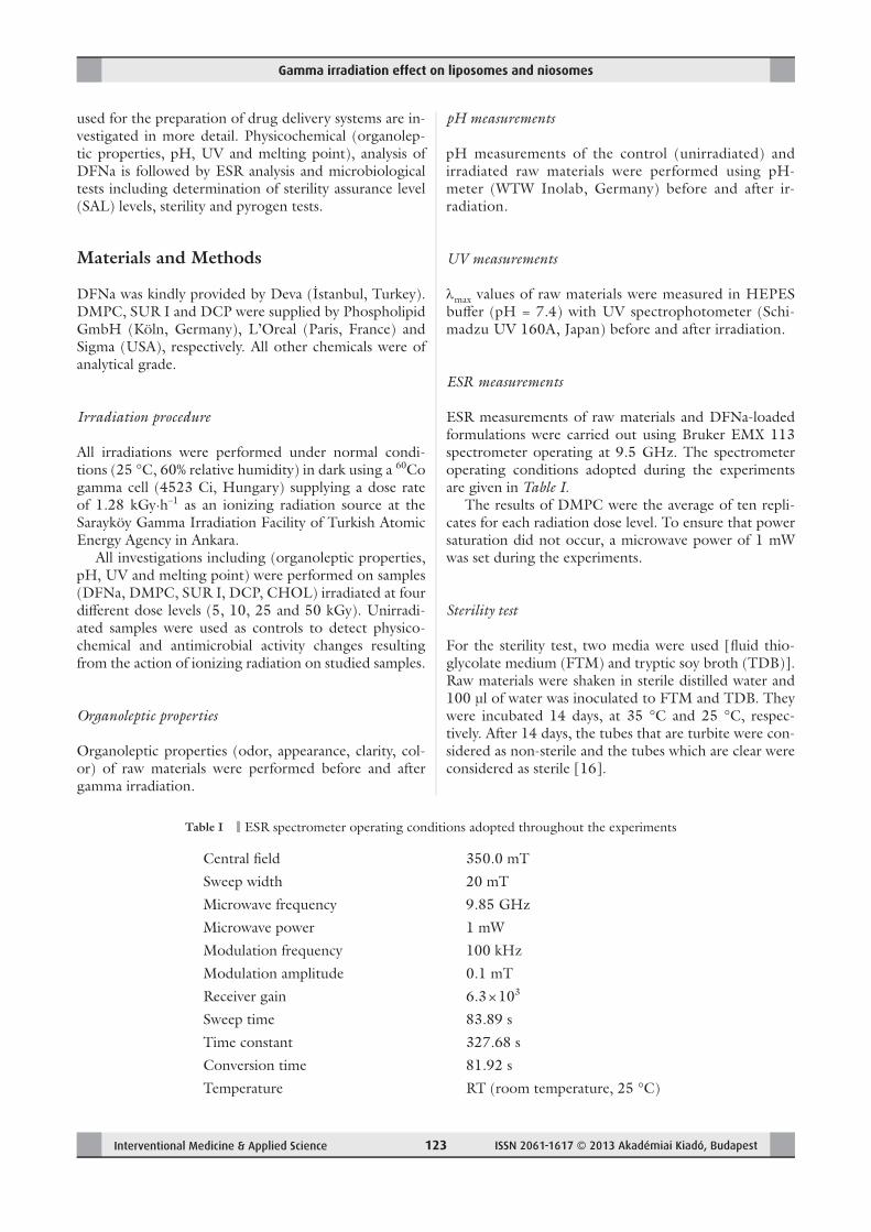
Table I ESR spectrometer operating conditions adopted throughout the experiments
Central fi eld 350.0 mT
Sweep width 20 mT
Microwave frequency 9.85 GHz
Microwave power 1 mW
Modulation frequency 100 kHz
Modulation amplitude 0.1 mT
Receiver gain 6.3 × 10³
Sweep time 83.89 s
Time constant 327.68 s
Conversion time 81.92 s
Temperature RT (room temperature, 25 °C)
Özer et al.
ISSN 2061-1617 © 2013 Akadémiai Kiadó, Budapest Interventional Medicine & Applied Science124
Pyrogen (LAL) test
The gel-clot method for assay of bacterial endotoxins
(the most common pyrogens) was examined for the
above-mentioned substances [16].
SAL determination
The samples were infected with Bacillus pumilus spore
suspension [6 × 10⁶ colony-forming unit (cfu·m1–1)]
and irradiated with various radiation dose levels (1, 5,
10, 25 and 50 kGy) and incubated in TSB plates at
35–37 °C. Bacillus pumilus colonies were enumerated
and cfu in 1 mL were calculated. SAL 10–6 dose was
calculated from the logarithmic microorganism death
graphics [17].
Stability tests
Stability tests were performed under normal (25 °C, 40%
relative humidity) and accelerated (40 °C, 75% relative
humidity) conditions over a period of 3 months. For the
accelerated stability test, the samples were stored in the
climated chamber and aliquots were taken off for the
measurements. For comparison unirradiated samples
were used as negative control.
Results and Discussion
This study describes investigation on ionizing radiation
induced oxidative damage to DFNa and phospholipids/
surfactants following gamma irradiation. Color change
in the irradiated raw materials is a simple and helpful
observation to get information about possible radiolytic
intermediates produced in these substances upon irra-
diation. As no color change was observed in irradiated
solid materials in the applied dose region of 5–50 kGy,
it can be concluded that either radiolytic intermediates
are not produced by irradiation in studied samples or
created intermediates do not exhibit any absorption in
the visible region. The negative result in color change of
the present work is consistent with the results reported
in the literature for similar compounds [18]. Experi-
mental results showed that organoleptic features such as
color, odor and clarity and odor of control and irradiated
samples did not change under accelerated test conditions
before and after irradiation (p < 0.05). The results were
in accordance with the literature [19].
pH values of raw materials changed after irradiation
at four diff erent dose levels (p > 0.05). The results are
given in Table II.λmax values of DFNa, DMPC, DCP, SUR I and
CHOL are found as 275, 245, 271, 256 and 242 nm,
respectively, before gamma irradiation. The comparative
λmax values determined from UV spectrum of samples are
given in Table III. As seen from the table, λmax values
Table II Measured pH values for control and irradiated samples (n = 6)
Substance pH
Dose rate (kGy)
0 5 10 25 50
DFNa 7.44 ± 0.03 7.37 ± 0.01 7.34 ± 0.02 7.24 ± 0.02 7.12 ± 0.02
DMPC 7.41 ± 0.01 7.41 ± 0.06 7.38 ± 0.01 7.30 ± 0.01 7.22 ± 0.03
DCP 7.40 ± 0.05 7.35 ± 0.03 7.33 ± 0.03 7.27 ± 0.03 7.21 ± 0.03
SUR I 7.43 ± 0.02 7.40 ± 0.01 7.35 ± 0.01 7.31 ± 0.01 7.28 ± 0.03
CHOL 7.44 ± 0.01 7.41 ± 0.02 7.40 ± 0.01 7.34 ± 0.03 7.18 ± 0.04
Table III λmax values calculated from UV spectrum of unirradiated and irradiated samples (n = 6)
Substance λmax (nm)
Dose rate (kGy)
0 5 10 25 50
DFNa 275 ± 0.1 274 ± 0.3 274 ± 0.6 275 ± 0.1 275 ± 0.2
DMPC 245 ± 0.4 241 ± 0.1 243 ± 0.2 245 ± 0.5 244 ± 0.2
DCP 271 ± 0.1 270 ± 0.5 270 ± 0.4 273 ± 0.3 272 ± 0.4
SUR I 256 ± 0.3 256 ± 0.3 251 ± 0.1 254 ± 0.3 255 ± 0.3
CHOL 242 ± 0.1 240 ± 0.6 241 ± 0.2 240 ± 0.4 242 ± 0.2
Gamma irradiation effect on liposomes and niosomes
Interventional Medicine & Applied Science ISSN 2061-1617 © 2013 Akadémiai Kiadó, Budapest 125
calculated from UV spectrum did not change after ir-
radiation (p > 0.05). The results are in good agreement
with the literature [20–24].
Melting point values of DFNa, DMPC, DCP, SUR
I and CHOL are 271 °C, 22 °C, 171 °C, 50.9 °C and
148 °C, respectively. Results are given in Table IV and
were compared with unirradiated results.
According to organoleptic examination, pH, λmax and
melting point analysis, no signifi cant change was found
for all samples after irradiation with diff erent radiation
dose rates (p > 0.05).
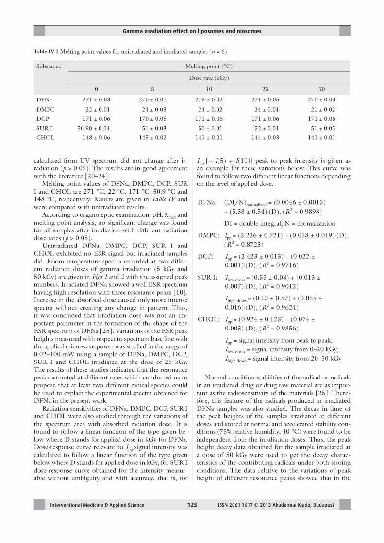
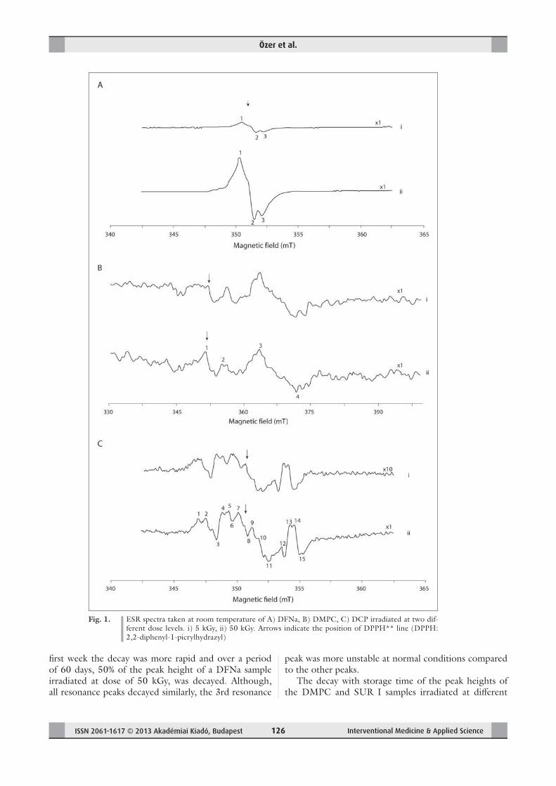
Unirradiated DFNa, DMPC, DCP, SUR I and
CHOL exhibited no ESR signal but irradiated samples
did. Room temperature spectra recorded at two diff er-
ent radiation doses of gamma irradiation (5 kGy and
50 kGy) are given in Figs 1 and 2 with the assigned peak
numbers. Irradiated DFNa showed a well ESR spectrum
having high resolution with three resonance peaks [10].
Increase in the absorbed dose caused only more intense
spectra without creating any change in pattern. Thus,
it was concluded that irradiation dose was not an im-
portant parameter in the formation of the shape of the
ESR spectrum of DFNa [25]. Variations of the ESR peak
heights measured with respect to spectrum base line with
the applied microwave power was studied in the range of
0.02–100 mW using a sample of DFNa, DMPC, DCP,
SUR I and CHOL irradiated at the dose of 25 kGy.
The results of these studies indicated that the resonance
peaks saturated at diff erent rates which conducted us to
propose that at least two diff erent radical species could
be used to explain the experimental spectra obtained for
DFNa in the present work.
Radiation sensitivities of DFNa, DMPC, DCP, SUR I
and CHOL were also studied through the variations of
the spectrum area with absorbed radiation dose. It is
found to follow a linear function of the type given be-
low where D stands for applied dose in kGy for DFNa.
Dose-response curve relevant to Ipp signal intensity was
calculated to follow a linear function of the type given
below where D stands for applied dose in kGy, for SUR I
dose-response curve obtained for the intensity measur-
able without ambiguity and with accuracy, that is, for
Ipp [= I(5) + I(11)] peak to peak intensity is given as
an example for these variations below. This curve was
found to follow two diff erent linear functions depending
on the level of applied dose.
DFNa: (DI/N)normalized = (0.0046 ± 0.0015)
+ (5.38 ± 0.54)·(D), (R2 = 0.9898)
DI = double integral; N = normalization
DMPC: Ipp = (2.226 ± 0.521) + (0.058 ± 0.019)·(D),
(R2 = 0.8723)
DCP: Ipp = (2.423 ± 0.013) + (0.022 ±
0.001)·(D), (R2 = 0.9716)
SUR I: Ilow doses = (0.55 ± 0.08) + (0.013 ±
0.007)·(D), (R2 = 0.9012)
Ihigh doses = (0.13 ± 0.57) + (0.055 ±
0.016)·(D), (R2 = 0.9624)
CHOL: Ipp = (0.924 ± 0.123) + (0.074 ±
0.003)·(D), (R2 = 0.9856)
Ipp signal intensity from peak to peak;
Ilow doses = signal intensity from 0–20 kGy;
Ihigh doses = signal intensity from 20–50 kGy
Normal condition stabilities of the radical or radicals
in an irradiated drug or drug raw material are as impor-
tant as the radiosensitivity of the materials [25]. There-
fore, this feature of the radicals produced in irradiated
DFNa samples was also studied. The decay in time of
the peak heights of the samples irradiated at diff erent
doses and stored at normal and accelerated stability con-
ditions (75% relative humidity, 40 °C) were found to be
independent from the irradiation doses. Thus, the peak
height decay data obtained for the sample irradiated at
a dose of 50 kGy were used to get the decay charac-
teristics of the contributing radicals under both storing
conditions. The data relative to the variations of peak
height of diff erent resonance peaks showed that in the
Table IV Melting point values for unirradiated and irradiated samples (n = 6)
Substance Melting point (°C)
Dose rate (kGy)
0 5 10 25 50
DFNa 271 ± 0.03 270 ± 0.01 273 ± 0.02 271 ± 0.05 270 ± 0.03
DMPC 22 ± 0.01 24 ± 0.03 24 ± 0.02 24 ± 0.01 21 ± 0.02
DCP 171 ± 0.06 170 ± 0.05 171 ± 0.06 171 ± 0.06 171 ± 0.06
SUR I 50.90 ± 0.04 51 ± 0.03 50 ± 0.01 52 ± 0.01 51 ± 0.05
CHOL 148 ± 0.06 145 ± 0.02 141 ± 0.01 144 ± 0.03 141 ± 0.01
=
Özer et al.
ISSN 2061-1617 © 2013 Akadémiai Kiadó, Budapest Interventional Medicine & Applied Science126
fi rst week the decay was more rapid and over a period
of 60 days, 50% of the peak height of a DFNa sample
irradiated at dose of 50 kGy, was decayed. Although,
all resonance peaks decayed similarly, the 3rd resonance
peak was more unstable at normal conditions compared
to the other peaks.
The decay with storage time of the peak heights of
the DMPC and SUR I samples irradiated at diff erent
Fig. 1. ESR spectra taken at room temperature of A) DFNa, B) DMPC, C) DCP irradiated at two dif-
ferent dose levels. i) 5 kGy, ii) 50 kGy. Arrows indicate the position of DPPH** line (DPPH:
2,2-diphenyl-1-picrylhydrazyl)
Gamma irradiation effect on liposomes and niosomes
Interventional Medicine & Applied Science ISSN 2061-1617 © 2013 Akadémiai Kiadó, Budapest 127
doses and stored at normal and stability conditions were
found to be independent from irradiation doses. Thus,
the peak height decay data obtained for a sample irradi-
ated at the dose of 50 kGy were used to get the decay
characteristics of the contributing radicals under both
storing conditions. The data associated with the varia-
tions of Ipp intensity showed that the species responsible
from experimental ESR signal were very unstable at nor-
mal conditions. Decay rate of the signal intensity was
very rapid during the fi rst hour just after irradiation and
72% decay in the signal intensity was calculated occur-
ring over a storage period of 90 days.
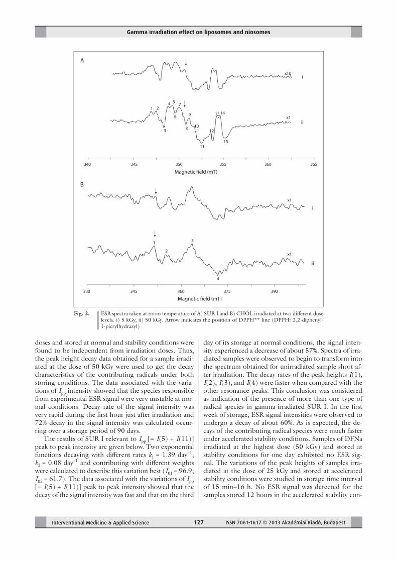
The results of SUR I relevant to Ipp [= I(5) + I(11)]
peak to peak intensity are given below. Two exponential
functions decaying with diff erent rates k1 = 1.39 day–1;
k2 = 0.08 day–1 and contributing with diff erent weights
were calculated to describe this variation best (I01 = 96.9;
I02 = 61.7). The data associated with the variations of Ipp
[= I(5) + I(11)] peak to peak intensity showed that the
decay of the signal intensity was fast and that on the third
day of its storage at normal conditions, the signal inten-
sity experienced a decrease of about 57%. Spectra of irra-
diated samples were observed to begin to transform into
the spectrum obtained for unirradiated sample short af-
ter irradiation. The decay rates of the peak heights I(1),
I(2), I(3), and I(4) were faster when compared with the
other resonance peaks. This conclusion was considered
as indication of the presence of more than one type of
radical species in gamma-irradiated SUR I. In the fi rst
week of storage, ESR signal intensities were observed to
undergo a decay of about 60%. As is expected, the de-
cays of the contributing radical species were much faster
under accelerated stability conditions. Samples of DFNa
irradiated at the highest dose (50 kGy) and stored at
stability conditions for one day exhibited no ESR sig-
nal. The variations of the peak heights of samples irra-
diated at the dose of 25 kGy and stored at accelerated
stability conditions were studied in storage time interval
of 15 min–16 h. No ESR signal was detected for the
samples stored 12 hours in the accelerated stability con-
Fig. 2. ESR spectra taken at room temperature of A) SUR I and B) CHOL irradiated at two diff erent dose
levels. i) 5 kGy, ii) 50 kGy. Arrow indicates the position of DPPH** line (DPPH: 2,2-diphenyl-
1-picrylhydrazyl)
Özer et al.
ISSN 2061-1617 © 2013 Akadémiai Kiadó, Budapest Interventional Medicine & Applied Science128
ditions. The peak height data derived from room tem-
perature ESR spectrum of DFNa sample irradiated at the
dose of 50 kGy was used as input for simulation calcula-
tions basing on a model of the presence of three radical
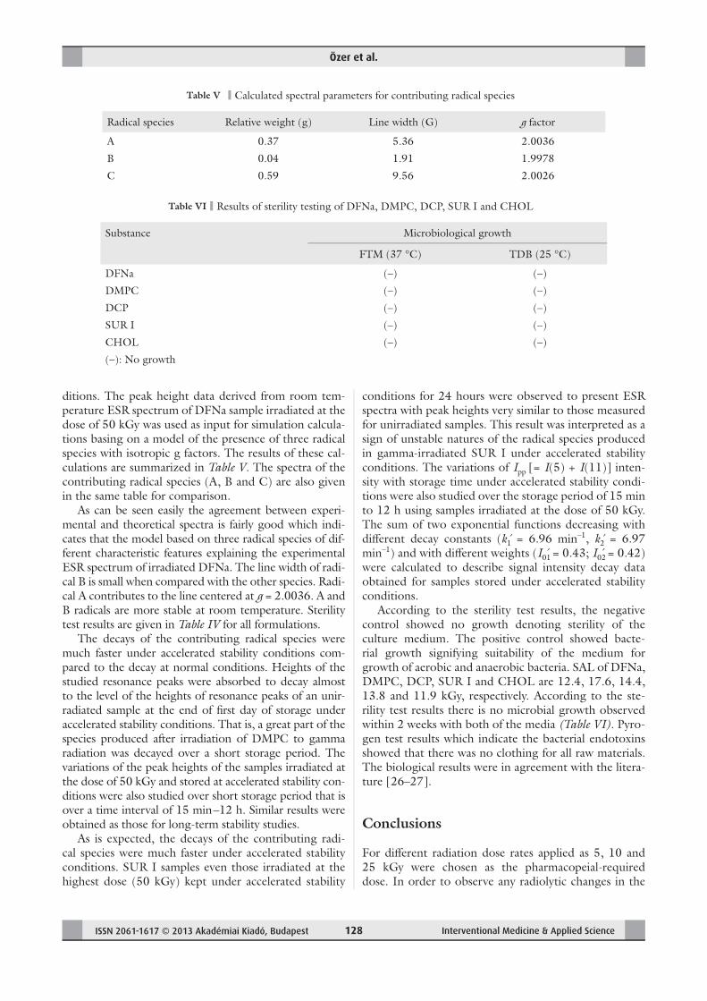
species with isotropic g factors. The results of these cal-
culations are summarized in Table V. The spectra of the
contributing radical species (A, B and C) are also given
in the same table for comparison.
As can be seen easily the agreement between experi-
mental and theoretical spectra is fairly good which indi-
cates that the model based on three radical species of dif-
ferent characteristic features explaining the experimental
ESR spectrum of irradiated DFNa. The line width of radi-
cal B is small when compared with the other species. Radi-
cal A contributes to the line centered at g = 2.0036. A and
B radicals are more stable at room temperature. Sterility
test results are given in Table IV for all formulations.
The decays of the contributing radical species were
much faster under accelerated stability conditions com-
pared to the decay at normal conditions. Heights of the
studied resonance peaks were absorbed to decay almost
to the level of the heights of resonance peaks of an unir-
radiated sample at the end of fi rst day of storage under
accelerated stability conditions. That is, a great part of the
species produced after irradiation of DMPC to gamma
radiation was decayed over a short storage period. The
variations of the peak heights of the samples irradiated at
the dose of 50 kGy and stored at accelerated stability con-
ditions were also studied over short storage period that is
over a time interval of 15 min –12 h. Similar results were
obtained as those for long-term stability studies.
As is expected, the decays of the contributing radi-
cal species were much faster under accelerated stability
conditions. SUR I samples even those irradiated at the
highest dose (50 kGy) kept under accelerated stability
conditions for 24 hours were observed to present ESR
spectra with peak heights very similar to those measured
for unirradiated samples. This result was interpreted as a
sign of unstable natures of the radical species produced
in gamma-irradiated SUR I under accelerated stability
conditions. The variations of Ipp [= I(5) + I(11)] inten-
sity with storage time under accelerated stability condi-
tions were also studied over the storage period of 15 min
to 12 h using samples irradiated at the dose of 50 kGy.
The sum of two exponential functions decreasing with
diff erent decay constants (k1́ = 6.96 min–1, k2́ = 6.97
min–1) and with diff erent weights (I01́ = 0.43; I02́ = 0.42)
were calculated to describe signal intensity decay data
obtained for samples stored under accelerated stability
conditions.
According to the sterility test results, the negative
control showed no growth denoting sterility of the
culture medium. The positive control showed bacte-
rial growth signifying suitability of the medium for
growth of aerobic and anaerobic bacteria. SAL of DFNa,
DMPC, DCP, SUR I and CHOL are 12.4, 17.6, 14.4,
13.8 and 11.9 kGy, respectively. According to the ste-
rility test results there is no microbial growth observed
within 2 weeks with both of the media (Table VI). Pyro-
gen test results which indicate the bacterial endotoxins
showed that there was no clothing for all raw materials.
The biological results were in agreement with the litera-
ture [26–27].
Conclusions
For diff erent radiation dose rates applied as 5, 10 and
25 kGy were chosen as the pharmacopeial-required
dose. In order to observe any radiolytic changes in the
Table V Calculated spectral parameters for contributing radical species
Radical species Relative weight (g) Line width (G) g factor
A 0.37 5.36 2.0036
B 0.04 1.91 1.9978
C 0.59 9.56 2.0026
Table VI Results of sterility testing of DFNa, DMPC, DCP, SUR I and CHOL
Substance Microbiological growth
FTM (37 °C) TDB (25 °C)
DFNa (–) (–)
DMPC (–) (–)
DCP (–) (–)
SUR I (–) (–)
CHOL (–) (–)
(–): No growth
Gamma irradiation effect on liposomes and niosomes
Interventional Medicine & Applied Science ISSN 2061-1617 © 2013 Akadémiai Kiadó, Budapest 129
samples, 50 kGy was employed as the accelerated condi-
tion [7–9, 28].
Physicochemical properties of the irradiated (5, 10,
25, 50 kGy) and unirradiated DFNa, DMPC, DCP,
CHOL and SUR I; were examined. Physicochemical
properties are organoleptic properties, pH, λmax and
melting point. During the experiments, the color of
the samples did not change after irradiation. pH values,
melting point, spectral properties (except ESR) were
determined with some changes which are independent
from the radiation dose [3].
According to ESR analysis, a model based on three
radical species was found to explain well the experimen-
tal results obtained for gamma-irradiated DFNa. Radia-
tion yield of solid DFNa was not high even at the dose
of 50 kGy and the radiolytical intermediates produced
in DFNa decayed fast at room temperature. The irradi-
ated sample stored in accelerated stability conditions for
12 hours exhibited no ESR signal. This means that solid
DFNa and drugs containing DFNa as active ingredient
could be safely sterilized by irradiation as the radicals de-
cay only in a day time when it is stored at stability condi-
tions and it has quenched rather fast even if it is stored at
normal stability conditions [29, 30].
Although very weak, gamma-irradiated DMPC ex-
hibits ESR signal. This signal is embedded in noise and
natural ESR signal of unirradiated DMPC. From varia-
tions of the heights of the assigned resonance peaks it
was concluded that more than one type of radicals could
contribute to the formation of experimental ESR spec-
trum. However, it was not possible to determine the
type of these radicals due to complex nature of recorded
experimental spectra.
Radiation yield of solid DMPC was found to be very
low even at the applied dose as high as 50 kGy and the
life times at normal and accelerated stability conditions
of the radical/radicals produced upon irradiation were
calculated to be very short. Based on these results, it was
concluded that solid DMPC and drug delivery systems
containing DMPC as active ingredient could be safely
sterilized by gamma irradiation and that ESR technique
could be used successfully in monitoring the radiosteril-
ization of this compound [3].
From microwave power saturation and stability re-
sults, it was concluded that more than two radicals of
diff erent types were produced upon gamma irradiation
of SUR I.
Spectrum simulation calculations were not carried
out due to complex nature of the experimental spectra.
Signal-to-noise ratio was too low even for samples ir-
radiated even at the highest dose (50 kGy) and there-
fore, it was concluded that SUR I was not a sensitive
compound to gamma irradiation. Besides, radicals pro-
duced upon irradiation were unstable under normal and
accelerated stability conditions. As expected, the decay
of radical species under stability conditions was calcu-
lated to be much faster under accelerated stability condi-
tions. Signal intensities of the samples irradiated at the
dose of 50 kGy were found to decrease to the levels of
the intensities recorded for unirradiated samples.
Thus, it was concluded that solid SUR I could not
be sterilized by itself gamma irradiation due to its poor
dosimetric feature, but drug delivery systems containing
SUR I as active ingredient could be safely sterilized by
radiation [3].
All the raw materials, including DFNa, DMPC, DCP
and SUR I, can be sterilized safely by gamma irradia-
tion at radiation doses of 12.4 kGy, 17.6 kGy, 14.4 kGy,
13.8 kGy and 11.9 kGy, respectively (below pharmaceu-
tical requirement of 25 kGy) without causing any no-
table changes. CHOL is the most sensitive substance for
the irradiation process in comparison with the others.
It might be sterilized below 10 kGy and further studies
should be carried out to evaluate the eff ect of irradiation
on the function of CHOL. The radicals of raw materi-
als produced upon irradiation were unstable under nor-
mal and accelerated stability conditions indicating that
gamma radiation sterilization can be used as an eff ective
method for the sterilization of those substances.
As conclusion, this study showed that DFNa, DMPC,
DCP, SUR I and CHOL can be sterilized with lower
doses, namely 12.4 kGy, 17.6 kGy, 14.4 kGy, 13.8 kGy
and 11.9 kGy, respectively. The confi rmation and the
validation of the SAL doses which are below 25 kGy is
a big advantage to the researcher or to the customer in
order to save money and time, because sterilization is
achieved by the irradiation of the products in relation
with time. Using lower doses instead of 25 kGy means
the reduction of irradiation time. The sterilization fee is
paid due to the sterilization time, thus, lower SAL values
cost less money. This study showed that gamma radia-
tion sterilization is a reliable sterilization method for a
wide range of raw materials.
Acknowledgements
We gratefully acknowledge the support of The Sci-
entifi c and Technological Research Council of Turkey
(TÜBİTAK) for their support (project no: SBAG 107
S 140). We also would like to thank Deva, L’Oreal, and
Phospholipid GmbH for supplying DFNa, SUR I and
phospholipids, respectively.
References
1. Bushell JA, Claybourn M, Wiliams HE, Murphy DM: An EPR
and ENDOR study of γ- and β-radiation sterilization in poly (lac-
tide-co-glycolide) polymers and microspheres. J Control Rel 110,
49–57 (2005)
2. Mohammed AR, Bramwell VW, Coombes AGA, Perrie Y: Lyophi-
lisation and sterilization of liposomal vaccines to produce stable
and sterile products. Methods 40, 30–38 (2006)
Özer et al.
ISSN 2061-1617 © 2013 Akadémiai Kiadó, Budapest Interventional Medicine & Applied Science130
3. Erdoğan S, Özer AY, Ekizoğlu M, Özalp M, Çolak Ş: Gamma
irradiation of liposomal phospholipids. FABAD J Pharm Sci 31,
182–190 (2006)
4. Olguner Mercanoğlu G, Özer AY, Çolak Ş, Korkmaz M, Özalp
M, Ekizoğlu M, Barbarin N, Tilquin B: Radiosterilization of sul-
fonamides I: Determination of the eff ects of gamma irradiation on
solid sulfonamids. Rad Phys Chem 69, 511–520 (2004)
5. Botelho ML, Verde SC, Alves L, Balchior A, Reymao J, Trabulo
S, Gaspar MM, Cruz MEM, Simoes S: Radiation sterilization of
antibiotic liposome formulations: A case study. Rad Phys Chem
76, 1542–1546 (2007)
6. Berk F, Özer AY: Radiation sterilization of medical devices.
FABAD J Pharm Sci 24, 223–231 (1999)
7. Berk F: Tek Kullanımlık tıbbi malzemelerin gama radyasyonu ile
sterilizasyonu ve diğer yöntemlerle karşılaştırılması. MSci thesis,
Institute of Health Science, Hacettepe Universty, Ankara, Turkey
(2002)
8. Liman V, Özer AY, Çolak S, Korkmaz M, Kılıç E, Özalp M: The
eff ects of gamma radiation sterilization on cefazolin sodium.
FABAD J Pharm Sci 30, 124–132 (2005)
9. Naki N: Kozmetikler ve kozmetik hammaddelerinin gama radyas-
yonla sterilizasyonu/dekontaminasyonu üzerinde çalışmalar. MSci
thesis, Institute of Health Science, Hacettepe University, Ankara,
Turkey (2003)
10. Olguner G: Radiation sterilization of sulfonamides and compari-
son with the other sterilization techniques. MSci thesis, Institute
of Health Science, Hacettepe University, Ankara, Turkey (2000)
11. Türker S, Erdoğan S, Özer AY, Ergün EL, Tuncel M, Bilgili H,
Deveci S: Scintigraphic imaging of radiolabelled drug delivery sys-
tems in rabbits with arthritis. Int J Pharm 296, 34–43 (2005)
12. Zuidam NJ, Gouw HK, Barenholz Y, Crommelin DJ: Physical (in)
stability of liposomes upon chemical hydrolysis: The role of lyso-
phosphalipids and fatty acids. Biochim Biophys Acta 22, 101–110
(1995)
13. Barbarin N, Tilquin B: Study of nonvolatile degradation com-
pounds produced by radiosterilization of cefatoxime. Radiat Phys
Chem 60, 359–367 (2001)
14. Gibella M, Crucq AS, Tilquin B, Stocker P, Lesgards G, Raffi
Y: ESR studies of some irradiated pharmaceuticals. Radiat Phys
Chem 58, 69–76 (2000)
15. Olguner Mercanoğlu G, Özer AY, Özalp M, Ekizoğlu M: Ra-
diosterilization of sulfonamides II: Determination of the eff ects
of gamma irradiation on commercial sulfanomide preparations.
Turkish J Pharm Sci 4, 159–170 (2007)
16. British Pharmacopoeia (2004): University Press, Cambridge
17. Kantaglu O (2004): Endüstride sterilizasyon kursu, TAEK, Ankara
18. Phillips GO, Power DM, Sewart MCG: Eff ects of gamma irradia-
tion on sodium sulphacetamide. Radiat Res 46, 236–250 (1971)
19. Amin GAM, Spyrou NM: Study of gamma-radiation-induced op-
tical eff ects in Ge-Se-Cd for possible industrial dosimetric applica-
tions. Rad Phys Chem 72, 419–422 (2007)
20. Pandey BN, Lathika KM, Mishra KP: Radiation induced oxidative
damage modifi cation by cholesterol in liposomal membrane. Rad
Phys Chem 54, 481–489 (1999)
21. Pandey BN, Lathika KM, Mishra KP: Modifi cation of radiation-
induced oxidative damage in liposomal and microsomal mem-
brane by eugenol. Rad Phys Chem 75, 384–391 (2006)
22. Plikk P, Odelius K, Hakkarainen M, Albertsson AC: Finalizing the
properties of porous scaff olds of aliphatic polyesters through radia-
tion sterilization. Biomaterials 27, 5335–5347 (2006)
23. Sprinz H, Schwinn J, Naumov S, Brede O: Mechanism of thiyl
radical-catalyzed isomerization of unsaturated fatty acid residues
in homogeneous solution and in liposomes. BBA 1483, 91–100
(2000)
24. Varshney L, Dodke PB: Radiation eff ect studies on anticancer
drugs, cyclophosphamide and doxorubicin for radiation steriliza-
tion. Rad Phys Chem 71, 1103–1111 (2004)
25. Deeley CM: A basic interpretation of the technical language of
radiation processing. Rad Phys Chem 71, 503–507 (2004)
26. El-Ridy MS, Mostafa DM, Shehab A, Nasr EA, El-Alim SA:
Biological evaluation of pyrazinamide liposomes for treatment of
Mycobacterium tuberculosis. Int J Pharm 330, 82–88 (2007)
27. Katusin-Razem B, Mihaljevic B, Razem D: Microbial decontami-
nation of cosmetic raw materials and personal care products by
irradiation. Rad Phys Chem 66, 309–316 (2003)
28. Dam AM, Gazso LG, Kaewpila S, Maschek I: Radiation treatment
of pharmaceuticals. Radiat Chem 47, 515–517 (1996)
29. Da Costa ZM, Pontusehka WM, Campos LL: Study of the ESR
signal of gamma irradiated hydroxyapatite for dose assessment.
Nucl Instrum Meth B 218, 283–288 (2004)
30. Faucitano A, Buttafava A, Montanari L, Cilurzo F, Conti B, Genta
I, Valvo L: Radiation-induced free radical reactions in polymer/
drug systems for controlled release: An EPR investigation. Radiat
Phys Chem 67, 61–72 (2003)