Embed Size (px)
Citation preview
The effects of cholinergic neuromodulation on neuronalphase-response curves of modeled cortical neurons
Klaus M. Stiefel & Boris S. Gutkin &
Terrence J. Sejnowski
Received: 3 September 2007 /Revised: 23 June 2008 /Accepted: 9 July 2008 / Published online: 11 September 2008# The Author(s) 2008. This article is published with open access at Springerlink.com
Abstract The response of an oscillator to perturbations isdescribed by its phase-response curve (PRC), which isrelated to the type of bifurcation leading from rest to tonicspiking. In a recent experimental study, we have shown thatthe type of PRC in cortical pyramidal neurons can beswitched by cholinergic neuromodulation from type II(biphasic) to type I (monophasic). We explored howintrinsic mechanisms affected by acetylcholine influencethe PRC using three different types of neuronal models: atheta neuron, single-compartment neurons and a multi-compartment neuron. In all of these models a decrease inthe amount of a spike-frequency adaptation current was a
necessary and sufficient condition for the shape of the PRCto change from biphasic (type II) to purely positive (type I).
Keywords Phase response curves . Cortex .
Neuromodulation .Muscarine . Acetylcholine .
Pyramidal neuron . Conductance-based model .
Multi-compartmental model .M-current
1 Introduction
Acetylcholine, a central nervous system neuromodulator, isfurnished to almost the entire cortex through widelydiverging inputs from a number of subcortical cholinergicnuclei. At the behavioral level acetylcholine plays animportant role in several aspects of cognitive function suchas arousal levels, attention and vigilance, as well asmemory and learning (Hasselmo 2006; Steriade 2004).One theory of learning and memory suggests that levels ofcholinergic neuromodulation may switch cortical networksbetween learning and memory-recall modes (Hasselmo 1999).At the circuit level, cholinergic modulation appears to controlthe frequency content and the coherence of corticaloscillations. For example gamma-band synchronizedoscillations are observed under increased cholinergic levelsin the cortex (Rodriguez et al. 2004)
In vitro, experiments have shown that cholinergicantagonists promote synchronized high frequency oscilla-tory activity (Buhl et al. 1998; Fisahn et al. 1998). Thissuggests that acetylcholine has a significant modulatorycontrol over the emergence of rhythmic activity in corticalcircuits. Certainly such effects are in part due to theacetylcholine modulation of synaptic transmission (Auerbachand Segal 1996) and plasticity (Artola and Singer 1993;
J Comput Neurosci (2009) 26:289–301DOI 10.1007/s10827-008-0111-9
DO00111; No of Pages
Action Editor: G. Bard Ermentrout
K. M. Stiefel : T. J. SejnowskiHoward Hughes Medical Institute,The Salk Institute for Biological Studies,La Jolla, CA 92037, USA
B. S. GutkinNeural Theory Group, DEC, ENS,CNRS and the College de France,Paris, Francee-mail: [email protected]
T. J. SejnowskiDivision of Biological Sciences,University of California at San Diego,La Jolla, CA 92093, USAe-mail: [email protected]
Present address:K. M. Stiefel (*)Okinawa Institute of Science and Technology,Okinawa, Japane-mail: [email protected]
Stiefel et al. 2005) and in part due to cholinergicmodulation of intrinsic cell excitability (McCormick1993). Cholinergic modulation affects a variety of bothionotropic (nicotinic) and metabotropic (muscarinic)receptor targets (Schoebesberger et al. 2000). Acetylcholineacting on muscarinic receptors downregulates several slowpotassium currents (IKM, IKAHP (Madison et al. 1987;McCormick 1993)), and changes the input resistance bydown regulating the leak conductance. These currents controlcellular excitability and repetitive firing of cortical pyramidalneurons.
Given such diverse acetylcholine action at the single celllevel, an interesting issue is which of the effects mediatedby the muscarinic acetylcholine-receptor modulation can beresponsible for the emergence of collective behaviors, suchas synchrony and oscillations. In fact, previous theoreticalwork has hinted that suppression of slow cation channels canbias pyramidal neurons toward synchrony with excitatorysynaptic coupling by modulating the dynamics of spikegeneration (Crook et al. 1998a, b; Ermentrout et al. 2001). Inthis report our goal is to systematically examine the variousmechanisms of cholinergic action and to identify those thathave the greatest impact on the dynamics of spike generationin cortical neurons.
We focus on the cholinergic effects on the excitability ofregular spiking (pyramidal) neurons that can be describedas oscillators. Specifically, we examine the dynamicsunderlying the spike generation and how the cell goes fromexcitable to periodically firing. Knowing the type of thistransition can provide insight not only about the input-output function of the neurons (the FI curve) but also aboutthe potential presence of subthreshold oscillations as wellas the synaptic mechanisms that can potentially lead tosynchrony (Ermentrout 1996). This transition from rest (astable fixed point and a threshold) to spiking (following alimit cycle) can occur via two major dynamical mecha-nisms: the “saddle-node on an invariant cycle” bifurcation(from here on referred to as “saddle-node” bifurcation) andthe subcritical Hopf-Andronov bifurcation. In the case ofthe saddle-node bifurcation, a stable fixed point (a node)and a saddle-point collide into a saddle-node and disappearto leave a stable limit cycle. In the case of the subcriticalHopf-Andronov bifurcation, the loss of stability occurswhen the real parts of conjugate eigenvalues grow positive,hence destabilizing the fixed point (see Izhikevich (2007)for further discussion). These different bifurcations affect theshape of the FI curve, which is continuous for the SNICbifurcation and discontinuous for the Hopf-Andronov case.Furthermore Hopf-Andronov systems can exhibit significantsubthreshold oscillations, and hysteresis.
These two classes of bifurcations also result in qualita-tively different phase response curves (PRCs). The PRCscharacterize the phase shift the results when a neuronal
oscillator is perturbed by an input as a function of the phaseof the perturbation. (Although the PRC can be constructedfor both excitatory and inhibitory perturbations, thestandard convention is to report results for depolarizinginputs.) In the case of a saddle-node bifurcation, the PRC ispurely positive, or type I (except possibly for an arbitrarilysmall region near the origin Hansel and Mato 1995), with aphase advance in response to perturbations at all phases(Ermentrout 1996). In the case of a Hopf-Andronovbifurcation, the PRC is biphasic, or type II, with a phasedelay in response to perturbations early in the period and aphase advance in response to perturbations late in theperiod (Hansel and Mato 1995; Ermentrout 1996; Moehliset al. 2006).
Computational studies have previously observed thatunder block of slow voltage dependent potassium currents(IM), the PRC changed quantitatively, associated with achange in the bifurcation structure (Ermentrout et al. 2001).The PRCs went from biphasic with full current conductanceto a monophasic when the current was sufficientlyweakened. Gutkin et al. (2005) further hypothesized thatthe differences seen in the PRCs of various pyramidalneurons experimentally (Reyes and Fetz 1993) can beexplained by the relative levels of the slow low-thresholdvoltage-dependent potassium current. Modeling studies alsosuggest that block of the high threshold, Ca2+ dependentslow potassium current (IAHP) cannot change the PRC type,but affects only the relative skew of the PRC peak. We thushypothesized that the IM is the key component necessary toinduce the switch of PRC type. In a recent experimentalstudy, cholinergic neuromodulation shifted the type of PRCfrom type II to type I in cortical pyramidal neurons (Stiefelet al. 2003). The decrease in the IM current leads to adecrease in spike-frequency adaptation and is also a primecandidate for the switch from type II to type I PRC.
However, as pointed out above, acetylcholine changes amultitude of mechanisms, so modulation of IM may not be anecessary or sufficient factor in switching the PRC type.We address this question systematically with simulationsusing successively more complex neuronal models. Thefirst model is the theta-neuron, for which the switch ofPRCs can be shown directly. We then examine a simplemodel (containing a restricted set of voltage dependentchannels) and a more complex single-compartment model(with a larger set of channels) and a multi-compartmentneuron.
For each of the above model neurons we show that thesum total of cholinergic effects causes a switch of thePRC. We further identified the down-regulation of a low-threshold spike-frequency adaptation current (IM) as thecause for the PRC type to switch from type II to type I,and that the other mechanisms are not able to induce theswitch.
290 J Comput Neurosci (2009) 26:289–301
2 Methods
All simulations were carried out with the NEURONcomputer program (Hines and Carnevale 1997, 2000). Thesimulations used the CNexp numerical integration methodwith dt=0.025 ms. The source code for these simulations(NEURON hoc and mod files) containing all simulationparameters is available as supplementary material or byrequest from K.M.S. and can be obtained from the SenseLabmodel database (http://senselab.med.yale.edu/modeldb/). Allsimulations were run in NEURON version 5.x either underWindows XP on an Athlon AMD 64 processor or underFreeBSD 4.4 on a Pentium IV processor.
We used four different neuronal models, with increasingdimensionality. These were the theta neuron (D=2), a simplesingle compartment neuron (D=5), a more complex single-compartment neuron (D=9) and a multi-compartment neuron(D>100). The latter neuron model contained 1,393 compart-ments with up to six currents per compartment (see below).Because the currents in the different compartments are notindependent (and thus not every state variable is a separatedimension), the dimensionality is ill defined.
2.1 Theta-neuron
The theta-neuron, the simplest neural model that spikes, isan oscillator with a single phase variable, θ (see e.g. Gutkinand Ermentrout 1998). The addition of an adapting currentmakes it a two-dimensional model following the equations:
dD
dt¼ 1� cosDþ 1þ cosDð Þ b þ sgM þ Sð Þ ð1Þ
tMds
dt¼ kMDð Þ 1� sð Þ � s ð2Þ
kM ¼ ae�b 1�cos D�Dthð Þð Þ ð3Þwhere in Eq. (1) is β the tonic drive, S the synaptic drive; inEq. (2) s is the state variable for the adaptation, Θth, b, gMand CM are threshold, activation slope, maximal conductanceand time-constant of the adapting current respectively.
2.2 Simple single-compartment neuron
The single-compartmental neuron model was geometricallysimple, but included biophysically realistic mechanisms forneuronal spiking based on Hodgkin and Huxley (1952).This model pyramidal neuron had identical equations forthe current kinetics as in (Golomb and Amitai 1997) butincluded, only the fast Na+, leak, delayed rectifier K+
currents, and a slower K+ current (IKslow), responsible forspike frequency adaptation. This last current was an
amalgam of low threshold slow potassium currents, ofwhich IM is an example. Since we did not adjust theparameters to fit precisely the data on the muscarinicsensitive IM, we refer to this current generically as IKslow.The voltage equation is:
CMdV
dt¼ �m3hgNa V � ENað Þ � ngKDR V � EKð Þ
� sgKs V � EKð Þ � gleak V � Eleakð Þ � Iinj; ð4Þwith m, n, h and s the voltage-dependent activationvariables that follow equations of the form:
dx
dt¼ t Vð Þ x� x1 Vð Þð Þ; ð5Þ
where τ(V) and x∞(V) are experimentally determinedfunctions for the time constant and infinite activation stateof the state variables, Cm the membrane capacitance, V themembrane potential, m, h, n and s the state variables of theNa+ current activation/inactivation and the delayed rectifier/slow K+ current, gNa, gKDR, gKs and gleak the conductancesof the respective currents, and ENa, EK and Eleak theirrespective reversal potentials. Iinj is the injected current.
2.3 Complex single-compartment neuron
The single-compartment model of neuronal spiking is anexpanded version of a previous model (Golomb and Amitai1997, Eqs. (4–5)). IM and IAHP K+ currents were included togenerate an after-hyperpolarization (AHP). Because the IAHPis Ca2+-dependent, an IL Ca
2+-current and an exponential Ca2+
decay representing a Ca2+-buffer were also included. Also, ahyperpolarization-activated mixed cation (depolarizing)current, IH, and an inactivating fast K+-current, IKA, wereincluded. All these currents occur in cortical pyramidalneurons (Johnston and Wu 1994). The formalisms for thecurrents are identical to the previous model. Note that thismodel simulates neuronal dynamics of both the membranepotential and the Ca2+ concentration. The formalism for theCa2+ dynamics follows:
dCa2þ
dt¼ 2ICa2þ
dþ Ca2þ0 � Ca2þ
� �
tCa2þ; ð6Þ
with F the Faraday constant (96,500 C) and δ the fraction ofthe intracellular space affecting the Ca2+ dependent current (a1 μm shell). This term couples the Ca2+ current to the Ca2+
concentration, which in the absence of any influx relaxesback to Ca2+0 with the time constant τCa2+=35 ms.
2.4 Complex multi-compartment neuron
This model, in contrast to the previous ones, incorporatesthe spatial structure of the dendrites and is based on a
J Comput Neurosci (2009) 26:289–301 291
reconstructed cortical layer II pyramidal neuron (Mainenand Sejnowski 1995). The shape of the neuron is depictedin Fig. 4(a). The neuronal morphology had 257 compart-ments. When investigating the effects of local dendriticchanges, the number of compartments was increased to1,393, to assure that each compartment was sufficientlysmall (<8 μm) to be approximately isopotential.
The voltage equation for each compartment contained anadditional term for current flow from the neighboringcompartments (for the dendritic compartment n):
CmdVn
dt¼
XI � Iinj � gax Vn � Vn�1ð Þ � gax Vnþ1 � Vnð Þ;
ð7Þ
where gax is the axial conductance and ΣI is the sum of allintrinsic ionic currents. The model consisted of axonal,somatic and dendritic regions with distinct properties.These membrane properties were adjusted from theiroriginal values in order to more realistically reproducethe spike and AHP-shape and pattern seen in our record-ings. Specifically, we increased the somato-dendriticconductances gM=2×−4 S cm−2 and gleak=2×10
−5 S cm−2
and decreased the somatic and somato-dendritic conductan-ces to gAHP(Ca)=1×10
−2 S cm−2 and gCaL=1×10⁻⁵ S cm−2.
In addition, the time constant of the Ca2+-decay wasdecreased to 35 ms to reproduce the shape of the recordedAHP more closely and to match the complex single-compartment model. gKDR in the soma was decreased from200 to 100 S cm−2. The axonal conductances remainedunchanged.
2.5 Determination of phase-response curves
We determined the PRCs in all models by first simulatingspiking until the spike discharge frequency reachedstationarity (ISIn/ISIn−1<1.01). Then we perturbed thevoltage by injecting a small (<1 mV) and short (<20 ms)exponentially decaying depolarizing synaptic event. The Δtof the ISI as a result of the synaptic input was determinedand normalized to the control (unperturbed) ISI duration.This was repeated at 64 phase positions spanning the entireperiod of the oscillation. Changing conductances to mimiccholinergic effects can alter (increase) the firing frequencyof the neuron. When we intended to keep the firingfrequency of the model neuron constant during a parametersweep, we determined a firing frequency/current curve (FIcurve) for each parameter value and used it as a lookup-table to determine the appropriate current for the desiredfrequency.
3 Results
3.1 Theta-neuron
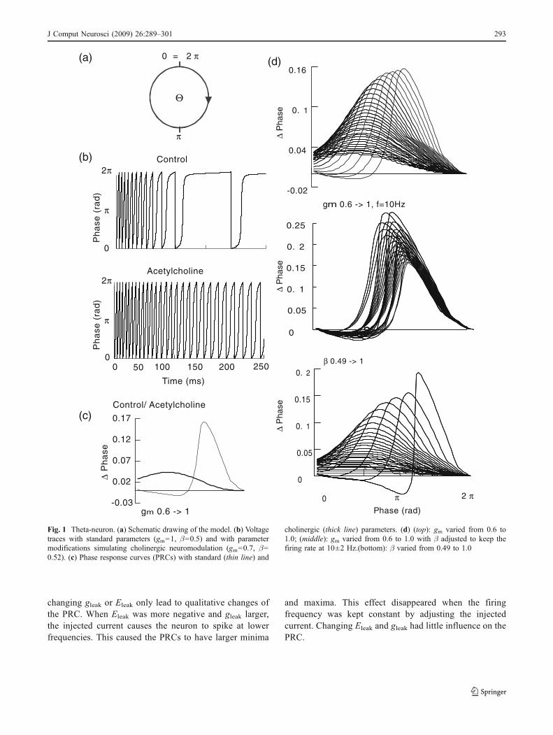
Using the theta-neuron, we modeled the effects of cholinergicmodulation by decreasing the adaptation current (gm from 1to 0.7) and by increasing the drive (β from 0.5 to 0.52).These manipulations mimicked the experimentallyobserved decrease in IM, the depolarization and theincrease in the input resistance. In response to thesemanipulations, the steady-state firing frequency increasedfrom 8.6 to 69.1 Hz and the PRC of the theta neuronswitched from type II to type I (Fig. 1(b, c)). This switchfrom type II to type I was observed both when loweringgm (1 to 0.6, Fig. 1(d), top) and when increasing β (0.49 to1, Fig. 1(d), bottom). During these parameter sweeps, thefiring frequency increased from 10.2 to 93 Hz and from5.8 to 196 Hz, respectively.
High firing frequencies can mask type II dynamics whenthe frequency is so high that the dynamics of the adaptationcurrent become much slower than the spiking dynamics.We thus performed a parameter sweep over gm (1 to 0.6)with β adjusted to keep the firing frequency at 10±2 Hz(Fig. 1(d), center). With decreasing gm, the negative part ofthe PRC decreased and its maximum, minimum and zero-crossing shifted to the left (earlier parts of the phase). Thezero-crossing, when present, also shifted to the left. Thisshows that a change in gm is sufficient to alter the PRC ofthe theta neuron.
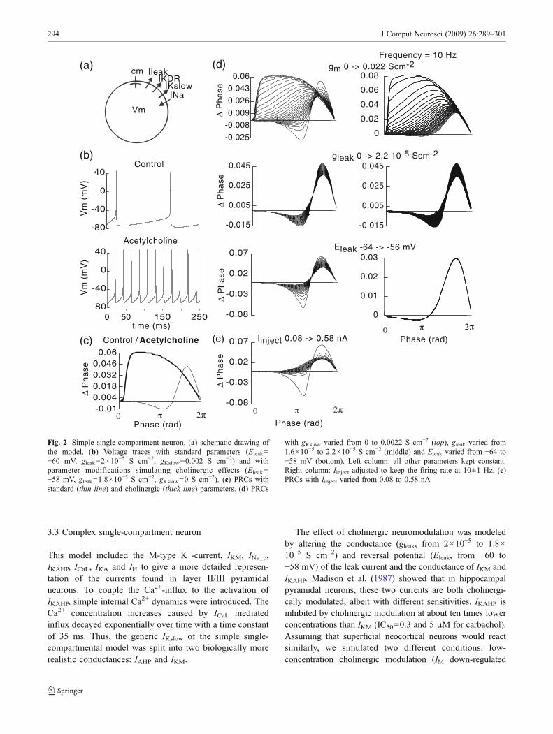
3.2 Simple single-compartment neuron
To model cholinergic neuromodulation in a single com-partment model with Ileak, INa, IKDR and IKslow, we alteredthe leak conductance (gleak, 2×10
−5 to 1.8×10⁻⁵ S cm−2),reversal potential of the leak current (Eleak, −60 to −58 mV)and the conductance of IKslow (gKslow, 2×10
−3 to 0 S cm−2).These modifications led to a switch of the PRC of themodel neuron from type II to type I (Fig. 2(b)).
We conducted parameter sweeps over all the parametersmanipulated to model cholinergic neuromodulation, bothwith all other parameters kept at default values and with theinjected current adjusted to keep the firing frequency at10±1 Hz (gleak: 1.6×10
−5 to 2.2×10⁻⁵ S cm−2, Eleak: −64 to56 mV, gKslow: 0 to 2.2×10⁻³ S cm−2). We found that only achange in the value of gm lead to a qualitative change of thePRC, a switch from type II to I. This was observed bothwith and without an adjustment of the injected current tokeep the firing frequency at 10 Hz. In both cases, themaximum, minimum and zero-crossing of the PRC shiftedto the right with higher values of gKslow. In contrast,
292 J Comput Neurosci (2009) 26:289–301
changing gleak or Eleak only lead to qualitative changes ofthe PRC. When Eleak was more negative and gleak larger,the injected current causes the neuron to spike at lowerfrequencies. This caused the PRCs to have larger minima
and maxima. This effect disappeared when the firingfrequency was kept constant by adjusting the injectedcurrent. Changing Eleak and gleak had little influence on thePRC.
50 100 150 200 2500
0
π
2π
0
π
2π
0 = 2 π
π
Θ
Time (ms)
Ph
ase
(ra
d)
Ph
ase
(ra
d)
(a)
(b)
Acetylcholine
Control
-0.03
0.02
0.07
0.12
0.17
∆ P
ha
se
(c)Control/ Acetylcholine
gm 0.6 -> 1
-0.02
0.04
0. 1
0.16
2
0.05
0. 1
0.15
0. 2
0.25
0 2
0
0
0.05
0. 1
0.15
0. 2
π 2 π
Phase (rad)
(d)
gm 0.6 -> 1, f=10Hz
β 0.49 -> 1
∆ P
hase
∆ P
hase
∆ P
hase
Fig. 1 Theta-neuron. (a) Schematic drawing of the model. (b) Voltagetraces with standard parameters (gm=1, β=0.5) and with parametermodifications simulating cholinergic neuromodulation (gm=0.7, β=0.52). (c) Phase response curves (PRCs) with standard (thin line) and
cholinergic (thick line) parameters. (d) (top): gm varied from 0.6 to1.0; (middle): gm varied from 0.6 to 1.0 with β adjusted to keep thefiring rate at 10±2 Hz.(bottom): β varied from 0.49 to 1.0
J Comput Neurosci (2009) 26:289–301 293
3.3 Complex single-compartment neuron
This model included the M-type K+-current, IKM, INa_p,IKAHP, ICaL, IKA and IH to give a more detailed represen-tation of the currents found in layer II/III pyramidalneurons. To couple the Ca2+-influx to the activation ofIKAHP, simple internal Ca2+ dynamics were introduced. TheCa2+ concentration increases caused by ICaL mediatedinflux decayed exponentially over time with a time constantof 35 ms. Thus, the generic IKslow of the simple single-compartmental model was split into two biologically morerealistic conductances: IAHP and IKM.
The effect of cholinergic neuromodulation was modeledby altering the conductance (gleak, from 2×10⁻⁵ to 1.8×10⁻⁵ S cm−2) and reversal potential (Eleak, from −60 to−58 mV) of the leak current and the conductance of IKM andIKAHP. Madison et al. (1987) showed that in hippocampalpyramidal neurons, these two currents are both cholinergi-cally modulated, albeit with different sensitivities. IKAHP isinhibited by cholinergic modulation at about ten times lowerconcentrations than IKM (IC50=0.3 and 5 μM for carbachol).Assuming that superficial neocortical neurons would reactsimilarly, we simulated two different conditions: low-concentration cholinergic modulation (IM down-regulated
-0.025-0.0080.0090.0260.043
0.06
0
0.02
0.04
0.06
0.08
4-0.015
0.005
0.025
0.045
4 4-0.015
0.005
0.025
0.045
4
4
-0.08
-0.03
0.02
0.07
4
0
0.01
0.02
0.03
-0.08
-0.03
0.02
0.07
cm IleakIKDR
IKslowINa
Vm
-80
-40
40
0 150 250-80
-40
0
40
0
-0.010.0040.0180.0320.046
0.06
π 2πPhase (rad)
0π 2π
Phase (rad)0
π 2πPhase (rad)
0
∆ P
ha
se∆
Ph
ase
∆ P
ha
se∆
Ph
ase
∆ P
ha
seV
m (
mV
)V
m (
mV
)
time (ms)
(a)
(b)
(c)
(d)Frequency = 10 Hz
gm 0 -> 0.022 Scm-2
gleak 0 -> 2.2 10-5 Scm-2
Eleak -64 -> -56 mV
Iinject 0.08 -> 0.58 nA
Control
Acetylcholine
Control / Acetylcholine (e)
50
Fig. 2 Simple single-compartment neuron. (a) schematic drawing ofthe model. (b) Voltage traces with standard parameters (Eleak=−60 mV, gleak=2×10
−5 S cm−2, gKslow=0.002 S cm−2) and withparameter modifications simulating cholinergic effects (Eleak=−58 mV, gleak=1.8×10
−5 S cm−2, gKslow=0 S cm−2). (c) PRCs withstandard (thin line) and cholinergic (thick line) parameters. (d) PRCs
with gKslow varied from 0 to 0.0022 S cm−2 (top), gleak varied from1.6×10−5 to 2.2×10⁻⁵ S cm−2 (middle) and Eleak varied from −64 to−58 mV (bottom). Left column: all other parameters kept constant.Right column: Iinject adjusted to keep the firing rate at 10±1 Hz. (e)PRCs with Iinject varied from 0.08 to 0.58 nA
294 J Comput Neurosci (2009) 26:289–301
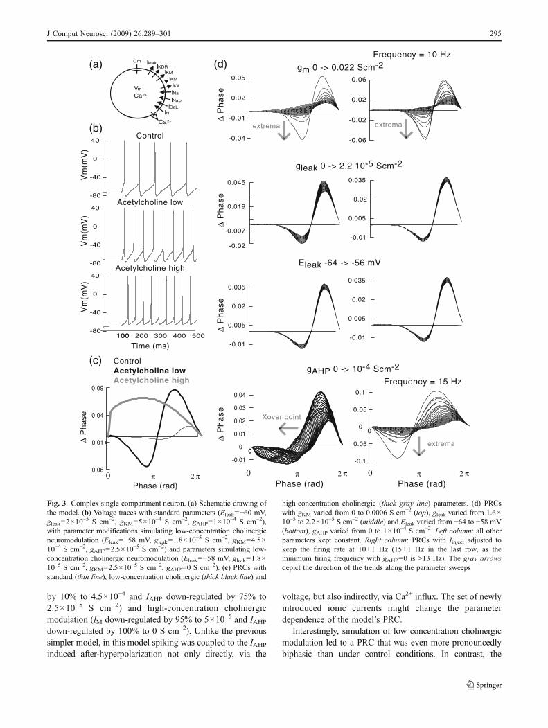
by 10% to 4.5×10⁻⁴ and IAHP down-regulated by 75% to2.5×10−5 S cm−2) and high-concentration cholinergicmodulation (IM down-regulated by 95% to 5×10⁻⁵ and IAHPdown-regulated by 100% to 0 S cm−2). Unlike the previoussimpler model, in this model spiking was coupled to the IAHPinduced after-hyperpolarization not only directly, via the
voltage, but also indirectly, via Ca2+ influx. The set of newlyintroduced ionic currents might change the parameterdependence of the model’s PRC.
Interestingly, simulation of low concentration cholinergicmodulation led to a PRC that was even more pronouncedlybiphasic than under control conditions. In contrast, the
cm IleakIKDR
IKM
INa p
Vm
IKM
INa
ICaLIH
IKA
Ca2+
Ca2+
∆ P
ha
se
(a)
Control Acetylcholine lowAcetylcholine high
-0.04
-0.01
0.02
0.05
-0.06
-0.02
0.02
0.06
-0.02
-0.007
0.019
0.045
-0.01
0.005
0.02
0.035
-0.01
0.005
0.02
0.035
-0.01
0.005
0.02
0.035
(b)
(c)
Phase (rad)π 2 π
Phase (rad)0
∆ P
ha
se∆
Ph
ase
∆ P
ha
se
(d)Frequency = 10 Hz
gm 0 -> 0.022 Scm-2
gleak 0 -> 2.2 10-5 Scm-2
Eleak -64 -> -56 mV
Control
Acetylcholine low
Vm
(mV
)V
m(m
V)
Time (ms)
0 2-0.01
0
0.01
0.02
0.03
0.04
0 2
0 2
-0.1
0.05
0.05
0.1
20
Frequency = 15 Hz
π 2 π0
∆ P
ha
se
gAHP 0 -> 10-4 Scm-2
Xover point
extrema
extremaextrema
0
0.06
0.01
0.04
0.09
0
Phase (rad)π 2 π0
100100 200 300 400 500
-40
0
40
-80
-40
0
40
-80
-80
-40
0
40
Vm
(mV
)
Acetylcholine high
Fig. 3 Complex single-compartment neuron. (a) Schematic drawing ofthe model. (b) Voltage traces with standard parameters (Eleak=−60 mV,gleak=2×10
−5 S cm−2, gKM=5×10⁻⁴ S cm−2, gAHP=1×10⁻⁴ S cm−2),
with parameter modifications simulating low-concentration cholinergicneuromodulation (Eleak=−58 mV, gleak=1.8×10
−5 S cm−2, gKM=4.5×10⁻⁴ S cm−2, gAHP=2.5×10
−5 S cm−2) and parameters simulating low-concentration cholinergic neuromodulation (Eleak=−58 mV, gleak=1.8×10⁻⁵ S cm−2, gKM=2.5×10⁻⁵ S cm−2, gAHP=0 S cm−2). (c) PRCs withstandard (thin line), low-concentration cholinergic (thick black line) and
high-concentration cholinergic (thick gray line) parameters. (d) PRCswith gKM varied from 0 to 0.0006 S cm−2 (top), gleak varied from 1.6×10⁻⁵ to 2.2×10⁻⁵ S cm−2 (middle) and Eleak varied from −64 to −58 mV(bottom), gAHP varied from 0 to 1×10⁻⁴ S cm−2. Left column: all otherparameters kept constant. Right column: PRCs with Iinject adjusted tokeep the firing rate at 10±1 Hz (15±1 Hz in the last row, as theminimum firing frequency with gAHP=0 is >13 Hz). The gray arrowsdepict the direction of the trends along the parameter sweeps
J Comput Neurosci (2009) 26:289–301 295
simulation of high-concentration cholinergic modulationproduced a transition from a type II to a type I PRC(Fig. 3(c); the firing frequencies in these simulations werekept at 12±1 Hz in order to ensure comparability).
Parameter sweeps gleak, Eleak, gM, and gAHP (gleak: from1.6×10−5 to 2.2×10⁻⁵ S cm−2, Eleak: from −64 to 56 mV,gM: from 0 to 6×10⁻⁴ S cm−2, gAHP 0 to 10−4 S cm−2)revealed that the qualitative change in the PRC was solely aconsequence of changes in gM. This occurred regardless ofwhether the firing frequency was kept constant by adjustingcurrent injection. No switch from a biphasic to a purelypositive PRC occurred when gleak, Ileak or gAHP were varied.The sweeps over the parameters of the leak current hadlittle effect on the PRCs. Increasing gAHP moved thecrossover point between the negative and the positive partof the PRC to the left (towards earlier phases). When thefiring frequency during the determination of the PRCs waskept constant, this effect was greatly reduced and theextrema of the PRCs decreased with an increasing amountof gAHP.
In conclusion, the effects of cholinergic neuromodula-tion on the PRC were biphasic in simulations of thecomplex single-compartmental model. An increase of thenegative part of the PRC mediated by low modulatorconcentration was followed by a disappearance of thisnegative part mediated by a high acetylcholine concentra-tion. The switch from a type II to a type I PRC was due tothe decrease of IM. However, under control conditions, theIAHP masked a part of the negative portion of the PRC.
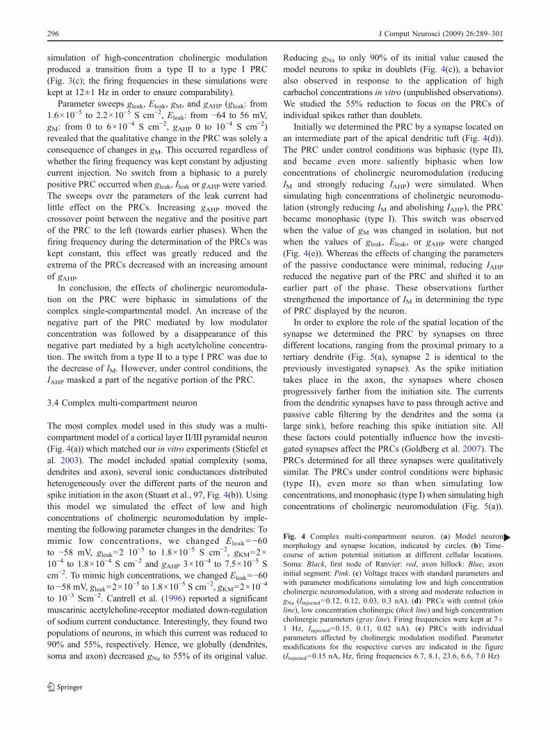
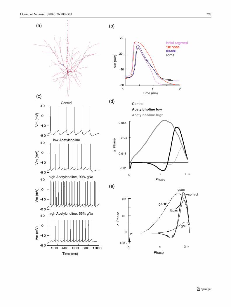
3.4 Complex multi-compartment neuron
The most complex model used in this study was a multi-compartment model of a cortical layer II/III pyramidal neuron(Fig. 4(a)) which matched our in vitro experiments (Stiefel etal. 2003). The model included spatial complexity (soma,dendrites and axon), several ionic conductances distributedheterogeneously over the different parts of the neuron andspike initiation in the axon (Stuart et al., 97, Fig. 4(b)). Usingthis model we simulated the effect of low and highconcentrations of cholinergic neuromodulation by imple-menting the following parameter changes in the dendrites: Tomimic low concentrations, we changed Eleak=−60to −58 mV, gleak=2 10−5 to 1.8×10⁻⁵ S cm−2, gKM=2×10⁻⁴ to 1.8×10⁻⁴ S cm−2 and gAHP 3×10⁻⁴ to 7.5×10⁻⁵ Scm−2. To mimic high concentrations, we changed Eleak=−60to −58 mV, gleak=2×10
−5 to 1.8×10⁻⁵ S cm−2, gKM=2×10⁻⁴
to 10−3 Scm−2. Cantrell et al. (1996) reported a significantmuscarinic acetylcholine-receptor mediated down-regulationof sodium current conductance. Interestingly, they found twopopulations of neurons, in which this current was reduced to90% and 55%, respectively. Hence, we globally (dendrites,soma and axon) decreased gNa to 55% of its original value.
Reducing gNa to only 90% of its initial value caused themodel neurons to spike in doublets (Fig. 4(c)), a behavioralso observed in response to the application of highcarbachol concentrations in vitro (unpublished observations).We studied the 55% reduction to focus on the PRCs ofindividual spikes rather than doublets.
Initially we determined the PRC by a synapse located onan intermediate part of the apical dendritic tuft (Fig. 4(d)).The PRC under control conditions was biphasic (type II),and became even more saliently biphasic when lowconcentrations of cholinergic neuromodulation (reducingIM and strongly reducing IAHP) were simulated. Whensimulating high concentrations of cholinergic neuromodu-lation (strongly reducing IM and abolishing IAHP), the PRCbecame monophasic (type I). This switch was observedwhen the value of gM was changed in isolation, but notwhen the values of gleak, Eleak, or gAHP were changed(Fig. 4(e)). Whereas the effects of changing the parametersof the passive conductance were minimal, reducing IAHPreduced the negative part of the PRC and shifted it to anearlier part of the phase. These observations furtherstrengthened the importance of IM in determining the typeof PRC displayed by the neuron.
In order to explore the role of the spatial location of thesynapse we determined the PRC by synapses on threedifferent locations, ranging from the proximal primary to atertiary dendrite (Fig. 5(a), synapse 2 is identical to thepreviously investigated synapse). As the spike initiationtakes place in the axon, the synapses where chosenprogressively farther from the initiation site. The currentsfrom the dendritic synapses have to pass through active andpassive cable filtering by the dendrites and the soma (alarge sink), before reaching this spike initiation site. Allthese factors could potentially influence how the investi-gated synapses affect the PRCs (Goldberg et al. 2007). ThePRCs determined for all three synapses were qualitativelysimilar. The PRCs under control conditions were biphasic(type II), even more so than when simulating lowconcentrations, and monophasic (type I) when simulating highconcentrations of cholinergic neuromodulation (Fig. 5(a)).
Fig. 4 Complex multi-compartment neuron. (a) Model neuronmorphology and synapse location, indicated by circles. (b) Time-course of action potential initiation at different cellular locations.Soma: Black, first node of Ranvier: red, axon hillock: Blue, axoninitial segment: Pink. (c) Voltage traces with standard parameters andwith parameter modifications simulating low and high concentrationcholinergic neuromodulation, with a strong and moderate reduction ingNa (Iinjected=0.12, 0.12, 0.03, 0.3 nA). (d): PRCs with control (thinline), low concentration cholinergic (thick line) and high concentrationcholinergic parameters (gray line). Firing frequencies were kept at 7±1 Hz, Iinjected=0.15, 0.11, 0.02 nA). (e) PRCs with individualparameters affected by cholinergic modulation modified. Parametermodifications for the respective curves are indicated in the figure(Iinjected=0.15 nA, Hz, firing frequencies 6.7, 8.1, 23.6, 6.6, 7.0 Hz)
b
296 J Comput Neurosci (2009) 26:289–301
(a)
Phase0 π 2 π
∆ P
ha
se
Control
20
70
-80
-30
1 20
Vm
(m
V)
Time (ms)
soma
-80
-40
0
40
-80
-40
0
40
-80
-40
0
40
200 400 600 800 1000-80
-40
0
40
low Acetylcholine
high Acetylcholine, 90% gNa
high Acetylcholine, 55% gNa
(b)
(c)
Time (ms)
Vm
(m
V)
Vm
(m
V)
Vm
(m
V)
Vm
(m
V)
(d)
(e)
-0.01
0.015
0.04
0.065
Control
Acetylcholine low
Acetylcholine high
.0.005
0
0.01
0.02
.
∆ P
ha
se
Phase
0 π 2 π
gM
gpas
Epas
gAHP
control
J Comput Neurosci (2009) 26:289–301 297
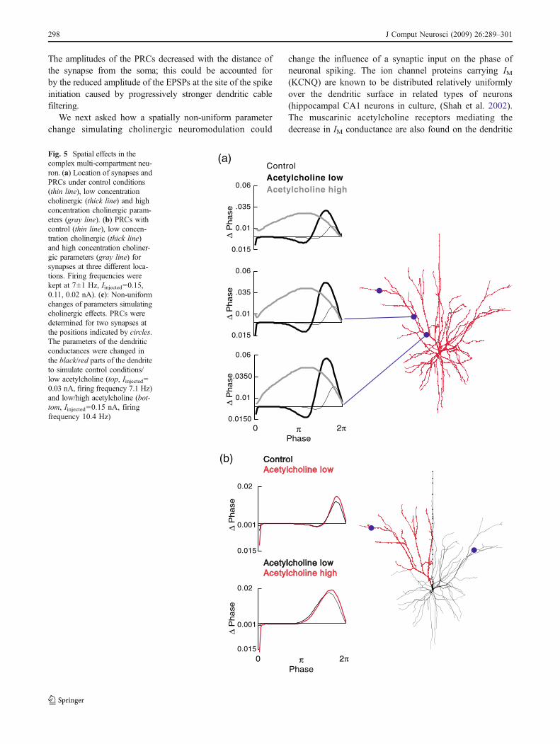
The amplitudes of the PRCs decreased with the distance ofthe synapse from the soma; this could be accounted forby the reduced amplitude of the EPSPs at the site of the spikeinitiation caused by progressively stronger dendritic cablefiltering.
We next asked how a spatially non-uniform parameterchange simulating cholinergic neuromodulation could
change the influence of a synaptic input on the phase ofneuronal spiking. The ion channel proteins carrying IM(KCNQ) are known to be distributed relatively uniformlyover the dendritic surface in related types of neurons(hippocampal CA1 neurons in culture, (Shah et al. 2002).The muscarinic acetylcholine receptors mediating thedecrease in IM conductance are also found on the dendritic
(a)
(b)
Phase0 π 2π
∆ P
hase
∆ P
hase
∆ P
hase
0.0150
0.01
.0350
0.06
0.015
0.01
.035
0.06
0.015
0.01
.035
0.06
Control Acetylcholine lowAcetylcholine high
Control Acetylcholine low
Acetylcholine lowAcetylcholine high
Phase0 π 2π
∆ P
hase
∆ P
hase
0.015
0.001
0.02
0.015
0.001
0.02
Fig. 5 Spatial effects in thecomplex multi-compartment neu-ron. (a) Location of synapses andPRCs under control conditions(thin line), low concentrationcholinergic (thick line) and highconcentration cholinergic param-eters (gray line). (b) PRCs withcontrol (thin line), low concen-tration cholinergic (thick line)and high concentration choliner-gic parameters (gray line) forsynapses at three different loca-tions. Firing frequencies werekept at 7±1 Hz, Iinjected=0.15,0.11, 0.02 nA). (c): Non-uniformchanges of parameters simulatingcholinergic effects. PRCs weredetermined for two synapses atthe positions indicated by circles.The parameters of the dendriticconductances were changed inthe black/red parts of the dendriteto simulate control conditions/low acetylcholine (top, Iinjected=0.03 nA, firing frequency 7.1 Hz)and low/high acetylcholine (bot-tom, Iinjected=0.15 nA, firingfrequency 10.4 Hz)
298 J Comput Neurosci (2009) 26:289–301
surface, and the G-protein mediated signal transductionbetween muscarinic receptors. On the other hand, KCNQ ishighly local (spread of the signal ∼1 μm). Because thecholinergic projections arising from the nucleus basalis orfrom subtypes of cholinergic interneurons could activatemuscarinic receptors in a spatially limited dendritic region,and subcellular signaling cascades mediating between themare local, cholinergic effects are likely to be localized.Similar reasoning holds for the modulation of the ionchannels responsible for the leak current (GIRK).
We therefore simulated spatially heterogeneous cholin-ergic modulation of dendritic conductances (Fig. 5(c)). Wefirst simulated a low concentration cholinergic modulationin a single dendritic tree (18% of the neuron’s surface area)with no cholinergic effects in the rest of the neuron. Underthese conditions, the PRC for a synapse located within andoutside of the region experiencing changes was intermedi-ate between the PRCs determined for homogeneousparameter changes. The PRC were biphasic, with thecrossover point shifted to earlier phases as compared to aneuron under homogeneous control conditions. This wasexpected, as the dendritic tree with cholinergic parameterscontained a significant fraction of the neuron’s membranesurface. Surprisingly, the PRCs determined for the synapseoutside the region with the cholinergically modifiedparameters were quite similar, although the PRC of thesynapse inside the dendritic region modulated by acetyl-choline had a slightly higher positive peak.
Next, we modified the parameters in the same dendritic treeto simulate a high, and in the remaining neuron a lowconcentration of cholinergic modulation. Again, we deter-mined the PRCs of a synapse within and outside of thisdendritic tree. As before, the PRCs were intermediate betweenthe PRCs obtainedwith each parameter regime homogenouslyimplemented over the complete dendritic tree. The negativepart of the PRCs was strongly reduced. As in the previouscase, the PRCs were very similar for the synapses inside andoutside of the dendritic tree with different parameter mod-ifications. The positive peak of the former PRC was slightlyhigher in amplitude and shifted to later phases.
The chief conclusion is that the modulation of PRC isglobal to a neuron. Changes of membrane properties insmall dendritic regions were insufficient to switch the typeof PRC of a synapse, whereas changes in large regionscause enough difference in the total currents to affectsynapses outside of the region of change.
4 Discussion
Acetylcholine is a powerful neuromodulator that altersbehavior and affects the nervous systems at both the circuitand cellular levels. In particular, some of the more
prominent effects of acetylcholine at the cellar level are toreduce the spike frequency adaptation by blocking currentsresponsible for it, as well as to increase the resting potentialof the cell, in part by a partial block of leak currents. Suchmodulation of cell excitability has been hypothesized to leadto changes in the structure of collective network activity inthe cortex (Ermentrout et al. 2001; Crook et al. 1998b). Inparticular, theoretical studies have suggested that blockingadaptation can change synchronization of neurons invarious frequency bands (e.g. gamma and beta; Ermentroutet al. 2001). This is supported by EEG studies in attentivevs. non-attending conditions (Fan et al. 2006), and in vitroacetylcholine modulation produces coherent oscillations inlocal cortical circuits (Buhl et al. 1998; Fisahn et al. 1998).In this report we focused on examining the outcome ofcholinergic modulation of the various potassium and leakcurrents on the spike generating dynamics in model corticalpyramidal neurons by using the PRC as an assay.
We found that in a number of models the simulation ofthe effects of cholinergic neuromodulation on corticalpyramidal neurons led to a shift from a biphasic (type II)to a purely positive (type I) PRC. The dynamics werealready present in the simplest model (theta neuron). Noqualitatively new features with respect to the phenomenonof spike time resetting occur in the more sophisticatedmodels that included all the elements necessary for spikeinitiation and after-hyperpolarization.
In the more complex models, cholinergic modulationrevealed an unexpected interplay between the IKAHP andIKM. Although previous studies have suggested that bothcurrents might have similar effects on the PRC shape,producing a rightward skew in the peak (Ermentrout et al.2001), we found that partial block of IKAHP with IKM fullyactive uncovered the negative portion of the PRC. In thelow acetylcholine condition we found that PRC lookedeven more type II then in control; the negative lobe of thePRC increased significantly in the high acetylcholinecondition, with both potassium currents the PRC switchedto type I. This occurred because IKAHP flattens out the PRCin the initial portion of the firing cycle by reducing the cellsexcitability in a voltage independent manner, since itsactivation is largely uncoupled form the voltage at therelatively hyperpolarized and partially depolarized poten-tials. The decrease in the excitability and cell responses tothe input could occur because of the additional conductanceload on the membrane by the activated IKAHP, as noted byErmentrout et al. (2001). Thus, IKAHP reduced the ampli-tude of the PRC in the initial portion of the inter-spikeinterval, including the negative lobe of the potentially typeII PRC. Once this voltage-independent “break” on theexcitability is removed, by partial block of IKAHP, thenegative lobe (due to IKM recruitment by the input inducedvoltage transients) was uncovered. These results suggest
J Comput Neurosci (2009) 26:289–301 299
that acetylcholine should have a biphasic effect on the spikegenerating dynamics in pyramidal neurons, perhaps similarto the inverted U-curve suggested previously for actions ofdopamine (Seamans and Yang 2004).
PRCs of spatially extended neuronal models have beendetermined previously (Keck et al. 2003; Goldberg et al.2007). Here we used complex compartmental models to studywhat would be the effect of confining the cholinergicmodulation to a particular portion of the dendritic tree. Wefound effect of spatially confined cholinergic changes inmembrane properties were not restricted to the part of thedendritic tree subject to the changes. In fact the PRC changedfrom type I to type II for the full dendritic tree, even whenonly one of the dendrites received the simulated cholinergicmodulations. This is in contrast to other electrophysiologicalfeatures, such as the normalization of EPSP shapes by Ih,which have been shown to be highly localized (Magee 1999).
Using the complex compartmental model we alsoaddressed the question of how synaptic location at differentdendritic site may influence the type II to type I change inthe PRC. Recently, Goldberg et al. (2007) showed thatactive dendritic conductances can potentially shift the shapeof the PRC measured at the soma, when the perturbation isdelivered distally in the synapse. Their modeling studiespredicted a shift from type I to type II (as the stimulationmoved from the soma to the dendrite) that wasconfirmed by in vitro experiments. Our goal was toexamine the shift from type II to type I induced bycholinergic modulation following dendritic stimulation.We found that the cholinergic modulation of the PRC typewas independent of the synapse location.
Although the qualitative switch from biphasic (type II) tomonophasic (type I) PRCs was seen in all models, theshape and development of the shape as a function ofparameter change differed in the models used. In particular,the extrema and the zero-crossing shifted to later phasesonly in the theta and simple single-compartment model, notin the complex single-compartment model (the parameterdependence was not investigated in the complex multi-compartment model). The integral of the PRCs undercontrol conditions (type II) was strongly positive in thetheta and simple single-compartment model. In contrast, inthe complex single-compartment model and in the complexmulti-compartment model, the regions under the negativeand the positive parts of the PRC were quantitatively morebalanced. The added features of the more complex modelscould also influence other phenomena, such as theintegration of multiple synaptic inputs. Other extensions,such as the introduction of a stochastic component, mightlead to the emergence of qualitatively new phenomena.These questions remain as topics for future investigation.
We conclude that the type and shape of the PRCs isqualitatively insensitive to changes in a number of param-
eters but highly sensitive to changes in the amount of twoadapting currents, IM and IAHP. This allows a key feature ofspiking, the PRC, to be manipulated without influencingother aspects of it's functioning, such as the integration ofsynaptic potentials. This supports our hypothesis that IMand IAHP cholinergic modulation is necessary and sufficientto change the PRC shape and hence the spike-generatingdynamics of cortical pyramidal neurons.
An important question is what these results mean for thesynchronization of neurons in the cortex in vivo. This has tobe addressed in the context of the complete corticalnetwork, which in addition to the pyramids, contains anumber of types of interneurons. Different types ofsynchronous oscillations (delta, theta, gamma) result fromdifferent dynamics involving different currents and celltypes (Tiesinga et al. 2001). The gamma oscillations,which are evoked by elevated acetylcholine (Rodriguez etal. 2004) are driven by synchronized interneural networks(Buhl et al. 1998). These interneurons then entrain thepyramids. Without interneural help, a network of reciprocallyexcitatory connected layer II pyramidal neurons would notsynchronize when the acetylcholine concentration is high.This is predicted by theoretical results which show thatneurons with a type I PRC (like layer II pyramids under theinfluence of acetylcholine) coupled with excitatory synapsesdon’t synchronize (Ermentrout et al. 2001).
The picture which thus emerges is that networks ofcortical layer II pyramidal neurons synchronize well whensubjected to no acetylcholine and even better when exposedto low concentrations of this neuromodulator. This type ofsynchronization breaks down when the cholinergic neuro-modulation becomes strong. Under such conditions, thepyramidal neurons are entrained into a gamma-rhythm byinhibitory interneurons. They then show no intrinsictendency to form synchronously oscillating assemblies.
This picture is consistent with the patchy and local natureof gamma oscillations, into which pyramidal neurons areforced by local interneural circuits, and the more globalnature of low-frequency oscillations (delta, theta), whichresult from the properties of the pyramidal neurons and theirnetworks themselves (Tiesinga et al. 2001).
Acknowledgements This work was supported by the DeutscheForschungsgemeinschaft (K.M.S.), the Gatsby Foundation, CNRS andMarie Curie EXT “BIND” (B.S.G.) and the Howard Hughes MedicalInstitute (T.J.S.). We would like to thank the organizers of and all ourcolleagues at the workshop “spiking neurons” at the 2004 ComputationalNeuroscience Meeting in Baltimore, MD, USA, where preliminaryresults of this work were presented.
Open Access This article is distributed under the terms of theCreative Commons Attribution Noncommercial License which per-mits any noncommercial use, distribution, and reproduction in anymedium, provided the original author(s) and source are credited.
300 J Comput Neurosci (2009) 26:289–301
References
Artola, A., & Singer, W. (1993). Long-term depression of excitatorysynaptic transmission and its relationship to long-term potentia-tion. Trends in Neurosciences, 16(11), 480–487. doi:10.1016/0166-2236(93)90081-V.
Auerbach, J. M., & Segal, M. (1996). Muscarinic receptors mediatingdepression and long-term potentiation in rat hippocampus. TheJournal of Physiology, 492(Pt 2), 479–493.
Buhl, E. H., Tamas, G., & Fisahn, A. (1998). Cholinergicactivation and tonic excitation induce persistent gammaoscillations in mouse somatosensory cortex in vitro. TheJournal of Physiology, 513, 117–126. doi:10.1111/j.1469-7793.1998.117by.x.
Cantrell, A. R., Ma, J. Y., Scheuer, T., & Catterall, W. A. (1996).Muscarinic modulation of sodium current by activation of proteinkinase C in rat hippocampal neurons. Neuron, 16, 1019–1026.doi:10.1016/S0896-6273(00)80125-7.
Crook, S. M., Ermentrout, G. B., & Bower, J. M. (1998a). Dendriticand synaptic effects in systems of coupled cortical oscillators.Journal of Computational Neuroscience, 5(3), 315–329.doi:10.1023/A:1008839112707.
Crook, S. M., Ermentrout, G. B., & Bower, J. M. (1998b). Spikefrequency adaptation affects the synchronization properties ofnetworks of cortical oscillations. Neural Computation, 10(4),837–854. doi:10.1162/089976698300017511.
Ermentrout, G. B. (1996). Type I membranes, phase resetting curves,and synchrony. Neural Computation, 8(5), 979–1001.doi:10.1162/neco.1996.8.5.979.
Ermentrout, G. B., Pascal, M., & Gutkin, B. S. (2001). The effects ofspike frequency adaptation and negative feedback on thesynchronization of neural oscillators. Neural Computation, 13,1285–1310. doi:10.1162/08997660152002861.
Fan, J., Byrne, J., Worden, M. S., Guise, K. G., McCandliss, B. D.,Fossella, J., et al. (2006). The relation of brain oscillations toattentional networks. The Journal of Neuroscience, 27(23),6197–6206. doi:10.1523/JNEUROSCI.1833-07.2007.
Fisahn, A., Pike, F. G., Buhl, E. H., & Paulsen, O. (1998). Cholinergicinduction of network oscillations at 40 Hz in the hippocampus invitro. Nature, 394, 186–189. doi:10.1038/28179.
Goldberg, J. A., Deister, C. A., & Wilson, C. J. (2007). Responseproperties and synchronization of rhythmically firing dendriticneurons. Journal of Neurophysiology, 97, 208–219. doi:10.1152/jn.00810.2006.
Golomb, D., & Amitai, Y. (1997). Propagating neuronal discharges inneocortical slices: computational and experimental study. Journalof Neurophysiology, 78, 1199–1211.
Gutkin, B. S., & Ermentrout, G. B. (1998). Dynamics of membraneexcitability determine interspike interval variability: a linkbetween spike generation mechanisms and cortical spike trainstatistics. Neural Computation, 10(5), 1047–1065. doi:10.1162/089976698300017331.
Gutkin, B. S., Ermentrout, G. B., & Reyes, A. (2005). Phase responsecurves determine the responses of neurons to transient inputs.Journal of Neurophysiology, 94(2), 1623–1635. doi:10.1152/jn.00359.2004.
Hansel, D., & Mato, G. (1995). Synchrony in excitatory neuralnetworks. Neural Computation, 7, 307–337. doi:10.1162/neco.1995.7.2.307.
Hasselmo, M. E. (1999). Neuromodulation: acetylcholine and memoryconsolidation. Trends in Cognitive Sciences, 3, 351–359.doi:10.1016/S1364-6613(99)01365-0.
Hasselmo, M. E. (2006). The role of acetylcholine in learning andmemory. Current Opinion in Neurobiology, 16(6), 710–715.doi:10.1016/j.conb.2006.09.002.
Hines, M. L., & Carnevale, N. T. (1997). The NEURON simulationenvironment. Neural Computation, 9, 1179–1209. doi:10.1162/neco.1997.9.6.1179.
Hines, M. L., & Carnevale, N. T. (2000). Expanding NEURON'srepertoire of mechanisms with NMODL. Neural Computation,12, 995–1007. doi:10.1162/089976600300015475.
Hodgkin, A. L., & Huxley, A. F. (1952). A quantitative description ofmembrane current and its application to conduction andexcitation in nerve. The Journal of Physiology, 117, 500–544.
Izhikevich, E. M. (2007). Dynamical systems in neuroscience: thegeometry of excitability and bursting. Cambridge: MIT Press.
Johnston, D., &Wu, S. (1994). Foundations of cellular neurophysiology.Cambridge, MA: The MIT Press.
Keck, T., Netoff, T. I., & White, J. A. (2003) Comparing dynamicresponses of neurons to dendritic and somatic inputs. Society forNeuroscience Abstracts, 31.
Madison, D. V., Lancaster, B., & Nicoll, R. A. (1987). Voltage clampanalysis of cholinergic action in the hippocampus. The Journal ofNeuroscience, 7(3), 733–741.
Magee, J. C. (1999). Dendritic Ih normalizes temporal summation inhippocampal CA1 neurons. Nature Neuroscience, 2(9), 848.doi:10.1038/12229.
Mainen, Z. F., & Sejnowski, T. J. (1995). Reliability of spike timing inneocortical neurons. Science, 268, 1503–1506. doi:10.1126/science.7770778.
McCormick, D. A. (1993). Actions of acetylcholine in the cerebralcortex and thalamus and implications for function. Progress inBrain Research, 98, 303–308.
Moehlis, J., Shea-Brown, E., & Rabitz, H. (2006). Optimal inputs forphase models of spiking neurons. Journal of Computational andNonlinear Dynamics, 1(4), 358–367. Transactions of the ASME.doi:10.1115/1.2338654.
Reyes, A. D., & Fetz, E. E. (1993). Effects of transient depolarizingpotentials on the firing rate of cat neocortical neurons. Journal ofNeurophysiology, 69, 1673–1683.
Rodriguez, R., Kallenbach, U., Singer, W., & Munk, M. H. (2004).Short- and long-term effects of cholinergic modulation on gammaoscillations and response synchronization in the visual cortex.The Journal of Neuroscience, 24(46), 10369–10378. doi:10.1523/JNEUROSCI.1839-04.2004.
Schobesberger, H., Wheeler, D. W., & Horn, J. P. (2000). A model forpleiotropic muscarinic potentiation of fast synaptic transmission.Journal of Neurophysiology, 83(4), 1912–1923.
Seamans, J. K., & Yang, C. R. (2004). The principal features andmechanisms of dopamine modulation in the prefrontalcortex. Progress in Neurobiology, 74, 1–57. doi:10.1016/j.pneurobio.2004.05.006.
Shah, M. M., Mistry, M., Marsh, S. J., Brown, D. A., & Delmas, P.(2002). Molecular correlates of the M-current in cultured rathippocampal neurons. The Journal of Physiology, 544, 29–37.doi:10.1113/jphysiol.2002.028571.
Steriade, M. (2004). Acetylcholine systems and rhythmic activitiesduring the waking–sleep cycle. Progress in Brain Research, 145,179–196.
Stiefel, K., Gutkin, B. S., & Sejnowski, T. J. (2003) Cholinergicmodulation of spike generating dynamics in cortical pyramidalneurons. Society for Neuroscience Abstract, 29.
Stiefel, K. M., Tennigkeit, F., & Singer, W. (2005). Synaptic plasticityin the absence of backpropagating spikes of layer II inputs tolayer V pyramidal cells in rat visual cortex. The EuropeanJournal of Neuroscience, 21(9), 2605–2610. doi:10.1111/j.1460-9568.2005.04094.x.
Tiesinga, P. H. E., Fellous, J. M., Jose, J. V., & Sejnowski, T. J.(2001). Computational model of carbachol-induced delta, theta,and gamma oscillations in the hippocampus. Hippocampus, 11,251–274. doi:10.1002/hipo.1041.
J Comput Neurosci (2009) 26:289–301 301


![Neurovascular-modulation...2020/07/21 · Introduction Vascular responses are ubiquitous across neuromodulation [1–6], but are considered epiphenomena to neuronal stimulation. Common](https://img.pdfslide.us/doc/110x75/60721ffc1eead12dfa46dec5/neurovascular-modulation-20200721-introduction-vascular-responses-are-ubiquitous.jpg)