Embed Size (px)
Citation preview
187
Nigerian Journal of Physiological Sciences 24 (2): 187 -194 ©Physiological Society of Nigeria, 2009
Available online/abstracted at http://www.bioline.org.br/np; www.ajol.info/journals.njps; www.cas.org
THE EFFECTS OF AMMONIUM METAVANADATE ON BIOCHEMICAL
HORMONAL, HAEMATOLOGICAL AND HISTOPATHOLOGICAL
PARAMETERS OF THE FEMALE WISTAR RATS
A. W. OBIANIME*, M. GOGO-ABITE1, AND I. I. ROBERTS
2
Department of Pharmacology*, Department of Anatomical Pathology1, and Department of Clinical Pharmacy
2,
University of Port Harcourt, Port Harcourt, Nigeria E-mail: [email protected] Tel: +234 (0) 8037452182
Summary: The effects of different doses of Ammonium metavanadate on the biochemical, haematological,
hormonal and histopathological parameters of stilbesterol treated female Wistar rats were investigated.
Ammonium metavanadate in the dose- range 0-6mg/kg caused a bi-phasic and time-dependent response on the
acid (total and prostate) phosphatase.. Furthermore ammonium metavanadate caused a dose-dependent
inhibition of the serum alkaline phosphatases. The maximal inhibitory response at 5mg/kg of ammonium
metavanadate was 40.0 ± 1.69 compared to 65.0 ± 0.94 control values. Ammonium metavanadate also caused a
positively correlated biphasic response in the serum female hormonal concentrations with an initial increase,
followed by a time-dependent decrease in the serum values of luteinizing (LH), follicle stimulating hormone
(FSH), prolactin . Furthermore ammonium metavanadate also caused time- and dose- dependent effects on the
haematological parameters. The effects were biphasic-increase within 72 hours and a reduction in the values of
haemoglobin and packed cell volume within 7-28 days. The white blood count and lymphocyte counts were also
reduced significantly at P ≤ 0.05. However the neutrophil counts were increased dose- and time-dependently.
Finally, ammonium metavanadate caused a dose-dependent destruction of the liver and female reproductive
organs namely the uterus, ovary and fallopian tubes. These were characterized by necrosis, oedema, eosinophilic
deposits and vacuolation. These results may be explained by the oxidative effects caused by the free oxygen
(O2) radical generated by the metavanadate ions.
Key words: vanadium, female hormones, blood count, histology, uterus
Introduction Vanadium is a transitional metal that exists in
different oxidational states (Barceloux & Barceloux
1999) in fossil fuels, industrial and environmentally
degraded areas (Osuji & Adesiyan 2005). It is also
a trace element found in the catalytic distillation of
crude oil. Absorption of vanadium compounds
depends on the chemical composition of the
compound as well as species and the route of
exposure (Conklin et al 1982). The toxicity of
vanadium varies with the chemical form and the
oxidational states (Waters et al 1974).
Animal studies with vanadate show that the
compound of vanadate caused haemorrhagic
exudates from the nose, marked diarrhea,
respiratory distress and convulsion (Gossellin et al
1984). The pathological effects include diffuse
desquamative enteritis of lungs, liver, kidneys,
adrenal cortex, brain, spinal cord and bone marrow
(Gosellin et al 1984). Higher concentrations may
cause irreversible kidney damage (Kumar & Corder
1980). The compounds also caused
vasoconstriction of vessels in the lungs (Erdmann
et al 1984). Sodium metavanadate has also been
shown to cause increase in blood Urea, Uric acid
and histopathological lesions in the kidneys and
spleen (Domingo et al 1985), decreases in
erythrocyte count and haemoglobin level
(Zaprowska and Wasilewski 1991), inhibition of
mating and reproductive toxicity (Domingo, 1996),
pulmonary Oedema (Sjoberg 1950), increased
leucocytes in bronchiolar lavage (Knecht et al
1992), increased alveolar macrophages (Lee and
gillies 1986), reduced oxyhaemoglobin and
pathological changes in the lungs (pazyniah 1966),
increased fatty changes in the liver of rats, reduced
fertilization of females mated to exposed males
(Hacket and Kalman 1983).However, most of these
reports were single doses of the metavanadate and
thus not qualitative. There were no correlation
studies between the phosphatase increases, tissue
organ toxicities and dysfunctions in addition to
histopathological effects on the blood cells and
hormonal dysfunction.
It is therefore in that light that we seek to
investigate the qualitative pharmacological effects
of ammonium metavanadate on some biochemical
(phosphatases), histological (the liver, blood cells,
kidney, ovary, oviduct and uterus) and the female
hormonal parameters of the female wister rats. In
other words, what is the relationship between
detailed biochemical responses and histological
structure and function of the female wister rats
traumatised with ammonium metavanadate. Also,
what is the correlation with the hormones studies?

188
Finally in this study, we have also investigated the
dose- and time- dependency of ammonium
metavanadate on phosphatases, hormones,
haematological and histological changes on tissues.
What are the various forms of correlations of these
parameters if any?
Materials and methods
Dose-dependent Studies.
The effect of intraperitoneal administration of
different doses of ammonium metavanadate on the
biochemical, haematological, hormonal and
histopathological parameters of the female
stilbesterol treated albino rats were investigated.
Thirty-five albino rats of average weight 250±6gms
were divided into seven cages of five each. The
first was administered with distilled water as
control, the second to the sixth cages were
administered with different doses of ammonium
metavanadate in the dose range 0-6mg/kg. The rats
were anaethesized, blood and tissue samples were
removed and subjected to various forms of
analysis.
Time-dependent Studies:
The effects of time of administration of a
single dose of ammonium metavanadate on various
responses of the female albino rats were
investigated. Twenty 25mg stilbesterol-treated
female rats, divided into four cages of five rats each
were administered with single doses of ammonium
metavanadated intraperitoneally. The blood and
tissue samples were harvested from the first cage
on the first day after four (4) hours, the rest were
harvested after 24 hours, 4 days and seven days
respectively. These were then analysed for
biochemical, hormonal and histopathological
parameters.
Animals
The average weight of animals used in this
study was 250±6gms. All animals used in this
study were handled with the international, natural
and institutional guidelines for care and use of
laboratory animals in biomedical research as
promulgated by the Canadian Council of Animal
Care (1984). Outbred strains of the female Wister
rats of average weight 250mg±6gms aged between
15-20 weeks were obtained from the animal house
of the University of Port Harcourt and allowed to
acclimatise for 14 days. They were housed in cages
with wire bar lids used to hold the water bottle and
feed to prevent contamination with urine or faeces.
Bedding was placed directly into the shoe box cage
to allow the absorption of urine. They were kept
well ventilated room at ambient temperature of
28.0±2.0oC under 12hr light/dark cycle and well
provided with food and tap water ad libitum.
Generally the study was conducted in accordance
with the recommendations from the declaration of
Helsinki on guiding principles in care and use of
animals.
Phosphatase Analysis
Determination of serum alkaline phosphatase
(ALP), serum acid phosphatase tartrate labile
(AcPT) and the prostatic acid phosphatases (AcPP)
was carried out using the hydrolysed phenol
method (Kind & King 1954).
Hormonal Analysis
Blood samples were collected in a 10 ml
plastic syringe. The peptide hormones namely
follicle stimulating hormone, Luteinizing hormone
and Prolactin were measured by radioimmunoassay
as described by Banu et al (2002) and
concentration expressed as ng/ml. while the steroid
hormone oestrogen was measured by
radioimmunoassay as described by Banu et al
(2002) in ng/ml.
Histological analysis
The tissues were isolated and preserved in
10% formalin and routinely processed for
histopathological examination using H&E stain
technique for paraffin embedded tissue sections as
described by Benjamin (2001). The various tissue
sections were viewed and photographed with the
DMLS camera and digital microscope (LEICA).
Haematological Analysis
In the morning hours, about 5 ml of blood was
collected in standing position from right jugular
vein of the female Wister rats and placed in citrated
vials. Blood samples were analyzed for
Haemoglobin (Hb) by acid heamatin (Sahali’s
haemoglobinometer) method and Packed Cell
Volume (PCV) by Wintrobes method. These
methods are standard methods used for
haematological parameters (Schalm, 1986).
Analysis of Urea and Creatinine
Urea measurements were performed using the
diacetyl monoxime, a Total urinary excretion
method (Toro & Ackermann 1975), creatinine was
assayed using the Jaffe alkaline picrate method
(Annino & Giese 1979).
Statistical analysis:
All values were represented as mean ± SEM
of n=5 and the values were taken as significant at p
≤ 0.05 ANOVA (analysis of variance).
Results: The effects of ammonium metavanadate on the
biochemical, hormonal, haematological and
histopathological parameters of the female Wistar
rats were investigated.
A. W. Obianeme et al

189
0 2 4 60
20
40
60
80
ALP
Total ACP
Prostatic ACP*
**
* **
* *
Dose (Mg/kg)
Seru
n C
on
cen
trati
on
of
Ph
osp
hata
ses
0 50 100 150 2000
20
40
60
80
Alkaline Phosphatase
*
*
Time (Hrs)
Seru
m C
on
cen
trati
on
of
Alk
alin
e P
ho
sp
hata
se
(IU
/L)
0 50 100 150 2000
5
10
15
20
25
Total ACP
Prostatic
*
*
*
*
*
*
Time (Hrs)
Resp
on
se
(IU
/L)
a)
b)
c)
Fig 1. The effects of (a) different doses of the
ammonium metavanadate-induced responses on
acid, alkaline and prostatic phosphatases (b) Time
on a single dose of Ammonium metavanadate on
alkaline phosphatases (ALP) and (c) Total and
prostatic phosphatases (AcPT & AcPP) Data is
presented as mean ± SEM * shows significance at
P ≤ 0.05 (ANOVA).
Biochemical effects:
Ammonium metavanadate caused a dose-
dependent decrease in the alkaline phosphatase
levels (fig 1a) and biphasic responses in the total
and prostatic acid phosphatases (fig 1b&c).
Ammonium metavanadate at the lower doses
caused a decrease which was followed at higher
doses by a stimulation or increase in phosphatase
levels (fig 1b&c). The responses showed that the
basal values of ALP changed from 65.0 ± 0.94 to
40 ± 1.89 IU/L.
I)
II)
II)
IV)
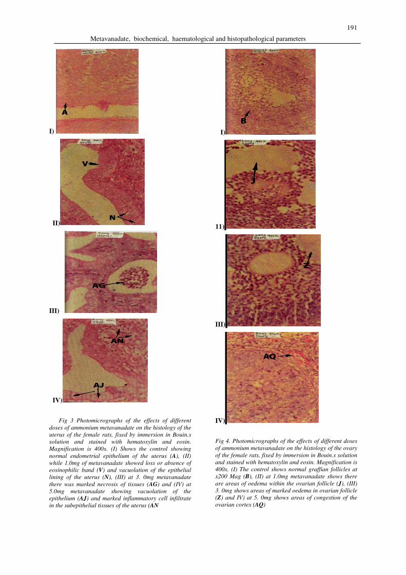
Fig 2. Photomicrographs of the effects of different
doses of ammonium metavanadate on the histology
of the oviduct of the female rats fixed by immersion
in Bouin.s solution and stained with hematoxylin
and eosin. Magnification is 400x. (i) The control
shows areas of normal epithelium of the oviduct
(C), while (ii) with 1mg metavanadate shows there
was increased proliferation of the epithelial lining
of the oviduct (K), (iii) at 3mg metavanadate
showed areas of vascular congestion in the wall
(AA) and (iv) with 5mg metavanadate showed
gradual loss of surface eosinophilic band (AP).
Similarly the basal values of the total acid
phospatase (ACP) changed from 15.4 ± 2.2 to 21.0
Metavanadate, biochemical, haematological and histopathological parameters
190
± 1.1 IU/L and 3.3 ± 0.1 to 9.4 ± 0.3 IU/L for
prostatic acid phosphatase (fig 1a). These values
were significant at p ≤ 0.005 ANOVA. These
effects on the biochemical phosphatases parameters
were also time- dependent maximizing at 7 (seven)
days of treatment (figs 1b&c).
Hormonal effects:
Stilbesterol-treated female Wister rats,
ammonium metavanadate in the dose range 0-6mg
caused a positively correlated dose-dependent
biphasic responses in the serum concentration of
the hormonal profiles of the female wister rats
studied (Table 1 & 2). Ammonium metavanadate
at the initial lower doses caused an increase (a
spike) in the hormonal concentrations measured.
This was followed by a dose-dependent decrease in
the basal serum hormonal levels (Table 1). The
maximal inhibitory responses at 5mg/kg of
ammonium metavanadate were 2.7± 0.34, 2.1 ±
0.06, 72.0 ± 2.0 and 0.14 ± 0.01 for LH, FSH,
PROL and Oest respectively (Table 1). These
effects were also time-dependent (Table 2)
maximizing at 7 days of treatment. The effects
were characterized by an initial surge in hormonal
effects within 48 hours followed by a time-
dependent decrease in seven days.
Haematological parameters:
Ammonium metavanadate in the dose-range 0-
10mg/kg caused both dose- and time – dependent
effects on the haemoglobin and packed cell volume
(Table 3). In the short term (24-72 hours), vanadate
caused a transient increase in the neutrophils, Hb
and PCV values (Table 3). However after 7 to 28
days of persistent treatment with Vanadate,
ammonium metavanadate caused a dose-dependent
decrease in Hb, PCV, lymphocytes and white blood
cell values (Table 3). Finally the histological
examination of the red blood cells under the
microscope revealed that the red blood cells were
characterized by the presence of target cells,
acanthocytes, segmented cells of stave forms,
increased anisocytosis and segmented leukocytes
largely made up of band and juvenile forms when
viewed under the light electron microscope fitted
with DMLS camera. Showing that the agent caused
anaemic responses.
Histopathological effects:
Ammonium metavanadate in the dose range 0-
10mg/kg caused a dose-dependent destruction of
the female reproductive tract viz the uterus,
ovaries, and fallopian tubes (Figs 2, 3 & 4). These
results were positively correlated with the results of
the biochemical, haematological and hormonal
parameters. This was characterized by oedema of
the ovarian tissue, reduction in follicular cells,
abortive follicles, complete ablation of the ovaries
and para-ovarian tissue, congestion, small vessel
proliferation, thrombosis and increase in
inflammatory cells (figs 2, 3 & 4). These
toxic/destructive effects were dose-and time-
dependent and were completely
moderated/inhibited by vitamin E, vitamin C,
selenium and their combination (not shown).
Table 1: The effect of different doses (0 – 5mg/kg of
ammonium metadanadate on the serum
concentration of various female hormones in the
female Wistar rats
Dose of
Cadmium
(mg/kg)
Prolactin LH FSH Oestrogen
0 95.35
±12.73
6.32
±1.37
5.32±
1.23
0.21
±0.09
1 145.74
±18.93*
9.15
±1.89
*
5.59
±1.01
0.26
±0.07
2 141.92
±13.83*
8.27
±1.8*
5.89
±1.08
0.23
±0.01
3 99.29
±3.27
6.38
±1.1
4.98
±1.42
0.25
±0.11
4 81.97
±4.5*
6.09
±1.32
4.36±
0.97*
0.27
±0.06
5 90.23
±2.41
5.19
±0.99
3.95±
1.02*
0.25
±0.04
N=5 * Significance at P ≤ 0.05 (ANOVA).
TABLE 2: The Effects of Time on the Ammonium
Metavanadate Induced Hormonal Responses of the
Female Wistar Rats
Time
(Hrs)
Luteinising
hormone
Follicle
stimulating
hormone
Oestrogen
0 4.35±0.31 3.31±0.35 0.111±0.02
24 6.54±0.24* 4.01±0.29* 0.340±0.09
75 1.72±0.09* 1.25±0.12* 0.380±0.09
165 1.63±0.34* 0.83±0.13* 0.240±0.03
N=5 * Significance at P ≤ 0.05 (ANOVA).
Discussion In this study, the quantitative and qualitative
effects of ammonium metavanadate on the
biochemical, haematological, hormonal and
histopathological effects were investigated.
Furthermore, 25mg of stilbesterol was given to
each female rat to stabilize them at pre-oestrous
state to avoid hormonal and cyclic complications in
the interpretation of our results.
Ammonium metavanadate in these dose-range
measured caused a time-and dose-dependent
increase in total acid phosphatase and prostatic acid
phosphatase levels. This is consistent with works of
Lees et al, 2005; Pierce et al 1996. However these
results were not qualitative.
A. W. Obianeme et al
191
I)
II)
III)
IV)
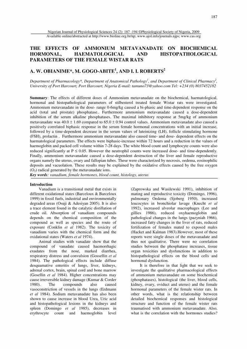
Fig 3 Photomicrographs of the effects of different
doses of ammonium metavanadate on the histology of the
uterus of the female rats, fixed by immersion in Bouin.s
solution and stained with hematoxylin and eosin.
Magnification is 400x. (I) Shows the control showing
normal endometrial epithelium of the uterus (A), (II)
while 1.0mg of metavanadate showed loss or absence of
eosinophilic band (V) and vacuolation of the epithelial
lining of the uterus (N), (III) at 3. 0mg metavanadate
there was marked necrosis of tissues (AG) and (IV) at
5.0mg metavanadate showing vacuolation of the
epithelium (AJ) and marked inflammatory cell infiltrate
in the subepithelial tissues of the uterus (AN
I)
11)
III)
IV)
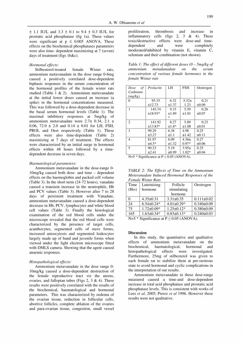
Fig 4. Photomicrographs of the effects of different doses
of ammonium metavanadate on the histology of the ovary
of the female rats, fixed by immersion in Bouin.s solution
and stained with hematoxylin and eosin. Magnification is
400x. (I) The control shows normal graffian follicles at
x200 Mag (B), (II) at 1.0mg metavanadate shows there
are areas of oedema within the ovarian follicle (J), (III)
3. 0mg shows areas of marked oedema in ovarian follicle
(Z) and IV) at 5. 0mg shows areas of congestion of the
ovarian cortex (AQ)
Metavanadate, biochemical, haematological and histopathological parameters

192
TABLE 3: The Effect of Dose and Time on the Ammonium Metavanadate-Induced Haematological (Neutrophil,
Lymphocyte, White blood cell count, Haemoglobin and Packed Cell Volume) Responses of the Stilbesterol
Treated Female Rats.
Dose
Cadmium
(mg/kg)
Neutrophil
count
Lymphocy
te count
White
Blood
Cell
Haemoglobin Packed Cell Volume
24Hrs 7 Days 28 Days 24 Hrs 7 Days 28 Days
0 38.1±1.3 62.20±2.6 5.21
±0.6
11.25
±1.2
11.29
±1.2
11.350
±1.4
34.76
±2.6
34.93
±2.6
34.35
±2.4
2.5 49.2±1.4* 6.56±0.9* 7.56
±0.5*
16.25
±0.9*
9.36
±1.3*
7.260
±1.8*
48.57
±2.9*
33.21
±2.9*
18.09
±1.9*
5 56.2±1.4* 40.90±5.5
*
3.42
±0.4*
14.78
±1.1*
7.14
±1.1*
5.350
±1.1*
44.76
±3.8*
21.73
±1.9*
10.31
±1.08*
17.5 60.2±1.2* 24.50±3.3
*
2.63
±0.5*
13.97
±1.3*
5.67
±0.9*
4.245
±1.02*
43.94
±3.3*
19.37
±1.9*
9.120
±1.03*
N=5 * Significance at P ≤ 0.05 (ANOVA).
Ammonium metavanadate also caused a time-
and dose-dependent decrease in the hormonal
concentrations studied. This is consistent with the
earlier works of Llobert et al (1993). These works
did not study the time-dependence neither did they
show the correlation between the biochemical and
hormonal changes. Ammonium metavanadate
further caused a biphasic response on the
haematological parameters. The agent stimulated
an increase in haemoglobin and packed cell volume
within 3-days and time-dependently in 30 days,
caused a decrease in the same parameters. The
stimulatory responses were novel since no works
had earlier reported these. However the inhibitory
responses (anaemia) recorded were consistent with
the earlier works of Knecht et al (1985), showing
that the results obtained with haematological
parameters are dependent on the time of havesting
of the blood cells.
The agent also caused a dose-and time-
dependent increase in neutrophils, and inhibition of
lymphocytes which is an indication of
immunological suppression (Pierce et al 1996,
Cohen et al 1996). Showing that this agent is
inflammatory and neutropenic. This is also novel.
The histological analysis of the blood cells showed
the presence of acanthocytes, anisocytes and
segmental leucocytes largely made up of band and
juvenile forms. This explains the initial increase of
haemoglobin and packed cell volume values,
showing that vanadate stimulates mitosis in the
short run and inhibit meiosis and maturation in the
long run which predisposes the animal to anaemia
(Yang et al 1986). The results are indicative of
maturation arrest of haematological cells. Thus no
matter the value of the Hb, the remaining cells are
physiologically not useful.
Vanadate caused dose-and time-dependent
pathological changes, to the histology of the female
reproductive system. Furthermore, it also caused
hormonal depressions/inhibitions which were
positively correlated to the histological damages to
uterus, ovaries and oviduct, showing that the
histological destruction resulted in the
pathophysiological decreases in hormonal
production and function which may lead to
infertility (Gupta et al 2004, Uche & Obianime
2008 and Lafuente 2000). This aspect of positive
correlation between structure and function is novel
as no other studies had ever undertaken such.
Furthermore, studies with oxidizing agents like cd,
oxolinic acid, lansoprazole and procymedone have
shown that these agents may in addition have direct
effects on the pituitary axis, leydig cell damage,
perturbation of testosterone production and
overstimulation of luteinising hormone (Fort et al
1995, Murakami 1995, Yamada 1994) thus
resulting in reduction in normal feedback inhibitory
mechanisms which will reduce hormonal
production.
Another school of thought also has it that these
hormonal disruptions precede tissue and uterine
damage and carcinogenesis (Waalkes et al 1997).
Also, these increases in the oxidative metabolism
of cells, may have resulted in the direct destruction
of the uterus, ovaries and fallopian tubes. These
destructions were characterized by oedema,
increase in fat tissue, necrosis of tissues,
vacuolation etc (this study). These results were
consistent with the earlier works of Obianime and
Aprioku (2008). Previous reports did not show the
detailed histological and micrographic results,
neither did they show correlation between the dose
and time of vanadate-induced effects on the
phosphatase, haematological and hormonal
responses.
Vanadate is known to cause an increase in the
serum concentration of free radicals H2O2, OH+
(Llobert et al 1993). These radicals are known to
cause various oxidative pathological destructions
(Kerchaert et al 1996, Rojas et al 1996, Nangi and
Hilter 1996, Lafuente (2001, 2003) which may
result in the compromising of the intergrity of the
female reproductive system and function.
A. W. Obianeme et al

193
Also vanadate caused a dose-and time-
dependent inhibition of the serum hormonal levels
and these results were consistent with previous
reports on the effects of vanadate on the animal
studies (Uche & Obianime 2008). These results
were not qualitative neither were they correlated to
histopathological stimulation of various
phosphatases, which are indices of general toxicity
and an increase which is an index of toxicity of the
prostate and testicular tissues (Eyo 2003, Hirano et
al 1990, Stohs et al 2001, Shimizu & Morita.
1990). The prostatic acid-induced toxicity is
positively correlated to damage of the ovarian and
uterine architecture and function, resulting in the
various histological destructions seen in this study.
Thus showing that vanadate would induce both
biochemical, hormonal and histopathological
alterations which are positively correlated to one
another.
Finally, pretreatment with individual and
combination studies of vitamin C, E and Selenium,
showed that the vitamins and selenium individually
caused a significant inhibition of the vanadate
responses but in combination totally blocked the
vanadate-induced toxicity (not shown). Showing
that, Ammonium metavanadate caused tissue
toxicities through the stimulation of the protein
kinase C (PKC) and calcium ion signal
transductional pathways (Bonkent et al 2007). This
is consistent with the results of Bonkent et al 2007
in the rat intestine.
Vitamin C, E and selenium are antioxidants with
effects on calcium and PKC pathways while
selenium is an important agent in the rate limiting
step of antioxidation (Niki et al 2000).
These antioxidants effect on calcium
metabolism and PKC reduced and inhibited the
generation of oxidative free radicals and also mop
up excessive peroxidation, thus their inhibitory and
remediating action.
References
Annino, J.S. Giese, R.W. (1979) Clinical
chemistry, 4th ed. Little Brown, Boston, pp
170-177.
Barceloux, D.G. Barceloux, D. (1999).
"Vanadium". Clinical Toxicology 37 (2): 265–
278.
Banu, S.K. Govindarajulu, P. Aruldhas, M.M.
(2002). Testosterone and estradiol up-regulate
androgen and estrogen receptors in immature
and adult rat thyroid glands in vivo. Steroids
67 1007–1014.
Benjamin, M.M. (2001). In: Outline of Veterinary
Clinical Pathology, 3rd
. ed. Kalyani
publishers, New Delhi.
Bolkent, S. Koyuturk, M. Bulan, O.K. Tunali, S.
Yanardag, R. Tabakoglu, A.O. (2007). The
effects of combined alpha-tocopherol,
ascorbic acid, and selenium against cadmium
toxicity in rat intestine. J. Environ. Pathol.
Toxicol. Oncol. 26(1):21–27.
Cohen, M., McManus, T. Yang, Z. Qu, Q.
Schlesinger, R. Zelikoff, J. (1996). Vanadium
effects macrophage interferon-gamma-binding
and –inducible responses. Toxicol. Appl.
Pharmacol. 138:110-120.
Conklin A, Skinner C, Felten T, Sanders C (1982)
Clearance and distribution of intratracheally
instilled 48
Vanadium salts in the rat. Toxicol.
Lett. 11:199–203.
Domingo, J.L. Llobet, J.M. Tomas, J.M. Corbella,
J. (1985) Shortterm toxicity studies of
vanadium in rats. J Appl Toxicol 5: 418–421.
Domingo, J.L. (1996). Vanadium: A review of the
reproductive and developmental toxicity.
Repr. Toxicol. 10: (3): 175-182.
Erdmann, E. Werdan, K. Krawietz, W. Schmitz, W.
Scholtz, H. (1984) Vanadate and its
significance in biochemistry and
pharmacology. Biochem Pharmacol. 33:945–
950.
Fort, F.L. Miyajima, H. Suzuki, T. Yamamoto, M.
Hamashima, T. Sato, S. Kitazaki, T. Mahony,
M.C. and Hogden, G.D. (1995). Mechanism
for species-specific induction of leydig cell
tumors in rat by lansoprazole. Fundam. Appl.
Toxicol. 26, 191-202.
Gupta, R. S., Kim, J., Gomes, C., et al. (2004).
Effect of ascorbic acid supplementation on
testicular steroidogenesis and germ cell death
in cadmium-treated male rats. Mol Cell
Endocrinol. 221:57-66.
Gosselin, R. E., Smith, R. P.and Hodge, H. C.
(1984). Clinical Toxicology of Commercial
Products. 5th ed. Baltimore, MD: Williams &
Wilkins. pp. 11-148-149.
Hackett, P. L. and B.J. Kelman. 1983. Availability
of toxic trace metals to the conceptus. Sci.
Total Environ. 28: 433-442.
Hirano, S. Tsukamoto, N. and Suzuki, K. T. (1990).
Biochemical changes in the rat lung and liver
following intratracheal instillation of
cadmium oxide. Toxicol Lett 50:97-105.
Kind, P. R. N. and King, E. J. (1954). Estimation of
plasma phosphatase by determination of
hydrolysed phenol with aminoantipyrine. J.
Clin. Path. 1954; 7, 322-326.
Knecht, E.A., Moorman, W. J., Clark, J. C., Lynch,
D. W. and Lewis, T. R. (1985). Pulmonary
effects of acute vanadium pentoxide
inhalation in monkeys. Am Rev Respir Dis
132:1181-1185.
Knecht, E. Moorman, W. Clark, J.C. Hull, R.D.
Biagini, R.E. Lynch, D.W. Boyle, T.J. Simon,
S.D. (1992). Pulmonary reactivity to
vanadium pentoxide following subchronic
inhalation exposure in a non-human primate
animal model. Journal of Applied Toxicology,
12:427-434.
Metavanadate, biochemical, haematological and histopathological parameters

194
Kerckaert, G., LeBouef, R. Isfort, R. (1996). Use of
the Syrian hamster embryo cell transformation
assay for determining the carcinogenic
potential of heavy metal compounds. Fund.
Applied Toxicol. 34:67-72.
Kumar, A. and Corder, C.N. (1980). Diuretic and
vasoconstrictor effects of sodium
orthovanadate on the isolated perfused rat
kidney. J. Pharmacol. Exp. Ther. 213: 85-90.
Lafuente, A. Marquez, N. Perez-Lorenzo, M. Pazo,
D. Esquifino, A.I. (2000). Pubertal and
postpubertal cadmium exposure differentially
affects the hypothalamic-pituitary-testicular
axis function in the rat. Food Chem Toxicol
38: 913–923.
Lee, J. S., Zhang, M. H., Yun, E. K., Geum, D.,
Kim, K., Kim, T. H., Lim, Y. S. and Seo, J. S.
(2005). Heat shock protein 27 interacts with
vimentin and prevents insolubilization of
vimentin subunits induced by cadmium. Exp
Mol Med. 37:427-435.
Lee, K. P. and Gillies, P. J. (1986). Pulmonary
response and intrapulmonary lipids in rats
exposed to bismuth orthovanadate dust by
inhalation. Environ. Res. 40:115-135.
Llobet, J. M., Colomina, M. T., Siruent, J. J.,
Domingo, J. L. and Corbella, J. (1993).
Reproductive toxicity evaluation of vanadium
in male mice. Toxicolog.y 80:199–206.
Murkami, M., Hosokawa, S., Yamada, T.,
Harakawa, M., Ito, M., Koyama, Y., Kimura,
J., Yoshitake, A. and Yamada, H. (1995).
Species-specific mechanism in rat Leydig cell
tumorigenesis by procynidone. Toxicol. Appl.
Pharmacol. 131. 244-252.
Nanji, A. A, and Hiller-Strurmhofel, (1997).
Apoptosis and necrosis: Two types of cell
death in alcoholic liver diseases. Alcoholic
Health Res. World. 21:325-330.
Osuji, L. C. and Adesiyan, S. O. (2005).
Extractable Hydrocarbons, Nickel and
Vanadium Contents of Ogbodo-Isiokpo Oil
Spill Polluted Soils in Niger Delta, Nigeria.
Env. Monitoring Assess. 110: (1-3)129-139.
Pazynich, V. M. (1966). Experimental data for the
hygienic substantiation of the maximum
permissible concentration of vanadium
pentoxide in the atmosphere. Gig. Sanit. 7: 8-
12.
Pierce, L. M., Alessandrini, F., Godleski, J. J. and
Paulauskis, J. D. (1996). Vanadium-induced
chemokine mRNA expression and pulmonary
inflammation. Toxicol Appl Pharmacol 138:
1-11.
Uche, F. I., Obianime, A. W. and Gogo-Abite, M.
(2008). Effects of vanadium pentoxide on the
histological and sperm parameters of male
Guinea pigs. J. Appl. Sci. Environ. Manage.
12 (3) 107 – 115.
Rojas, E., Valverde, M., Herrera, L., Altamirano-
Lozano, M. and Ostrsky-Wegman, P. (1996).
Genotoxicity of vanadium pentoxide
evaluated by the single gel electrphoresis
assay in human lymphocytes. Mutation Res.
359: 77-84.
Schalm, O. W., Jain, N. C. and Carrot, F. J. (1975).
Veterinary Heamatology, 3rd ed, Lee and
Febiger, Philadelphia.
Shimizu, M. and Morita, S. (1990). Effects of
fasting on cadmium toxicity, glutathione
metabolism, and metallothionein synthesis in
rats. Toxicol. Appl. Pharmacol. 103: 28–39.
Stohs, S. J. Bagchi, D., Hassoun, E. and Bagchi, M.
(2001). Oxidative mechanisms in the toxicity
of chromium and cadmium ions. J. Environ.
Pathol. Toxicol. Oncol. 20: 77-88.
Toro, G. and Ackermann, P. G. (1975). Practical
Clinical Chemistry. Little Brown, Boston, pp
148-156.
Waalkes, M. P., Rehm, S. and Devor, D.E. (1997).
The effects of continuous testosterone
exposure on spontaneous and cadmium-
induced tumors in the male Fisher rats
(F344/NCr) rat: Loss of testicular response.
Toxicol. Appl. Pharmacol. 142, 40-46.
Waters, M. D., Gardner, D. E. and Coffin, D. L.
(1974). Cytotoxic effects of vanadium on
rabbit alveolar macrophages in vitro. Toxicol.
Appl. Pharmacol. 28(2): 253-263.
Yamada, T., Nakamura, J., Murakami, M., Okuno,
Y., Hosokawa, S., Matsuo, M. and Yamada,
H. (1994). The correlation of serum
luteinising hormone levels with induction of
leydig cell tumors in rats by oxolinic acid.
Toxicol. Appl. Pharmacol. 129: 146-154.
Zaporowska, H. and Wasilewski, W. (1991).
Significance of reduced food and water
consumption in rats intoxicated with
vanadium. Comp. Biochem. Physiol. 99C (3):
349-352.
Received: October 12, 2009
Accepted: December 22, 2009
A. W. Obianeme et al