Embed Size (px)
Citation preview
INTRODUCTION
Adhesive dental restorative materials have some major advantages over non adhesive materials. For example, better sealing with tooth tissues, prevention to secondary caries and preservation of natural tooth structures as limited cavity preparation is needed. The idea of adhesive materials in dentistry was introduced by Buonocore in 19551). In order to obtain the adhesion between dental hard tissues and biomaterials, several modifications were explored such as alteration in material’s chemistry or modifying tooth surface properties by chemical treatment with acids1). Phosphoric acid (35–40% in the form of liquid or gel) is commonly used for chemical treatment (acid etching) of dental hard tissue. The acid etching treatment removes smear layer and provide relatively rough surface to create a better interface upon the application of bonding agents2). The surface roughness is resulted due to the loss of minerals from dissolution of tooth hydroxyapatite crystals. The hydrophilic bonding material penetrates in to the roughened tooth surface around etched hydroxyapatite crystals producing micro-tags. This whole phenomenon leads to formation of adhesive tooth-biomaterials interface and micromechanical retention2). Improvement in other physical characteristics such as cleansing of tooth surface, increased surface area, and surface energy as well as exposure of organic matrix to act as a scaffold are also contributing factors to reinforce the bonding strength1).
The adhesion using acid etching of tooth surfaces is a very sensitive technique procedure and can fail as
a result of certain reasons. For example, retention of pellicle or plaque, acid resistant tooth structures, inadequate efficiency of etchant, contamination of the surface and insufficient time for the etchant for appropriate action2). The etching time (the measure of time that acid etchant stays in direct contact with the dental hard tissues) is an important parameter that can influence the surface properties of etched tissues. An etching time of 15–30 s is usually recommended by manufacturer of various dental products. However, in order to get a sufficiently etched surface, a prolonged etching may be required in certain clinical scenarios such as fluorosis and amelogenesis imperfecta. Similarly an additional 30 s etching is suggested for enamel if uniform white frosted appearance is not obtained using the recommended etching time2).
In order to improve the adhesion of resin restorative materials to the tooth structure, the role of acid etching using phosphoric acid (35–38%) has been well documented and supported by a number of studies3-10). The effect of etching time on the final bond strength as well as underlying physical surface properties is not very well understood. The acid etching results in washout of inorganic minerals from the tooth surface hence the amount of the time tooth structure is exposed to acid can affect the underlying physical and mechanical surface properties as well. The objective of this study was to evaluate the effects of acid etching time on surface properties of dental hard tissues at micron and nano scale. The changes in surface roughness, surface hardness and Young’s modulus were studied as function of etching time.
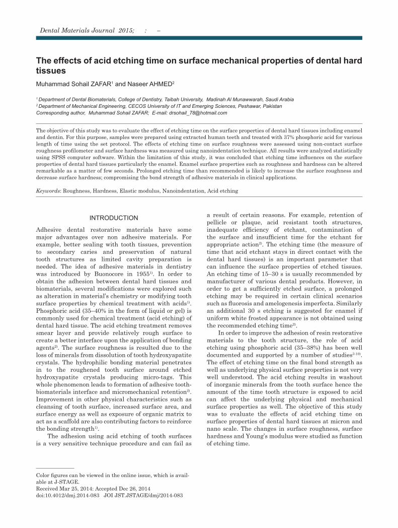
The effects of acid etching time on surface mechanical properties of dental hard tissuesMuhammad Sohail ZAFAR1 and Naseer AHMED2
1 Department of Dental Biomaterials, College of Dentistry, Taibah University, Madinah Al Munawwarah, Saudi Arabia2 Department of Mechanical Engineering, CECOS University of IT and Emerging Sciences, Peshawar, PakistanCorresponding author, Muhammad Sohail ZAFAR; E-mail: [email protected]
The objective of this study was to evaluate the effect of etching time on the surface properties of dental hard tissues including enamel and dentin. For this purpose, samples were prepared using extracted human teeth and treated with 37% phosphoric acid for various length of time using the set protocol. The effects of etching time on surface roughness were assessed using non-contact surface roughness profilometer and surface hardness was measured using nanoindentation technique. All results were analyzed statistically using SPSS computer software. Within the limitation of this study, it was concluded that etching time influences on the surface properties of dental hard tissues particularly the enamel. Enamel surface properties such as roughness and hardness can be altered remarkable as a matter of few seconds. Prolonged etching time than recommended is likely to increase the surface roughness and decrease surface hardness; compromising the bond strength of adhesive materials in clinical applications.
Keywords: Roughness, Hardness, Elastic modulus, Nanoindentation, Acid etching
Color figures can be viewed in the online issue, which is avail-able at J-STAGE.Received Mar 25, 2014: Accepted Dec 26, 2014doi:10.4012/dmj.2014-083 JOI JST.JSTAGE/dmj/2014-083
Dental Materials Journal 2015; : –
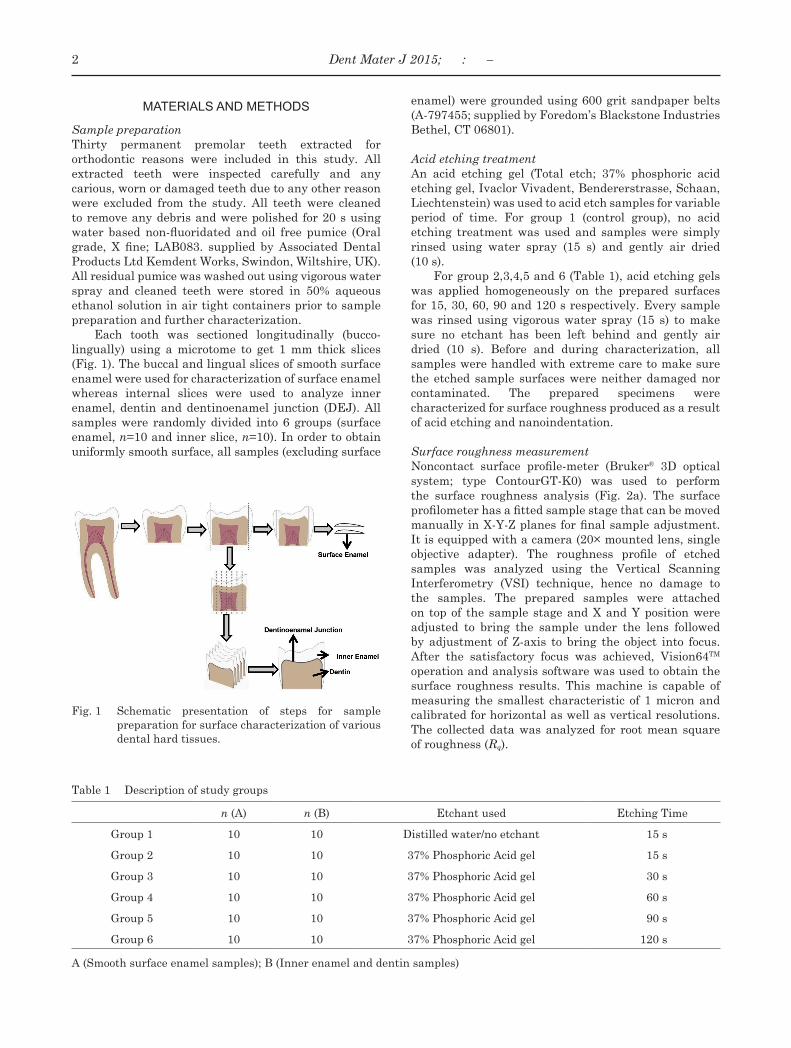
Fig. 1 Schematic presentation of steps for sample preparation for surface characterization of various dental hard tissues.
Table 1 Description of study groups
n (A) n (B) Etchant used Etching Time
Group 1 10 10 Distilled water/no etchant 15 s
Group 2 10 10 37% Phosphoric Acid gel 15 s
Group 3 10 10 37% Phosphoric Acid gel 30 s
Group 4 10 10 37% Phosphoric Acid gel 60 s
Group 5 10 10 37% Phosphoric Acid gel 90 s
Group 6 10 10 37% Phosphoric Acid gel 120 s
A (Smooth surface enamel samples); B (Inner enamel and dentin samples)
MATERIALS AND METHODS
Sample preparationThirty permanent premolar teeth extracted for orthodontic reasons were included in this study. All extracted teeth were inspected carefully and any carious, worn or damaged teeth due to any other reason were excluded from the study. All teeth were cleaned to remove any debris and were polished for 20 s using water based non-fluoridated and oil free pumice (Oral grade, X fine; LAB083. supplied by Associated Dental Products Ltd Kemdent Works, Swindon, Wiltshire, UK). All residual pumice was washed out using vigorous water spray and cleaned teeth were stored in 50% aqueous ethanol solution in air tight containers prior to sample preparation and further characterization.
Each tooth was sectioned longitudinally (bucco-lingually) using a microtome to get 1 mm thick slices (Fig. 1). The buccal and lingual slices of smooth surface enamel were used for characterization of surface enamel whereas internal slices were used to analyze inner enamel, dentin and dentinoenamel junction (DEJ). All samples were randomly divided into 6 groups (surface enamel, n=10 and inner slice, n=10). In order to obtain uniformly smooth surface, all samples (excluding surface
enamel) were grounded using 600 grit sandpaper belts (A-797455; supplied by Foredom’s Blackstone Industries Bethel, CT 06801).
Acid etching treatmentAn acid etching gel (Total etch; 37% phosphoric acid etching gel, Ivaclor Vivadent, Bendererstrasse, Schaan, Liechtenstein) was used to acid etch samples for variable period of time. For group 1 (control group), no acid etching treatment was used and samples were simply rinsed using water spray (15 s) and gently air dried (10 s).
For group 2,3,4,5 and 6 (Table 1), acid etching gels was applied homogeneously on the prepared surfaces for 15, 30, 60, 90 and 120 s respectively. Every sample was rinsed using vigorous water spray (15 s) to make sure no etchant has been left behind and gently air dried (10 s). Before and during characterization, all samples were handled with extreme care to make sure the etched sample surfaces were neither damaged nor contaminated. The prepared specimens were characterized for surface roughness produced as a result of acid etching and nanoindentation.
Surface roughness measurement Noncontact surface profile-meter (Bruker® 3D optical system; type ContourGT-K0) was used to perform the surface roughness analysis (Fig. 2a). The surface profilometer has a fitted sample stage that can be moved manually in X-Y-Z planes for final sample adjustment. It is equipped with a camera (20× mounted lens, single objective adapter). The roughness profile of etched samples was analyzed using the Vertical Scanning Interferometry (VSI) technique, hence no damage to the samples. The prepared samples were attached on top of the sample stage and X and Y position were adjusted to bring the sample under the lens followed by adjustment of Z-axis to bring the object into focus. After the satisfactory focus was achieved, Vision64TM operation and analysis software was used to obtain the surface roughness results. This machine is capable of measuring the smallest characteristic of 1 micron and calibrated for horizontal as well as vertical resolutions. The collected data was analyzed for root mean square of roughness (Rq).
2 Dent Mater J 2015; : –
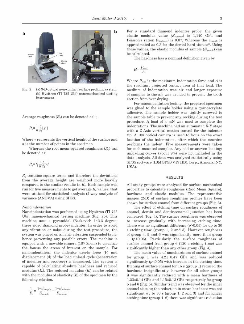
Fig. 2 (a) 3-D optical non-contact surface profiling system, (b) Hysitron (TI 725 Ubi) nanomechanical testing instrument.
Average roughness (Ra) can be denoted as11);
1 nRa= Σ|yi|n i=1
Where y represents the vertical height of the surface and n is the number of points in the specimen.
Whereas the root mean squared roughness (Rq) can be denoted as;
1 nRq=√ Σyi
2n i=1
Rq contains square terms and therefore the deviations from the average height are weighted more heavily compared to the similar results in Ra. Each sample was run for five measurements to get average Ra values; that were utilized for statistical analysis (2-way analysis of variance (ANOVA) using SPSS.
NanoindentationNanoindentation was performed using Hysitron (TI 725 Ubi) nanomechanical testing machine (Fig. 2b). This machine uses a pyramidal (Berkovich 142.3 degree, three sided diamond probe) indenter. In order to avoid any vibration or noise during the test procedure, the system was placed on an anti-vibration suspended table, hence preventing any possible errors. The machine is equiped with a movable camera (10× Zoom) to visualize the foucus the areas of interest on the sample. For nanoindentation, the indentor exerts force (F) and diaplacement (d) of the load unload cycle (penetration of indentor and recovery) is measured. The system is capable of calculating absolute hardness and reduced modulus (Er). The reduced modulus (Er) can be related with the modulus of elasticity (E) of the specimen by the following relation.
1 1−v2sample 1−v2
indenter = + Er Esample Eindenter
For a standard diamond indenter probe, the given elastic modulus value (Eindenter) is 1,140 GPa and Poisson’s ration (vindenter) is 0.07, Whereas the vsample is approximated as 0.3 for the dental hard tissues2). Using these values, the elastic modulus of sample (Esample) can be calculated.
The hardness has a nominal definition given by
PmaxH= A
Where Pmax is the maximum indentation force and A is the resultant projected contact area at that load. The medium of indentation was air and longer exposure of samples to the air was avoided to prevent the tooth section from over drying.
For nanoindentation testing, the prepared specimen was glued to the sample holder using a cyanoacrylate adhesive. The sample holder was tightly screwed to the sample table to prevent any rocking during the test procedure. A load of 4 mN was used to complete the indentations. The machine had an automated X-Y stage with a Z-Axis vertical motion control for the indenter tip. A 10× optical camera is used to focus on the exact location of the indentation, after which the machine performs the indent. Five measurements were taken for each mounted samples. Any odd or uneven loading/unloading curves (about 9%) were not included in the data analysis. All data was analyzed statistically using SPSS software (IBM SPSS V19 IBM Corp., Armonk, NY, USA).
RESULTS
All study groups were analyzed for surface mechanical properties to calculate roughness (Root Mean Square), hardness and elastic modulus. The representative images (2-D) of surface roughness profiles have been shown for surface enamel from different groups (Fig. 3).
The effect of etching time on surface roughness of enamel, dentin and dentinoenamel junction has been compared (Fig. 4). The surface roughness was observed to increase gradually with increasing etching time. There was no significant difference (p>0.05) for up to 30 s etching time (group 1, 2 and 3). However roughness of group 4, 5 and 6 was significantly more than group 1 (p<0.05). Particularly the surface roughness of surface enamel from group 6 (120 s etching time) was significantly higher than any other group (Fig. 4).
The mean value of nanohardness of surface enamel for group 1 was 4.21±0.47 GPa and was reduced significantly (p<0.05) with increase in the etching time. Etching of surface enamel for 15 s (group 1) reduced the hardness insignificantly, however for all other groups it was significantly reduced with a mean hardness of 1.23±0.14 GPa and 1.15±0.13 GPa respectively for group 5 and 6 (Fig. 5). Similar trend was observed for the inner enamel tissues; the reduction in mean hardness was not significant up to 30 s (group 1, 2 and 3) and for longer etching time (group 4–6) there was significant reduction
3Dent Mater J 2015; : –
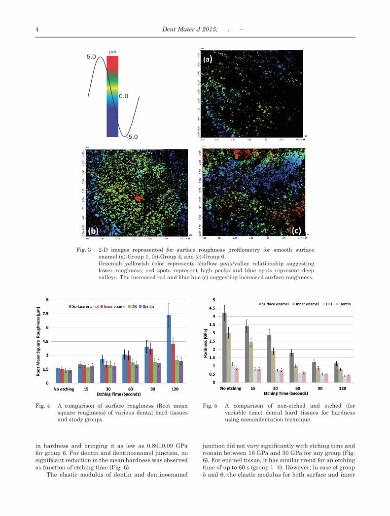
Fig. 3 2-D images represented for surface roughness profilometry for smooth surface enamel (a)-Group 1, (b)-Group 4, and (c)-Group 6.
Greenish yellowish color represents shallow peak/valley relationship suggesting lower roughness; red spots represent high peaks and blue spots represent deep valleys. The increased red and blue hue (c) suggesting increased surface roughness.
Fig. 4 A comparison of surface roughness (Root mean square roughness) of various dental hard tissues and study groups.
Fig. 5 A comparison of non-etched and etched (for variable time) dental hard tissues for hardness using nanoindentation technique.
in hardness and bringing it as low as 0.80±0.09 GPa for group 6. For dentin and dentinoenamel junction, no significant reduction in the mean hardness was observed as function of etching time (Fig. 6).
The elastic modulus of dentin and dentinoenamel
junction did not vary significantly with etching time and remain between 16 GPa and 30 GPa for any group (Fig. 6). For enamel tissue, it has similar trend for an etching time of up to 60 s (group 1–4). However, in case of group 5 and 6, the elastic modulus for both surface and inner
4 Dent Mater J 2015; : –
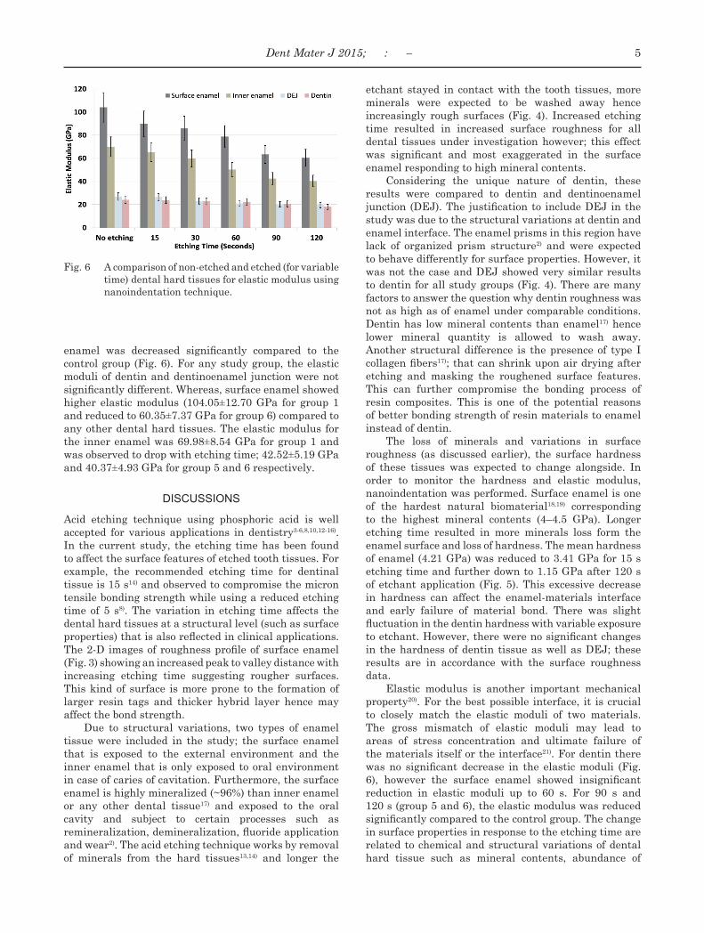
Fig. 6 A comparison of non-etched and etched (for variable time) dental hard tissues for elastic modulus using nanoindentation technique.
enamel was decreased significantly compared to the control group (Fig. 6). For any study group, the elastic moduli of dentin and dentinoenamel junction were not significantly different. Whereas, surface enamel showed higher elastic modulus (104.05±12.70 GPa for group 1 and reduced to 60.35±7.37 GPa for group 6) compared to any other dental hard tissues. The elastic modulus for the inner enamel was 69.98±8.54 GPa for group 1 and was observed to drop with etching time; 42.52±5.19 GPa and 40.37±4.93 GPa for group 5 and 6 respectively.
DISCUSSIONS
Acid etching technique using phosphoric acid is well accepted for various applications in dentistry3-6,8,10,12-16). In the current study, the etching time has been found to affect the surface features of etched tooth tissues. For example, the recommended etching time for dentinal tissue is 15 s14) and observed to compromise the micron tensile bonding strength while using a reduced etching time of 5 s8). The variation in etching time affects the dental hard tissues at a structural level (such as surface properties) that is also reflected in clinical applications. The 2-D images of roughness profile of surface enamel (Fig. 3) showing an increased peak to valley distance with increasing etching time suggesting rougher surfaces. This kind of surface is more prone to the formation of larger resin tags and thicker hybrid layer hence may affect the bond strength.
Due to structural variations, two types of enamel tissue were included in the study; the surface enamel that is exposed to the external environment and the inner enamel that is only exposed to oral environment in case of caries of cavitation. Furthermore, the surface enamel is highly mineralized (~96%) than inner enamel or any other dental tissue17) and exposed to the oral cavity and subject to certain processes such as remineralization, demineralization, fluoride application and wear2). The acid etching technique works by removal of minerals from the hard tissues13,14) and longer the
etchant stayed in contact with the tooth tissues, more minerals were expected to be washed away hence increasingly rough surfaces (Fig. 4). Increased etching time resulted in increased surface roughness for all dental tissues under investigation however; this effect was significant and most exaggerated in the surface enamel responding to high mineral contents.
Considering the unique nature of dentin, these results were compared to dentin and dentinoenamel junction (DEJ). The justification to include DEJ in the study was due to the structural variations at dentin and enamel interface. The enamel prisms in this region have lack of organized prism structure2) and were expected to behave differently for surface properties. However, it was not the case and DEJ showed very similar results to dentin for all study groups (Fig. 4). There are many factors to answer the question why dentin roughness was not as high as of enamel under comparable conditions. Dentin has low mineral contents than enamel17) hence lower mineral quantity is allowed to wash away. Another structural difference is the presence of type I collagen fibers17); that can shrink upon air drying after etching and masking the roughened surface features. This can further compromise the bonding process of resin composites. This is one of the potential reasons of better bonding strength of resin materials to enamel instead of dentin.
The loss of minerals and variations in surface roughness (as discussed earlier), the surface hardness of these tissues was expected to change alongside. In order to monitor the hardness and elastic modulus, nanoindentation was performed. Surface enamel is one of the hardest natural biomaterial18,19) corresponding to the highest mineral contents (4–4.5 GPa). Longer etching time resulted in more minerals loss form the enamel surface and loss of hardness. The mean hardness of enamel (4.21 GPa) was reduced to 3.41 GPa for 15 s etching time and further down to 1.15 GPa after 120 s of etchant application (Fig. 5). This excessive decrease in hardness can affect the enamel-materials interface and early failure of material bond. There was slight fluctuation in the dentin hardness with variable exposure to etchant. However, there were no significant changes in the hardness of dentin tissue as well as DEJ; these results are in accordance with the surface roughness data.
Elastic modulus is another important mechanical property20). For the best possible interface, it is crucial to closely match the elastic moduli of two materials. The gross mismatch of elastic moduli may lead to areas of stress concentration and ultimate failure of the materials itself or the interface21). For dentin there was no significant decrease in the elastic moduli (Fig. 6), however the surface enamel showed insignificant reduction in elastic moduli up to 60 s. For 90 s and 120 s (group 5 and 6), the elastic modulus was reduced significantly compared to the control group. The change in surface properties in response to the etching time are related to chemical and structural variations of dental hard tissue such as mineral contents, abundance of
5Dent Mater J 2015; : –

collagen fibers and must be considered while using acid etching technique to get the best clinical outcome.
Regarding the effect of structural domains such as enamel prisms and sheath on surface properties, the authors strongly agree with Ge et al.22). The differences in the orientation of structural units and apatite crystals affect the material properties such as hardness and roughness. In addition to anisotropic structure of enamel, the surface chemistry also affects the properties. For example, surface enamel remain protected by amealoblast cells prior to eruption and exposed to the oral environment after eruption and making it more mineralized (~9%) than inner enamel. Similarly, the amount of organic matrix in the enamel sheath is significantly higher with less mineral than enamel prisms hence affecting the properties such as hardness17). In order to evaluate the effects of etching time on structural units (such as enamel prism, rods or crystals), nanoindentation under high resolution magnification such as Atomic Force Microscopy (AFM) is required and can be considered for future work.
CONCLUSION
Within the limitation of this study, it can be concluded that the etching time influences on the surface properties of dental hard tissues particularly the surface enamel. Enamel surface properties such as roughness and hardness can be altered remarkably as a matter of few seconds, hence must not be etched for longer than 30 s. Similarly dentin should be etched for 15–30 s without any remarkable damage to the tissue surface. Prolonged etching time than recommended is likely to increase the surface roughness and decrease surface hardness hence compromising the bond strength of adhesive materials in clinical applications.
CONFLICT OF INTEREST
No conflict of interest for conducting this research.
REFERENCES
1) Buonocore MG. A simple method of increasing the adhesion of acrylic filling materials to enamel surfaces. J Dent Res 1955; 34: 849-853.
2) Sakaguchi RL, Powers JM. Craig’s restorative dental materials. Philadelphia, PA: Elsevier/Mosby; 2012.
3) Fernandes ACR, Bridi EC, do Amaral FLB, França FMG, Flório FM, Basting RT. Microtensile bond strength of silorane or methacrylate resin-based composites associated to self-etching or conventional adhesives to dentin after different storage times. Int J Adhes Adhes 2014; 48: 28-34.
4) Kayahan MB, Nekoofar MH, McCann A, Sunay H, Kaptan RF, Meraji N, Dummer PMH. Effect of acid etching procedures on the compressive strength of 4 calcium Silicate-based
endodontic cements. J Endod 2013; 39: 1646-1648. 5) Dieng-Sarr F, Sharrock P, Dabsie F, Grégoire G. Modifications
of the organic and mineral fractions of dental tissues following conditioning by self-etching adhesives. J Dent 2011; 39: 141-147.
6) Chasqueira F, Portugal J, Arantes SS, Oliveira, Lopes LP. Adhesion of dental sealants to enamel with self-etching adhesives in salivary contamination conditions: Influence of the light curing protocol. Revista Portuguesa de Estomatología, Medicina Dentária e Cirugia Maxilofacial 2011; 52: 2-6.
7) Choi S, Rhee Y, Park J, Lee G, Kim K, Park J, Park Y, Park H. Effects of fluoride treatment on phosphoric acid-etching in primary teeth: An AFM observation. Micron 2010; 41: 498-506.
8) Bolaños-Carmona V, González-López S, Briones-Luján T, De Haro-Muñoz C, de la Macorra JC. Effects of etching time of primary dentin on interface morphology and microtensile bond strength. Dent Mater 2006; 22: 1121-1129.
9) Ariyaratnam MT, Wilson MA, Mackie IC, Blinkhorn AS. A comparison of surface roughness and composite/enamel bond strength of human enamel following the application of the nd:YAG laser and etching with phosphoric acid. Dent Mater 1997; 13: 51-55.
10) Brännström M, Malmgren O, Nordenvall K. Etching of young permanent teeth with an acid gel. Am J Orthod 1982; 82: 379-383.
11) De Garmo Ernest P, Black JT, Kohser Ronald A. DeGarmo’s Materials and processes in manufacturing. Hoboken, NJ: Wiley; 2012.
12) Kukiattrakoon B, Thammasitboon K. The effect of different etching times of acidulated phosphate fluoride gel on the shear bond strength of high-leucite ceramics bonded to composite resin. J Prosthet Dent 2007; 98: 17-23.
13) Perdigão J, Lopes M. The effect of etching time on dentin demineralization. Quintessence Int 2001; 32: 19-26.
14) Pioch T, Stotz S, Buff E, Duschner H, Staehle HJ. Influence of different etching times on hybrid layer formation and tensile bond strength. Am J Dent 1998; 11: 202-206.
15) Garcia-Godoy F, Gwinnett AJ. Effect of etching times and mechanical pretreatment on the enamel of primary teeth: An SEM study. Am J Dent 1991; 4: 115-118.
16) Wilson NHF, Gordan VV, Brunton PA, Wilson MA, Crisp RJ, Mjor IA. Two-centre evaluation of a resin composite/self-etching restorative system: Three-year findings. J Adhes Dent 2006; 8: 47-51.
17) Nanci A. Ten Cate’s oral histology: development, structure, and function. St. Louis, Mo ; London: Mosby; 2012.
18) Zafar MS, Ahmed N. Nano-mechanical evaluation of dental hard tissues using indentation technique. World Appl Sci J 2013; 28: 1393-1399.
19) Zafar MS. A comparison of dental restorative materials and mineralized dental tissues for surface nanomechanical properties. Life Sci J 2014; 11: 19-24.
20) Zafar MS, Ahmed N. Nanoindentation and surface roughness profilometry of poly methyl methacrylate denture base materials. Technol Health Care 2014; 22: 573-581.
21) Shi L, Wang L, Duan Y, Lei W, Wang Z, Li J, Fan X, Li X, Li S, Guo Z. The improved biological performance of a novel low elastic modulus implant. PloS One 2013; 8: e55015.
22) Ge J, Cui F, Wang X, Feng H. Property variations in the prism and the organic sheath within enamel by nanoindentation. Biomaterials 2005; 26: 3333-3339.
6 Dent Mater J 2015; : –


![Index INSTRUCTIONS FOR USE - FLAVA217]_etching_gel_navod.pdf · 37% Etching Gel & 37% Phosphoric Acid 10% Etching Gel 10% Phosphoric Acid Wallingford, CT 06492 USA tel: 203.265.7397](https://img.pdfslide.us/doc/110x75/5e30b61787eaf86a72023ba7/index-instructions-for-use-flava-217etchinggelnavodpdf-37-etching-gel.jpg)