Embed Size (px)
Citation preview
Page 1
The Effect of Tobacco Extract and Nicotine Alone and in Combination
on Fracture Healing in Rats
Research Year Thesis
Stud. Med. Martin Skøtt
Faculty of Health Sciences University of Aarhus
Denmark 2004
Page 2
Corrospondance:
Martin Skøtt
Orthopeadic Research Laboratory
Department of Orthopeadic Surgery
Aarhus University Hospital
Nørrebrogade 44, building 1A
DK-8000 Aarhus C
Denmark
Phone: +45 89494162
Fax: + 45 89494150
E-mail: [email protected]
Page 3
Preface This research year thesis is based on scientific work performed at Orthopaedic Biomechanics
Laboratory, Hennipin County Medical Center, Minneapolis, MN, USA and Orthopaedic Research
Laboratory, Aarhus University Hospital.
I am very grateful to have had the opportunity to gain an insight into the fascinating world of
research. It has been exiting and challenging event, especially the 4 months in Minneapolis were the
experiment took place.
This specific research project was initiated by my supervisors Prof. Kjeld Søballe, MD, DMSc. and
Joan E. Bechtold, Ph.D. my deepest thanks go to them.
The result of my research year is a manuscript to an article, which has been submitted to the Journal
of Orthopaedic Research. I would therefore like to thank my co-authors, which are besides my two
supervisors, Troels T. Andreassen, Associate Professor MD, and Michael Ulrich-Vinther, MD,
Ph.D. A signed statement concerning my work during this research project can by found at the end
of this report.
The study was presented as a poster at the Annual meeting of the Orthopaedic Research Society,
Washington DC in February 2005. Abstracts submitted to DOS 2005 and SICOT 2005 can also be
found at the end of this report.
Financial support was gratefully received from Orthopaedic Research Foundation in Aarhus and by
the Danish Medical Research Council.
Page 4
Contents
Preface ................................................................................................................................ 3
Abstract ............................................................................................................................................6
Introduction......................................................................................................................................7
Materials and Methods.....................................................................................................................8
Results............................................................................................................................................12
Discussion ......................................................................................................................................14
Reference .......................................................................................................................................17
Appendix........................................................................................................................... 19
Table 1 MECHANICAL PROPERTIES OF THE FRACTURED FEMORA..............................19
Table 2 SERUM CONCENTRATIONS OF NICOTINE AND COTININE DURING THE EXPERIMENT (ng/ml) .................................................................................................................19
Table 3 CALLUS PROPERTIES OF THE FRACTURED FEMORAL.......................................20
Table 4 CALLUS PROPERTIES OF THE FRACTURED FEMORAL.......................................20
Poster to ORS, spring 2005............................................................................................................21
Abstract to DOS, August 2005 ......................................................................................................22
Abstract to SICOT, September 2005 .............................................................................................23
Page 5
The Effect of Tobacco Extract and Nicotine Alone and in Combination
on Fracture Healing in Rats
Martin Skoett1, 2, Troels T. Andreassen3, Michael Ulrich-Vinther1, X. Chen2, Dan Keyler4,
Mark LeSage4, Paul Pentel4, Joan E. Bechtold2, Kjeld Soeballe1
1 Orthopaedic Research Laboratory, Department of Orthopaedic Surgery, Aarhus University
Hospital, Aarhus, Denmark.
2 Orthopaedic Biomechanics Laboratory, Midwest Orthopaedic Research foundation and
Minneapolis Medical Research Foundation, Hennepin County Medical Center, Minneapolis, MN,
USA.
3 Department of Connective Tissue Biology, Institute of Anatomy, University of Aarhus, Denmark
4 Department of Medicine, Division of Clinical Pharmacology, Hennepin County Medical Center,
Minneapolis, MN, USA
Corresponding author: Martin Skoett Orthopeadic Research Laboratory Department of Orthopeadic Surgery Aarhus University Hospital Nørrebrogade 44, building 1A DK-8000 Aarhus C Denmark Phone: +45 89494162 Fax: + 45 89494150 E-mail: [email protected]
Page 6
Abstract The influence of nicotine and tobacco extract (without nicotine) alone and in combination on callus
formation and mechanical strength of femoral fractures in rats was investigated after 21 days of
healing. One hundred and four male Sprague-Dawley rats were divided into four groups receiving:
saline, nicotine, tobacco extract, and tobacco extract plus nicotine. One week prior to fracture
surgery Alzet miniosmotic pumps were implanted subcutaneously in all animals to administer saline
or nicotine. The tobacco extract was administered orally. Over an intramedullary pin, a closed
transverse femur fracture was performed and mechanically tested after 21 days of healing. Ultimate
torque and torque at yield point of the tobacco extract group was decreased by 21 % (p=0.010) and
23 % (p=0.056), respectively, compared with the vehicle group and by 20 % (p=0.023) and 26 %
(p=0.004), respectively, compared with the nicotine group. No difference was found between the
tobacco extract and tobacco extract plus nicotine groups. An 18 % (p=0.013) reduction in torque at
yield point was observed in the tobacco extract plus nicotine group compared with the nicotine
group. No differences in ultimate stiffness, energy absorption, callus bone mineral content, and
callus external dimensions at the fracture line were found between the groups. Serum levels of
nicotine were between 40 – 50 ng/mL. Equivalent serum levels are observed in persons smoking
one pack of cigarettes per day. In summary, tobacco extract decreased the mechanical properties of
healing fractures. Nicotine given in a dose inducing serum levels equivalent to those observed in
daily smokers, did not, however, affect mechanical properties of healing fractures.
Key words: tobacco extract, nicotine, fracture healing, mechanical strength, rat
Page 7
Introduction Fracture repair is slower in smokers compared to people who do not smoke, and the rate of non-
union is augmented among smokers. This has been shown both in clinical trials concerning
open/closed tibiae shaft fractures and spinal fusions (1-6).
Furthermore these findings have been supported by a number of fracture and spinal fusion studies in
rodents exposed to different levels of nicotine (7-9). The negative impact might be explained by
nicotine’s inhibition of osteoblastic activity (8;10) and by its vasoconstrictive action on the
microvasculature (11-13). The vasoconstrictive effect is seen during revascularization of cancellous
bone graft during spinal fusion (14). It might also be that nicotine attenuate a wide range of
cytokines which are normally expressed during bone formation (15).
One common feature of the animal studies was that the experiments used only nicotine and that the
nicotine usually was administered in doses causing higher serum levels than those observed in
smokers consuming a pack of cigarettes daily (12;16;17). Besides nicotine there are about 3500
other compounds in the particulate phase of cigarette smoke, including the lung carcinogenic agents
(11;12;18).
To the best of our knowledge, no experiment has been conducted to examine the effect of the
tobacco extracts without nicotine on fracture healing. Therefore the aim of this study was to
investigate the effects of tobacco extract and nicotine, when given separately and in combination,
on rat fracture healing. The tobacco extract was obtained from nicotine free cigarettes and
administered intermittently by the oral route. Nicotine was administered continuously in a dose by
which we achieved serum levels equivalent to those observed in daily smokers (11;12;16;17;19).
Page 8
Materials and Methods Animals and design
The experimental protocol was approved by the institutional review board (Animal Care, Use &
Research Committee, Minneapolis Medical Research Foundation).
One hundred and four male Sprague-Dawley rats with an initial weight of 250-275 gram were
randomized according to weight into four groups, receiving saline, nicotine, tobacco extract, and
tobacco extract plus nicotine. Sample size was based on the following equation: n= 2(t2α+tβ) ×
SD2/D2. Error of the first kind (2α) was selected to 5% and error of the second kind (β), the risk of
concluding that two effects are identical if in fact the difference is below the minimal clinically
relevant difference (D), was chosen to be 20 % which means a power of 80%. Based on previous
studies, a SD of 20 % and a D of 18% for all variables is justified. Based on these assumptions at
least 20 animals in each group had to be included. In the study we included 26, to allow for the loss
of 6 animals per group. The rats were housed individually and allowed feed and water ad libitum,
except the two groups that received tobacco extract orally during a 16-hour period. The room was
maintained on a twelve hour light/dark cycle (the light cycle began at 07.00 hr) at approximately
21°C. Cages, food, and bottles were changed once a week.
Treatment was administered for 4 weeks – corresponding to one week pre-fracture and 3 weeks
post-fracture.
Preparation and administration of nicotine
Nicotine barbiturate was obtained from Sigma Chemical Co. (St. Louis, MO). Osmotic minipumps
(Alzet 2ML4, Durect Cupertino, CA) were loaded with the nicotine preparation containing a
concentration of 14.00 mg/ml. The 2ML4 pumps delivered 2.5µL/day for four weeks, which
yielded a dosage of 3.0 mg/kg/day.
Page 9
The pumps were implanted subcutaneously (under i.p. ketamine and xylazine anesthesia at a dose of
80-100 mg/kg and 2-3 mg/kg, respectively) in the intrascapular area using aseptic conditions. The
incision sites were prepared by shaving followed by disinfection of the exposed skin with
Povidone-inodine USP 10 %. One incision of approximately 5 mm was made in the skin of the
dorsal mid-cervical region and the pump was implanted in the subcutaneous tissue of the lateral
neck area. The surgical wound was closed by 9-mm stamps and daubed with antiseptic crème
(Betadine). All animals received a pump containing either saline (vehicle) or the nicotine solution.
Preparation and administration of tobacco extract
Tobacco extract was prepared from nicotine-free Quest Cigarettes (Vector Tobacco Inc,
Timberlake, NC), as described by Demady et al. (26). Four cigarettes were cut open, the tobacco
ground with a mortar and pestle and placed in a 50-ml plastic tube containing 40 ml water. The
tubes were mixed overnight at room temperature using a tube tipper. The contents were filtered
through gauze and the liquid was centrifuged at 5000 rpm for 20 min. The supernatant was vacuum
filtered using z Buchner funnel and Whatman paper. The extract was filtered again using a 0.2-µm
sterile filter. Three batches were checked for uniformity by measuring anatabine on a GC-MS. The
extract was diluted 1/20 for use to correspond to the amount of nicotine used and stored at 4ºC. The
solution was administered orally by 30 ml. drinking bottles and the daily amount consumed at the
end of the 16-hour period was recorded.
Closed fracture surgery
The animals were anesthetized using intraperitoneal injection of ketamine/xylazine at a dose of 100
mg/kg and 2.0 mg/kg, respectively. When the animals reached the surgical stage of general
anesthesia, preparation for surgery was initiated, including shaving and disinfection of the left leg.
Page 10
A lateral parapatellar approach to the left stifle was performed by a 5-mm skin incision (without
damaging the patellar ligament). The longitudinal fibers of the quadriceps mechanism were divided
and the patella was dislocated medially. Internal rotation of the tibia and valgus of the knee resulted
in exposing of the femoral condyles. The marrow canal was reamed with a 1.5-mm 18 gauge
needle. A 1.4-mm K-wire (D. Trocar PT 9, 054 1.4 mm) of 33 mm in length was introduced in a
retrograde fashion. The pin was countersunk and seated in the proximal femur. Muscles and fascia
was reapposed using Ethilon 4-0 suture (Ethicon) and the skin was closed in standard fashion, also
using Ethilon 4-0 suture.
Immediately after K-wire placement, a closed transverse fracture in the mid-diaphyseal region of
the femur was performed using a fracture apparatus designed according to the original
specifications by Bonnarens and Einhorn (20). Presence of the transversal fracture line was
confirmed by palpation.
Serum Analysis
To determine nicotine and cotinine levels, blood samples were taken from a tail vein at: fracture,
after two weeks of healing, and at sacrifice. The blood samples were centrifuged at 3000 rpm for 15
min. Serum was then stored at –70ºC until the gas chromatography (Hewlett – Packed 5890 Series
II) with nitrogen-phosphours detection assay was performed (21).
In a separate study with a similar setup as in the main study the amount of tobacco extract was
determined by measuring the urine concentration of the tobacco alkaloids anabasine and anatabine.
These markers were determined by gas chromatography-mass spectrometry (22) on 24 hr urine
samples collected from rats in metabolic chambers on days 1, 7 and 14.
Page 11
Mechanical testing
The rats were euthanized with pentobarbital (100 mg/kg intraperitoneally; Beauthanasia-D) and X-
rays of the femurs were taken to inspect fracture healing and placement of the intra-medullary K-
wire. Femurs were stripped of soft tissue and the K-wire was carefully removed by gentle axial pull
with rotational motions. In the fractured femur, the external medial-lateral and anterior-posterior
diameters were measured at the fracture line using a sliding caliper. The proximal and distal ends
were potted in aluminum fixtures (Wood´s metal) with low melting temperature metal while
maintaining the callus areas moist. The fracture was tested on an axial-torsion materials testing
machine (model AT3045-2800, Bose EnduraTEC Systems Corp., Eden Prairie, MN) with a low
capacity 50 in-lb torque cell (Transducer Techniques, Temecula, CA).The femur was aligned so that
the longitudinal axis of the mid-femur was approximately collinear with the longitudinal axis of the
proximal and distal blocks of potting material and torsional actuator of the materials test machine.
The fractures were loaded to failure in torsion at a rate of 0.5 degree/sec. Thereafter, the femurs
were stored in 70% ethanol at 10ºC until further examination.
Torques were measured to within ± 0.002 Nm in 1.14 Nm range, and angles were measured to
within 0.25 % of the full scale (60º), or 0.15º. Torque and rotation were continuously recorded over
3 consecutive cycles with a personal computer and WinTest software (Bose EnduraTEC Systems
Corp.) at a rate of 20 Hz. The data were translated into a load-deformation curve where ultimate
torque (maximum load) and ultimate stiffness (equal to the maximum slope of the load-deformation
curve) were calculated. The energy absorptive capacity was measured as the area under the load-
formation curve until ultimate torque. The yield point was defined as the point at which a 20 %
reduction in maximum slope occurred.
Page 12
Dual-energy X-ray absorptiometry (DEXA)
For all fractures only one fracture line and no loose fragments was observed. The two ends of bones
tested were realigned at their original position. The femurs were placed in 70% ethanol with the
anteromedial surface downward and scanned by DEXA using the regional high resolution analysis
program for small animals (QRD-2000; Hologic, Inc. Waltham, MA). The amount of bone around
the fracture line was measured in an 8.0-mm-high diaphysial segment, 4 mm proximal and 4 mm
distal to the fracture line. BMC was measured in this segment.
Statistical Analysis
The data were tested for normality and homogeneity of variance and when these conditions were
fulfilled, parametric analyses were applied. Otherwise, nonparametric analyses were used.
The effects of the different exposure were evaluated using the following groups: Vehicle, Nicotine,
Tobacco extract, and Nicotine plus Tobacco extract. Differences between these groups were tested
by one-way analysis of variance or Kruskal-Wallis test. In cases in which differences occurred, all
pair wise multiple comparisons procedures were applied (unpaired t-test or Mann-Whitney Rank
Sum Test). Changes in weight in the individual groups during the experiment were analyzed by
using unpaired t-test as normality and homogeneity of variance were fulfilled.
The value of p < 0.05 (two-tailed) was considered statistically significant.
Results Out of 104 animals operated on, 89 were included in the subsequent experiment. Seven animals
died as a result of anesthesia, six as a consequence of insignificant fracture and two because of
pump malfunction. The number of animals in each group was: vehicle (n=22), nicotine (n=22),
tobacco extract (n=23) and nicotine plus tobacco extract (n=22).
Page 13
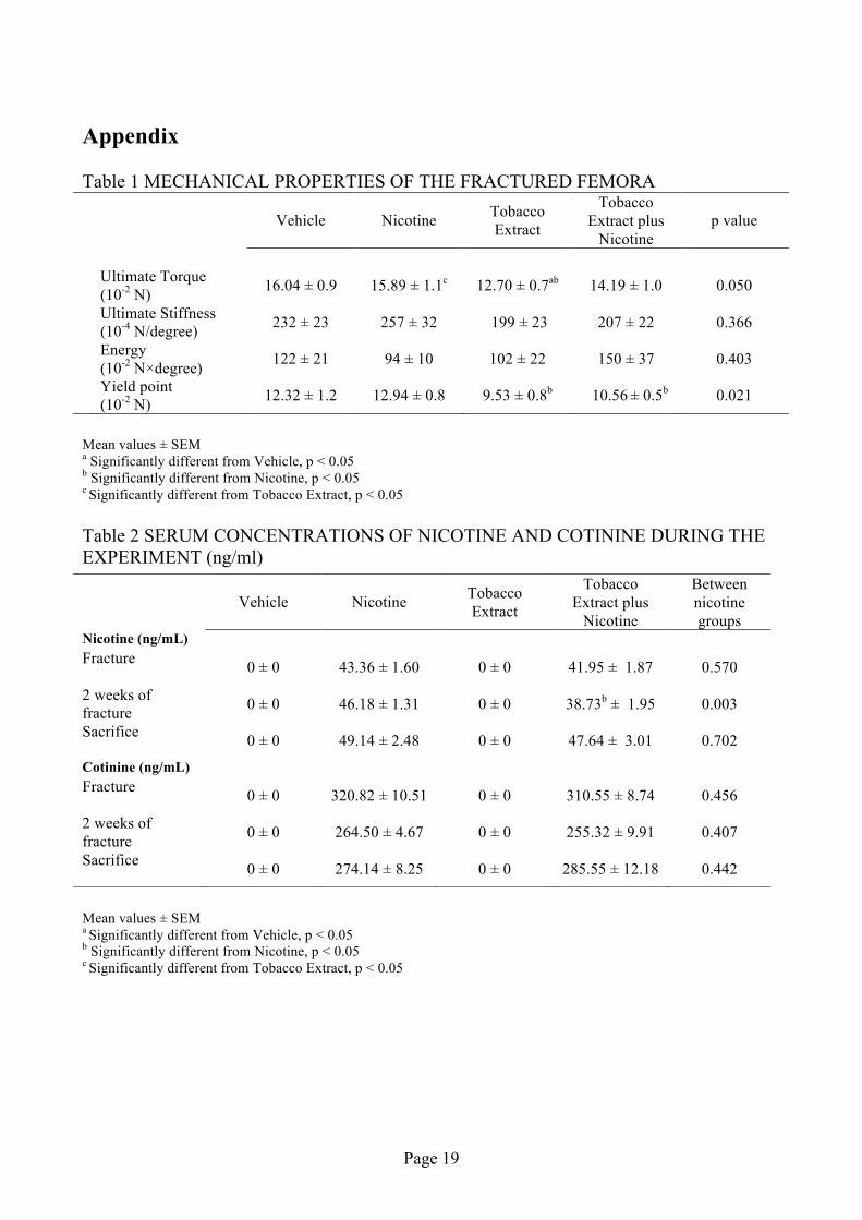
The results of the mechanical testing of the fractured femurs are given in Table 1. After 21 days of
healing, the ultimate torque of the fractures was decreased only in the group given tobacco extract
alone, both in comparison with the nicotine group and the vehicle group (20 %, p=0.023 and 21 %,
p=0.010, respectively). No differences were found between the tobacco extract group and the
tobacco extract plus nicotine group (10%, p = 0.239). The ultimate stiffness and energy revealed no
differences between any of the groups. Compared with the nicotine group and vehicle group, the
torque at yield point in the tobacco extract group was decreased by 26 % (p=0.004) and 23 %
(p=0.056), respectively. An 18 % (p=0.013) reduction in torque at yield point was observed in the
tobacco extract plus nicotine group compared with the nicotine group, as was a 14% reduction when
compared with vehicle group (p=0.167). Again, no difference was found between the tobacco
extract group and tobacco extract plus nicotine group (10 %, p=0.286).
Serum nicotine levels at fracture, 2 weeks of healing, and sacrifice are given in Table 2. The levels
of nicotine in the nicotine group and the tobacco extract plus nicotine group were in the range of 39
– 49 ng/mL during the observation period. Both in the vehicle group and the tobacco extract group
no measurable nicotine levels were observed during the observation period. The levels of cotinine
in the nicotine group and the tobacco extract plus nicotine group were in the range of 255 – 320
ng/mL during the observation period. No measurable cotinine levels were observed in either the
vehicle group or the tobacco extract group during the observation period.
The daily amount of tobacco extract consumed in the tobacco extract group and tobacco extract plus
nicotine group did not reveal any differences (data not shown).
Dimensions and BMC are given in Table 3. No significant differences in anterior-posterior or
medial-lateral external callus dimensions were observed at sacrifice. Neither did the callus DEXA-
BMC reveal any significant difference between the four groups.
Page 14
The mean body weights in each group at the beginning and the end of the study are given in Table
4. No differences in body weight were found between the groups at the day of pump implantation.
All animals gained weight during the study. In the nicotine group, the body weight at fracture was 4
% lower than in the vehicle group (p=0.03). However, at sacrifice no difference in body weight was
found between the nicotine group and the vehicle group. At fracture, the tobacco extract group and
the tobacco extract plus nicotine group had an increase in body weight compared with the vehicle
group (7%, p<0.001 and 5%, p<0.001, respectively). At sacrifice, no differences in body weight
were seen between the tobacco extract plus nicotine group and vehicle group, whereas the body
weight of the tobacco extract group was increased by 5% compared with the vehicle group
(p=0.01).
Discussion The present study shows that tobacco extract hampers fracture healing, whereas administration of
nicotine does not seem to influence fracture healing, when administered in a dose inducing serum
nicotine levels similar to those observed in daily smokers. Our nicotine data correspond with
Abulencia et al. (8), who showed that rats treated with the same amount of nicotine as used in our
experiment (3 mg/kg/day) had normal mechanical properties of healing fractures after 3 and 6
weeks. Abulencia et al. also used a 3 fold higher dose of nicotine and this treatment induced a
decrease in fracture strength after 3 weeks of healing. However, this higher dose of nicotine did not
influence mechanical strength after 6 weeks of healing.
Raikin et al. (7) treated rabbits with nicotine in a dose of 9 mg/kg/day and found that the
mechanical strength of a mid-shaft tibial osteotomy was decreased by 34% after 8 weeks of healing.
Spine fusion in rabbits treated with nicotine (6 mg/kg/day) was investigated after 5 weeks of
Page 15
healing (9). Nicotine did not influence the mechanical strength of the fused spine. In both studies
the administration of nicotine began at the initial surgery.
Daftari et al. (14) investigated the effect of nicotine (9 mg/kg/day) on revascularization of rabbit
autologous cancelleous bone implanted in the anterior eye chamber for 2 and 4 weeks. Nicotine
treatment delayed the revascularization and augmented necrosis within the graft.
The animal experiments show that nicotine would need to be given in a dose higher than 3-6
mg/kg/day to impair mechanical properties of healing bone. However, on gene expression levels,
nicotine in a dose of 3 mg/kg/day has been shown to decrease mRNA levels in rabbits with spine
fusion (15). Nicotine hampered the gene expression of a wide range of cytokines associated with
neovascularization and osteoblast differentiation (VEGF, BMP-2, BMP-4, BMP-6, bFGF and
collagen I).
The effects of tobacco extract on bone formation have also been investigated in vitro. Galvin et al.
(23) cultured chick embryonic tibiae in tobacco extract (with nicotine) and nicotine alone. They
found that tobacco extract markedly depressed bone collagen synthesis. Nicotine alone also
hampered collagen synthesis. However, the nicotine had to be present in a 12 fold higher
concentration to induce the same depression in collagen synthesis as the one observed when using
tobacco extract. Lenz et al. (24) cultured osteoblast-like cells obtained from chick embryo calvarias
in a medium containing increasing concentrations of tobacco extracts (with nicotine). They found
that collagen synthesis decreased with increasing concentrations of tobacco extract. They compared
the hampering effects of tobacco extract with their previously published data on nicotine alone (25)
and concluded that nicotine had to be present in at least a 3 fold higher concentration to induce the
same depression in collagen synthesis. Fang et al. (10) cultured UMR osteoblast-like cells in
varying concentrations of nicotine, and found that nicotine produced a decrease in DNA synthesis
and an increase in alkaline phosphatase activity. In contrast to the results reported by Fang et al., the
Page 16
experiment by Lenz et al. (25) showed that nicotine decreased alkaline phosphatase activity and
augmented DNA synthesis.
These data are to be interpreted within the confines of the experimental model, and the nicotine and
tobacco extract administration schedule and route. The closed fracture in the rat is a well-used
animal model for studying modalities affecting fracture healing. However, it bears emphasizing
bone healing in the rat is different than bone healing in humans.
The effect of pre-fracture exposure time and post-fracture healing time were not evaluated in this
study. Due to the ability of the pump to administer a total four week interval, we exposed the rats to
the nicotine and/or tobacco extract only for one week, followed the fracture healing for 3 weeks. It
is unknown what effect a longer pre-fracture administration time would have. Also unknown is
whether the decreased mechanical properties at 3 weeks with tobacco extract but not nicotine would
persist at later time points.
The effect of route of administration is not known. In this study, the nicotine was given with a mini
osmotic pump, using technique and equipment with which our group has years of familiarity. The
serum nicotine levels achieved with this route of administration were similar to those seen in
patients who smoke a pack of cigarettes a day. The tobacco extract was given to the rats in their
drinking water. The rats did consume their allotment of tobacco in this manner, and we did not see
appetite changes (data not shown).
In conclusion, our study shows that tobacco extract administration decreases mechanical strength of
healing rat femur fractures. However, nicotine treatment does not seem to depress the mechanical
strength of healing fractures, when given in a dose inducing serum levels equivalent to those
observed in daily smokers, for a one week period pre-fracture, and allowed for 3 weeks of healing.
Page 17
Reference
(1) Adams CI, Keating JF, Court-Brown CM. Cigarette smoking and open tibial fractures. Injury 2001 Jan;32(1):61-5.
(2) Harvey EJ, Agel J, Selznick HS, Chapman JR, Henley MB. Deleterious effect of smoking on healing of open tibia-shaft fractures. Am J Orthop 2002 Sep;31(9):518-21.
(3) Kyro A, Usenius JP, Aarnio M, Kunnamo I, Avikainen V. Are smokers a risk group for delayed healing of tibial shaft fractures? Ann Chir Gynaecol 1993;82(4):254-62.
(4) Schmitz MA, Finnegan M, Natarajan R, Champine J. Effect of smoking on tibial shaft fracture healing. Clin Orthop 1999 Aug;(365):184-200.
(5) Brown CW, Orme TJ, Richardson HD. The rate of pseudarthrosis (surgical nonunion) in patients who are smokers and patients who are nonsmokers: a comparison study. Spine 1986 Nov;11(9):942-3.
(6) Wing KJ, Fisher CG, O'Connell JX, Wing PC. Stopping nicotine exposure before surgery. The effect on spinal fusion in a rabbit model. Spine 2000 Jan;25(1):30-4.
(7) Raikin SM, Landsman JC, Alexander VA, Froimson MI, Plaxton NA. Effect of nicotine on the rate and strength of long bone fracture healing. Clin Orthop 1998 Aug;(353):231-7.
(8) Abulencia AE, Friedlaender GE, Troiano NW, Patel T.C. The Influence Of Nicotine On Fracture Repair In Rats. [Nicotine AND Fracture Repair]. 2-4-1999. Ref Type: Generic
(9) Silcox DH, III, Daftari T, Boden SD, Schimandle JH, Hutton WC, Whitesides TE, Jr. The effect of nicotine on spinal fusion. Spine 1995 Jul 15;20(14):1549-53.
(10) Fang MA, Frost PJ, Iidaklein A, Hahn TJ. Effects of Nicotine on Cellular Function in Umr 106-01 Osteoblast-Like Cells. Bone 1991;12(4):283-6.
(11) Zevin S, Gourlay SG, Benowitz NL. Clinical pharmacology of nicotine. Clin Dermatol 1998 Sep;16(5):557-64.
(12) Benowitz NL. Clinical pharmacology of nicotine. Annu Rev Med 1986;37:21-32.
(13) Krupski WC. The peripheral vascular consequences of smoking. Ann Vasc Surg 1991 May;5(3):291-304.
(14) Daftari TK, Whitesides TE, Jr., Heller JG, Goodrich AC, McCarey BE, Hutton WC. Nicotine on the revascularization of bone graft. An experimental study in rabbits. Spine 1994 Apr 15;19(8):904-11.
(15) Theiss SM, Boden SD, Hair G, Titus L, Morone MA, Ugbo J. The effect of nicotine on gene expression during spine fusion. Spine 2000 Oct 15;25(20):2588-94.
Page 18
(16) Armitage AK, Dollery CT, George CF, Houseman TH, Lewis PJ, Turner DM. Absorption and metabolism of nicotine from cigarettes. Br Med J 1975 Nov 8;4(5992):313-6.
(17) Windon RE. The Health Consequences Of Smoking. Nicotine Addiction, a report of The Surgeon General. 2004. U.S Goverment Printing Office, Washington DC 20402, US Department Of Health And Human Services . Ref Type: Generic
(18) Koop CE. The Health Consequences Of Using Smokeless Tobacco. A Report of the Advisory Committee To The Surgeon General. 1-4-1986. NIH Publication No. 86-2874 April 1986, US Department Of Health And Human Services, Publich Health Service. Ref Type: Generic
(19) Russell MA, Jarvis M, Iyer R, Feyerabend C. Relation of nicotine yield of cigarettes to blood nicotine concentrations in smokers. Br Med J 1980 Apr 5;280(6219):972-6.
(20) Bonnarens F, Einhorn TA. Production of a standard closed fracture in laboratory animal bone. J Orthop Res 1984;2(1):97-101.
(21) Jacob P, III, Wilson M, Benowitz NL. Improved gas chromatographic method for the determination of nicotine and cotinine in biologic fluids. J Chromatogr 1981 Jan 2;222(1):61-70.
(22) Jacob P, III, Yu L, Liang G, Shulgin AT, Benowitz NL. Gas chromatographic-mass spectrometric method for determination of anabasine, anatabine and other tobacco alkaloids in urine of smokers and smokeless tobacco users. J Chromatogr 1993 Sep 8;619(1):49-61.
(23) Galvin RJ, Ramp WK, Lenz LG. Smokeless tobacco contains a nonnicotine inhibitor of bone metabolism. Toxicol Appl Pharmacol 1988 Sep 15;95(2):292-300.
(24) Lenz LG, Ramp WK, Galvin RJ, Pierce WM, Jr. Inhibition of cell metabolism by a smokeless tobacco extract: tissue and species specificity. Proc Soc Exp Biol Med 1992 Feb;199(2):211-7.
(25) Ramp WK, Lenz LG, Galvin RJ. Nicotine inhibits collagen synthesis and alkaline phosphatase activity, but stimulates DNA synthesis in osteoblast-like cells. Proc Soc Exp Biol Med 1991 May;197(1):36-43.
Page 19
Appendix Table 1 MECHANICAL PROPERTIES OF THE FRACTURED FEMORA
Mean values ± SEM a Significantly different from Vehicle, p < 0.05 b Significantly different from Nicotine, p < 0.05 c Significantly different from Tobacco Extract, p < 0.05 Table 2 SERUM CONCENTRATIONS OF NICOTINE AND COTININE DURING THE EXPERIMENT (ng/ml)
Mean values ± SEM a Significantly different from Vehicle, p < 0.05 b Significantly different from Nicotine, p < 0.05 c Significantly different from Tobacco Extract, p < 0.05
Vehicle Nicotine Tobacco
Extract
Tobacco Extract plus
Nicotine p value
Ultimate Torque
(10-2 N) 16.04 ± 0.9 15.89 ± 1.1c 12.70 ± 0.7ab 14.19 ± 1.0 0.050
Ultimate Stiffness (10-4 N/degree) 232 ± 23 257 ± 32 199 ± 23 207 ± 22 0.366
Energy (10-2 N×degree) 122 ± 21 94 ± 10 102 ± 22 150 ± 37 0.403
Yield point (10-2 N) 12.32 ± 1.2 12.94 ± 0.8 9.53 ± 0.8b 10.56 ± 0.5b 0.021
Vehicle Nicotine Tobacco
Extract
Tobacco Extract plus
Nicotine
Between nicotine groups
Nicotine (ng/mL) Fracture 0 ± 0 43.36 ± 1.60 0 ± 0 41.95 ± 1.87 0.570
2 weeks of fracture 0 ± 0 46.18 ± 1.31 0 ± 0 38.73b ± 1.95 0.003
Sacrifice 0 ± 0 49.14 ± 2.48 0 ± 0 47.64 ± 3.01 0.702
Cotinine (ng/mL) Fracture 0 ± 0 320.82 ± 10.51 0 ± 0 310.55 ± 8.74 0.456
2 weeks of fracture 0 ± 0 264.50 ± 4.67 0 ± 0 255.32 ± 9.91 0.407
Sacrifice 0 ± 0 274.14 ± 8.25 0 ± 0 285.55 ± 12.18 0.442
Page 20
Table 3 CALLUS PROPERTIES OF THE FRACTURED FEMORAL
Mean values ± SEM a Significantly different from Vehicle, p < 0.05 b Significantly different from Nicotine, p < 0.05 c Significantly different from Tobacco Extract, p < 0.05 Table 4 CALLUS PROPERTIES OF THE FRACTURED FEMORAL
Mean values ± SEM a Significantly different from Vehicle, p < 0.05 b Significantly different from Nicotine, p < 0.05 c Significantly different from Tobacco Extract, p < 0.05
Vehicle Nicotine Tobacco Extract
Tobacco Extract plus Nicotine p value
BMC(mg) 109.16 ± 4.97 119.78 ±4.49 114.30 ± 3.12 114.96 ± 2.85 0.314 Anterior - Posterior diameter (mm) 6.72 ± 0.17 6.59 ± 0.14 6.45 ± 0.13 6.65 ± 0.11 0.584
Medial - Lateral diameter (mm) 6.94 ± 0.16 7.02 ± 0.12 7.21 ± 0.15 7.13 ± 0.12 0.544
Vehicle Nicotine Tobacco
Extract
Tobacco Extract plus
Nicotine p value
Body weight at (gram) pump implantation 253.0 ± 1.3 252.5 ± 1.7 252.8 ± 1.3 254.7 ±1.6 0.698 fracture surgery 307.0 ± 2.7 296.2ac ±2.1 328.2ab ± 2.5 321.7ab ± 2.4 < 0.001 sacrifice 362.1 ± 4.7 365.0c ± 5.2 379.0ab ± 3.8 370.9 ± 3.7 0.038
Page 21
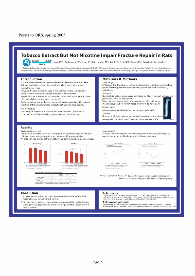
Poster to ORS, spring 2005
Orthopaedic Biomechanics Laboratory, Midwest Orthopaedic Research Foundation, Minneapolis, MN, USA; Orthopaedic Research Laboratory, Department of Orthopeadics, Aarhus University Hospital, Denmark;
Department of Connective Tissue Biology, Institute of Anatomy, University of Aarhus, Denmark; Department of Medicine, Division of Clinical Pharmacology, Hennepin County Medical Center, Minneapolis, MN, USA
Materials & Methods Introduction
Conclusion References:
Acknowledgement:
Yield Point
Nm
0.00
0.05
0.10
0.15
0.20
Nicotine Vehicle Tobacco Extract plus Nicotine
Tobacco Extract
p < 0.05
p < 0.05
Ultimate Torque
Nm
0.00
0.05
0.10
0.15
0.20
Vehicle Tobacco extract plus nicotine
Tobacco Extract
Nicotine
p < 0.05p < 0.05
Serum Cotinine
ng
/ml
200
250
300
350
400
Nicotine group Nicotine plus Tobacco Extract group
7th day 21st day 28th day
Serum Nicotine
ng
/ml
20
30
40
50
60
Nicotine group Nicotine plus Tobacco Extract group
7th day 21st day 28 th day
*
0.403150 ± 37102 ± 2294 ±10122 ± 21Energy(10-2N!degree)
0.366207 ± 22199 ± 23257 ± 32232 ± 23Ultimate Stiffness(10-4N/degree)
p valueTobacco
Extract and Nicotine
Tobacco Extract
NicotineVehicle
0.403150 ± 37102 ± 2294 ±10122 ± 21Energy(10-2N!degree)
0.366207 ± 22199 ± 23257 ± 32232 ± 23Ultimate Stiffness(10-4N/degree)
p valueTobacco
Extract and Nicotine
Tobacco Extract
NicotineVehicle
Results
Page 22
Abstract to DOS, August 2005 Introduction Fracture repair is slower and less complete in smokers compared to people who do not smoke. The influence of nicotine and tobacco extract (without nicotine) alone and in combination on callus formation and mechanical strength of femoral fracture in rats was investigated after 21 days of healing. Material and Methods 104 Sprague-Dawley rats were divided into four groups receiving; nicotine, tobacco extract, nicotine plus tobacco extract, and vehicle. One week prior to fracture surgery Alzet miniosmotic pumps were implanted subcutaneously to administer saline or nicotine, while tobacco extract were administered orally. A closed transversal fracture was performed as described by Bonnarens and Einhorn. Results After 21 days of healing ultimate torque and yield point of the tobacco extract group was decreased by 26 % (p=0.010) and 29 % (p=0.056), respectively compared to the vehicle and 25 % (0.023) and 36 % (p=0.004), respectively compared to the nicotine group, whereas no difference was found between the tobacco extract and tobacco extract plus nicotine group (p = 0.239). A 22 % (p=0.013) reduction in yield point were observed between the tobacco extract plus nicotine group and the nicotine group. No differences in ultimate stiffness and energy absorption were found between the groups. Serum levels of nicotine between 40 – 50 ng/ml were achieved. Conclusion Tobacco extract without nicotine decreases mechanical strength of the healing fracture compared with vehicle. Nicotine does not depress the mechanical strength of the healing fracture, when given in a dose inducing serum levels equivalent to those observed in daily smokers.

Page 23
Abstract to SICOT, September 2005 Introduction This study investigated the effects of nicotine and tobacco extract in combination and alone on fracture healing in rats. We hypothesized that mechanical parameters and callus properties of the fractured bone would be impaired by exposure to tobacco extracts and nicotine. Materials 89 rats were included in the study, divided into 4 groups; 1) nicotine, 2) tobacco extract, 3) nicotine and tobacco extract and 4) saline (control). Nicotine bitartrate or saline was administrated for 28 days through miniosmotic pumps (delivered 3.0 mg/kg/day). Tobacco extract orally in a 16 hour daily period. Methods A transverse, mid-diaphyseal fracture was induced and stabilized. 4 weeks observation period. By torsion to failure, ultimate torque, linear stiffness and energy absorption were derived. DEXA scans were performed using an 8 mm region of interest for BMC measurements. Callus volume and dimensions were obtained. Results ANOVA were performed on transformed data. Tobacco extract significantly lowered ultimate torque (P=0.006) but none of the other variables were impaired. Nicotine had no significant effect on any of the variables. Serum levels of nicotine and cotinine were between 40-50 ng/ml resp. 250-350 ng/ml, which are normal levels seen in smokers who smoke 1 to 1½ pack of cigarettes per day. Discussion Tobacco extract without nicotine impaired the mechanical properties of the fracture site. Nicotine alone had no adverse impact on fracture healing. Data suggests that nicotine may not be the only component impairing fracture healing in smokers