Embed Size (px)
Citation preview
! 1!
!PhD!Course!in!Molecular!and!Experimental!Medicine!!
Department!of!Experimental!Biomedicine!and!Clinical!Neuroscience!SSD!MED/18!>!Chirurgia!Generale!
!!!!!!!!
!!!!
The Effect of Probiotics in The Therapy of Ulcerative Colitis - a clinic study -
Il Dottore Dr. Marcello Romeo
Il Coordinatore Prof. Giovanni Zummo
Il Tutor Prof. Giovanni Tomasello
Ciclo XXV A.A. 2015

! 2!
Index Acknowledgements 4
1. Introduction 5
1.1 The Ulcerative Colitis 5
1.2 Epidemiology 6
1.3 Etiopathogenetic Mechanisms 7
1.3.1 Genetic Factors 8
1.3.2 Environmental Factors 11
1.4 Pathogenetic Pathophysiological Mechanisms 13
1.4.1 Intestinal Microbiota 14
1.4.2 Intestinal Mucus 20
1.4.3 Junctional Mucosal System 22

! 3!
1.4.4 Cytokine Network 27
1.4.5 MALT 29
1.5 Clinical Aspects of UC 33
1.6 The Diagnostic Path in The UC 37
1.7 The UC Therapeutic Management 39
1.8 Probiotics 42
1.9 Rationale for Integrated use of Probiotics in the UC 47
2. Aims 52
3. Materials and Methods 56
4. Results 57
5. Discussion 74
!

! 4!
Acknowledgements
Desidero esprimere il mio personale e doveroso ringraziamento a tutti coloro
che mi hanno offerto tutta la loro disponibilità e professionalità
nell’elaborazione di questo progetto di ricerca.
In particolare, ringrazio il Prof Giovanni Zummo, il Prof. Francesco
Cappello e il Prof Giovanni Tomasello che con immensa generosità mi hanno
offerto la loro grande esperienza e professionalità elargendomi preziosi
consigli che si riveleranno preziosi anche nel proseguo della mia carriera.
Un particolare ringraziamento va rivolto al Dr Vincenzo Palumbo per l’aiuto
nella raccolta dei dati e alla D.ssa Antonella Marino Gammazza per l’analisi
statistica.
Nel ringraziare le splendide persone che hanno contribuito alla stesura di
questo elaborato, auguro buona lettura e spero che i risultati di questa ricerca
possano contribuire ad offrire ai pazienti un ulteriore strumento terapeutico
per migliorare la loro qualità di vita.

! 5!
1. Introduction
1.1 The Ulcerative Colitis The Inflammatory Bowel Diseases (IBD) include two pathological entities of
multifactor etiology not completely understood characterized by a complex
immunological and inflammatory process in chronic form: Crohn's Disease
and Ulcerative Colitis (UC).
In the context of IBD, a different form of ulcerative indefinite etiology,
referred to as Indefinite Microscopic Colitis was also inserted.1
In particular, in the preparation of this thesis, attention has been made to the
UC whose chronic inflammatory process seems to depend on genetic
predisposition and epigenetic environmental factors are not yet completely
known.
!!!!!!!!!!!!!!!!!!!!!!!!!!!!!!!!!!!!!!!!!!!!!!!!!!!!!!!!1!Strober W, Fuss I, Manon P, De Foundmental Basis of IBD. J Clin Invest 2007; 117:

! 6!
1.2 Epidemiology
The highest rates of incidence and prevalence of UC are recorded in North
America and Northern Europe, with an impact ranging from 9 to 20 new
cases a year per 100,000 individuals and the prevalence rates range from 156
to 291 cases per 100,000 individuals.2
The recorded data indicates a higher impact in industrialized countries
confirming the extremely important role exercised by environmental factors
in the genesis of chronic inflammatory process, typical of the disease.
The UC reflects a characteristic bimodal pattern of incidence, with a peak
incidence occurring between the ages of 15 and 30 and a second less
significant peak of incidence between the ages of 50 and 70 with effects
slightly higher in males than females.3
The onset cases at an early age account for approximately 30% and similar to
that observed in adulthood, recent studies show a clear increase of the disease
in children.4
!!!!!!!!!!!!!!!!!!!!!!!!!!!!!!!!!!!!!!!!!!!!!!!!!!!!!!!!2 Ordas I, Eckmann L, Talamini M, Baumgart DC, Sandborn WJ. Ulcerative Colitis. Lancet 2012; 380: 1606-1619 3!Loftus EU JR, Sandborn WJ, Epidemiology of IBD. Gastroenterol Clin Nord AM 2002; 31: 1-20 4!Pappa HM, Semrin G, Walzer TR, Grand RJ. Pediatric Inflammatory Bowel Disease. Curr Opin Gastroenterol 2004; 20: 33-40

! 7!
1.3 Etiopathogenetic Mechanisms
The UC, a chronic inflammatory bowel disease in which etiopathogenesis is
not yet well defined, is certainly multifactorial. The UC consider the most
accredited etiopathogenetic hypothesis, as the result of the influence of
several environmental epigenetic factors on a predisposed genotype, capable
synergetically, to interact with the host immune system.
In this way, an inappropriate and persistent activation of the immunitary
response at the level of the intestinal mucosa would trigger, facilitated in turn,
by a pre-existing impairment of barrier functions. The latter would be due to a
reversal of the ultra structural junction of the mucosal system.
The result of these complex mechanisms would give rise to the characteristic
inflammatory phenotype of the UC.5
!!!!!!!!!!!!!!!!!!!!!!!!!!!!!!!!!!!!!!!!!!!!!!!!!!!!!!!!5 Hanauer SB. Inflammatory Bowel Disease: Epidemiology, Pathogenesis And Therapeutic Opportunities. Inflamm Bowel Dis 2006: 12S1

! 8!
1.3.1 Genetic Factors
Numerous epidemiological studies largely confirmed that the UC and in
general all the IBD, are genetically complex diseases characterized by high
genetic heterogeneity, able to interact with specific environmental factors
affecting the phenotypic characteristic appearance of the disease.
Hereditary studies show that about 5.7-15.5% of individuals with IBD have a
first-degree relative affected by UC or CD.6
The familial aggregation observed in IBD, confirms the hypothesis of the
presence of a genetic susceptibility, on which environmental factors
contribute to the onset of the disease.
Numerous studies on monozygotic and dizygotic twins, outline the different
influence of genetic profile and environmental factors on etiopathogenesis of
UC and CD.
In particular, studies show concordance rates for UC significantly higher for
monozygotic twins (6-17%) compared to dizygotic twins (0-5%), confirming
the role played by genetic factors.7
The relationship between patients with UC and unaffected members within
the same family, suggests a hereditary pattern that differs significantly from
the classical Mendelian model with complete penetration, confirming the
complex heterogeneity of the disease.
Recent Linkage studies have made it possible to acquire more precise
information on IBD genetic heterogeneity, identifying locus chromosomal
susceptibility and candidate genes for both the CD and UC.
!!!!!!!!!!!!!!!!!!!!!!!!!!!!!!!!!!!!!!!!!!!!!!!!!!!!!!!!6!Farmer RG, Michener WM, Mortimer EA. Studies of Family History Among Patients with IBD. Clin Gastroenterol 1980; 9: 271-277 7!Satsangi J, Morecroft J, Shah NB. Genetics of Inflammatory Bowel Disease: Scientific and Clinical Implications. Best Pract Res Clin Gastroenterol 2003; 17: 3-18 !

! 9!
In particular, genes responsible for susceptibility to IBD in general exist,
disease-specific genes that can influence the onset of CD or UC and
phenotype-specific genes that influence the location, the natural history of the
disease and the different individual capacity to respond to therapeutic
treatment.
Linkage studies of the whole genome has allowed the identification of several
locus susceptibility for IBD.8 (Table 1)
Locus Cromosome Disease
IBD 1 16q12 MC
IBD 2 12p13-q24 CU
IBD 3 6p CU, MC
IBD 4 14q11-12 MC
IBD 5 5q31-33 MC
IBD 6 19p13 MC, CU
IBD 7 1p36 MC, CU
Table 1: Genetic Linkage Studys and candidate genes. IBD Year Book 2005
Additional studies have identified other chromosomal areas with greater
evidence of linkage to UC.9 (Table 2)
!!!!!!!!!!!!!!!!!!!!!!!!!!!!!!!!!!!!!!!!!!!!!!!!!!!!!!!!8 Brant SR, Shugart YY. Inflammatory Bowel Disease Gene Hunting by Linkage Analysis. Rationale Methodology and Present status of the Field. Inflamm Bowel Dis 2004ì; 10: 300-311 9!Van Heel Da, Fisher SA, Kirby A, Daly MJ Riouxjd, Lewis CM. Inflammatory Bowel Disease Susceptility Loci Defined by Genome Scan Meta-Analysis of 1592 Affected Relative Pairs. Hum Mol Genet 2004; 13: 763-770

! 10!
Locus IBD CD UC
2q24-q34 0.004 0.04 0.01
3q27-qter 0.005 0.02
4q23-q28 0.05
4q28-q32 0.04
5q13-q15 0.02
6pter-p23 0.005 0.01
6p23-p21.1
(IBD3)
0.0001 0.004 0.05
7q11.1-q21 0.02
12p12-q13
(IBD 2)
0.05 0.04 0.02
16p13.1-q12.2
(IBD 1)
0.008 0.003
16q12.2-q22 0.01
17q21-q24 0.01
19pter-p13.2 0.007
19p13.2-q13.2 (IBD 6) 0.01
19q13.2-qter 0.03
Table 2: Cromosomic Locus and Genetic Linkage. IBD Year Book 2005!

! 11!
1.3.2 Environmental Factors
Although the existence of a genotype susceptibility and specific "candidate
genes" in IBD was confirmed, several studies emphasize the importance of
environmental factors that can provoke or act as protector in the UC.
Breastfeeding for example, especially if more than 3 months, certainly acts as
a protective factor against the UC.10
Smoking also appears to play a protective role for RCU. In fact, studies show
that the course of the disease seems to be slower in smokers than non-
smokers.11
Previous episodes of bacterial gastroenteritis supported by Campylobacter,
Salmonella and Shigella can determine a significant modification of the
intestinal flora by acting as triggers on the onset of the chronic inflammatory
process of IBD. 12
Numerous studies have previously demonstrated the protective role for UC of
appendectomy especially if the person is subjected to surgery within the first
20 years of age.13
The scientific literature data show a 69% reduction in the risk of developing
UC after appendectomy.14
!!!!!!!!!!!!!!!!!!!!!!!!!!!!!!!!!!!!!!!!!!!!!!!!!!!!!!!!10!Klement E, Cohen RV, Boxman J, Joseph A, Reif S. Breastfeeding and Risk of Inflammatory Bowal Disease: A Systematic Review with Meta-Analysis. AM J Clin Nutr 2004; 80: 1342-1352 11 Ordas I, Eckmann L, Talamini N, Baumgart DC, Sandborn J. Ulcerative Colitis. Lancet 2012; 380: 1606-1619 12!Garcia Rodriguez LA, Rui Gomez A, Panes J. Acute Gastroenteriti is Followed by Anicreased Risk of Inflammatory Bowel Disease. Gastroenterology 2006; 130: 1588-1594 13!Duggan AE, Usmani I, Neal KR, Logan RF. Appendicectomy, Childhood Hygiene, H. Pylori Status and Risk of Inflammatory Bowel Disiase: A Case-Control Study. Gut 1998; 43: 494-498 14!Kout Roubakis IE, Vlachonikolis IG. Appendicectomy and the Development of UC: Resuls of A Metanalysis of Published Case Control Studies . AM J Gastroenterol 2000; 95 171-176

! 12!
Subsequent studies have confirmed this protective role of appendectomy in
the onset of UC proving even a phenotype disease significantly less
aggressive than in UC patients not undergoing appendectomy.15
!!!!!!!!!!!!!!!!!!!!!!!!!!!!!!!!!!!!!!!!!!!!!!!!!!!!!!!!15!Randford- Smith GL, Edwards JE, Purdie DM, Pandeya N, Watson M, Martin NG. Protective Role of Appendicectomi on Onset and Severity of Ulcerative Colitis and Crohn’s Disease. Gut 2002; 51: 808-813 !

! 13!
1.4 Pathophysiological Mechanisms
The altered intestinal permeability, which originated from the destruction of
the junctional mucosa (TJ) and the mucus film that covers the epithelium, in
the presence of significant alterations of the microbiota, stimulate a
disordered local immune response, by producing pro-inflammatory factors
and recruiting specific cellular elements.
This should be a summary of the pathophysiological framework that underlies
the characteristic chronic inflammatory process of IBD. (Fig.1)
Fig. 1 Pathophysiology of Ulcerative Colitis. Ingrid Ordás, Lars Eckmann, Mark Talamini, Daniel C Baumgart, William J Sandborn. Ulcerative colitis. Lancet 2012; 380: 1606–19

! 14!
1.4.1 Intestinal Microbiota
Several studies published in recent years have confirmed the role of the
intestinal microbiota as an important epigenetic factor that can affect the
appearance of specific modifications of the intestinal micro-environmental
characteristics (intestinal permeability, altered cytokine network) resulting in
inappropriate bacterial translocation and persistent activation of the local
immune response widely recognized in patients suffering from IBD.
The presence of persistent intestinal dismicrobism characterized by
qualitative and quantitative alterations of the normal resident bacterial flora,
and the presence of a microbial pathogen load in a position to contribute
significantly to the onset of the typical inflammation process of IBD is
observed in patients with CD and UC.16
The hypothesis of an intestinal dismicrobism typical of patients with UC is
supported by studies showing the up-regulation of the expression of beta-
defensins (Beta-Difensina 2).17
It has been demonstrated that, in patients with IBD, the intestinal microbiota
loses the ability to down-regulate inflammatory genes and to inhibit the
activation of NF-KB by triggering an inflammatory response loading the cells
of the intestinal mucosa so as to encourage a chronic alteration of the immune
response.18
!!!!!!!!!!!!!!!!!!!!!!!!!!!!!!!!!!!!!!!!!!!!!!!!!!!!!!!!16!Morgan XC, Tickle TL, Sokol H, Gevers D, Devaney KL, Ward DV, Reyes JA Shah SA, Leleiko N, Snapper SB, Bousvaros A, Korzenik J, Sands BE, Xavier RJ, Huttenhower C. Dysfunction of the Intestinal Microbiome in Inflammatory Bowel Disease and Treatment. Genome Biology 2012; 13: R79 17!Rahman A, Fahl Gren A, Sundstedt C, Hammarstrom S, Danielson A, Hammarstrom MI. Crhonic Colitis Induces Expression of Beta-Defensins in Murine Intestinal Epithelial Cells. Clin Exp Immunol 2011; 163: 123-130 18!Colletti T. IBD-Recognition, Diagnodis and Therapeutics. JAAPA 2004; 7: 16-24

! 15!
The intestinal microbiota is a complex ecosystem characterized by the
coexistence of diverse ecological niches, which are home to a microbial
population, consisting of numerous bacterial species and strains.
Recent advances in molecular biology techniques applied to genomic
sequencing and studies of Metagenomics, have allowed us to make a more
precise and detailed classification of microbiota.
In this way, it was possible to greatly expand the knowledge already acquired,
both in terms of its genotypic characterization and of its genotypic
characteristics, and potential metabolic capacity to exercise it.
The results of two major research projects, the Human Microbiome Project
(HMP), performed in the USA, and the great Metagenomics of Human
Intestine (METAHIT), taking place in Europe, have expanded the knowledge
about the composition of the bacterial flora present along our entire
gastrointestinal tract, allowing to typify over 1100 bacterial species and over
15000 different bacterial strains, able to constitute a biomass of about 1 Kg.19
In the light of present knowledge, approximately 90% of the intestinal
microbial population is represented by 2 dominant phylum formed by
Bacteroidetes (Bacteroides and Prevotella), while the remaining 10% is
variously divided into 5 sub-dominant phylum: Actinobacteria,
Proteobacteria, Fusobacteria, Cyanobacteria and Verrucomicrobia.20
Subsequent studies have identified a specific "enterotype" characteristic of
our species, likely to meet a qualitative and quantitative modifications in the
natural course of life to constitute a different enterotype, both phylogenetic
!!!!!!!!!!!!!!!!!!!!!!!!!!!!!!!!!!!!!!!!!!!!!!!!!!!!!!!!19 !Qin J, Li R, Raes J, Arumugam M. A Human Gut Microbial Gene Catalogue Established by Metagenomic Sequencing. Nature 2010; 4, 464: 59-65 20!Kuczynski J, Costello EK, Nemergui DR, Zaneveld J, Lauber CL, Knigcts D, Coren O, Fierer N, Kelley ST, Ley RE, Gordon JI, Knight R. Direct Sequencing of the Human Microbioma Readily Reveals Communty Differences. Genome Biology 2010; 11: 210

! 16!
and functional terms, capable of preparing the subject towards specific
pathological conditions.21
Studies in mouse models have shown the possibility of enterotype to evolve
towards a predominant variant: "enterotype 2" (Bacteroitedes).
This eventuality is positively correlated with a high concentration of fecal
Calprotectin, known index of the intestinal mucosa, found in high
concentrations in patients with UC. Previous studies have allowed the
identification of a modification of the intestinal microbiota, both in patients
with CD and UC.22
In particular, some studies show a clear reduction of Lactobacilli and
Bifidobacteria, in histological specimens and cultivation of patients with
UC.23 The reduction of the normal intestinal flora, is accompanied by the
bacterial pathogen overgowth responsible for carrying out the pro-
inflammatory action in consideration of the intestinal mucosa with
consequent onset of UC.24
A demonstration of bacterial pathogen overgrowth of pro-inflammatory
action found in IBD stems from the discovery in the urine of patients of
specific metabolites (Hippurate hydrolases), produced by bacterial
fermentation processes.25
!!!!!!!!!!!!!!!!!!!!!!!!!!!!!!!!!!!!!!!!!!!!!!!!!!!!!!!!21!Arumugam M, Raes J, Pelletier E, Le Paslier D, Yamada T, Mende RD, at al. Enteroypes of the Human Gut Microbiome. Nature 2011; 473: 174-180 22!Hildebrand F, Brinkman B, Yunta RG, Cauwe B, Vandenabeele P, Liston A, Raes J. Inflammetion-Associated Enterotypes, Host Genotype Cage and Inter-Individual Effects Drive Gut Microbiota Variation in Common Laboratory Mice. Genome Biology 2013; 14: R4 23!Ouwshand AC, Salminem S, Roberts PJ. Disease-Dependent Adesion of Lactic Acid Bacteria to the Human Intestinal Mucosa. Clin Diagn Lab Immunol 2003; 10 (4): 643-646 24!Gionchetti P, Rizzello F, Lammers KM. Antibiotics and Probiotics in Treatment of IBD. World J Gastroenterol 2006; 12: 3306-3313 25 Williams HR, Cox IJ, Walker DG. Characterization of IBD with Urinary Metabolic Profiling. Am J Gastroenterol 2009; 104 (6): 1435-1444
! 17!
In terms of bacterial phylum, recent metagenomic studies carried out on
individuals with IBD, detect a significant alteration of the two dominant
phylum: Bacteroidetes and Firmicutes.26
It has also been demonstrated recently that a significant reduction of bacterial
strains belonging to the genus Roseburia and Phascolarctobacterium, strains
responsible to the production of short chain fatty acids (SCFA) in a particular
way of butyrate and propionate, important in terms of trophism and protection
of intestinal mucosa and to stimulate the production of regulatory T cells to
anti-inflammatory action.27
It is also found in patients suffering from UC, a decreased concentration of
bacterial species belonging to the genus Leuconostocaceae producing acetate
and lactate.28
A significant reduction of concentration also affects the Odoribacter genus
belonging to the family of Porphyromonadaceae.
It can be observed especially in patients with Pancolitis, a more severe
clinical form of UC.
The reduced presence of these bacterial strains lays, not only in favor of an
intestinal dysbiosis, but also in favor of metabolic modifications, of a reduced
production of acetate in particular, propionate and butyrate, able to influence
the inflammatory process of the intestinal mucosa.29
!!!!!!!!!!!!!!!!!!!!!!!!!!!!!!!!!!!!!!!!!!!!!!!!!!!!!!!!26!Frank DN at al. Molecular-Phylogenetic Characterization of Microbial Community in Balances in Human Inflammatory Bowel Diseases. Proc Natl Acad Sci USA 2007; 104: 13780-13785!27!Atarashi K, Tanoue T, Shima T, Imaoka A, Kuwattara T, Momose Y, Cheng G, Yamasaki S, Saito T, Ohba Y. Induction of Colonic Regulatory T Cells by Indigenous Clostridium Species. Science 2011; 331: 337-341 28!Cogan JG, Jordan KN. Metabolism of Leuconostoc Bacteria. J Dairy Sci 1994; 77: 2704-2717 29!Goker M, Gronow S, Zeytun A, Nolan M, Lucas S, Lapidus A, Hammon N, Deshpande S, Cheng JF, Pitluck S. Complete Genome Sequence of Odoribacter Splanchnicus Type Strain (1651/6). Stand Genomic Sci 2011; 4: 200-209

! 18!
The SCFA and, in particular, the butyrate, have shown to possess anti-
inflammatory action but also to inhibit the processes of bacterial
translocation.30
Subsequent studies have highlighted the close relationship between butyrate
and barrier function of the intestinal mucosa.
Research shows in particular how the butyrate is able to regulate the
expression of Claudina-2.31
The metabolic changes resulting from alterations typical of the intestinal
microbiota of patients with IBD, does not only involve the short-chain fatty
acids (SCFA), but also the metabolism of Glutathione synthesized by
Proteobacteria, Streptococcus and Enterococcus, responsible to actively
combat oxidative stress load of intestinal epithelial cells found in IBD.32
In fact, high concentrations of highly reactive species of oxygen and nitrogen
in patients with CD and UC are found.33
To further confirm the role played by the intestinal flora in the UC, it is noted
in the in vitro studies that the bacterial residents, due to high adhesion and
invasiveness, are capable of inducing the production of toxins and pro-
inflammatory mediators (IL -1, IL-6, IL-8) responsible for the transmigration
!!!!!!!!!!!!!!!!!!!!!!!!!!!!!!!!!!!!!!!!!!!!!!!!!!!!!!!!30!Lewis K, Lutgendorff F, Phan U, Soderholm JD, Sherman PM, Mckay DM. Enhanced Translocation of Bacteria Across Metabolically Stressed Epithelia is Reduced by Butyrate . Inflamm Bowel Dis 2010; 16: 1138-1148 31!Ploger S, Bergann T, Fromm A, Bucker R, Fromm M, Schulzke JD. Effect of Sodium Butyrate on Barrier Function of HT-29/B6-TET-ON-GR-MR Cells in Conjunction with Down Regulation of Claudin-2. Genes Nutr 2010; 5 (S1): S28 32 Sherrill C, Fahey RC. Import and Metabolism of Glutathione by Streptococcus Mutans. J Bacteriol 1998; 180: 1454-1459 33 Keshavarzian A, Banan A, Farhadi A, Komanduri S, Mutlu E, Zhang Y, Fields JZ. Increases in Free Radicals and Cytoskeletal Protein Oxidation and Nitration in the Colon of Patients with Inflammatory Bowl Disease. Gut 2003; 720-728

! 19!
of neutrophils across the intestinal epithelium with the appearance of the
typical crypt abscesses of the UC.34
Subsequent studies confirm that the HLA-B27 transgenic mice and mice
deficient in IL-2 and IL-10 develop chronic colitis.35
!!!!!!!!!!!!!!!!!!!!!!!!!!!!!!!!!!!!!!!!!!!!!!!!!!!!!!!!34!Meijssen MAC at al. Altereted Cytockine and CD14 Expression by Intestinal Epithelial Cells of IL-2 Deficient Mice. Gastroenterology 1996; 110: A966 35!Rath HC. Role of Commensal Bacteria in Chronic Experimental Colitis: Lessons from the HLA-B27 Transegenic Rat. Pathobiology 2002; 70: 131-138

! 20!
1.4.2 Intestinal Mucus
The gastrointestinal epithelium is continuously exposed to the insult
exercised by a number of chemical, physical and biological agents.
The intestinal mucus represents an important defensive barrier of the
epithelium against the potential harmful effect exerted by these agents.
It appears to be variously distributed along the various sections of our
gastrointestinal system, arranged in a double stratification: an inner layer
perfectly adherent to the epithelial surface and almost free of bacteria, and an
outer layer in direct contact with the intestinal lumen.36
Its adequate physico-chemical composition and its perfect layering
throughout the epithelium, are the essential elements for effective barrier
action against damaging agents.37
The mucus produced by globet cells, which are present in the intestinal
epithelium, is richly peppered with mucins or glycosylated proteins that
impart trophic lubricity and are protective towards epithelium.38
It is detected in patients suffering from UC a reduced representation of goblet
cells, of the quantities of mucus and the amount of mucin that it forms. In
fact, it has been demonstrated in IBD and in particular in the UC, the reduced
expression of specific genes responsible for the adequate more or less
adequate expression of globet cells.39
!!!!!!!!!!!!!!!!!!!!!!!!!!!!!!!!!!!!!!!!!!!!!!!!!!!!!!!!36!O’Hara AM, Shanahan F. The Gut Flora as a Forgotten Organ. EMBO 2006; 7: 688–693 !37!Matsuo K at al. Histochemistry of the Surface Mucous Gel Layer of the Human Colon. Gut 1997; 40: 782-789 38!Johansson ME at al. The Two Mucus Layer of Colon are Organized by the Muc2 Mucin, Whereas the Outer layer is a Legislator of Host-Microbial Interactions. Proc Natl Acad Sci USA 2011; 108 (S1): 4659-4665!39!Gersemann M, Becker S, Kubler I. Differences in Globet Cell Differentiation Between Crohn’s Disease and UC. Differentiation 2009; 77 (1): 84-94

! 21!
It is witnessed in the mucin deficient mice (MUC-2) or with altered
glycosylation patterns, a major pathogenic bacterial proliferation and
tendency to spontaneous colitis and colitis induced.
This study confirms the importance of an adequate production of mucus and a
rich amount of mucin in countering the onset of mucosal intestinal
inflammation.40
GF animal studies demonstrate a quantitative reduction of the globet cells and
the thickness of the mucus layer in colonic level, confirming the important
role exercised by the intestinal bacterial flora in the maintenance of the
physical and chemical mucus characteristics, of its quantity produced and its
functional potentials.41
!!!!!!!!!!!!!!!!!!!!!!!!!!!!!!!!!!!!!!!!!!!!!!!!!!!!!!!!40!Fu J at al. Loss of Intestinal Core1-Derived O-Glicans Causes Spontaneous Colitis in Nice. J Clin Invest 2011; 121: 1657-1666 41!Sharma R at al. Rat Intestinal Mucosal Responses to A Microbial Flora and Difference Diets. Gut 1995; 36: 209-214 !

! 22!
1.4.3 Junctional Mucosal System
Under physiological conditions, the intestinal epithelium plays an efficient
barrier function allowing the transportation of selective paracellular and
transcellular of specific ions and molecules, thanks to the intervention of
highly specialized structures, which together constitute the defense system of
the intestinal mucosa.
This system is responsible in maintaining an appropriate pattern of intestinal
permeability.42
The structural alteration of this complex defense system involves the onset of
a state of altered permeability, contributing to the onset of intestinal and extra
intestinal pathological conditions.
Despite the immune dysregulation, inflammation and mucosal barrier
function abnormalities are common factors found in patients with IBD, it is
not yet clear which of these three mechanisms actually precedes the onset of
the disease. Studies show that the only structural alteration of the defense
system of the mucosa with consequent increased of intestinal permeability is
not able to trigger the onset of IBD.43
On the contrary, the appearance of altered intestinal permeability is
accompanied by immune dysregulation.
The defense system of the intestinal mucosa, from the structural point of
view, consists of structures of a protein nature, adequately distributed along
the side of the apical, basal and latero-lateral of epithelial cells (tight junction,
!!!!!!!!!!!!!!!!!!!!!!!!!!!!!!!!!!!!!!!!!!!!!!!!!!!!!!!!42!Turner JR. Molecular Basis of Epithelial Barrier Regulation From Basic Mechanisms to Clinical Application. AM J Pathol 2006; 169: 1901-1909 43!Edelblum KL, Turner JR. The Tight Junction in IBD: Communication Breakdown. Curr Opin Farmacol 2009; 9(6): 715-720

! 23!
adherens Junction, maculae adherens, gap Junction, hemidesmosomes).
(Fig.2)
Fig. 2: Schematics showing composition and organization of adherens junctions (AJs) and tight junctions
(TJs). JAM: Junction Adhesion Molecule. J. Miyoshi, Y. Takai, Advanced Drug Delivery Reviews, 2005)
From the biomolecular point of view, it deals with the complex structures made up of specific components of
protein-based nature in a trans membrane confinement, among which Junctional Adhesion Molecules (JAM),
Tricellulina, Occludin and Claudina can be distinguished.

! 24!
Through their intracellular component, they are capable of interacting closely
with specific membrane proteins among which Zonuline and Cinguline can
be distinguished.44
The transcriptional processes of these complex protein molecules,
constitutive of the mucosal defense system and responsible for its barrier
function may be due to specific genomic sequences.
It has been demonstrated in patients with IBD a reduced expression of these
proteins due to alterations of some genomic sequences.45
There is a reduced expression of Occludin in the gut epithelium in IBD.46
Further studies have also demonstrated the presence of altered transcriptional
regulation and consequent altered expression of Claudine.47
The alterations of the defense system of the intestinal mucosa, responsible for
determining the appearance of increased permeability in IBD, are directly
attributable to dysregulated expression of specific pro-inflammatory
cytokines: TNF-ALPHA, GAMMA-INF, IL-1beta and IL-13.48
In this regard, recent studies have made it possible to distinguish the various
forms of IBD, the cytokine profile characteristic and specific alterations of
the proteins of the defense system of the intestinal mucosa. (Tab. 3)
!!!!!!!!!!!!!!!!!!!!!!!!!!!!!!!!!!!!!!!!!!!!!!!!!!!!!!!!44!Mitic LL, Anderson JM. Molecular Architecture of TJ. Ann Rev Physiol 1998; 60: 121-142 45!Mees ST, Mennigen R, Spieker T. Expression of Tight and Adherens Junction Proteins in UC Associated Colorectal-Carcinoma Up-Regulation of Claudin-1, 3, 4 and Beta-Catenin. Int J Colorectal Dis 2009; 24(4): 361-368 46!Heller F, Florian P, Bojarski C, Richter J, Christ M, Hillenbrand B, Makertz J, Gitter AH, Burgel N, From M. Interleukin-13 is the Key Effector TH2 Citokine in UC that Affects Epitelial TJ, Apoptosis and Cell Restitution. Gastroenterology 2005; 129: 550-564 47!Prasad S, Mingrino R, Kaukinen K, Hayes KL, Pouel RM, Mac Donald, Collins JE. Inflammatory Processes have Differential Effects on Claudins 2, 3 and 4 in Colonic Epitelial Cells. Lab Invest 2005; 85: 1139-1162 48!Hering NA, Fromm M, Schulzke JD. Determinats of Colonic Barrier Function in IBD And Potential Therapeutics. J Physiol 2012; 5: 1035-1044

! 25!
The alterations of barrier function may also be due to the intestinal
dismicrobism. Recent studies have in fact demonstrated in mouse models
deficient in IL-2 or IL-10 suffering from chronic inflammatory bowel
condition that the transition to a GF, does not lead to the appearance of
clinical symptoms typical of chronic inflammation.49
Subsequent studies demonstrate the involvement of Junctional Adhesion
Molecule (JAM-A) in regulating the state of permeability of the intestinal
mucosa and inflammation. (Fig. 2)
JAM-A is a protein associated with TJ, which is extremely important in terms
of adjustment of the barrier function and, where the reduced expression is
!!!!!!!!!!!!!!!!!!!!!!!!!!!!!!!!!!!!!!!!!!!!!!!!!!!!!!!!49!Elson CO, Cong Y, Mc Cracken VJ, Dimmitt RA, Lorenz RG, Weaver T. Experimental Models of IBD Reveal Innate, Adapative and Regultory Mechanisms of Host Dialogue with the Microbiota. Immunol Rev 2005; 206: 260-276 !
Table 3: Aspects of barrier dysfunction in IBD. Nina A. Hering, Michael Fromm, Jorg-
Dieter Schulzke. J Physiol 2012; 590.5: pp 1035–1044
The Journal of Physiology, 2012 !

! 26!
accompanied by high intestinal permeability, recruitment and activation of
leukocytes with consequent increase in the mucosal inflammatory process.50
!!!!!!!!!!!!!!!!!!!!!!!!!!!!!!!!!!!!!!!!!!!!!!!!!!!!!!!!50!Laukoetter GM, Nava P, Lee YW, Severson EA, Capaldo CT, Babbin BA, Williams IR, Koval M, Parkos C. JAM-A Regulates Permeability and Inflammation in the Intestine in –Vivo. JEM 2007; 204 (13): 3067-3076

! 27!
1.4.4 Cytokine Network
The impressive and chronic inflammatory process seen in mucosal UC and in
general in IBD, is the result, as a co-factor, also of alterations of the cytokine
network.51
In particular, it has been demonstrated the presence of a richly peppered
epithelium highly pro-inflammatory and damaging cytokines, in which the
concentration depends on the extent of the inflammatory process
characteristic of the UC.52
A widely-related cytokine, in this sense, is definitely the Tumor Necrosis
Factor Alpha (TNF-A).
In fact, in patients with UC, it is detected a high concentration of TNF-Alpha
in serum, faeces and at the level of the intestinal mucosa.53
Studies show that the magnitude, extent and progression of mucosal
inflammation, is significantly related to the concentration of TNF-Alpha.
The impressive role played by TNF-A in the pathogenesis of chronic
inflammatory process of IBD, has stimulated the execution of countless
studies, wherein data have led to a breakthrough in the treatment of these
diseases with the advent of therapies that exploit the action of specific anti-
bodies monoclonal anti-TNF, with very encouraging results.54
!!!!!!!!!!!!!!!!!!!!!!!!!!!!!!!!!!!!!!!!!!!!!!!!!!!!!!!!51!Schulzke JD, Plogers S, Amasheh M. Epithelia TJ in Intestinal Inflammation. Ann NY Acad Sci 2009; 1165: 294-300 52!Jalocha L, Wojtun S, Dyrla P. TNF-Alpha Polymorphism and Course of UC. Pol Merkur Lekarski 2009; 26(155): 444-445 53!Komatsu M, Kobayashi D, Saito K, Furuya D, Yagihashi A, Araake H, Tsuji N, Sakamaki S, Niitsu Y, Watanabe N. TNF-Alpha in Serum of Patients with IBD as Measured by a Highly Sensitive Immuno-PCR. Clin Chem 2001; 47: 1297-1301 54!Ordas I, Mould DR, Feagan BG, Sandborn WJ. Anti-TNF Monoclonal Antibodies in IBD: Pharmacokinetics-Based Dosing Paradigms. Clin Pharmacology and Therapeutics 2012; 1-11

! 28!
Several studies show also an involvement in the inflammatory process of IL-
32. It is a cytokine synthesized by monocytes, natural killer cells, T
lymphocytes and epithelial cells.55
In particular, IL-32 is able to stimulate the synthesis of pro-inflammatory
cytokines and chemokines thanks to the intervention of NF-KB and MAPK
thus, stimulating the onset of IBD.56
The epithelial cells of the intestinal mucosa of these patients show a high
concentration of a specific variant of IL-32: IL-32 Alpha.57
Other cytokines seem to play an important role in the pathophysiology of IBD
and, in this sense, studies show an increased concentration of IL-17.58
The same pro-inflammatory role seems to be played also by IL-31, which is
also increased in the CD and in the UC.59
!!!!!!!!!!!!!!!!!!!!!!!!!!!!!!!!!!!!!!!!!!!!!!!!!!!!!!!!55!Kim SH, Han SY, Azam T, Yoon DY, Dinarello CA. IL-32: a Cytokine and Inducer of TNF-Alpha. Immunity 2005; 22: 131-142 56 Shioya M, Nishida A, Yagi A, Ogawa A, Tsujikawa T, Kim-Mitsuyama S, Takayanagi A, Shimizu N, Fujiyama Y, Andoh A. Epithelial Overexpression of IL-32 Alpha in IBD. Clin Exp Immunol 2007; 149: 480-486 57!Andoh A, Yagi Y, Shioya M, Nishida A, Tsujikawa T, Fujiyama Y. Mucosal Cytokine Network in IBD. World J Gastroenterol 2008; 14(33): 5154-5161 58!Fujino S, Andoh A, Bamba S, Ogawa A, Hata K, Araki Y, Bamba T, Fujiyama Y. Increased Expression of IL-17 in IBD. Gut 2003; 52: 65-70 59!Diveu C, Lak-Hal AH, Froger J, Ravon E, Grimaud L, Barbier F, Hermann J, Gascan H, Chevalier S. Predominant Expression of the Long Isoform of GP130-Like (GPL) Receptor is Required for IL-31 Signaling. Eur Cytokine Netw 2004; 15: 291-302 !

! 29!
1.4.5 MALT (Mucose-Associated Lymphoid Tissue)
The immune system can perform a defensive action of the body against
pathogens in which it comes in contact with. This defense action takes place
thanks to the synergy of the inherent components constituent of Immunity
Innate and Adaptive Immunity.60
The most important defense mechanism against pathogens is put in place by
the Mucosal Immune System (MALT - Mucosa-Associated Lymphoid
Tissue).
The MALT, present in our intestinal mucosa, is indicated in a specific manner
like GALT (Gut-Associated Lymphoid Tissue).
It is made up of specific cellular components (Peyer's Plaques) and
mesenteric lymph nodes.
The immune cellular components that characterize it are represented by: T
and B Lymphocytes, Dendritic cells, M cells, Paneth cells and Intraepithelial
Lymphocytes.61
Their role is to ensure a full immune response both in the humoral and cell
type against all antigenic exogenous and endogenous stimuli which is
continually exposed to the intestinal mucosa.62
The intestinal microbiota has demonstrated the ability to interact closely with
the GALT ensuring proper and effective immuno-biological modulation of
the microenvironment of the intestine.63
!!!!!!!!!!!!!!!!!!!!!!!!!!!!!!!!!!!!!!!!!!!!!!!!!!!!!!!!
60 Abbas, A., & Lichtman, A. Cellular and Molecular Immunology, 5th ed. (2003).
61Wershil BK, Furuta GT. Gastrointestinal mucosal immunity. J Allergy Clin Immunol 2008 Feb;121(2 Suppl): S380-3 62!Jump RL, Levine AD. Mechanisms of natural tolerance in the intestine. Implications for inflammatory bowel disease. 2004; Vol. 10: pp 462–478

! 30!
In particular, the resident bacterial flora in our intestines exerts its influence
on the local immune system, due to its interaction with specific receptors:
Pattern Recognition Receptors (PRRs).
Specific PRRs referred to as Toll Like Receptors (TLR), are localized on the
surface of intestinal epithelial cells can recognize specific molecular
determinants of microbial origin called Pathogen-Associated molecular
Patterns (PAMPs).
Recognition of microbial antigens takes place thanks to the identification by
TLR of specific components of the bacterial cell: Peptidoglycan, Lipoproteins
surface, Lipoteichoic Acid and Lipopolysaccharide (LPS).
The phase following the recognition of these antigens, is characterized by the
detection by TLR factors of antimicrobial actions to stop the microbial
growth and the subsequent infection process that follows.
The reduced expression of TLRs in the intestine is responsible for the
appearance of dismicrobism resulting in mucosal inflammation.
In fact, studies show that the quali-quantitative alteration of the intestinal
bacterial flora is one of the most important trigger factors in determining a
chronic overstimulation of the GALT .64
This dysregulated local immune response is characterized by the involvement
of specific components (leukocytes, lymphocytes, CD4, arachidonic acid,
reactive species and stress proteins) that, peppering the intestinal mucosa, are
!!!!!!!!!!!!!!!!!!!!!!!!!!!!!!!!!!!!!!!!!!!!!!!!!!!!!!!!!!!!!!!!!!!!!!!!!!!!!!!!!!!!!!!!!!!!!!!!!!!!!!!!!!!!!!!!!!!!!!!!!!!!!!!!!!!!!!!!!!!!!!!!!!!!!!!!!!!!!!!!!!!!!!!63!Gasbarrini A, Lauritano EC, Garcovich M. New Insights into the Pathophysiology of IBS: Intestinal Microflora, Gas Production and Gut Motility. Eur Rev Med Pharmacol Sci 2008; 1: 111-117 64 !Llopis M, Antolin M, Carol M. Lactobacillus Casei Downregulates Commensals Inflammatory Signals in Crohn’s Disease Mucosa. Inflamm Bowel Dis 2009; 15(2): 275-283

! 31!
responsible for the continuous stimulation and maintenance of the chronic
inflammatory process, characteristic of the UC.65
Studies also show that in these cases, the immune structure is characterized
by an overexpression of the Th2 response mediated by atypical NK cells
responsible for the stimulated production of pro-inflammatory specific
cytokines.66
!!!!!!!!!!!!!!!!!!!!!!!!!!!!!!!!!!!!!!!!!!!!!!!!!!!!!!!!65 !Arseneau KO, Cominelli F. Leukocytaferesis in Ulcerative Colitis: a Possible Alternative to Biological Therapy? Dig and Liver Dis 2009; 41: 551-552 66!Heller F, Florian P, Bojarski C. IL-13 is the Key Effector Th2 Cytokine in Ulcerative Colitis that Affects Epithelial Tight Junctions, Apoptosis and Cell Restitution. Gastroenterology 2005; 129: 550-564

! 32!
1.5 Clinical Aspects of the UC
The UC is a chronic inflammatory bowel disease with a quite heterogeneous
clinical course. 67
At a clinical level, patients with UC have a course of generally chronic-
recurrent in which periods of acute symptoms alternate with periods of
remission, with significant reduction in the accompanying clinical symptoms
or even complete absence of symptoms.68
The abdominal pain, the tenesmus, the urgency and bloody diarrhea are the
most common symptoms reported by the patient with UC.69
The inflammatory process that characterizes the disease has only partial
involvement of the colonic wall, involving only the mucosal and submucosal
layer.
The intensity and extent of inflammation allow the definition of different
forms of UC. In particular, the inflammatory process can affect exclusively
the rectal mucosa by determining, in this case, the onset of a typical clinical
picture of proctitis or in certain cases, may extend proximally to the sigmoid
colon, presenting a picture of distal colitis.
Other times, it can affect the left colon with a pattern of the left colitis or
involving the entire colon with a pattern of pancolitis.70
!!!!!!!!!!!!!!!!!!!!!!!!!!!!!!!!!!!!!!!!!!!!!!!!!!!!!!!!67!Yamamoto-Furusho JK. Clinical Epidemiology of UC in Mexico: a Single Hospital-Based Study in a 20-year Period. J Clin Gastroenterol 2009; 43(3): 221-224 68!Gurel S, Kiyici M. UC Activity Index: a Useful Prognostic Factor for Predicting UC Outcome. The Journal of International Medical Research 2005; 33: 103-110 69!Carter MJ, Lobo J, Travis SPL. Guidelines for the Management of IBD in Adults. www.gut.bmj.com 70!Silverberg MS, Satsangi J, Ahmad T. Toward an Integrated Clinical, Molecular and Sierological Classification of IBD: Report of a Working Party of the 2005 Montreal World Congress of Gastroenterology. Can J Gastroenterol 2005; 19(SA): 5-36

! 33!
Recent data indicate that, at the time of diagnosis, 30-50% of patients have a
distal colitis with involvement of the rectosigmoid, 20-30% have a picture of
left ulcerative colitis while 20% have pancolitis.71
The degree of extension of the mucosal inflammatory process certainly
represents one of the most important predictors about the course and severity
of the disease.
In this regard, the data suggest that the extent of the disease is crucial in terms
of colectomy, in fact, patients with pancolitis have a 3.5-4 times higher risk
than in patients with proctitis.72
In relation to the degree of extent of the disease and the extent of the clinical
pattern that goes with it, different forms of UC are identified: a chronic-
recurrent forms, remission shapes and fulminanting forms.73
The Ulcerative Colitis Severity Index Score, an important predictor of clinical
evolution of the disease, is used to distinguish a mild, moderate and severe
UC.74
Recent data indicate that 64% of patients have a mild form of UC, 7% have
moderate and 17% a severe form.75
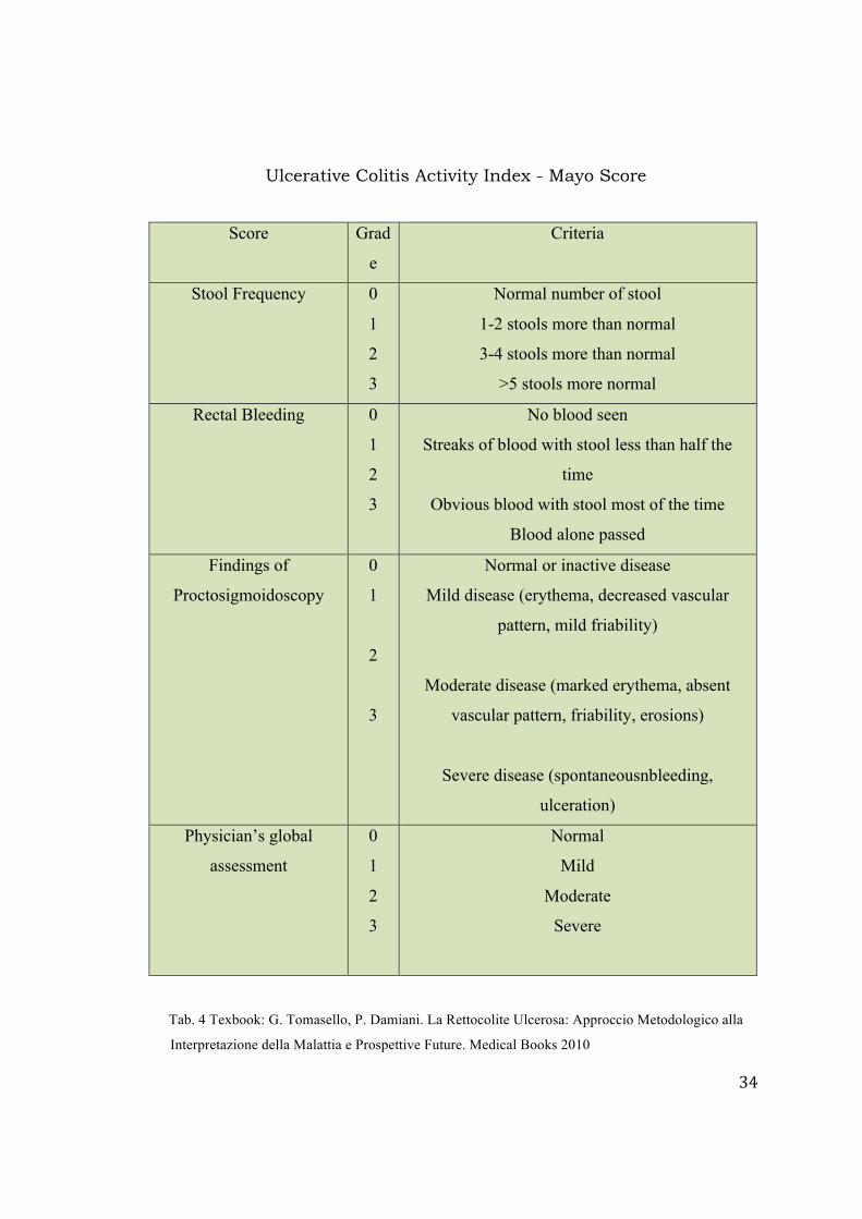
The degree of severity of the disease can also be evaluated through the Mayo
Score.76 (Table 4) !!!!!!!!!!!!!!!!!!!!!!!!!!!!!!!!!!!!!!!!!!!!!!!!!!!!!!!!71!Ordas I, Eckmann L, Talamini M, Baumgart DG, Sandborn JW. Ulcerative Colitis. Lancet 2012; 380: 1606-1609 72!Hoie o, Wolters FL, Riis L, and the European Collaborative Study Group of IBD. Low Colectomy Rates in UC in an Unselected European Cohort Followed for 10 years. Gastroenterology 2007; 132: 507-515 73!Stenson WF, Korzenik J at al. IBD. Textbook of Gastroenterology 2003; 4Th ED: 1699-1759 74!Jun Yun, Chang-Tai XU, Bo-Rong Pan. Epidemiology and Gene Markers of UC in the Cinese. Wolrd J Gastroenterol 2009; 21: 15(7) 788-803 75!Paiotti AP, Artigiani Neto R, Forones NM. Immuno Expression of Cyclooxygenase-1 and 2 in UC. Brazilian J Med Biol Res 2007; 4(7): 911-918 76!Chang Hwan Choi, Sung AE Jung, BO IN LEE. Diagnostic Guideline of UC. Korean J Gastroenterology 2009; 53: 145-160 !
! 34!
Tab. 4 Texbook: G. Tomasello, P. Damiani. La Rettocolite Ulcerosa: Approccio Metodologico alla Interpretazione della Malattia e Prospettive Future. Medical Books 2010
Score Grad
e
Criteria
Stool Frequency 0
1
2
3
Normal number of stool
1-2 stools more than normal
3-4 stools more than normal
>5 stools more normal
Rectal Bleeding 0
1
2
3
No blood seen
Streaks of blood with stool less than half the
time
Obvious blood with stool most of the time
Blood alone passed
Findings of
Proctosigmoidoscopy
0
1
2
3
Normal or inactive disease
Mild disease (erythema, decreased vascular
pattern, mild friability)
Moderate disease (marked erythema, absent
vascular pattern, friability, erosions)
Severe disease (spontaneousnbleeding,
ulceration)
Physician’s global
assessment
0
1
2
3
Normal
Mild
Moderate
Severe
Ulcerative Colitis Activity Index - Mayo Score

! 35!
The UC, and in general IBD, are characterized not only by a clinical pattern
of intestinal involvement, but also for the appearance of symptoms related to
the involvement of other organs.
For this reason, the detection of extraintestinal manifestations allows the
consideration of the IBD as systemic diseases.77
The data indicate that extraintestinal manifestations may report directly to the
chronic inflammatory process characteristic of the disease. And so, they are
manifestations of specific diseases which require a common pathogenic
mechanism with IBD.78
Other events seem, however, not directly related to intestinal disease but the
genetic susceptibility for common autoimmunity.79
Published studies indicate that 6-47% of patients with UC have extraintestinal
manifestations.80
These events may affect different organs and systems compromising
significantly the clinical picture of IBD. (Table 5)
!!!!!!!!!!!!!!!!!!!!!!!!!!!!!!!!!!!!!!!!!!!!!!!!!!!!!!!!77!Danese S, Semeraro S, Papa A, Roberto I, Scaldaferri F, Fedeli G, Gasbarrini A. Extraintestinal Manifestations in IBD. World J Gastroenterol 2005; 11(46): 7227-7236!78!Das Km, Relationship of Extraintestinal Involvements in IBD: New Insights Into Autoimmune Pathogenesis. Dig Dis Sci 1999; 44: 1-13 79!Veloso FI, Carvalho J, Magro F. Immune-Related Systemic Manifestations of IBD. A Prospective Study of 792 Patience. J Clin Gastroenterol 1996; 23: 29-34 80!Robert C, Langan MD, Patricia B. UC: Diagnosis and Treattment. American Family Physician 2007; 76: 1323-1330

! 36!
Pericholangitis
Sclerosing cholangitis
30
1
Hepatocellular fatty infiltration 30
Chronic active hepatitis
Cirrhosis
Amyloidosis
5
3
1
Anemia 15
Iron deficiency 5
Autoimmune hemolytic anemia 1
Thrombocytosis <1
Thromboembolic disease 20
Peripheral arthritis, migratory, nondeforming,
large-joint seronegative
2
Ankylosing spondylitis, sacroilitis 20
Erythema nodosum 20
Pyoderma Gangrenosum 3
Episcleritis, uveitis 4
Iritis 5
Extraintestinal Complicantions and frequency
Table 5: First Principles of Gastroenterology 5Th Ed. Jansenn Ortho

! 37!
1.6 The Diagnostic Path in the UC
The diagnostic uses of UC implementation of different laboratory tests,
instrumental and imaging that, in a synergistic way, enable a precise
classification of the disease.
Their purpose is to make a certain diagnosis of the disease, to assess their
activity status, to delineate the degree of extension and severity of the
inflammatory process as well as the possible presence of extraintestinal
manifestations.
The Gold Standard diagnosis of the UC is surely represented by colonoscopy
and biopsy with histological examination of the intestinal mucosa.81
A colonoscopy allows a direct view of the intestinal mucosa, to determine the
extent of the disease and the severity of the inflammatory process based on
the assessment of the Mayo Endoscopic Score. (Fig.5)
!!!!!!!!!!!!!!!!!!!!!!!!!!!!!!!!!!!!!!!!!!!!!!!!!!!!!!!!81 !D’Argenio G, Cosenza V, Riegler G. Serum Transglutaminase Correlates with Endoscopic And Histopathologic Grading in Patients with Ulcerative Colitis. Dig Dis Sci 2001; 46: 649-657 !
Fig.5: Mayo endoscopic score for ulcerative colitis
(A) Score 0=normal; endoscopic remission. (B) Score 1= mild; erythema, decreased vascular pattern, mild friability. (C) Score 2=moderate; marked erythema, absent vascular pattern, friability, erosions. (D) Score 3=severe; spontaneous bleeding, ulceration. Ingrid Ordás, Lars Eckmann, Mark Talamini, Daniel C Baumgart, William J Sandborn. Ulcerative colitis. Lancet 2012; 380: 1606–19
!

! 38!
Histopathologic biopsy examination of the intestinal mucosa appears to be
essential for the diagnosis of certainty of UC and for a precise histological
characterization of the lesions of the colonic mucosa.82
It also determines the extent of mucosal inflammatory process allowing to
classify the patients according to the degree of disease activity in accordance
to the Histological Activity Index (HAI). (Tab. 6)
The information obtained from the colonoscopy and histopathological biopsy
examination helps to determine the Disease Activity Index.83
The endoscopic and histopathological survey appear to be necessary not only
in the diagnostic phase but also during the follow-up to monitor response to
treatment of the patient.84
!!!!!!!!!!!!!!!!!!!!!!!!!!!!!!!!!!!!!!!!!!!!!!!!!!!!!!!!82!Goetz M, Kiesslich R. Advanced Imaging of the Gastrointestinal Tract: Research vs Clinical Tools? Curr Opin Gastroenterol 2009; 25(5): 412-421 !83!Osada T, Ohkusa T, Yokoyama T. Comparison of Several Activity Indices for the Evaluation of Endoscopic Activity in Ulcerative Colitis: Inter-and Intraobserver Consistency. IBD 2010; 16(2): 192-197 84!Manes G, Imbevi V, Ardizzone S. Appropriateness and Diagnostic Yield of Colonscopy in the Management of Patients with Ulcerative Colitis: a Prospective Study in a Open Access Endoscopy Service. IBD 2008; 14(8): 1133-1138 !
Inflammatory Activity Score Histopathologic Defining Characteristics
Inactive/quiescent/normal 0 No epithelial infiltration by neutrophils
Mildly active 1 Neutrophils infiltration of <50% of sampled
crypts or cross section, no ulcers or erosions
Moderately active 2 Neutrophils infiltration of >50% of sampled
crypts or cross sections, no ulcers or erosions
Table 6: Histological Activity Index (HAI) – Gastroenterology 2007

! 39!
1.7 The UC Therapeutic Management
The UC therapeutic management requires a long-term treatment, very often
based on the use of different drugs to control the disease better.85
In fact, the therapeutic treatment arises with the potential achievement of
several objectives:
1. To reduce mucosal inflammation and try to get a restitutio ad integrum of
the intestinal mucosa
2. To improve the quality of patient's life by intervening on functional
intestinal discomfort (abdominal colic, dysentery, abdominal bloating)
3. To reduce to a minimum the side effects and adverse reactions related to a
long therapeutic treatment
4. To maintain long stages of disease remission
5. To reduce the possibility of complications of the disease
In relation to the diverse manifestation of the disease, to the seriousness and
severity of the clinical pattern, to the possible presence of extraintestinal
manifestations and / or complications, the management of RCU appears to be
significantly diversified and is imprinted on the application of the already
well established therapeutic protocols and others with a more recent
formulation.
Corticosteroids Numerous studies confirm the effectiveness of steroids in the UC treatment in
the acute stage.86
!!!!!!!!!!!!!!!!!!!!!!!!!!!!!!!!!!!!!!!!!!!!!!!!!!!!!!!!85!Worl Gastroenterology Organisation Global Guidelines 2009 86 !Richard P, MacDermott MD, Jesse A. Wath is the Optimal Therapy for severe Ulcerative Colitis? Inflamm Bowel Dis 2008; 14 S2 !

! 40!
In particular, approximately 70% of patients go to clinical remission
following treatment with corticosteroids even if this treatment is not
particularly effective in maintaining the remission stage.87
Acid 5-Aminosalicylic Acid (5-ASA) The elective use of Aminosalicylates in the UC is the maintenance stage of
clinical remission.88
Studies show that taking 5-ASA may reduce the risk of colorectal cancer by
up to 75% and this justifies the long-term treatment of patients with UC.89
Thiopurines The Thiopurines are effective both for the treatment of UC in the active stage
and in the maintenance stage of remission.
The indication for use of Thiopurines should include patients who undergo
multiple relapse episodes of the disease during the year or a relapse within 6
weeks from the interruption of corticosteroids.90
Methotrexate The Methotrexate therapy is reserved for the treatment of patients with a
severe and complicated form of UC with extraintestinal manifestations.91
!!!!!!!!!!!!!!!!!!!!!!!!!!!!!!!!!!!!!!!!!!!!!!!!!!!!!!!!87!Gurel S, Kiyici M. Ulcerative Colitis Activity Index: a Useful Prognostic Factor for Predicting Ulcerative Colitis Outcome. The Journal of International Medical Res 2005; 33: 103-110 88 !Sutherland LR, Roth D, Beck P. Oral 5-Aminosalicyclic Acid for Maintaining Remission in Ulcerative Colitis. Cochrane Database Syst Rev 2002 89!Eaden J, Abrams K, Ekbom A. Colorectal Cancer Prevention in Ulcerative Colitis: a Case-Control Study. Aliment Pharmacol Ther 2000; 14: 145-153 90!McGovern DPB, Travis SPL. Thiopurine therapy: When to Start and When to Stop. Eur J Gastroenterol Hepatol 2003;15: 219–24 91!Peluso R, Atteno M, Iervolino S. Methotrexate in the Treatment of Pheripheral Arthritis in Ulcerative Colitis. Reum 2009; 61(1): 15-20

! 41!
Ciclosporina
The immunosuppressive therapy with this drug allows to obtain an
improvement of the mucosal inflammatory thanks to its action of inhibition of
the production of pro-inflammatory cytokines and the activation of remedy
processes of the intestinal epithelium.92
Immunobiological Therapy (Infliximab) It is an emerging therapeutic possibility that involves the use of specific
monoclonal antibodies with anti-TNF-Alpha.
Its use is widely used in patients with UC in the active stage immune to
steroid therapy and other conventional treatments.93
Surgical Treatment The surgical treatment of the UC does not surely reppresents the first choice
treatment but it is reserved to selected patients.
Several parameters are considered during the assessment of a possible
surgical treatment: high scores of Ulcerative Disease Activity Index, the
extension and severity of the mucosal inflammatory process, the possible
complications, the non responsiveness to conventional drug treatment and the
neoplastic transformation.94
!!!!!!!!!!!!!!!!!!!!!!!!!!!!!!!!!!!!!!!!!!!!!!!!!!!!!!!!!!!!!!!!!!!!!!!!!!!!!!!!!!!!!!!!!!!!!!!!!!!!!!!!!!!!!!!!!!!!!!!!!!!!!!!!!!!!!!!!!!!!!!!!!!!!!!!!!!!!!!!!!!!!!!!!92 !Satoh Y, Ishiguro Y, Sakuraba H. Cyclosporin Regulates in Intestinal Epithelial Apoptosis Via TGF-(Beta) Related Signalling. Am J Physiol Gastrointest Liver Physiol 2009; 297(3): 514-519 93!Rutgeerts P, Sandborn WJ, Feagan BG. Infliximab for Induction and Maintenance Therapy for Ulcerative Colitis. N E J Med 2005; 353: 2462-2476 !94!Filik L, Dagli U. A Retrospective Evaluation of the History of Treated Ulcerative Colitis. Bratisl Lek Listy 2009; 110(4): 253-257

! 42!
1.8 Probiotics
In years, various scientific traces have widely demonstrated the rational use
of Probiotics in the prevention and treatment of numerous affection.95
The term probiotic was used for the first time at the beginning of the last
century by Nobel laureate Elie Metchnikoff who, following a number of
observational studies, suggested that the longevity of the Caucasian
shepherds, could be due to the assumption of foods containing live lactic
bacteria.96
These observations gave rise to a long series of scientific and research studies
aimed to study the composition of the human intestinal microbiota and the
potential use of bacterial strains with probiotic action to stimulate and secure
the natural well-being of our body.
The definition of Probiotics, as provided by current guidelines published by
the Ministry of Health, is the one adopted in 2001 by the Expert Consultation
FAO / WHO. It defines Probiotics as "live and vital microorganisms which,
when administered in adequate amounts as part of a food or a supplement,
gives benefit on the human health."97
The same guidelines define Food / Supplements with Probiotics those
containing micro-organisms, in sufficiently large number that can reach the
intestine, multiply and exercise a beneficial effect on the human health / well-
being.
!!!!!!!!!!!!!!!!!!!!!!!!!!!!!!!!!!!!!!!!!!!!!!!!!!!!!!!!95!Kaur IP, Chopra K, Saini A. Probiotics: Potentiale Pharmaceutical Applications. Eur J Pharm Sci 2002; 15: 1-9 96!Metchnikoff E. The Prolungation of Life. Putman and Sons, 1907 97!FAO/WHO Expert Consultation. Health and Nutritional Properties of Probiotics in Food Including Powder Milk with Live Lactic Acid Bacteria, 2001. www.fao.org !

! 43!
A careful examination of the international scientific literature shows a
growing number of micro-organisms considered as Probiotics.
Notwithstanding, it is difficult to list thoroughly all the bacterial species in
use considered as Probiotics. At present, the probiotic activity has been
recognized only for specific strains. (Tab. 7)
Table 7: Heyman M and Menard S. Cellular and Molecular Life Sciences.
Vol. 59, 2002
One of the key features that a Probiotic should have is the precise
identification of the bacterial strains it contains.
A careful reading of the guidelines prepared by FAO / WHO (2001- 2002),
shows how this feature turns out to be absolutely necessary and that the
Bifidobacterium Species Lactobacillus Species Other Species
B. Bifidum L. Acidophilus B. Cereus
B. Longum L. Rhamnosus Enterococcus Faecium
B. Breve L. Gasseri Enterococcus Faecalis
B. Infantis L. Reuteri Streptococcus
Thermophilus
B. Lactis L. Bulgaricus Clostridium Butyricum
B. Adolescentis L. Johnsonii Escherichia Coli
L. Paracasei Saccharomyces
Boulardii
L. Casei Saccharomyces
Cerevisiae
L. Salivarius
L. Lactis

! 44!
selection of strains to be used in Probiotic preparations must follow a
meticulous process of selection to ensure the taxonomic identity, the specific
phenotypic characteristics, security of use and their potential effectiveness.98
The bacterial microorganisms must meet several other requirements to be
considered as Probiotics: it must be safe for human use, it must be active and
vital when administered in sufficient quantity so as to carry out the beneficial
effects, it must be able to reproduce as much as possible permanently in time
and finally it must be able to confer a benefit to the body.
The effects of a probiotic and the therapeutic response are strictly dependent
on the interaction between its metabolic functional activities and those of the
host.
Studies confirm that they appear to be strain-specific.
The importance of the strain specificity is considered one of the fundamental
requirements to obtain an effective therapeutic response, as documented by
FAO and WHO: "data Obtained with one specific probiotic food can not be
extrapolated to other foods containing that particular probiotic strain or to
other probiotic microorganisms”.
The AFSSA (Agencie Francaise de Sécurité Sanitaire des Aliments) states
that: "the quantity of live probiotics passing through the gut depends on the
strain, the dose ingested, factors related to the host and the vector food".
Therefore, in the light of these observations, the treatment of both intestinal
pathological conditions and extraintestinal bowel with probiotics, can not in
any way regardless of the exact taxonomic definition and the amount of
bacteria strains administered.
!!!!!!!!!!!!!!!!!!!!!!!!!!!!!!!!!!!!!!!!!!!!!!!!!!!!!!!!98!FAO/WHO Expert Consultation. Health and Nutritional Properties of Probiotics in Food Including Powder Milk with Live Lactic Acid Bacteria. 2001. www.fao.org !

! 45!
In relation to the amount of bacterial strains to be administered to the patient,
the document drafted by the AFSSA shows that:
1)“the dose of probiotics ingested is an important factor to obtain high
concentrantions in the various compartments of the gastroentestinal tract”;
2)“it is often said that probiotic concentrantions must be greater than or equal
to 106 CFU/ml in the small intestine (ileum) and 108 CFU/g in the colon”.99
From what can be deduced from published studies, the bacterial strains
considered to be probiotics and therefore could potentially be used in humans
for the treatment of specific medical conditions, must be distinguished from
other bacterial strains erroneously considered probiotics and used for
fermentation of dairy products .
The latter, in fact, are defined as "lactic acid bacteria", they are not human-
derived and are exclusively used as food supplements.100
In the light of all the studies published up to now, it is clear that, for some
specific pathological conditions, the probiotics find a real scientific rationale
use while, for others, the therapeutic efficacy has not yet been fully clarified
and validated.
For many years researchers have recommended the use of probiotics almost
exclusively for the treatment of gastroenteritis, limiting its use in other
pathological conditions, due to the limited knowledge of their functional
abilities and their action mechanisms.
At present, new scientific evidence shed light on new potential ability of
probiotics, expanding its range of use. Some bacterial strains have, in fact,
demonstrated the ability to strictly adhere to the intestinal mucosa by
antagonizing the invasion of pathogenic microorganisms not only thanks to a !!!!!!!!!!!!!!!!!!!!!!!!!!!!!!!!!!!!!!!!!!!!!!!!!!!!!!!!99 !AFSSA. Effecs Des Probiotiques et Prebiotiques sur la Flore et L’Immunitè de L’Homme Adulte. 2005 100!Caramia G. Probiotics: From Metchnikoff to the Current Preventive and Therapeutic Possibilities. Med Surg Ped 2004; 26: 19-33

! 46!
mechanism of territorial competition but also to the production of specific
antibacterial and antifungal substances.
This way, they can correct the intestinal dismicrobism found in various
pathological conditions.
Other probiotic bacterial strains possess the ability to modulate the response
of our immune system by directly influencing the cytokine network.
Still others, contribute significantly to the maintenance of the physiological
pattern of intestinal mucosal permeability. Lastly, it is clear that their role in
the metabolic processes of proteins, carbohydrates and fats as well as in the
synthesis of specific vitamins are useful for the good functioning of our
body.101
!!!!!!!!!!!!!!!!!!!!!!!!!!!!!!!!!!!!!!!!!!!!!!!!!!!!!!!!101!Resta SC. Effects of Probiotics and Commensals on Intestinal Epithelial Physiologi: implications for Nutrient Handling. J Physiol 2008; 587: 4169-4174 !

! 47!
1.9 Rationale for integrated use of probiotics in the UC
In relation to etiopathogenetic factors and the pathophysiological mechanisms
implicated in the onset of IBD, and in particular of the UC, over the years
numerous experimental studies have been performed, both in vitro and in
vivo, with the objective of verifying the existence of a potential rationale for
integrated use of probiotic bacterial strains for their treatment.
The results obtained from these trials indicate that specific probiotics
integration may be useful in the management of patients with UC.102
A careful analysis of the scientific literature shows that the use of probiotic
preparations can be considered a potential aid in the treatment of IBD in
combination with conventional therapies.103
A randomized study n particular, conducted on a sample of 120 patients with
UC and treated with probiotics and symbiotics, has shown an improvement in
the clinical symptoms of patients associated with a significant reduction in
inflammatory markers.104
Probiotics are particularly useful in reducing the rate of recurrence after
surgical treatment as well as in reducing the appearance of pauchite.105
Further study has also validated the efficacy of probiotics in the maintenance
of clinical remission of inflammatory disease.106
!!!!!!!!!!!!!!!!!!!!!!!!!!!!!!!!!!!!!!!!!!!!!!!!!!!!!!!!102!Kanauchi O, Mitsuyama K, Araki Y, Andoh A. Modification of Intestinal Flora in the Treatment of Inflammatory Bowel Disease. Curr Pharm Des 2003; 9: 333-346 103!Hedin C, Welan K, Lindsay JO. Evidence for the Use of Probiotics and Prebiotics in IBD: A Review of Clinical Trials. Proc Nutr Soc 2007; 66 (3): 307-315 104!Fujimori S, Gudis K, Mitsu K. A Randomized Controlled Trial on the Efficacy of Symbiotic Versus Probiotic or Prebiotic Treattment to Improve the Quality of Life in Patients with Ulcerative Colitis. Nutrition 2009; 25 (5): 520-525 !105!Mach T. Clinical Usefulnes of Probiotics in IBD. Journal of Physiology 2006; 57: (S9) 23-33 106!Zocco MA, Dal Verme LZ, Cremonini F. Efficacy of Lactobacillus GG in Maintaining Remission of Ulcerative Colites. Aliment Pharmacol Ter 2006; 23: 1567-1574!

! 48!
Other studies have confirmed that probiotics can intervene in the damage
repair processes at the expense of the intestinal mucosa.107
In particular, it was shown that their repairing action of the intestinal mucosa
stems from their production of SCFA.108
Probiotics also intervene, enhancing the expression of constitutive proteins of
the TJ improving, in this way, the barrier function of the intestinal mucosa.109
Experimental evidence suggests a stimulation action exerted by some
probiotics against goblet cell ensuring adequate production of intestinal
mucus.110
In particular, it has been demonstrated that some of them stimulate the
expression of genes responsible for the production of mucin (MUC2 -
MUC3) resulting in an increase of approximately 60% of mucin.111
Another important rationale for the use of probiotics in the UC is their ability
to correct the status of intestinal dysbiosis through several mechanisms:
competition for nutrient substrates, production of substances with
antibacterial and antifungal and territorial competition.112
!!!!!!!!!!!!!!!!!!!!!!!!!!!!!!!!!!!!!!!!!!!!!!!!!!!!!!!!107!Canonici C, Siret C, Pellegrino E, Pontier-Bres R at al. Saccharomyces Boulardii Improves Intestinal Cell Restitution Through Activation of the Alpha2 Beta1 Integrin Collagen Receptor. Plos One 2011; 31: 18427 108!Sakata T, Kojima T, Fujieda M, Miyakozawa M, Takahashi M, Ushida K. Probiotic Preparations Dose-Dependentily Increase Net Production Rates of Organic Acids and Decrease that of Ammonia by Pig Cecal Bacteria in Batch Culture Dig Dis Sci 1999; 44: 1485-1493 109!Mennigen R, Bruewe M. Effect of Probiotics on Intestinal Barrier Function, Molecular Structure and Function of the TJ. Ann N Y Sci 2009; 1165, 183-189 110!Mattar AF, Tehelbaum DH, Drongo Wski RA, Yongyi F, Harmon CM, Coran AG. Probiotics Up-regulate MUC-2 Mucin Gene Expression in a Caco-2 Cell-Culture Model. Pediatr Surg Int 2002; 18: 586-590 111!Caballerp-Franco C, Keller K, De Simone C, Chadee K. The VSL3 Probiotic Formula Induces Mucin Gene Expression and Secretion in Colonic Epithelial Cells. Am J Physiol Gastrointest Liver Physiol 2007; 292: 215-232 112!Matur E, Eraslan E. The Impact of Probiotics on the Gastrointestinal Physiology. 2012 – www.intechopen.com

! 49!
In particular, Saccharomyces Boulardii has demonstrated the ability to
produce specific protease capable of degrading the toxins produced by
Clostridium Difficile and a phosphatase of 63 kDa capable of destroying the
endotoxin produced by Escherichia Coli.113
Previous studies had demonstrated the ability of S. Boulardii to inhibit the
growth of other pathogens: Salmonella Typhimurium and Yersinia
Enterocoliticum.114
The use of probiotics in the integrated therapy of patients with UC may
reduce the activity of the disease down-regulating mucosal inflammation.115
The ability of some strains of probiotics to significantly influence the
cytokine network through the production of anti-inflammatory cytokines,
suggested their further potential use in the UC.
The inflammatory process characteristic of IBD also stems from the
activation of an important transcription factor present in the cytoplasm cell:
the Nuclear Factor kappa-light-chain-enhancer of activated B cells (NF-KB).
Its activation occurs following the stimulation exerted by inflammatory
agents causing detachment from its precursor: IKB.116
In particular, its activation is influenced by inflammatory cytokines such as
IL-1 and TNF-Alpha and endotoxin represented by lipopolysaccharides (LPS)
of the cell wall of gram-bacteria.117
!!!!!!!!!!!!!!!!!!!!!!!!!!!!!!!!!!!!!!!!!!!!!!!!!!!!!!!!113!MC Farland VL. Systematic Review and Meta-Analysis of Saccharomyces Boulardii in Adult Patients. World Journal of Gastroenterology 2010; 16(18): 2202-2222 !114!Zibden R, Bonczi E, Altewegg M. Inhibition of S. Boulardii on Cell Invasion of Salmonella Typhimurium and Yersinia Enterocolitica. Micro Ecol Health Dis 1999; 11: 158-162 115 !Hanauer SB. IBD: Epidemiology, Pathogenesis and Therapeutic Opportunities. Inflamm Bowel Dis 2006; 12(S1): 3-9 116!Neish AS, Gewirtz AT, Zeng H. Prokaryotic Regulation of Epithelial Responses by Inhibition of IKB-Alpha Ubiquitination. Science 2000; 289: 1560-1563 117!Benelli R, Gavazzi M. La via di segnale IKK/NF-KB. Natural Medicine 2007

! 50!
Some studies show that the initiator mechanism can stimulate the activation
could be represented just by LPS and TLR expressed on the epithelial cell
surface.118
Following its activation, it relocates in the cell nucleus by interacting directly
with the DNA thus stimulating, the expression of specific genes.
In a specific way, it assists the expression of adhesion molecules,
inflammatory cytokines, chemokines, factors growth, cyclooxygenase Cox-2
and metalloproteinases.
Recent studies identify the NF-KB as inflammatory factor common to various
pathological conditions including IBD, rheumatoid arthritis, Lupus, and
diabetes.119
Some probiotic bacterial strains, particularly Lactobacillus Plantarum, have
shown to intervene on these mechanisms inhibiting the activity of NF-KB.120
The activity of NF-KB may also be inhibited by the activation of another
factor: the Peroxisome Proliferator-Activated Receptor Gamma (PPAR-
gamma).
The PPAR-Gamma is a nuclear receptor expressed in cells of different tissues
and organs, but especially in the epithelial cells of the colon, where its
expression appears to be regulated by the intestinal flora.121Its concentration
appears to be significantly reduced in IBD.122
!!!!!!!!!!!!!!!!!!!!!!!!!!!!!!!!!!!!!!!!!!!!!!!!!!!!!!!!118!Chen R, Alvero AB, Silasi DA, Mor G. Inflammation, Cancer and Chemioresistance: Taking Advantage of the Toll Like Receptor Signaling Pathway. Am J Rep Imm 2007; 57: 93-107 119!Aggarwal BB, Sethi G, Nair A, Ichikawa H. Nuclear Factor-KB: a Holy Grail in Cancer Prevention and Therapy. Curr Signal Transduction Therapy 2006; 1: 25-52 120!Petrof EO, Claud EC, Sun J. Bacteria-Free Solution Derived from Lactobacillus Plantarum Inhibits Multiple NF-KB Pathways and Inhibits Proteasome Function. Inflamm Bowel Dis 2009; 15: 1537 121!Dubuquoy L, Dharancy S, Nutten S, Auwerx J, Desreumaux P. PPAR-Gamma/RXR in IBD: a Critical Review. Best Pract Res Clin Gastroenterol 2003; 17: 805-820 122 !Dubuquoy L, Jansson EA, Deeb S. Impaired Expression of PPAR-Gamma in Ulcerative Colitis. Gastroenterology 2003; 124: 1265-1276

! 51!
In particular, in the RCU, its reduced concentration stimulates the
inflammatory process.123
The integration of specific probiotics has shown the increase, in the intestinal
epithelial cells, the expression of PPAR-GAMMA inhibiting, in this way, the
activation of NF-KB and significantly reducing the inflammatory process in
patients with IBD.124
It was also demonstrated that the chronic inflammatory process resulting from
iperexpression of TNF-ALPHA, IFN-GAMMA and other inflammatory
cytokines, is able to activate specific pro-apoptotic protein.
Some probiotics inhibit their expression by adjusting, in a way, the process of
cell proliferation.125
!!!!!!!!!!!!!!!!!!!!!!!!!!!!!!!!!!!!!!!!!!!!!!!!!!!!!!!!123!Bianchi Porro G, Ardizzone S, Colombo E. IBD: Year Book 2005 !124 !Kelly D, Campbell JI, King TP. Commensal Anaerobic Gut Bacteria Attenuate Inflammation By Regulating Nuclear-Cytoplasmic Shuttling of PPAR-GAMMA and RELA. Nut Immunol 2004; 5: 104-112 125!Yan F, Cao H, Cover TL. Soluble Proteins Produced by Probiotic Bacteria Regulate Intestinal Epithelial Cell Survival and Growth. Gastroeterology 2007; 132: 562-575!

! 52!
2. Aims In the last decade, in relation to the numerous published scientific evidence,
the management of UC has undergone considerable evolutions, offering a
wide range of therapeutic options that are based on the application of already
well established protocols and the introduction of new options of recent
formulation.
The correct therapeutic strategy to be considered in the treatment of patients
with UC can not be ignored in taking into account the possible and desirable
goals to achieve or improve the quality and life expectancy of the patient,
obtain a greater control and a possible and rapid resolution of clinical
symptoms in addition to achieving a significant improvement in patient’s
compliance minimizing the side effects of the therapy.
The therapeutic choice should strictly depend on the history and clinical
evolution of the disease in the individual patient.
Therefore, during the acute stage patients require pharmacological treatment
that strictly depends on the severity of the clinical chart which, in severe
cases, may require hospitalization and / or surgery.
During the remission period, the pharmacological approach aims to maintain
clinical quiescence.
Careful analysis of the international scientific literature shows that the
different therapeutic strategies adopted in recent years for the treatment of
UC, despite offering a significant improvement in the clinical symptoms,
have not showed their capacity to change radically the history of the disease
in the long term clinical evolution.
In relation to numerous scientific papers published in recent years that
demonstrate the potential function performed by a number of probiotic
bacterial strains and the results obtained from using them, the objective of

! 53!
this thesis, is to verify the effective usefulness of probiotics integration in the
treatment of UC, confirming, in this way, this potential and innovative
therapeutic approach to the treatment of the disease.
In the execution of this clinical study, the patients were subjected to a drug
treatment of Mesalazine (Mesavancol®) and a probiotic (Acronelle®) intake,
a mixture of: Lactobacillus Acidophilus, Lactobacillus Salivarius and
Bifidobacterium Bifidum s.p. BGN4.
The choice of this probiotics formulation sprung from several observations on
metabolic functional capabilities, already known, performed by these
bacterial strains.
The choice of this probiotics formulation sprung from several observations on
metabolic functional capabilities, already known, performed by these
bacterial strains.
In particular, previous studies have demonstrated an important role of L.
Acidophilus in IBD. Integration with Salivarius L. and B. Bifidum BGN4 has
shown to boost its action.
The L. Salivarius has, in fact, demonstrated high adhesion to the intestinal
epithelium providing an important trophic function and repair of the
mucosa.126
The Bifidobacteria and in particular the B. Bifidum BGN4, proved to be
extremely important in reducing the inflammatory processes of the intestinal
mucosa, in the modulation of the immune response and in actively
!!!!!!!!!!!!!!!!!!!!!!!!!!!!!!!!!!!!!!!!!!!!!!!!!!!!!!!!126!Collins JK. Demonstration of functional properties of probiotics lactic acid bacteria (L.
Salivarius). Ind. Milk 2001; 39-61 Clinical studies show the therapeutic efficacy of
Lactobacillus Salivarius in IBD. (Harrison's Principles of Internal Medicine, 15th Ed, Vol
2. MC Graw Hill 2001; 1679-1695 !

! 54!
combatting the intestinal dysbiosis detected in multiple disorders such as
IBD.
Numerous studies have demonstrated that bifidobacteria has the capacity to
play an important antigenotoxic role compared to the cells of the intestinal
mucosa.127
The antigenotoxicity represents, in light of the extensive scientific evidence
published in recent years, one of the most important potential function
performed by selected probiotics bacterial strains.128
In vitro studies demonstrate the role played by bacterial pathogen overgrowth
in the intestine in inducing carcinogenesis processes.129
In particular, it has been shown how the presence of Bacteroides and
Clostridium overgrowth is associated with a higher incidence and speed of
growth of colon cancer while on the contrary an adequate presence of
Lactobacilli and Bifidobacteria results to play a protective role.
Experimental and epidemiological studies have demonstrated the ability of
some strains of bacteria (bifidobacteria) to reduce the expression of several
enzymes (azoreduttase, β-glucuronidase, nitroreductase, 7-α-dehydroxylase)
involved in the synthesis of genotoxic compounds capable of promoting the
beginning of the typical carcinogenesis processes.
!!!!!!!!!!!!!!!!!!!!!!!!!!!!!!!!!!!!!!!!!!!!!!!!!!!!!!!!127!Burns AJ, Rowland IR. Anti-Carcinogenicity of Probiotics and Prebiotics. Curr Issues
Header Microbiol 2000; 1: 13-24 128!O'Mahony L, M Feeney, O'Halloran S, Murphy L, Kiely B, Fitzgibbon J, Lee G,
O'Sullivan G, Shanahan F, Collins JK. Probiotic impact on microbial flora, inflammation
and tumor development in IL 10 knockout mice. Aliment Pharmacol Ther 2001; 15 (8):
1219-1225 129!Cenci G, Caldini G, F Trotta, Bosi P. In vitro inhibitory activity of probiotic spore-
forming bacilli against genotoxins. Applied Microbiology 2008; 46 (3): 331-337
!

! 55!
Recent studies show that B. Bifidum BGN4 can perform antigenotoxic action,
anti-inflammatory and immune-modulator thanks to its ability to produce a
complex polysaccharide (BB-Pol) consisting of Chiroinositolo, Rhamnose,
Glucose, Galactose and Ribose.
The BB-pol produced by B. bifidum BGN4 has shown to inhibit in vitro, the
growth of specific tumor cell lines of human colon.

! 56!
3. Materials and Methods For our study which lasted for two years (2011-2013), we recruited 60
patients with UC at the Clinic of Digestive Endoscopy of AOUP “P.
Giaccone” in Palermo, and were divided into two groups of 30 elements each.
Group A included 22 men and 8 women aged between 35 and 69 (average:
43) with an index of activity (Mayo Endoscopic Scoring System) between 2
and 3 (moderate-to-severe disease), treated pharmacologically with once-
daily dose of 1200 mg of oral mesalazine (Mesavancol® 1200 mg cpr,
Giuliani spa, Milan). 19 men and 11 women suffering from UC aged between
28 and 71 (average: 46) were included in group B and activity index always
lies between 2 and 3 (moderate-to-severe disease), treated again with 1,200
mg of mesalazine per os (Mesavancol® 1200 mg cpr, Giuliani spa, Milan),
this time assisted in taking daily double administration of a probiotic blend
Lactobacillus salivarius and Lactobacillus acidophilus based, specific strain
of Bifido bifidum BGN4 (Acronelle®, Bromatech srl, Milan, Italy).
A series of endoscopic surveillance during the first visit (t0) were
programmed for each group and later 6 (t1), 12 (t2), 18 (t3) and 24 (t4)
months. A new reassessment was carried out for every check-ups in
accordance with Mayo endoscopic score for ulcerative colitis.
The results were used to build a database on excel sheet and the data were
evaluated statistically using the ANOVA method for repeated measures
(Repeated Measures ANOVA), while the two groups were compared using
the t test (unpaired t test).

! 57!
4. Results Analysis of the results (and subsequent follow-up t0 t1-t4 - range: 24 months)
shows that patients treated with mesalazine and probiotic blend had a better
clinic-endoscopic score (Mayo) than patients treated with mesalazine alone
(Fig. 6-20).
In particular, Figure 16 suggests that the benefits of probiotic blend in
combination with the anti-inflammatory are more obvious and significant
after a period of at least 18 months.
We can see from the graphs that both considered treatments have some
effectiveness or otherwise provide a distinct advantage to patients with UC
who use it. Between the two groups, however, the one that gets the most
benefit is definitely group B, in which of course, the use of probiotic blend
enhances the effects which are already positive in the anti-inflammatory.
Considering the individual parameters which are taken into account to
calculate the Mayo Score, it is enough to look at figures 6 and 7 to understand
out how mesalazine has positive effects in both groups, but these are already
significant as early as 6 months of treatment for only group B, the one treated
with the addition of the probiotic blend.
We can also see from the figures how, in reality, the effect of therapy is much
more constant in B, where the values used in assessing the overall clinical
condition remain consistently low, while there is a slight deterioration in A.
Figure 4 corroborates as specified, showing a slight benefit of the
combination therapy compared with the sole use of mesalazine, with a
statistically significant benefit after only 2 years of treatment.
The alvo frequency shows a significant reduction, this time in both groups,
although it is more pronounced in the second (Fig. 9-10). It is strangely
interesting to note that there is a slight deterioration at 18 months in group B,

! 58!
while in group A, even in this case, is encumbered by a moderate loss of
effectiveness of anti-inflammatory therapy. Comparing the two groups
(Fig.11), the reduction of alvo frequency becomes statistically significant at 6
and 24 months always in favor of group B. Even in this case it detects a small
reversal of the trend at 18 months when the patients of group B seem to lose
the advantage supplied by the probiotics.
Regarding the endoscopic picture (Fig. 12-13-14), both treatments are already
valid after 6 months. In group B, however, in addition to showing greater
improvement of the intestinal mucosa conditions studied, it is noted, as for
the overall clinical condition, a prolonged beneficial effects of combination
therapy for up to two years from the beginning of the treatment, that also in
this case, does not occur for the group, which, indeed, loses some of the
benefits of the therapy already at t3.
Finally, as for the endoscopic picture, the evaluation of rectal bleeding more
or less follows the same time lapse (Fig. 15-16-17). In fact, it is noted that
there is a minor bleeding in both groups, already at t1, but while group B
enjoys the positive effects of probiotics up to 2 years, showing a certain
constancy of values at the time of check-up, group A, once again, gets
progressively worse from 18 months of treatment onwards.

! 59!
Fig. 6 Statistical analysis of the "general clinical condition" parameter
of endoscopic Mayo Score for Ulcerative Colitis (UC) for group A. The
use of mesalazine (1200 mg cpr Mesavancol® rp, Giuliani SpA, Milan, Italy)
in patients with moderate and severe UC at t0, implies an improvement in
overall clinical condition statistically significant at 18 and 24 months
(Repeated Measures ANOVA).
General clinical conditions A
T0 T1 T2 T3 T40
1
2
3
4Score * #
T0 vs T4 p<0.001 ; T1 vs T3 p<0.05
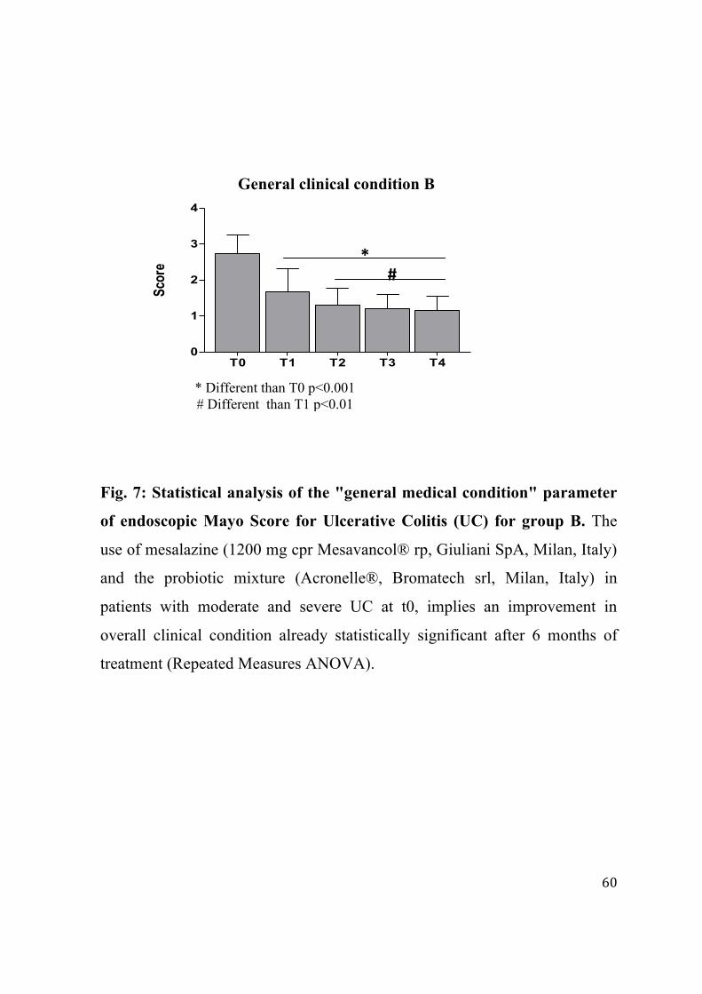
! 60!
Fig. 7: Statistical analysis of the "general medical condition" parameter
of endoscopic Mayo Score for Ulcerative Colitis (UC) for group B. The
use of mesalazine (1200 mg cpr Mesavancol® rp, Giuliani SpA, Milan, Italy)
and the probiotic mixture (Acronelle®, Bromatech srl, Milan, Italy) in
patients with moderate and severe UC at t0, implies an improvement in
overall clinical condition already statistically significant after 6 months of
treatment (Repeated Measures ANOVA).
General clinical condition B
* Different than T0 p<0.001 # Different than T1 p<0.01
T0 T1 T2 T3 T40
1
2
3
4
Score *
#

! 61!
Fig. 8: Comparison statistics between group A and group B of "general
medical condition" parameter of endoscopic Mayo Score for Ulcerative
Colitis (UC). In patients with moderate and severe UC at t0, group B shows a
significant improvement in overall clinical condition compared to group A,
after 2 years of treatment (Repeated Measures ANOVA).
General'clinical'condition'A'vs'B
*'Different'than'T47A'p=0.0159
T0-A
T0-B
T1-A
T1-B
T2-A
T2-B
T3-A
T3-B
T4-A
T4-B
0
1
2
3
4
Score
*

! 62!
Fig. 9: Statistical analysis of the "alvo frequency " parameter of
endoscopic Mayo Score for Ulcerative Colitis (UC) for group A. The use
of mesalazine (1200 mg cpr Mesavancol® rp, Giuliani SpA, Milan, Italy) in
patients with moderate and severe UC at t0, leads to a significant reduction in
the frequency of the alvo after 6 months of treatment, with a statistically
higher significance at t2 and t3 (Repeated Measures ANOVA).
T0 T1 T2 T3 T40
1
2
3
4Score
Alvo frequency A
B*
* Different than T0 p<0.001 # Different than T1 p<0.01
#

! 63!
Fig. 10: Statistical analysis of the "alvo frequency " parameter of
endoscopic Mayo Score for Ulcerative Colitis (UC) for group B. The use
of mesalazine (1200 mg cpr Mesavancol® rp, Giuliani SpA, Milan, Italy) and
probiotic blend (Acronelle®, Bromatech srl, Milan, Italy) in patients with
moderate and severe UC at t0, leads to a significant reduction in the
frequency of the alvo after 6 months of treatment, with a statistically
significant increase after 2 years of initiation of treatment (Repeated
Measures ANOVA).
T0 T1 T2 T3 T40
1
2
3
4
Score
Alvo frequency B
*
*'Different'than'T0'p<0.001
#'Different'than'T1'p<0.05
#

! 64!
!
Fig. 11: Comparison statistics between group A and group B of the "alvo
frequency" parameter of endoscopic Mayo Score for Ulcerative Colitis
(UC). In patients with moderate and severe UC at t0, group B shows a
significant reduction in the frequency of the alvo, compared with group A, at
6 months and 2 years after initiation of treatment (Repeated Measures
ANOVA).
Alvo Frequency A vs B

! 65!
!!!!!!!!!!
Fig. 12: Statistical analysis of the "endoscopic picture" parameter of
endoscopic Mayo Score for Ulcerative Colitis (UC) for group A. The use
of mesalazine (1200 mg cpr Mesavancol® rp, Giuliani SpA, Milan, Italy) in
patients with moderate and severe UC at t0, implies a statistically significant
improvement in endoscopic picture after only 6 months of treatment, with
greater statistical significance than t2 (Repeated Measures ANOVA).
Endoscopic Picture A

! 66!
Fig. 13: Statistical analysis of the "endoscopic picture" parameter of
endoscopic Mayo Score for Ulcerative Colitis (UC) for group B. The use
of mesalazine (1200 mg cpr Mesavancol® rp, Giuliani SpA, Milan, Italy) and
the probiotic blend (Acronelle®, Bromatech srl, Milan, Italy) in patients with
moderate and severe UC at t0, leads to a statistically significant improvement
in endoscopic picture after 6 months of treatment, with even greater
significance at 18 and 24 months (Repeated Measures ANOVA).
T0 T1 T2 T3 T40
1
2
3
4
Score
Endoscopic'picture'B''
*'
#
#'Different'than'T1'p<0.001

! 67!
!
Fig. 14: Statistical Comparison between group A and group B of the
"endoscopic picture" parameter of endoscopic Mayo Score for
Ulcerative Colitis (UC). In patients with moderate and severe UC at t0,
group B shows a significant improvement in the endoscopic picture,
compared to group A, starting from t3 (Repeated Measures ANOVA).
Endoscopic Picture A vs B
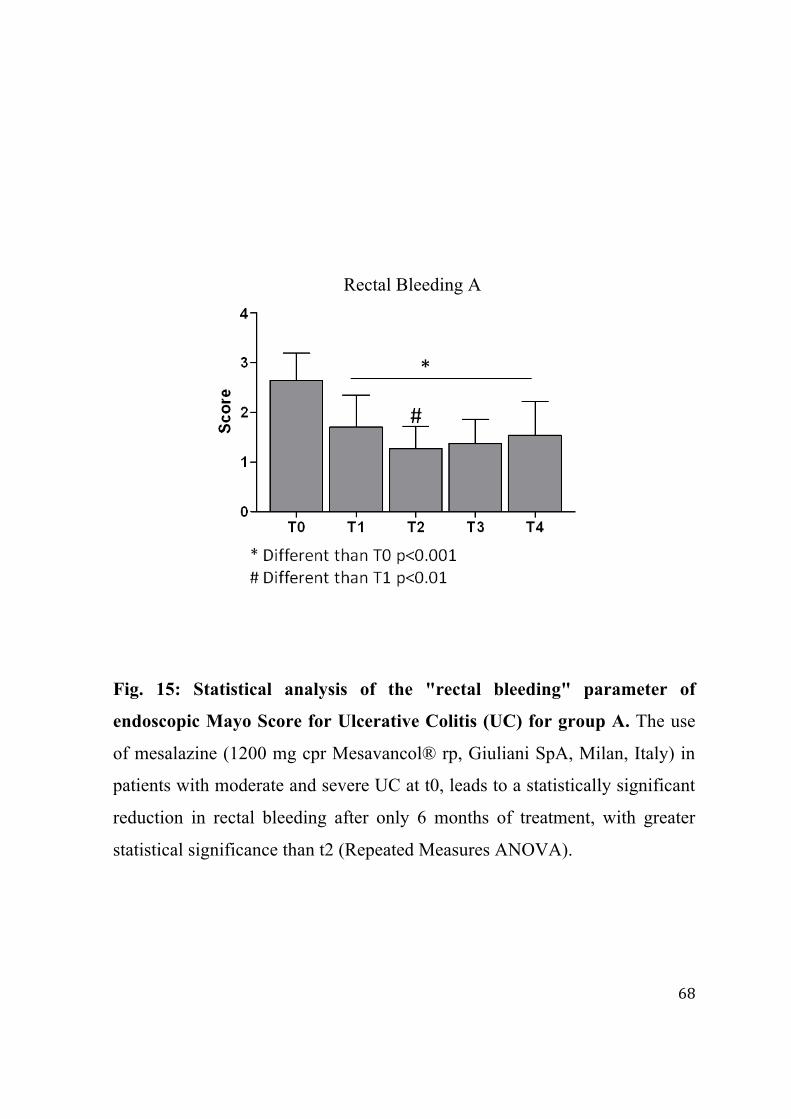
! 68!
!
Fig. 15: Statistical analysis of the "rectal bleeding" parameter of
endoscopic Mayo Score for Ulcerative Colitis (UC) for group A. The use
of mesalazine (1200 mg cpr Mesavancol® rp, Giuliani SpA, Milan, Italy) in
patients with moderate and severe UC at t0, leads to a statistically significant
reduction in rectal bleeding after only 6 months of treatment, with greater
statistical significance than t2 (Repeated Measures ANOVA).
Rectal Bleeding A

! 69!
!
Fig. 16: Statistical analysis of the "rectal bleeding" parameter of
endoscopic Mayo Score for Ulcerative Colitis (UC) for group B. The use
of mesalazine (1200 mg cpr Mesavancol® rp, Giuliani SpA, Milan, Italy) and
the probiotic blend (Acronelle®, Bromatech srl, Milan, Italy) in patients with
moderate and severe UC at t0, leads to a statistically significant reduction in
rectal bleeding after only 6 months of treatment (Repeated Measures
ANOVA).
Rectal Bleeding B

! 70!
!
Fig. 17: Comparison statistics between group A and group B of the
"rectal bleeding" parameter of endoscopic Mayo Score for Ulcerative
Colitis (UC). In patients with moderate and severe UC at t0, group B shows a
significant reduction in rectal bleeding after only 6 months of treatment
compared to group A and then at t3 and t4, losing significance only at 1 year
of treatment.
Rectal Bleeding A vs B

! 71!
Fig. 18: Statistical analysis of changes in the endoscopic Mayo Score for
Ulcerative Colitis (UC) over time (t0-t4) relating to group A. The use of
mesalazine (1200 mg cpr Mesavancol® rp, Giuliani SpA, Milan, Italy) in
patients with moderate and severe UC at t0, leads to a statistically significant
reduction of the score after only 6 months of treatment, with greater statistical
significance than t2 (Repeated Measures).
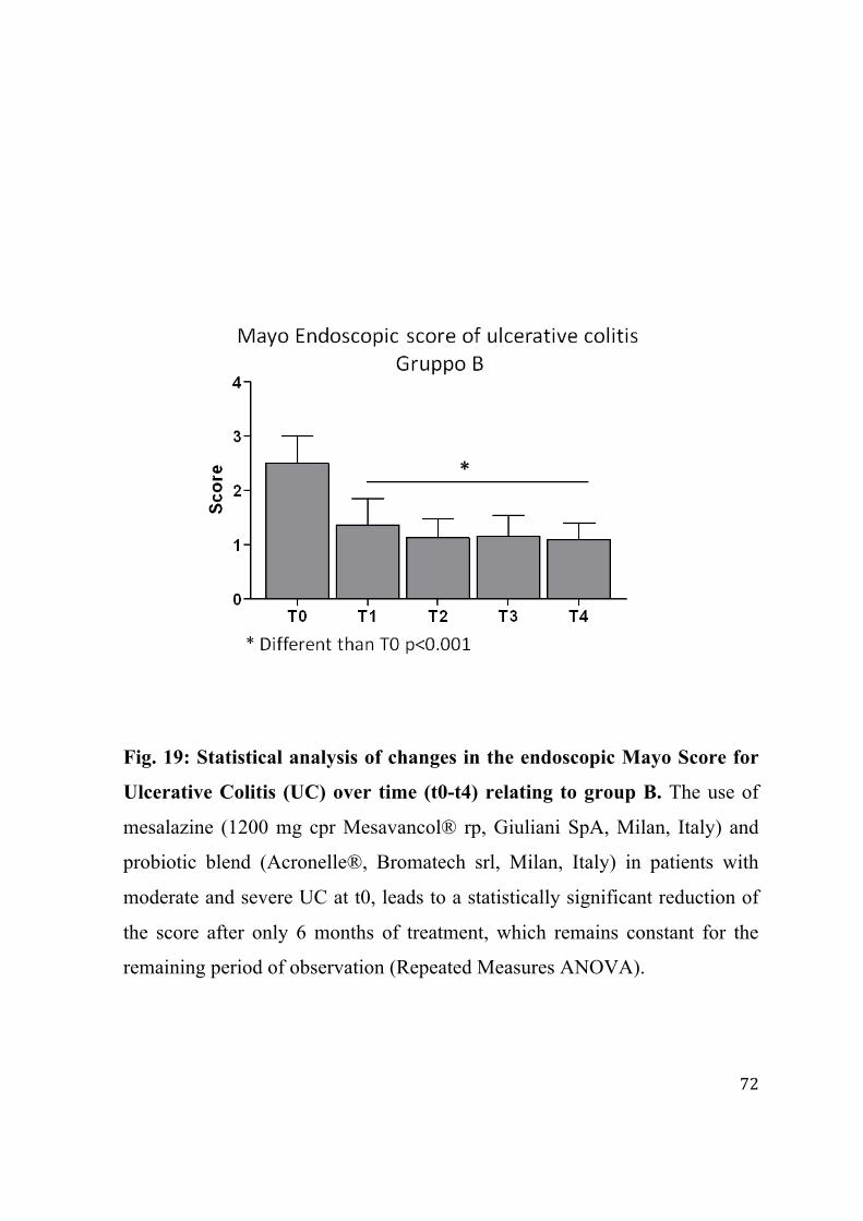
! 72!
Fig. 19: Statistical analysis of changes in the endoscopic Mayo Score for
Ulcerative Colitis (UC) over time (t0-t4) relating to group B. The use of
mesalazine (1200 mg cpr Mesavancol® rp, Giuliani SpA, Milan, Italy) and
probiotic blend (Acronelle®, Bromatech srl, Milan, Italy) in patients with
moderate and severe UC at t0, leads to a statistically significant reduction of
the score after only 6 months of treatment, which remains constant for the
remaining period of observation (Repeated Measures ANOVA).
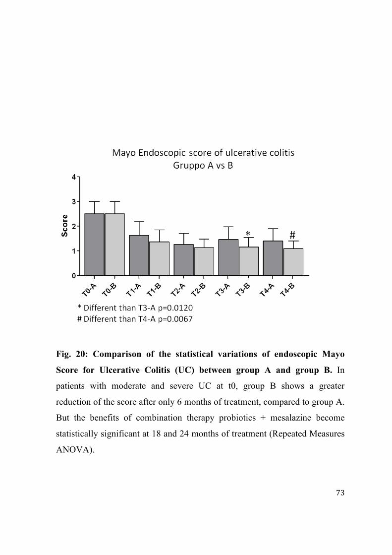
! 73!
Fig. 20: Comparison of the statistical variations of endoscopic Mayo
Score for Ulcerative Colitis (UC) between group A and group B. In
patients with moderate and severe UC at t0, group B shows a greater
reduction of the score after only 6 months of treatment, compared to group A.
But the benefits of combination therapy probiotics + mesalazine become
statistically significant at 18 and 24 months of treatment (Repeated Measures
ANOVA).

! 74!
5. Discussion
The UC as the CD are considered multifactor etiopathogenesis pathologies.
The scientific literature data identify the presence of a genotype susceptibility
which, together with various environmental factors, are capable of interacting
with the immune system to give rise to a chronic inflammation to
significantly damage the intestinal mucosa of these patients.
Although the possible genetic alterations responsible for providing for UC
have been identified, the role played by environmental factors in the
pathogenesis of the disease should not be underestimated.
Among the various epigenetic factors identified so far, the intestinal
microbiota seems to represent one of the most important factors.
The characteristic recurrent chronic trend of the UC has led to the hypothesis
that the inflammation process of the intestinal mucosa is due to the constant
exposure of the mucosa to antigenic endoluminal stimuli that can stimulate
the activation of an exaggerated local immune response.
Numerous studies published over the years have identified in the intestinal
dismicrobism the most important endoluminal antigenic stimulus that, along
with the overthrow of the whole intestinal microenvironment, can trigger the
inflammatory process and hyperactivation of the immune system typical of
the UC.
It is also observed in the course of IBD and in particular of UC, in addition to
a state of intestinal dysbiosis, a modification of other components of the
mucosa.
In particular, we can see a significant reduction in the thickness of the mucus
layer associated with a reduced representation of goblet cells in addition to a

! 75!
reduced synthesis of mucin resulting in the impairment of the barrier function
carried out by the intestinal mucosa.
In IBD, particularly in the UC it is found, in fact, the reduced expression of
specific genes responsible for the more or less adequate expression of the
goblet cells.
The mucin deficiciency appears to be related to the onset of intestinal
dysbiosis and increased tendency to colitis.
Many studies confirm the importance of a proper production of mucous and a
rich amount of mucin to avoid the onset of intestinal musale inflammation.
Studies also show a modification of the mucous system. In fact, it is observed
in patients with UC an increased intestinal permeability due to the destruction
of the Tight Junction.
In fact, it is detected in UC a reduced transcription of specific gene sequences
assigned to the synthesis of the protein constituent of the TJ.
The alterations of the defense system of the intestinal mucous are directly
attributable to the disregulated expression of specific cytokines inflammatory.
Other than these specific alterations, a change is detected in both quality and
quantity of the normal intestinal flora bacteria.
Recent studies show, in fact, that the state of intestinal dysbiosis, typical of
patients with UC, is characterized by enterotype alteration with the
establishment of a specific and characteristic enterotype of the IBD.130
This specific enterotype seems to be directly related to the degree of
inflammation of the intestinal mucous and the extent of the clinical-
symptomatic chart.
!!!!!!!!!!!!!!!!!!!!!!!!!!!!!!!!!!!!!!!!!!!!!!!!!!!!!!!!130!Hildebrand F, Brinkman B, Yunta RG, Cauwe B, Vandenabeele P, Liston A, Raes J. Inflammetion-Associated Enterotypes, Host Genotype Cage and Inter-Individual Effects Drive Gut Microbiota Variation in Common Laboratory Mice. Genome Biology 2013; 14: R4 !
! 76!
In particular, the establishment of this specific enterotype results to be
positively correlated with a high concentration of Fecal Calprotectin, known
index of the intestinal mucous found in high concentrations in patients with
UC.
Microbiological studies detect, in particular, a significant reduction of
Bifidobacteria and Lactobacilli and the growth of bacterial pathogens
responsible not only for the onset of persistent intestinal infection but also of
micro-lesions of the mucous and of the continuous characteristic
inflammatory process.
In fact, it is demonstrated in patients with IBD, the overgrowth of specific
pathogenic strains as some members of the Deltaproteobacteria phylum like
Bilophila Wadsworthia.
It is a gram-negative bacteria resistant to bile hardly found as a intestinal
commensal particularly abundant in healthy subjects and in patients with IBD
and responsible to subvert immune homeostasis resulting in the appearance of
a low-grade mucosal inflammatory process with increase in INF-G, IL-12 and
reduced IL-6, 17 and 23.
Bilophila Wadsworthia has shown to metabolize Sulfates producing
Hydrogen Sulfide responsible for lesions of the intestinal mucous and to
inhibit the production of SCFA (A. Butyric) resulting in inhibition of the
processes of repair of the intestinal mucous.131
The alteration of the normal intestinal flora seems to be responsible to
participate in the dysregulated immune response characteristic of local IBD.
!!!!!!!!!!!!!!!!!!!!!!!!!!!!!!!!!!!!!!!!!!!!!!!!!!!!!!!!131!Dietary-fat-induced taurocholic acid promotes pathobiont expansion and colitis in Il10−/− mice. Suzanne Devkota, Yunwei Wang, Mark W. Musch, Vanessa Leone, Hannah Fehlner-Peach, Anuradha Nadimpalli, Dionysios A. Antonopoulos, Bana Jabri & Eugene B. Chang Nature 487, 104–108

! 77!
Studies show that micro-intestinal mucous mostly involved in the
inflammation are those on which there is an increased bacterial pathogens.
Histological analysis and biopsy cultivation of the patients’ mucous with UC
detect a significant reduction of Bifidobacteria and lactobacilli and an
increase in the concentration of pathogenic bacteria capable of inducing a
pro-inflammatory effect.
The persistent intestinal dysbiosis detectable in the UC is responsible for the
activation of the GALT directly connected on the onset of the inflammatory
process.
Contextually, there is an alteration in the immune-tolerance biological
mucous capable in inducing significant modifications of the normal resident
bacterial flora.
The result of these processes is the appearance of chronic inflammation and
persistent intestinal dismicrobism.
The studies show that the persistent intestinal disbiosis subtends, at least in
the 70% of the patients to a reduced or non expression of the TSLP (Thymic
Stromal Lymphopoietin) which is defined as “protein keeper” of our
intestine, responsible for the discriminating action between beneficial resident
flora and pathogen flora associated with reduced expression of the Difensins,
protein responsible for the destruction of the pathogen bacteria present in our
intestine.132
The clinical experience gained over the years has made it possible to identify
different therapeutic protocols for the treatment of UC but none of them has
proven so far to be able to significantly change the natural history of the
disease.
!!!!!!!!!!!!!!!!!!!!!!!!!!!!!!!!!!!!!!!!!!!!!!!!!!!!!!!!132!Allakhverdi, Z., M. R. Comeau, et al. "Thymic stromal lymphopoietin is released by human epithelial cells in response to microbes, trauma, or inflammation and potently activates mast cells." J Exp Med 2007. 204(2): 253-8.

! 78!
Studies have shown great efficacy of anti-inflammatory drugs such as 5-
aminosalicylic acid and corticosteroids, but these are not always sufficient in
controlling the clinical course of the disease.133
In recent years a widespread use immunosuppressive drugs have been found.
These biological agents potentially capable of blocking the biochemical
events which support a periodic inflammatory process characteristic of the
intestinal mucosa of UC but these drugs do not always guarantee a
satisfactory therapeutic result.134
These observations have prompted research to identify new therapeutic treatments that allow to act on the pathophysiological mechanisms characteristic of UC and capable of improving the quality and life expectancy of patients, to reduce significantly the clinical symptoms and to reduce to the minimum side effects. In relation to the extensive scientific evidence that over the years have amply demonstrated and confirmed the efficacy of probiotics in the treatment of various diseases, it has been suggested its potential use in the treatment integrated with IBD, and particularly in the UC . The use of probiotics in the clinical setting is now widely documented in both therapeutic and preventive setting. Their potential rationale use stems from a number of studies that over the years have demonstrated the specific metabolic functional potentials they completed. In particular, it has been repeatedly demonstrated and confirmed their ability to counteract the state of intestinal dysbiosis significantly reducing the presence of pathogenic bacteria and increasing the concentration of Bifidobacteria and Lactobacilli constituent of a normal intestinal bacterial flora. !!!!!!!!!!!!!!!!!!!!!!!!!!!!!!!!!!!!!!!!!!!!!!!!!!!!!!!!133!Robert C, Langan MD, Patricia B. UC: diagnosis and treatment. (AAFP) American Family Physician 2007; 76: 1323-1330!134!Richard P, MacDermott MD, Jesse A. What is the optimal therapy for UC? Inflamm Bowel Dis 2008. Vol 14(S2)

! 79!
Specific probiotic bacterial strains such as Enterococcus Faecium and S. Boulardii are, in fact, capable of producing substances with antibacterial and antifungal which reduces the presence of E. Coli, of Clostridium D., Candida Albicans.135 Other probiotics have demonstrated an intervention in the processes of production of intestinal mucus, to produce SCFA such as acetic, propionic and butyric with the capacity to nourish the intestinal mucosa and restore the barrier functions.136 Numerous studies have also demonstrated the ability of specific probiotics to go unto the cytokine network reducing mucosal inflammation process and modulating the local immune response. A careful analysis of the scientific literature shows that the use of probiotic preparations, can be considered a potential aid in the treatment of IBD in combination with conventional therapies. A randomized study, conducted on a sample of 120 patients with UC and treated with probiotic and symbiotic, showed an improvement of the clinical symptoms of patients associated with a significant reduction in inflammatory markers.137 Probiotics are found particularly useful in reducing the rate of recurrence after surgical treatment and to reduce the appearance of pauchite.138 A further study also validated the efficacy of probiotics in maintaining clinical remission of inflammatory disease.139
!!!!!!!!!!!!!!!!!!!!!!!!!!!!!!!!!!!!!!!!!!!!!!!!!!!!!!!!135!Borgia M, Sepe N, Brancato V. A controlled clinical study on streptococcus faecium preparation for the prevention of side reactions during long term antibiotic treatments. Curr Ther Res 1982; 31(2) 136!Collins JK. Demonstration of functional properties of probiotics lactic acid bacteria. Ind Latte 2010; 39-61 137!Fujimori S, Gudis K, Mitsu K. A randomized controlled trial on the efficacy of symbiotic versus probiotic or prebiotic treatment to improve the quality of life in patients with UC. Nutrition 2009; 25(5): 520-525 138!Mach T. Clinical Usefulness of probiotics in inflammatory bowel diseases. Journal of Physiology 2006; 57(S9): 23-33 139!Schwartz M, Cohen R. Optimizing conventional therapy for inflammatory bowel disease. Curr Gastroenterol Rep. 2008; 10(6): 585-590!

! 80!
Other studies have confirmed that probiotics are able to intervene in the process of repairing the damage to the intestinal mucosa.140 In particular, it was shown that their repairing action of the intestinal mucosa stems from their production of SCFA. In light of these observations, the origin of this thesis, it was decided to use a specific probiotic, Acronelle (Acronelle®) containing L. Salivarius and Bifidobatterium Bifidum BGN4, in the integrated treatment of patients with UC and treated with mesalazine (Mesavancol®) with the objective of verifying the potential benefits achieved on the clinical symptoms. Lactobacillus Salivarius showed to be capable of producing peptides and other antimicrobial agents with high contrast activity against numerous intestinal pathogens.141 It has a very high adhesive intestinal epithelium with respect to which it
exerts trophic, restorative and anti-inflammatory functions.
The Bifidobacterium bifidum BGN 4 is able to perform in a synergetic
manner with L. Salivarius anti-inflammatory action against the intestinal
mucosa thanks to the production of a polysaccharide (BB-Pol) containing
Chiroinositolo capable of reducing the production of cytokines acting as
proinflammatory and increasing the anti-inflammatory action.142
The conclusions of this study seem to confirm the initial hypothesis of a
potential rationale for the use of probiotics in the management of UC and that
!!!!!!!!!!!!!!!!!!!!!!!!!!!!!!!!!!!!!!!!!!!!!!!!!!!!!!!!140!Tomasello G, Damiani P, Novi L. Intestinal bacteria and bowel disease: role of probiotics. Capsula Eburnea 2010; 5(20): 116-119 141!Collins JK, Demonstration of functional properties of probiotics lactic acid bacteria (L. Salivarius). Ind. Latte 2001; XXXVII(1-2): 39-61 142 !Hyun Ju You, Deok-Kun Oh, Geun Eog Ji. Anticancerogenic effect of a novel chiroinositol-containing polysaccharide from Bifidobacterium bifidum BGN4. FEMS Microbiology Letters 240 (2004) 131–136
!!!

! 81!
the combined treatment with them is significantly beneficial for patients
suffering from the disease.
In particular, considering the individual parameters on the basis of Mayo
Score, it shows that combination treatment in patients with moderate and
severe UC involves:
! an improvement in the general clinical conditions already statistically
significant after 6 months of treatment with a neat improvement at 18
and 24 months;
! an already significant reduction in the alvus frequency after 6 months
of treatment, with a greater statistical significance at 18 and 24 months;
! an already statistically significant improvement in endoscopic chart
after 6 months of treatment, with a greater statistical significance at 18
months and 24 months;
! an already statistically significant decrease of rectal bleeding after 6
months of treatment, with a greater statistical significance at 18 months
! a statistically significant reduction of the score as early as 6 months of
treatment, with a greater statistical significance at 18 months;
The results of the study therefore show, a significant benefit of the treatment
of the the UC with integrated probiotics confirming, in this way, the results
published from previous scientific works which justify the rationale for the
use of specific probiotic bacterial strains in the management of patients with
UC.
The encouraging results obtained seem to justify further studies which would
increase the knowledge about the genotypic and phenotypic characteristics of
our intestinal microbiota and specific bacterial probiotics capable of

! 82!
enhancing their integrated use not only in IBD but also in other pathological
conditions.