Embed Size (px)
Citation preview

Crit Care Shock (2018) 21:25-32
The effect of mild hypothermia therapy in the level of MMP-9 pro-tein and the Marshall CT score in high risk traumatic brain injury Eko Prasetyo, Andi Asadul Islam, Mochammad Hatta, Djoko Widodo, Ilhamjaya Pattelongi
Abstract The effect of mild hypothermia therapy (34°-36°C) and alterations of matrix metalloprotein-ase-9 (MMP-9) were examined in 20 patients with high risk traumatic brain injury (TBI). The neurologic status was assessed using the Full Outline of UnResponsiveness (FOUR) score and the outcome using the Marshall CT score. The objective of this study was to determine serum MMP-9 level and the Marshall CT score. This research used a prospective randomized controlled study and was conducted in RD Kandou Hospital Manado. Patients with high risk TBI (the FOUR score ≤7) were randomized into two groups, with and without mild hypo- .
thermia therapy, and were investigated within 24 and 72 hours. The MMP-9 protein levels were estimated using enzyme-linked immuno-sorbent assay (ELISA). Different levels of these variables were compared in the two groups. The results showed that the level of MMP-9 protein significantly decreased (p<0.05) in the hypo-thermia group; however, there was no signifi-cant improvement of the Marshall CT score (p>0.05) within 24-72 hours. The study conclud-ed that mild hypothermia therapy had a signifi-cant influence on the alteration of biomarkers rather than the alteration of anatomical imag-ing in high risk TBI patients.
Key words: Mild hypothermia, MMP-9, Marshall CT score, TBI.
Crit Care Shock 2018 Vol. 21 No. 1 25
Address for correspondence: Eko Prasetyo Departement of Surgery, Division of Neurosurgery, Faculty of Medicine, Samratulangi University/RD Kandou Hospital, Manado, Indonesia Tel: +628114300131 Email: [email protected]
From Faculty of Medicine, Samratulangi University/RD Kan-dou Hospital, Manado, Indonesia (Eko Prasetyo) and Faculty of Medicine, Hasanuddin University, Makassar, Indonesia (Andi Asadul Islam, Mochammad Hatta, Djoko Widodo, Ilhamjaya Pattelongi).
Introduction Traumatic brain injury (TBI) is a changed brain function that manifests in decreased consciousness, seizures, coma, or neurologic deficits caused by an impact on the head. (1) In the USA, this type of injury caused 290,000 hospital admissions, 51,000 deaths, and 80,000 permanently disabled survivors. (2) In Indonesia, the prevalence of national injuries was 8.2% with the following details: 14.9% of head injury with a concussion of 0.4% where the victim of motorcyclist was 40.6% with the largest age group of 15-24 year-old (11.7%) and the prev-alence of male by 10.1%. (3) TBI continues to be a .
significant cause of mortality, morbidity, and eco-nomic burden globally. (4) Computerized tomography (CT) scan of the brain remains a standard diagnostic tool for assessing TBI, and it is also used for outcome prediction. In the acute stage of TBI, brain CT scan is the most frequently used neuroimaging method. The brain injury may be characterized based on its finding, e.g. presence of focal lesion, mass lesion, or dif-fuse brain injury. In order to systematize such pathological changes after TBI, imaging features have been combined into classification systems such as Marshall CT score. The Marshall CT score class I-IV comprises a diffuse injury severity rating scale and class V-VI reflects a mass lesion. (5-7) The principle treatment of the TBI patients was to prevent the process of secondary brain injury. Bio-chemical changes and cellular metabolism lead to increased intra-cranial pressure, damage to blood-brain barrier (BBB), neuroinflammation, cerebral edema, brain hypoxia, ischemia, and neurodegen-eration. (8) In many types of brain pathologies, including TBI, matrix metalloproteinase-9 (MMP-9) is markedly upregulated 24-72 hours and this is thought to cause BBB disruption. (9) MMP-9 may be in-volved in the pathophysiology of neural damage .

26 Crit Care Shock 2018 Vol. 21 No. 1
after TBI. MMP-9 is a family of zinc dependent endopetidases enzyme, degrading various compo-nents of extracellular matrix. This enzyme causes endothelial lamina degradation, e.g. laminin, fi-bronectin, collagen, proteo glycans, zonula oc-cludens-I (ZO-I), occludin and claudin. The degra-dation process will cause cytoskeletal damage, dis-rupting cellular homeostasis, ischemia, inflamma-tion, tissue necrosis, and cell death or apoptosis. (10) The elevation of MMP-9 contributes to cere-bral edema, neuronal death, and the associated se-verity of neurological deficits. The important effort in the treatment of secondary brain injury is to provide neuroprotectors, and the selected ones are able to inhibit many cascades with mild hypother-mia. (11,12) Hypothermia is believed to reduce neuroinflammatory processes as an integral part of secondary brain injury. (13) Pardamean concluded that mild hypothermia therapy improved neurolog-ical outcome in patients with severe TBI. (14) The aim of this study was to demonstrate the bene-ficial effect of the mild hypothermia therapy in high risk TBI patients. We performed evaluation based on anatomical imaging assessment using the Marshall CT score and the level of MMP-9 pro-tein. Materials and methods The study covered a period of time, that was, from September to December 2016 in RD Kandou Hos-pital Manado. The samples were patients with high risk TBI who came to the Emergency Unit of RD Kandou Hospital Manado. All of the patients re-ceived a standard therapy, which is recommended by Brain Trauma Foundation Guidelines 2007. The protocol and consent procedures were approved by the Human Research Review Committee of RD Kandou Hospital. Written informed consents were obtained from patients’ family members for inclu-sion in the study. All patients were with high risk isolated closed TBI (Full Outline of UnRespon-siveness [FOUR] score ≤7) and confined the Mar-shall CT score (class I-III). They had fulfilled the inclusion criteria, and then were divided into 2 groups: the control and the hypothermia treatment, which was performed with the Marshall CT score and the level of MMP-9 protein. These patients were randomly put into the mild hypothermia ther-apy group (n=10) and the control group (n=10). After 24 and 72 hours, the Marshall CT score and the level of MMP-9 protein were examined. The mild hypothermia therapy on patients with a high risk TBI, with a decrease in normal body temperature to 34-36°C, was carried out with an ice packed on the entire body of the patients (sur- .
face cooling). The mild hypothermia therapy con-sists of three phases: the induction, maintenance, and rewarming phase (0.5 to 1°C/hour); and the total overall time was 72 hours. SPSS software V.20.0 (SPSS Inc., Chicago, IL, USA) was employed to input and analyze data. Univariate and bivariate analysis was performed on significant boundaries where p-values were con-sidered when p<0.05. The tests of different chang-es in the level of MMP-9 and the Marshall CT score used independent t test or Mann-Whitney U and Wilcoxon rank test. Results Basic characteristics Subject characteristics and group homogeneity can be seen in the analysis summary in Table 1. As shown in Table 1, there was no significant differ-ence in characteristics (p>0.05) between the two groups. The two groups can be considered as ho-mogenous data based on the characteristics of sex, age, and onset of hospitalization. Mild hypothermia therapy and the serum MMP-9 level Mild hypothermia therapy influenced the protein content of serum MMP-9 at the time of 24 and 72 hours, which can be seen in Table 2 and Figure 1. Table 2 shows that there was a significant increase in MMP-9 protein (p<0.05) of 98.10 pg/ml in the control group, whereas in the group of patients re-ceiving mild hypothermia, there was no significant change (p>0.05) within 24 hours. There was a sig-nificant difference in the level of MMP-9 (p<0.05) between the two groups within 72 hours. The level of MMP-9 protein in the hypothermia group (309.98±226.84 pg/ml) was lower than in the con-trol group (553.37±198.87 pg/ml) within 72 hours. Figure 1 presents that within the period of 24-72 hours, the level of MMP-9 protein tended to de-crease in the mild hypothermia therapy group and contrariwise in the control group. The obvious dif-ference of level of MMP-9 protein occurred within 72 hours. Mild hypothermia therapy and the Marshall CT score The effect of mild hypothermia therapy on the al-teration of the Marshall CT score can be seen in Table 3 and Figure 2. Table 3 shows that there was a slight improvement of the Marshall CT score. However, there was no significant difference (p>0.05) within 24 and 72 hours in the mild hypo-thermia therapy group and the control group. Fig-ure 2 presents that the Marshall CT score was .

Crit Care Shock 2018 Vol. 21 No. 1 27
starting to coincide during the period of 24 hours in the mild hypothermia therapy group and the control group. It was clear that the Marshall CT score decreased in the mild hypothermia group within 72 hours. Figure 3 shows the comparative alteration of each mean variable in both groups and there was a sig-nificant change in mean variable of the serum MMP-9 level (p<0.05), while the Marshall CT score mean variable did not change significantly (p>0.05). Discussion There was a significant increase in level of MMP-9 protein in the control group; whereas in the mild hypothermia therapy group, there was no signifi-cant decrease within 24-72 hours. There was a sig-nificant decrease in the level of MMP-9 protein in both control and mild hypothermia groups within 72 hours. This was in accordance with the nature of synthesis of the MMP-9 enzyme, which is tem-perature sensitive. By providing mild hypothermia therapy action, there will be an inhibition of MMP-9 synthesis and regulation. Hypothermia provokes down-regulation of extracellular signal-regulated kinase (ERK) phosphorylation. Degradation of ERK results in reduced nuclear factor kappa-light-chain-enhancer of activated B cells (NFkB) de-pendent proinflammatory gene expression in MMP-9 synthesis. (15-17) Hypothermia appeared to regulate MMP-9 expression in particular at the transcription level and suppressed its protease ac-tivity. (18,19) In the acute period of TBI, brain CT is sensitive in detecting skull fractures, intracranial lesions, intra-cranial hematomas such as subarachnoid hemor-rhages and subdural hematomas, and mass effect. (20) Detection of these macroscopic lesions in the acute setting of a trauma is crucial for patient prognosis and provides essential information for determining correct patient management. Intracra-nial hemorrhages as a result of trauma will appear hyperdense compared to gray manner in the acute setting, but transitions to isodense during the sub-acute and chronic periods. (21) This study proves that the effect of mild hypother-mia therapy improved the Marshall CT score; how- .
ever, it was not significant. The results evaluate that the anatomical radiological use of the Marshall CT score did not indicate considerable improve-ment of the anatomical features due to the mild hypothermia within the periods of 24 and 72 hours. This possibility can be attributed to the improve-ment of pathological anatomical structures includ-ing bleeding, edema, and ischemia that require longer mild hypothermia therapy action time. An-other possibility is that more than 72 hours of ob-servation are needed to determine the improvement of the pathological process on CT scan. This is because the alteration of CT scan images in pa-tients with TBI occurs within 3-5 days. (22,23) On the results of CT scan, it was found 35 to 60% of brain contusions picture. The brain contusions are a density mixture consisting of a picture of necro-sis, bleeding, infraction, and ischemia that deter-mine the degree of lightness of a trauma. Contu-sions are formed from areas of white color or hy-perdensity that describe bleeding areas on CT scan images. Hemoglobin in the blood will lose oxygen and become deoxyhemoglobin within 10-15 days and will dissolve into methhemoglobin. (24) Anatomical imaging and time processes are nota-ble part of the secondary brain injury. The mecha-nism of secondary damage is an important point of neuroprotective treatments of TBI that should be extended up to 72 hours. A reason for using more long-term cooling is that cerebral swelling is often the greatest in 3-5 days after injury. (25) Peterson reported that reduction in risk mortality was the greatest and most favorable neurologic outcome, much more common when hypothermia was main-tained for more than 48 hours. (26) Conclusion The mild hypothermia therapy in high risk TBI patients significantly decreased the level of MMP-9 protein and did not significantly improve the Marshall CT score within 72 hours. Conflict of interest disclosure The authors report no conflicts of interest. The au-thors have no personal, financial, or institutional interest in any of the drugs, materials or devices used in the study.

Table 1. Characteristics of subjects Characteristics Group P
Control (n=10) Hypothermia (n=10) Gender (M/F) 6/4 7/3 0.500* Age (years): mean (SD)/min-max 29.1 (8.5)/20-44 29.3 (8.4)/20-43 0.958** Onset (min): mean (SD)/min-max 75.0 (21.2)/45-120 79.0 (22.8)/45-120 0.690**
Legend: M=male; F=female; SD=standard deviation; *=Fisher's exact; **=Independent t test. Table 2. The changes in the level of MMP-9 protein in subgroups
Group MMP-9 (pg/ml); mean±SD P 24 hours 72 hours Δ
Control 455.27±74.76b 553.37±198.87a 98.10q 0.037** Hypothermia 460.57±62.00b 309.98±226.84c -150.59p 0.203**
Legend: MMP-9=matrix metalloproteinase-9; SD=standard deviation; Δ=time differences 24 and 72 hours; a,b,c,p,q=same letter indicates not significant (p>0.05) and different letter indicates significant (p<0.05); **=Wilcoxon test. Table 3. The changes in the Marshall CT score in subgroups
Group Marshall CT score (mean±SD) P 24 hours 72 hours Δ
Control 2.7±0.5a 2.6±0.5a -0.1a 0.317**
Hypothermia 2.7±0.5a 2.3±0.8a -0.4a 0.102** Legend: SD=standard deviation; Δ=time differences 24 and 72 hours; a=not significant; **=Wilcoxon test.
28 Crit Care Shock 2018 Vol. 21 No. 1

Figure 1. The changes in the level of MMP-9 protein serum in the two groups within 24-72 hours
Legend: MMP-9=matrix metalloproteinase-9. Figure 2. The changes in the Marshall CT score in the two groups within 24-72 hours
Crit Care Shock 2018 Vol. 21 No. 1 29
Mea
n M
arsh
all C
T sc
ore
Time
Mea
n M
MP-
9 se
rum
(pg/
ml)
Time
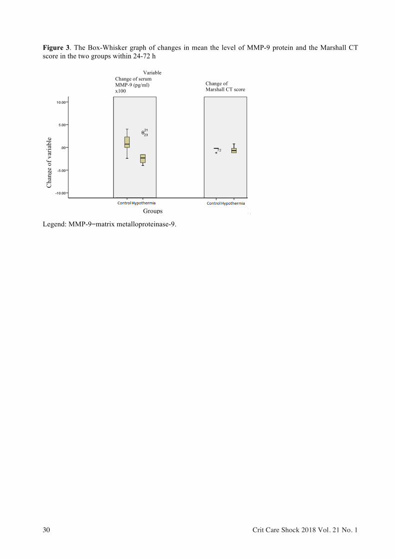
Figure 3. The Box-Whisker graph of changes in mean the level of MMP-9 protein and the Marshall CT score in the two groups within 24-72 h
Legend: MMP-9=matrix metalloproteinase-9.
30 Crit Care Shock 2018 Vol. 21 No. 1
Cha
nge
of v
aria
ble
Groups
Variable Change of serum MMP-9 (pg/ml) x100
Change of Marshall CT score

1. Bruns J Jr, Hauser WA. The Epidemiology of traumatic brain injury: A review. Epilepsia 2003;44:2-10.
2. Sadaka F, Patel D, Lakshmanan R. The FOUR score predicts outcome in patients after trau-matic brain injury. Neurocrit Care 2012;16:95-101.
3. Riyadina W, Sirait AM, Tuminah S, Suharyan-to FX, Nantabah Z. Cedera. Jakarta: Badan Penelitian dan Pengembangan Kesehatan Ke-menterian Kesehatan RI; 2013 Dec 1. 268 p.
4. Stein DM, Feather CB, Napolitano LM. Trau-matic brain injury advances. Crit Care Clin 2017;33:1-13.
5. Stenberg M, Koskinen LD, Jonasson P, Levi R, Stålnacke BM. Computed tomography and clinical outcome in patients with severe trau-matic brain injury. Brain Inj 2017;31:351-8.
6. Marshall LF, Marshall SB, Klauber MR, van Berkum Clark M, Eisenberg HM, Jane JA, et al. A new classification of head injury based on computerized tomography. J Neurosurg 1991;75:S14-20.
7. Zhu GW, Wang F, Liu WG. Classification and prediction of outcome in traumatic brain injury based on computed tomographic imaging. J Int Med Res 2009;37:983-95.
8. Loane DJ, Faden AI. Neuroprotection for traumatic brain injury: translational challenges and emerging therapeutic strategies. Trends Pharmacol Sci 2010;31:596-604.
9. Jia F, Pan YH, Mao Q, Liang YM, Jiang JY. Matrix metalloproteinase-9 expression and protein levels after fluid percussion injury in rats: The effect of injury severity and brain temperature. J Neurotrauma 2010;27:1059-68.
10. Copin JC, Rebetez MM, Turck N, Robin X, Sanchez JC, Schaller K, et al. Matrix metallo-proteinase 9 and cellular fibronectin plasma concentrations are predictors of the composite endpoint of length of stay and death in the in-tensive care unit after severe traumatic brain injury. Scand J Trauma Resusc Emerg Med 2012;20:83.
11. Suehiro E, Fujisawa H, Akimura T, Ishihara H, Kajiwara K, Kato S, et al. Increased matrix metalloproteinase-9 in blood in association with activation of interleukin-6 after traumatic brain injury: influence of hypothermic therapy. J Neurotrauma 2004;21:1706-11.
12. Yokobori S, Yokota H. Targeted temperature management in traumatic brain injury. J Inten-sive Care 2016;4:28.
Crit Care Shock 2018 Vol. 21 No. 1 31
13. Haji S, Ling GSF. Therapeutic hypothermia for traumatic brain injury and spinal cord inju-ry. In: Ecklund JM, Moores LE, editors. Neu-rotrauma management for severely injured polytrauma patient. Switzerland: Springer In-ternational Publishing AG; 2017. P. 247-52.
14. Pardamean DT, Prasetyo E, Oley M. Thera-peutic mild hypothermia towards blood lactate levels and Glasgow Coma Score in severe traumatic brain injury. Bali Med J 2015;4:82-5.
15. Mori T, Wang X, Aoki T, Lo EH. Downregu-lation of matrix metalloproteinase-9 and atten-uation of edema via inhibition of ERK mitogen activated protein kinase in traumatic brain in-jury. J Neurotrauma 2002;19:1411-9.
16. Schmitt KR,Diestel A,Lehnardt S, Schwart-lander R, Lange PE, Berger F, et al. Hypo-thermia suppresses inflammation via ERK sig-naling pathway in stimulated microglial cells. J Neuroimmunol 2007;189:7-16.
17. Diestel A, Roessler J, Berger F, Schmitt Kristus. Hypothermia downregulates inflam-mation but enhances IL-6 secretion by stimu-lated endothelial cells. Cryobiology 2008;57: 216-22.
18. Lee JE, Yoon YJ, Moseley ME, Yenari MA. Reduction in levels of matrix metalloprotein-ases and increased expression of tissue inhibi-tor of metalloproteinase-2 in response to mild hypothermia therapy in experimental stroke. J Neurosurg 2005;103:289-97.
19. Prasetyo E, Islam AA, Hatta M, Widodo D, Pattelongi I. Downregulation of MMP-9 level and GCS score improvement in severe trau-matic brain injury due to the mild hypothermia therapy. Am J Med Biol Res 2017;5:18-22.
20. Edlow BL, Wu O. Advanced neuroimaging in traumatic brain injury. Semin Neurol 2012;32: 374-400.
21. Zhang J, Puvenna V, Janigro D. Biomarkers of traumatic brain injury and their relationship to pathology. In: Laskowitz D, Grant G, editors. Translational research in traumatic brain inju-ry. New York: CRC Press; 2016. P. 263-76.
22. Lipper MH, Kishore PR, Enas GG, Domingues da Silva AA, Choi SC, Becker DP. Computed tomography in the prediction of outcome in head injury. AJR Am J Roentgenol 1985;144: 483-6.
23. Muakkassa FF, Marley RA, Paranjape C, Horattas E, Salvator A, Muakkassa K. Predic-tors of new findings on repeat head CT scan in .
References

blunt trauma patients with an initially negative head CT scan. J Am Coll Surg 2012;214:965-72.
24. Toyama Y, Kobayashi T, Nishiyama Y, Satoh K, Ohkawa M, Seki K. CT for acute stage of closed head injury. Radiat Med 2005;23:309-16.
25. Andresen M, Gazmuri JT, Marín A, Regueira .
32 Crit Care Shock 2018 Vol. 21 No. 1
T, Rovegno M. Therapeutic hypothermia for acute brain injuries. Scan J Trauma Resusc Emerg Med 2015;23:42.
26. Peterson K, Carson S, Carney N. Hypothermia treatment for traumatic brain injury: A system-atic review and meta-analysis. J Neurotrauma 2008;25:62-71.