Embed Size (px)
Citation preview

© 2015 DAR Publishers ⁄ The University of Jordan. All Rights Reserved.
The Effect of Local Injection of Mesenchymal Stem Cells and IL-10 with Platelet Lysate on Fracture
Healing in a Rat Model
Khaldoon Bashaireh 1*, Nizar M. Abuharfeil 2, Ziad Oudat 1, Wael M. Hananeh 3
Abstract Objective: This study investigates the effect of mesenchymal stem cells (MSC), an immune-privileged cell source for tissue repair, Interluken (IL-10), an immune inhibitory factor, and platelet lysate (PL) on the fracture healing process. Methods: Forty adult male Sprague-Dawley rats were anesthetized, and left tibia mid shaft fractures were created. The rats were randomly assigned to 5 groups and received injections with saline ( A); MSC and PL (B); IL-10 and PL (C); PL (D); and MSC, PL, and IL-10 (E). Lateral radiographs of four randomly selected rats in each group were taken at different days post-fracture. Four rats from each group were sacrificed at day twenty one post-fracture, with left tibiae harvested for histological analysis. Results: At 7 days post-fracture, the highest level of callus formation and bone healing was observed in group E (treated with MSC, PL, and IL-10) followed by groups B (treated with MSC and PL). The same results were obtained at days 14, 21 and 28 post-fracture. At 21days post-fracture, histological analysis revealed that the callus of fractured tibiae in group A consisted mostly of chondrocytes, while in groups C and D, extensive remodeling with a limited amount of chondrocytes present at the fracture. Combination of MSC, PL, and IL-10was associated with the greatest fracture healing performance. After 3 weeks of treatment in group E and B, trabecular bone formation and cortex remodeling were observed. Conclusions: The use of MSC in combination with IL-10 and PL is a promising alternative treatment to promote fracture healing. Keywords: Bone regeneration, Mesenchymal stem cells, IL-10, Platelet lysate.
(J Med J 2015; Vol. 49 (4):227-239)
Received Accepted
July 22, 2015 Aug 20, 2015
Introduction
Bone is a unique physiological human
tissue. There are many stages of fracture
healing starting with hematoma and progressing to an acute inflammatory stage at the fracture site and then callus formation, ending with remodeling. Several factors are
1. Department of Orthopedic Surgery, King Abdullah University Hospital, Jordan University of Science and Technology, Irbid,Jordan.
2. Department of Applied Biological Sciences, Jordan University of Science and Technology, Irbid, Jordan. 3. Department of Pathology and Public Health, Veterinary Medicine, Jordan University of Science and Technology, Irbid, Jordan.
* Correspondence should be addressed to: Khaldoon Bashaireh. E-mail: [email protected] P.O. Box 3030, 22110 Irbid, Jordan.

The Effect of Local … Khaldoon Bashaireh et al.
J Med J 2015; December: Vol. 49 (4) http:⁄⁄ journals.ju.edu.jo⁄jmj 228
known to have a positive effect on the bone healing process. Platelets and platelet derivatives has acquired clinical relevance as a means of accelerating bone healing. Platelet beneficial effect is attributed to the release of growth factors and other bioactive substances able to wound and bone healing.
The process of bone healing is finely orchestrated by the action of locally released growth factors and cytokines at the site of fracture. Studies have documented the effect of several growth factors including PDGF, TGF-β, IGF, and b-FGF as well other hormones and cytokines on bone regeneration both in vitro and in vivo(1,2,3,4,5,6).
All of these growth factors and others are present at high concentrations in platelets. Platelet rich plasma (PRP) is defined as “the volume of plasma fraction of autologous blood having a platelet concentration above baseline”(7). PRP has a wide range of applications, in particular for the treatment of muscular and skeletal injures(8). Gandhi et al. demonstrated that PRP has the potential to serve as a concentrated autologous source of growth factors through local percutaneous administration of PRP at the site of fracture in a diabetic rat model of femur fracture(9). Animal fracture models in both normal and osteoporotic rats treated with PRP reported increased callus to cortex width ratio and higher trabecular bone mass formation compared to control groups, indicating considerable enhanced healing over controls(3,10). The presence of PRP not only accelerated fracture healing but also regeneration in bone defects in an ovariectomized osteoporotic rat model(11). Early in vitro experiments assessing the effect of platelet lysate (PL) on bone marrow mesenchymal stem cell differentiation into osteogenic lineage revealed that PL increased
cellular proliferation and expansion at the expense of cellular differentiation(12). Subsequently, PL has been used as a serum substitute(13), and use of PL from the beginning of culture and during induction of the differentiation program resulted in superior osteoblastic differentiation and increased expression of osteogenic marker genes(14). In addition, clinical trials have been conducted confirming the efficacy of administering PRP on alveolar bone regeneration following removal of the third molar(15), intra-articular calcaneal fracture healing in combination with allograft(16), and relieving early knee pain associated with osteoarthritis(17).
Earlier data indicated that treating mesenchymal stem cells with IL-10 suppressed alkaline phosphatase (ALP), osteocalcin, and matrix mineralization formation, and this inhibitory effect was mediated through the suppression of TGF-β(18,19,20). However, IL-10 exerts this inhibitory function at the stage of stem cell specification to osteoblasts and is incapable of this effect in early committed osteoblastic cultures already expressing ALP, osteocalcin, and collagen type1(20).
Two recent lines of evidence have suggested that IL-10 plays a role in enhancing bone growth and healing. Toben et al. demonstrated that RAG-/- mice have enhanced bone growth, endochondral ossification, and remodeling. The cytokine profile in RAG-/- mice revealed decreased expression of pro-inflammatory cytokines including TNF and INF-γ and T-cell specific cytokines IL-2 and IL-4. By contrast, expression of the anti-inflammatory cytokine IL-10 was significantly higher(21). Thus, it is highly possible that the shift in cytokine profile from pro-inflammatory to anti-inflammatory plays a crucial role in enhancing fracture healing, which highlights IL-10 as a central player in

The Effect of Local … Khaldoon Bashaireh et al.
J Med J 2015; December: Vol. 49 (4) http:⁄⁄ journals.ju.edu.jo⁄jmj 229
accelerating fracture healing due to its immunomodulatory function. Iannone et al. have shown that treating osteoarthritis osteoblast cultures with extra-corporeal shock waves (ESW), which enhance osteoblast regeneration, enhanced the expression of osteoblastic markers such as CD29/β1 and CD105. In addition, along with the increase in cell surface osteogenic markers, IL-10 expression was significantly increased compared with control healthy osteoblast cultures(22).
Mesenchymal stem cells (MSC) have therapeutic potential to reduce the time of healing in patients with fractures(23). It was demonstrated that MSC transplant was shown to induce a biomechanical improvement in the healing process associated with increases in callus volume and cartilaginous and bone content(24). Intravenously administered MSC were capable of specifically homing to the fracture site and normalizing biomechanical, histological, and microcomputed tomography parameters of healing in animals(25).
The use of different local factors on the fracture to accelerate healing is attractive and could be implemented with adequate clinical evidence from clinical studies. In this study we aimed to analyze the effect of local injections of different combinations of MSC, IL-10, and PL on the fracture healing process.
Materials and Methods Stem cells isolation and differentiation
Animal experiments were approved by Jordan University of Science and Technology Institutional Animal Care and Use Committee (IACUC).
Stem cells were isolated according to the method of Lennon & Caplan(26). Briefly, four Sprague–Dawley rats were sacrificed, both
tibial and femur bones were harvested, the marrow was flushed with 5ml basal α-minimal essential medium (MEM) (Hyclone, USA), and the cell suspension was centrifuged at 1000 rpm for 5 minutes. The cell pellet was re-suspended in α-MEM supplemented with 10% fetal bovine serum (FBS) and 1% streptomycin, penicillin, and amphotericin B (Hyclone, USA) and seeded at a cell density of 5000 cells/cm2. Cultures were maintained in a 5% CO2 humidified atmosphere at 37ºC. Cells were allowed to adhere for 3 days; the first media change took place on the third day. Upon reaching 80% cell confluence, cells were passaged with 0.25% trypsin containing 1mM EDTA (Hyclone, US). Cells at passage three were used for downstream experiments.
Stem cells were characterized through verification of their multi-potent capacity. Cells were differentiated towards the osteogenic cells. MSC were seeded at a density of 5000 cells/ml, and after reaching 100% cell confluence, culture medium was switched to osteogenic differentiation medium (Hyclone, US) supplemented with 10% FBS and 1% streptomycin, penicillin, and amphotericin B. After 2 weeks, cells were washed with PBS, fixed with 10% formalin, and then washed twice with water. Alizarin red S staining was performer according to the manufacturer’s protocol. Stained cells were observed under a phase-contrast microscope. After1 week of osteogenic induction, cells were trypsinized and prepared for injection. Rat platelet lysate preparation
PRP was prepared according to the method of Ghandi et al.(9). Briefly, whole blood was collected via cardiac puncture from 15 adult Sprague-Dawley rats in sodium citrate-treated tubes and centrifuged at 1800 rpm for 10 minutes. Plasma and buffy coats were pooled

The Effect of Local … Khaldoon Bashaireh et al.
J Med J 2015; December: Vol. 49 (4) http:⁄⁄ journals.ju.edu.jo⁄jmj 230
in a 15-ml centrifuge tube and centrifuged at 3600 rpm for 10 minutes. A platelet pellet was formed, and the supernatant consisted of platelet poor plasma (PPP). At least half the PPP was discarded, and the rest was used to resuspend the platelet pellet to form PRP. PRP was subjected to 3-5 freeze-thaw cycles to break up platelets and release their contents, with freezing performed at -20ºC and thawing in a 37ºC water bath, followed by centrifugation at 4300 rpm for 15 minutes to remove platelet fragments. The supernatant contained platelet lysate. Fracture model and treatment groups
Forty Sprague-Dawley rats (average weight 110g) were anesthetized with an intraperitoneal injection of a mixture of ketamine hydrochloride (60 mg/kg body weight) and 2% xylazine (10 mg/kg body weight). The fracture site was labeled with a marker according to anatomical landmarks and left tibial midshaft fractures were created with a special device as described by Schmidmaier et al.(13). The fractured tibia was stabilized with intramedullary 0.45 mm Kirschner wire.
Animals were divided into five groups: group A treated with 0.3 ml PBS; group B treated with MSC (5105 cells suspended in 0.15 ml PBS) and 0.15ml PL; group C treated with 0.15 ml PL mixed with 75 µl IL-10 (10 ng/ml) and 75 µl PBS; group D treated with 0.15 ml PL and 0.15 ml PBS; and group E treated with MSC (5105 cells suspended in 0.15 ml PL) followed by 75 µl IL-10 (10 ng/ml) and 75 µl PBS). Local injections at the fracture site were performed on day 0, 3, and 5 after induced fracture. Radiographic analysis
Lateral radiographs of four randomly chosen rats in each group were taken at days 0,
7, 14, 21, and 28 post-fracture. Tibial radiographs were taken at 40 kV and 4 mA.X-ray meter and films were processed and developed using a Kodak RP X-omat Processor. Callus diameter was calculated at time of radiograph. Callus and tibia diameters were measured using Auto CAD software. Callus index, defined as the maximum diameter of the callus divided by the diameter of the bone, was calculated to stage the fracture healing. Histological analysis
Four rats from each group were sacrificed at day 21 post-fracture, and the left tibia was harvested for histological analysis. Adequate biopsies were taken from fracture site and fixed immediately in 10% formalin solution for 24 hours. The tissues were then decalcified in 10% EDTA (pH 7.4) for 7 days, processed routinely, and stained with H&E and Safranin O stains. Examination was performed by a pathologist with no prior knowledge of the treatment groups. A modified Allen et al. grading system was adopted for histopathological union assessment(27). The grade of histopathological change ranged from 0 to V as follows: 0, no healing or only fibrous tissue union; I, cartilage union with presence of fibrous tissues; II, only cartilage union; III, cartilage union with some bony ossification; IV, approximately equal amount of trabecular bone and cartilage union; and V, complete bony union
Statistical analysis
A two-tailed t-test was used to compare groups.
Results Stem cell isolation and differentiation
Stem cells cultures were initiated from rat
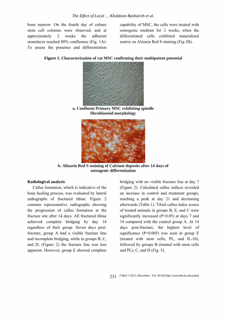
bone mstem ceapproximmonolayTo asse
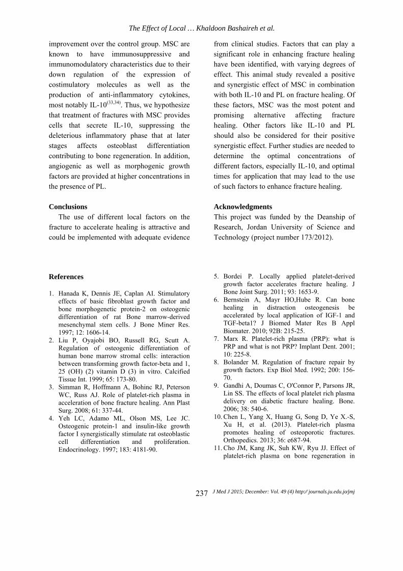
Radiolo
Callubone heradiogracontainsthe profracture achievedregardlefracture,and incoand D, apparent
marrow. On tell colonies mately 2 yer reached 8ess the pre
Figure 1.
b.
ogical analysus formationaling proces
aphs of fras representatgression of site after 14
d completeess of their , group A homplete bridg(Figure 2) t. However,
The Ef
the fourth dwere obseweeks t
80% conflueesence and
Characteriz
a. Co
Alizarin Re
sis , which is ins, was evaluactured tibitive radiogr
f callus form4 days. All fe bridging
group. Sevhad a visibleging, while ithe fracture group E sho
Effect of Loca
day of cultuerved, and the adhereence (Fig. 1Adifferentiati
zation of rat
nfluent Primfibrobl
ed S stainingosteoge
ndicative of tuated by laterae. Figure
raphs showimation at tfractured tibi
by day ven days poe fracture liin groups B,
line was leowed comple
al … Khaldoo
J M231
ure at
ent A). ion
caosdim
t MSC confi
mary MSC elastoid morp
g of Calciumenic differen
the ral
2 ing the iae 14
ost-ine C,
ess ete
br(Fanreafofsi14dasi(trfoan
on Bashaireh
Med J 2015; Dece
apability of Msteogenic mifferentiated
matrix on Aliz
irming their
exhibiting spphology
m deposits antiation
ridging with Figure 2). Cn increase ineaching a pfterwards (Taf treated animgnificantly i4 compared ays post-fragnificance (reated with
ollowed by gnd PL), C, an
h et al.
ember: Vol. 49 (4)
MSC, the cemedium for
cells exhzarin Red S s
r multipoten
pindle
fter 14 days
no visible falculated can control an
peak at day able 1). Tibiamals in grouincreased (P<with the conacture, the (P<0.005) w
h stem cellsgroups B (trend D (Fig. 3)
4) http:⁄⁄ journals.j
ells were trea2 weeks, whibited minstaining (Fig
nt potential
s of
fracture line allus indices nd treatment
21 and deal callus inde
ups B, E, and<0.05) at dantrol group A
highest lwas seen in s, PL, andeated with st).
ju.edu.jo⁄jmj
ated with when the neralized
g.1B).
at day 7 revealed
t groups, ecreasing ex scores d C were ays 7 and A. At 14 level of group E
d IL-10), tem cells

The Effect of Local … Khaldoon Bashaireh et al.
J Med J 2015; December: Vol. 49 (4) http:⁄⁄ journals.ju.edu.jo⁄jmj 232
Figure 2. Representative radiographs of fixed fractures at day 14 post-fracture
a. Control group treated with PBS
b. MSC and PL treatment group
c. PL and IL-10 treatment group
d. PL-only treatment group 2D

Histolog
Safrarevealedgroup A21 days and D, amount and calcdays po
Fi
gical analysianin O/methd that the caA consisted m
post-fractureextensive rof chondroccified tissue
ost-fracture.
The Ef
e
igure 3. The gr
is hylene light allus of fracmostly of che (Figure 4Aemodeling w
cytes present e site was oThe combin
Effect of Loca
e. MSC, IL-1
line graph roups A, B,
green stainitured tibiae hondrocytes
A). In Groupswith a limitat the fractu
observed at nation of ste
al … Khaldoo
J M233
0, and PL tre
depicts the mC, D, and E
ing in at
s C ted ure 21 em
ceentrfoobrecopr(F
on Bashaireh
Med J 2015; Dece
eatment grou
mean CallusE over 28 da
ells, PL, andnhancing fraeatment in g
ormation anbserved, suesorptive ostontrast, grouresence of wFig.4B and 4
h et al.
ember: Vol. 49 (4)
up
s Index (CI)ays
d IL-10 had tacture healingroup E andnd cortex
urrounded bteoclasts (Fiups C and woven bone C).
4) http:⁄⁄ journals.j
) in
the greatest ng. After 2d B, trabecu
remodelinby osteoblaig. 4D and D demonstrand cartilag
ju.edu.jo⁄jmj
effect on 1days of ular bone ng were asts and 4E). By
rated the e islands

The Effect of Local … Khaldoon Bashaireh et al.
J Med J 2015; December: Vol. 49 (4) http:⁄⁄ journals.ju.edu.jo⁄jmj 234
Figure 4. Safranin O staining at 21 days post-fracture
a. Score II, only cartilage union
b. Score III, artilage union with ossification
c. Score IV, approximately equal amounts of trabecular bone and cartilage union

The Effect of Local … Khaldoon Bashaireh et al.
J Med J 2015; December: Vol. 49 (4) http:⁄⁄ journals.ju.edu.jo⁄jmj 235
d. Score V, complete bony union
e. Score V, complete bony union
Discussion
Rat MSC, PRP, and the lack of adaptive immune response in association with the upregulation of IL-10 have been implicated in the acceleration of bone healing. In this study, the interplay of these factors and their combined effect on the process of fracture healing in a rat model were analyzed. The aims of this study were to generate a clinically relevant model of fracture and to evaluate the functional contributions of these factors to
fracture repair after local injection. Currently, use of stem cells has been a
subject of special interest to improve the fracture healing process. MSC can differentiate into several different cell types. The ability of MSC to differentiate into bone tissue, which enhances fracture healing and bone regeneration, has been established through in vitro and in vivo studies.
Inflammation is the first reaction elicited from the immune system immediately after a

The Effect of Local … Khaldoon Bashaireh et al.
J Med J 2015; December: Vol. 49 (4) http:⁄⁄ journals.ju.edu.jo⁄jmj 236
fracture occurs in order to combat pathogenic microorganisms when the fracture is associated with an infection. However, in cases where there is no simultaneous infection, this process is dispensable and in fact has been shown to be depressing bone regeneration(28). RAG knockout mice exhibit enhanced fracture healing and bone regeneration that is coupled with lower pro-inflammatory cytokine secretion and higher anti-inflammatory cytokine production, most importantly of IL-10(21). Several lines of evidence indicate that IL-10 is a positive regulator of bone regeneration. IL-10 influences the expression of genes that regulate osteoclastogenesis, thus exerting control over the process of bone resorption(29,30). In addition, IL-10-targeted gene knockout mice demonstrate reduced mechanical strength of the bone, osteopenia, and defects in bone remodeling(31). However, the role of IL-10 in the process of bone regeneration remains controversial, as in vitro studies conducted on bone marrow MSC revealed reduced expression of critical osteogenic proteins including ALP, collagen type I, and osteocalcin(18-20).
Recently, PRP has been shown to have an antibacterial effect: local application of PRP into the sternal wound in patients undergoing heart surgery reduced the incidence of both deep and superficial sternal wound infections(32). On the other hand, the effect of PL on bone regeneration and the osteogenic differentiation of MSC is well established, and PRP is widely used in the treatment of muscular and skeletal injuries(8).
In this study, we found that using either PL alone or combined with IL-10 or MSC led to increased evidence of healing compared to the control group, with MSC and PL demonstrating the highest callus indices compared to other groups. MSC injected in combination with IL-10 and PL showed the highest callus-to-cortex index, an indication of enhanced fracture healing, compared to controls and all other groups. This confirms the synergistic effect exerted by MSC and IL-10 on the process of bone regeneration. This effect is either due to the anti-inflammatory properties of IL-10 or its effect on other processes such as bone resorption and callus mineralization.
Table 1. Callus indices of tibia fracture healing
Day 0 Day 7 Day 14 Day 21 Day 28
Group A 1.00 1.15±0.23 1.32±0.29 1.45±0.20 1.35±0.21
Group B 1.00 1.21±0.26* 1.65±0.22* 1.62±0.20* 1.41±0.23*
Group C 1.00 1.16±0.28* 1.44±0.21* 1.51±0.26* 1.36±0.25*
Group D 1.00 1.18±0.24 1.42±0.28* 1.53±0.24 1.34±0.21
Group E 1.00 1.24±0.21* 1.68±0.24* 1.68±0.24* 1.45±0.22*
Date expressed as mean ± SD, n=4 per group Group A: placebo, Group B: MSC and PL, Group C: IL-10 and PL, Group D: PL, Group E: MSC and IL-10 *P<0.05 vs. group A N.S.: not significant
The obtained results indicate a synergistic
complimentary role of MSC and IL-10. Treatment of fractured tibia with stem cells in PL and IL-10 resulted in the most significant
The Effect of Local … Khaldoon Bashaireh et al.
J Med J 2015; December: Vol. 49 (4) http:⁄⁄ journals.ju.edu.jo⁄jmj 237
improvement over the control group. MSC are known to have immunosuppressive and immunomodulatory characteristics due to their down regulation of the expression of costimulatory molecules as well as the production of anti-inflammatory cytokines, most notably IL-10(33,34). Thus, we hypothesize that treatment of fractures with MSC provides cells that secrete IL-10, suppressing the deleterious inflammatory phase that at later stages affects osteoblast differentiation contributing to bone regeneration. In addition, angiogenic as well as morphogenic growth factors are provided at higher concentrations in the presence of PL.
Conclusions
The use of different local factors on the fracture to accelerate healing is attractive and could be implemented with adequate evidence
from clinical studies. Factors that can play a significant role in enhancing fracture healing have been identified, with varying degrees of effect. This animal study revealed a positive and synergistic effect of MSC in combination with both IL-10 and PL on fracture healing. Of these factors, MSC was the most potent and promising alternative affecting fracture healing. Other factors like IL-10 and PL should also be considered for their positive synergistic effect. Further studies are needed to determine the optimal concentrations of different factors, especially IL-10, and optimal times for application that may lead to the use of such factors to enhance fracture healing.
Acknowledgments This project was funded by the Deanship of Research, Jordan University of Science and Technology (project number 173/2012).
References 1. Hanada K, Dennis JE, Caplan AI. Stimulatory
effects of basic fibroblast growth factor and bone morphogenetic protein-2 on osteogenic differentiation of rat Bone marrow-derived mesenchymal stem cells. J Bone Miner Res. 1997; 12: 1606-14.
2. Liu P, Oyajobi BO, Russell RG, Scutt A. Regulation of osteogenic differentiation of human bone marrow stromal cells: interaction between transforming growth factor-beta and 1, 25 (OH) (2) vitamin D (3) in vitro. Calcified Tissue Int. 1999; 65: 173-80.
3. Simman R, Hoffmann A, Bohinc RJ, Peterson WC, Russ AJ. Role of platelet-rich plasma in acceleration of bone fracture healing. Ann Plast Surg. 2008; 61: 337-44.
4. Yeh LC, Adamo ML, Olson MS, Lee JC. Osteogenic protein-1 and insulin-like growth factor I synergistically stimulate rat osteoblastic cell differentiation and proliferation. Endocrinology. 1997; 183: 4181-90.
5. Bordei P. Locally applied platelet-derived growth factor accelerates fracture healing. J Bone Joint Surg. 2011; 93: 1653-9.
6. Bernstein A, Mayr HO,Hube R. Can bone healing in distraction osteogenesis be accelerated by local application of IGF-1 and TGF-beta1? J Biomed Mater Res B Appl Biomater. 2010; 92B: 215-25.
7. Marx R. Platelet-rich plasma (PRP): what is PRP and what is not PRP? Implant Dent. 2001; 10: 225-8.
8. Bolander M. Regulation of fracture repair by growth factors. Exp Biol Med. 1992; 200: 156-70.
9. Gandhi A, Doumas C, O'Connor P, Parsons JR, Lin SS. The effects of local platelet rich plasma delivery on diabetic fracture healing. Bone. 2006; 38: 540-6.
10. Chen L, Yang X, Huang G, Song D, Ye X.-S, Xu H, et al. (2013). Platelet-rich plasma promotes healing of osteoporotic fractures. Orthopedics. 2013; 36: e687-94.
11. Cho JM, Kang JK, Suh KW, Ryu JJ. Effect of platelet-rich plasma on bone regeneration in

The Effect of Local … Khaldoon Bashaireh et al.
J Med J 2015; December: Vol. 49 (4) http:⁄⁄ journals.ju.edu.jo⁄jmj 238
ovariectomized osteoporotic rats. J Korean Acad Prosthodont. 2010; 48: 16-27.
12. Arpornmaeklong P, Kochel M, Depprich R, Kubler N, Wurzler K. Influence of platelet-rich plasma (PRP) on osteogenic differentiation of rat bone marrow stromal cells. An in vitro study. Int J Oral Maxillofac Surg. 2004; 33 (1): 60-70.
13. Schmidmaier G, Wildemann B, Heeger J, Gäbelein T, Flyvbjerg A, Bail H, et al. Improvement of fracture healing by systemic administration of growth hormone and local application of insulin-like growth factor-1 and transforming growth factor-beta1. Bone. 2002; 31: 165-72.
14. Chevallier N, Anagnostou F, Zilber S, Bodivit G, Maurin S, Barrault A, et al. Osteoblastic differentiation of human mesenchymal stem cells with platelet lysate. Biomaterials. 2010; 31: 270-8.
15. AntonelloGde M, Torres do Couto R, Giongo C, Corrêa M, Chagas Júnior O, LemesCH. Evaluation of the effects of the use of platelet-rich plasma (PRP) on alveolar bone repair following extraction of impacted third molars: prospective study. J Craniomaxillofac Surg. 2013; 41: e70-5.
16. Wei L, Lei G, Sheng P, Gao S, Xu M, Jiang W, et al. (2012). Efficacy of platelet-rich plasma combined with allograft bone in the management of displaced intra-articular calcaneal fractures: a prospective cohort study. J Orthop Res. 2012; 30: 1570-6.
17. Patel S, Dhillon M, Aggarwal S, Marwaha N, Jain A. Treatment with platelet-rich plasma is more effective than placebo for knee osteoarthritis: a prospective, double-blind, randomized trial. Am J Sports Med. 2013; 41: 356-64.
18. Ivashkiv LB, Zhao B, Park-Min K,Takami M. Feedback inhibition of osteoclastogenesis during inflammation by IL-10, M-CSF receptor shedding, and induction of IRF8. Ann N Y Acad Sci. 2011; 1237: 88-94.
19. Van Vlasselaer P, Borremans B, Van Den Heuvel R, Van Gorp U, de Waal Malefyt R. Interleukin-10 inhibits the osteogenic activity of mouse bone marrow. Blood. 1993; 82: 2361-70.
20. Vlasselaer PV, Borremans BU, van Gorp JR, Waal-Malefyt RD. Interleukin 10 inhibits transforming growth factor-beta (TGF-beta) synthesis required for osteogenic commitment of mouse bone marrow cells. J Cell Biol. 1994; 124: 569-77.
21. Toben D, Schroeder I, Khassawna TE, Mehta M, Hoffmann JE, Frisch JT, et al. Fracture healing is accelerated in the absence of the adaptive immune system. J Bone Miner Res. 2011; 26: 113-24.
22. Iannone F, Moretti B, Notarnicola A, Moretti L, Patella S, Patella V, et al. Extracorporeal shock waves increase interleukin-10 expression by human osteoarthritic and healthy osteoblasts in vitro. Clin Exp Rheumatol. 2009; 27: 794-9.
23. Granero-Molto F, Weis JA, Longobardi L, et al. Role of mesenchymal stem cells in regenerative medicine: application to bone and cartilage repair. Expert OpinBiolTher. 2008; 8: 255-68.
24. .Granero-Moltó F, Weis JA, Miga MI, Landis B, Myers TJ, O'Rear L, et al. Regenerative effects of transplanted MSCs in fracture healing. Stem Cells. 2009; 27: 1887-98.
25. ObermeyerTS, Yonick D, Lauing K, Stock SR, Nauer R, Strotman P, et al.Mesenchymal stem cells facilitate fracture repair in an alcohol-induced impaired healing model. J Orthop Trauma. 2012; 26: 712-8.
26. Lennon DP, Caplan AI. Isolation of rat marrow-derived mesenchymal stem cells. Exp Hematol. 2006; 34: 1606-7.
27. Allen HL, Wase A, Bear WT. Indomethacin and aspirin: effect of nonsteroidal anti-inflammatory agents on the rate of fracture repair in the rat. ActaOrthop Scand. 1980; 51: 595-600.
28. Colburn N, Zaal K, Wang F, Tuan P. A role for gamma/delta T cells in a mouse model of fracture healing. Arthritis Rheum. 2009; 60: 1694-703.
29. Liu D, Yao S, Wise G. Effect of interleukin-10 on gene expression of osteoclastogenic regulatory molecules in the rat dental follicle. Eur J Oral Sci. 2006; 114: 42-9.
30. Sasaki H, Hou L,Belani A. IL-10, but not IL-4, suppresses infection-stimulated bone resorption in vivo. J Immunol. 2000; 165: 3626-30.
31. Dresner-Pollak R, Gelb N, Rachmilewitz D, Karmeli F,Weinreb M. Interleukin 10-deficient mice develop osteopenia, decreased bone formation, and mechanical fragility of long bones. Gastroenterology. 2004; 127: 792-801.
32. Serraino GF, Dominijanni A, Jiritano F, Rossi M, Cuda A, Caroleo S, et al. Platelet-rich plasma inside the sternotomy wound reduces the incidence of sternal wound infections. Int Wound J. 2013; doi: 10.1111/iwj.12087.
33. Sheng H, Wang Y, Jin Y, Zhang Q, Zhang Y, Wang L, et al. A critical role of IFN gamma in priming MSC-mediated suppression of T cell

The Effect of Local … Khaldoon Bashaireh et al.
J Med J 2015; December: Vol. 49 (4) http:⁄⁄ journals.ju.edu.jo⁄jmj 239
proliferation through up-regulation of B7-H1. Cell Res. 2008; 8: 846-57.
34. Klyushnenkova E, Mosca JD, Zernetkina V, Majumdar MK, Beggs KJ, Simonetti DW, et al.
T cell responses to allogeneic human mesenchymal stem cells: immunogenicity, tolerance, and suppression. J Biomed Sci. 2005; 12: 47-57.
ىعل 10لصفائح الدموية ومادة إنترلوكين واالحقن الموضعي للخلايا الجذعية تأثير شفاء عظمة الساق المكسورة عند الجراذين
3وائل حننة 1زياد عودات، 2،نزار أبو هرفيل 1،خلدون بشايرة
إربد، الأردن؛أمراض وجراحة العظام والمفاصل، مستشفى الملك المؤسس عبداالله الجامعي، قسم -1 العلوم والتكنولوجيا، إربد، الأردن؛قسم الأحياء التطبيقية، جامعة -2 .قسم الباثولوجيا والصحة العامة، الطب البيطري، جامعة العلوم والتكنولوجيا، إربد، الأردن -3
الملخصعلى شفاء 10فحص تأثير استخدام الحقن الموضعي للخلايا الجذعية والصفائح الدموية ومادة إنترلوكين إلىدف الدراسة :الهدف
.ينفي عظمة الساق عند الجراذلكسور ات عشوائية وحقن كل مجموعة خمس مجموعا إلىمن الجراذين بعد تخديرهم ومن ثم تم تقسيمهم 40تم كسر عظمة الساق عند :الطريقةمع محلول الصفائح إنترلوكين : الخلايا الجذعية مع محلول الصفائح الدموية، الثالثة: محلول ملحي طبيعي، الثانية: الأولىالمجموعة : كالآتي
تم عمل صورة شعاعية جانبية 10الخلايا الجذعية مع محلول الصفائح الدموية وانترلوكين : الخامسة محلول الصفائح الدموية،: الدموية،الرابعةمن عمل يوما 21لعظمة الساق وقت الكسر وعلى فترات مختلفة لمتابعة شفاء العظمة ثم تم اختيار أربعة جرذان من كل مجموعة بعد
.عةالكسر ودراسة التغير النسيجي في منطقة الكسر عند كل مجمو الخلايا الجذعية ( من تاريخ الكسر في المجموعة الخامسة أيامأظهرت النتائج وجود تكوين تكلسي واضح كدلالة للشفاء بعد مرور سبعة :النتائج
نفس . الأخرىمقارنة مع المجاميع ) الخلايا الجذعية مع محلول الصفائح الدموية(ومن ثم المجموع الثانية 10مع محلول الصفائح الدموية وانتيرلوكين أظهرت نتائج فحص النسيج وجود تغيرات تدل على حدوث الأمركذلك .عوثلاثة أسابيع وأربعة أسابي النتائج ظهرت بعد مرور أسبوعين
.الأخرىللمجموعة الخامسة مقارنة بالمجاميع أفضليةوعتين الخامسة والثانية مع التئام للكسر وتكلّس منطقة الكسر في المجم .عظميةلعلاج الكسور القد يكون خيار مستقبلي 10لوكين استخدام الخلايا الجذعية مع محلول الصفائح الدموية وانتير :الاستنتاجات
.الصفائح الدموية، 10شفاء الكسور، الخلايا الجذعية، إنترلوكين :الكلمات الدالة