Embed Size (px)
Citation preview

Ann. N.Y. Acad. Sci. ISSN 0077-8923
ANNALS OF THE NEW YORK ACADEMY OF SCIENCESIssue: Glycobiology of the Immune Response
The effect of galectins on leukocyte traffickingin inflammation: sweet or sour?
Dianne Cooper, Asif J. Iqbal, Beatrice R. Gittens, Carmela Cervone, and Mauro PerrettiWilliam Harvey Research Institute, Barts and The London School of Medicine, Queen Mary University of London, London,United Kingdom
Address for correspondence: Dianne Cooper or Mauro Perretti, William Harvey Research Institute, Barts and The LondonSchool of Medicine and Dentistry, Queen Mary University of London, Charterhouse Square, London EC1M 6BQ,United Kingdom. [email protected], [email protected]
The trafficking of leukocytes from the blood stream to the surrounding tissue is a fundamental feature of aninflammatory response. Although many of the adhesion molecules and chemokines that direct leukocyte traffickinghave been identified, there is still much to be discovered, particularly with regard to the persistence of leukocyteinfiltrates in chronic inflammation. Elucidating the molecular mechanisms involved in this process is critical tounderstanding and treating inflammatory pathologies. Recent studies have identified members of the galectin familyas immunoregulatory proteins. Included among the actions of galectins are modulatory effects, both positive andnegative, on leukocyte recruitment. The focus of this review is to summarize current knowledge on the role of galectinsin leukocyte trafficking during inflammation. A better understanding of the function of this family of endogenouslectins will open new avenues for innovative drug discovery.
Keywords: galectin; leukocyte; inflammation; adhesion molecule
Introduction
Inflammation is elicited as a response to tissue in-jury or infection. It is generally a protective responseof the host that serves to maintain tissue homeosta-sis. The symptoms of inflammation—pain, fever,redness, swelling, and, in chronic cases, loss offunction—occur as a result of a complex sequence ofevents that take place at the site of inflammation andsystemically. The recruitment of leukocytes from theblood stream to the surrounding tissue is the hall-mark of an inflammatory response. The first cells totraverse the endothelial barrier are neutrophils, thefoot soldiers of the inflammatory response. Thesecells are armed with powerful enzymes and oxidantswith antimicrobial properties. This is followed by awave of mononuclear leukocytes that, depending onthe initiating stimulus, might be T lymphocytes ormonocytes. In acute inflammation, the inflamma-tory response resolves by the coordinated release ofpro-resolution mediators that terminate neutrophilrecruitment and promote the non-phlogisitic clear-ance of the spent neutrophils by tissue macrophages;
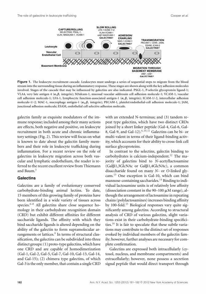
the end result being a return to tissue homeosta-sis.1,2 In some circumstances, restoration is notachieved, and the inflammation becomes chronicin nature. Chronic inflammation is characterizedby persistent leukocytic infiltrates that differ in na-ture depending on the initiating stimulus of theinflammation. As leukocyte trafficking is criticalto mounting an inflammatory response and per-sistence of chronic inflammation, the mechanismsby which leukocytes traverse the endothelium hasbeen the focus of intense research over the pasttwo decades. This has led to the identification ofmany of the molecular determinants involved in theparadigm that is the leukocyte recruitment cascade(Fig. 1). For excellent reviews on the mechanismsof leukocyte trafficking, the reader is directed toLey et al.3 and Luster et al.4
Although many of the adhesion molecules andchemokines that direct leukocyte trafficking havebeen identified, there is still much to be understood,particularly with regard to chronic inflammatorypathologies and the persistence of leukocytic infil-trates. Recent studies have identified members of the
doi: 10.1111/j.1749-6632.2011.06291.xAnn. N.Y. Acad. Sci. 1253 (2012) 181–192 c© 2012 New York Academy of Sciences. 181
The role of galectins in leukocyte trafficking Cooper et al.
Figure 1. The leukocyte recruitment cascade. Leukocytes must undergo a series of sequential steps to migrate from the bloodstream into the surrounding tissue during an inflammatory response. These stages are shown along with the key adhesion moleculesinvolved. Stages of the cascade that may be influenced by galectins are also indicated. PSGL-1, P-selectin glycoprotein ligand-1;VLA4, very late antigen 4 (α4β1 integrin); MAdcam-1, mucosal vascular addressin cell adhesion molecule-1; VCAM-1, vascularcell adhesion molecule-1; LFA-1, lymphocyte function-associated antigen-1 (αLβ2 integrin); ICAM-1/-2, intercellular adhesionmolecule-1/-2; MAC-1, macrophage antigen-1 (αMβ2 integrin); PECAM-1, platelet/endothelial-cell adhesion molecule-1; JAM,junctional adhesion molecule; ESAM, endothelial cell selective adhesion molecule.
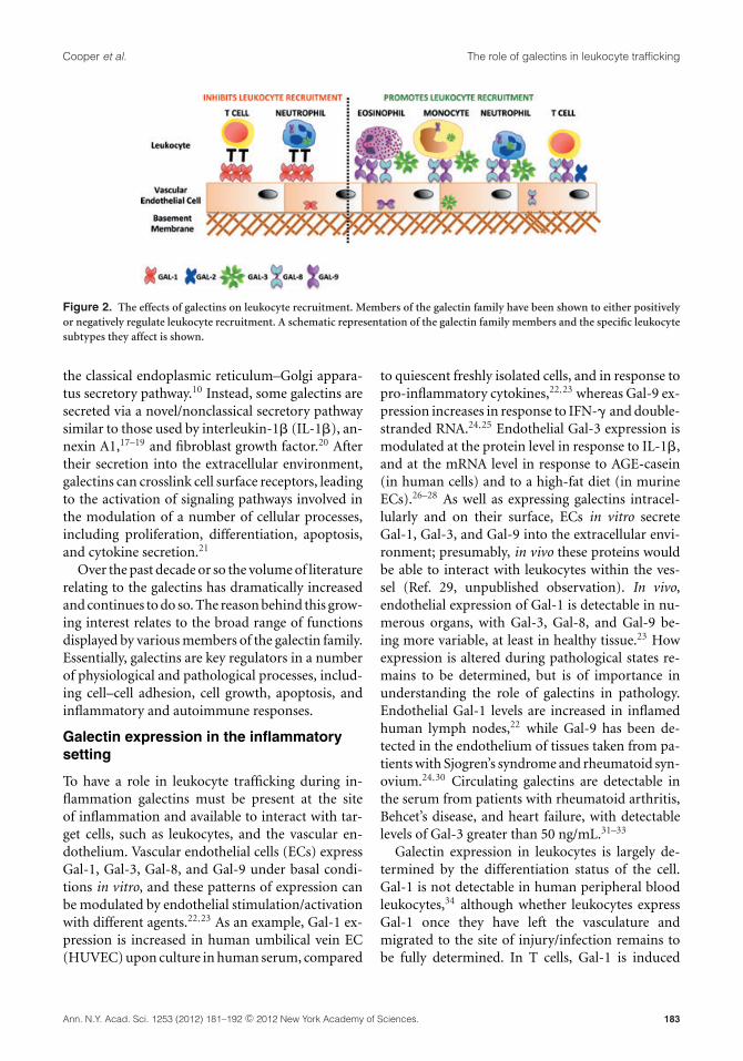
galectin family as exquisite modulators of the im-mune response; included among their many actionsare effects, both negative and positive, on leukocyterecruitment in both acute and chronic inflamma-tory settings (Fig. 2). This review will focus on whatis known to date about the galectin family mem-bers and their role in leukocyte trafficking duringinflammation. For a recent review on the role ofgalectins in leukocyte migration across both vas-cular and lymphatic endothelium, the reader is re-ferred to the recent excellent review from Thiemannand Baum.5
Galectins
Galectins are a family of evolutionary conservedcarbohydrate-binding animal lectins. To date,15 members of this growing family of proteins havebeen identified in a wide variety of tissues acrossspecies.6–8 All galectins share close sequence ho-mology in their carbohydrate recognition domain(CRD) but exhibit different affinities for differentsaccharide ligands. The affinity with which theybind saccharide ligands is altered depending on theability of the galectin to form supramolecular ar-rangements or lattices.9 In terms of structural clas-sification, the galectins can be subdivided into threedistinct groups: (1) proto-type galectins, which haveone CRD and are capable of homodimerization(Gal-1, Gal-2, Gal-5, Gal-7, Gal-10, Gal-13, Gal-14,and Gal-15); (2) chimera type galectins, of whichGal-3 is the only member, that contain a single CRD
with an extended N-terminus; and (3) tandem re-peat type galectins, which have two distinct CRDsjoined by a short linker peptide (Gal-4, Gal-6, Gal-8, Gal-9, and Gal-12).6,10,11 Galectins can be bi- ormulti-valent in terms of their ligand-binding activ-ity, which accounts for their ability to cross-link cellsurface glycoproteins.
In contrast to the selectins, galectin binding tocarbohydrates is calcium-independent.12 The ma-jority of galectins bind to N-acetyllactosamine(Gal�1,3GlcNAc or Gal�1,4GlcNAc), a commondisaccharide found on many N- or O-linked gly-cans.13 One exception is Gal-10, which can bindmannose-containing saccharides.14 Binding to indi-vidual lactosamine units is of relatively low affinity(dissociation constant in the 90–100 �M range), al-though the arrangement of lactosamine in repeatingchains (polylactosamines) increases binding affinityby 100-fold.15 Biological responses vary quite sig-nificantly among galectins. According to structuralanalysis of CRD of various galectins, slight varia-tions exist in their carbohydrate-binding specifici-ties.16 It is fair to speculate that these subtle varia-tions may contribute to the distinct set of responsesevoked by individual members of the galectin fam-ily; however, further analyses are necessary for com-plete confirmation.
Galectins are expressed both intracellularly (cy-tosol, nucleus, and membrane compartments) andextracellularly; however, none possess a secretionsignal peptide that would direct transport through
182 Ann. N.Y. Acad. Sci. 1253 (2012) 181–192 c© 2012 New York Academy of Sciences.
Cooper et al. The role of galectins in leukocyte trafficking
Figure 2. The effects of galectins on leukocyte recruitment. Members of the galectin family have been shown to either positivelyor negatively regulate leukocyte recruitment. A schematic representation of the galectin family members and the specific leukocytesubtypes they affect is shown.
the classical endoplasmic reticulum–Golgi appara-tus secretory pathway.10 Instead, some galectins aresecreted via a novel/nonclassical secretory pathwaysimilar to those used by interleukin-1� (IL-1�), an-nexin A1,17–19 and fibroblast growth factor.20 Aftertheir secretion into the extracellular environment,galectins can crosslink cell surface receptors, leadingto the activation of signaling pathways involved inthe modulation of a number of cellular processes,including proliferation, differentiation, apoptosis,and cytokine secretion.21
Over the past decade or so the volume of literaturerelating to the galectins has dramatically increasedand continues to do so. The reason behind this grow-ing interest relates to the broad range of functionsdisplayed by various members of the galectin family.Essentially, galectins are key regulators in a numberof physiological and pathological processes, includ-ing cell–cell adhesion, cell growth, apoptosis, andinflammatory and autoimmune responses.
Galectin expression in the inflammatorysetting
To have a role in leukocyte trafficking during in-flammation galectins must be present at the siteof inflammation and available to interact with tar-get cells, such as leukocytes, and the vascular en-dothelium. Vascular endothelial cells (ECs) expressGal-1, Gal-3, Gal-8, and Gal-9 under basal condi-tions in vitro, and these patterns of expression canbe modulated by endothelial stimulation/activationwith different agents.22,23 As an example, Gal-1 ex-pression is increased in human umbilical vein EC(HUVEC) upon culture in human serum, compared
to quiescent freshly isolated cells, and in response topro-inflammatory cytokines,22,23 whereas Gal-9 ex-pression increases in response to IFN-� and double-stranded RNA.24,25 Endothelial Gal-3 expression ismodulated at the protein level in response to IL-1�,and at the mRNA level in response to AGE-casein(in human cells) and to a high-fat diet (in murineECs).26–28 As well as expressing galectins intracel-lularly and on their surface, ECs in vitro secreteGal-1, Gal-3, and Gal-9 into the extracellular envi-ronment; presumably, in vivo these proteins wouldbe able to interact with leukocytes within the ves-sel (Ref. 29, unpublished observation). In vivo,endothelial expression of Gal-1 is detectable in nu-merous organs, with Gal-3, Gal-8, and Gal-9 be-ing more variable, at least in healthy tissue.23 Howexpression is altered during pathological states re-mains to be determined, but is of importance inunderstanding the role of galectins in pathology.Endothelial Gal-1 levels are increased in inflamedhuman lymph nodes,22 while Gal-9 has been de-tected in the endothelium of tissues taken from pa-tients with Sjogren’s syndrome and rheumatoid syn-ovium.24,30 Circulating galectins are detectable inthe serum from patients with rheumatoid arthritis,Behcet’s disease, and heart failure, with detectablelevels of Gal-3 greater than 50 ng/mL.31–33
Galectin expression in leukocytes is largely de-termined by the differentiation status of the cell.Gal-1 is not detectable in human peripheral bloodleukocytes,34 although whether leukocytes expressGal-1 once they have left the vasculature andmigrated to the site of injury/infection remains tobe fully determined. In T cells, Gal-1 is induced
Ann. N.Y. Acad. Sci. 1253 (2012) 181–192 c© 2012 New York Academy of Sciences. 183
The role of galectins in leukocyte trafficking Cooper et al.
upon stimulation of peripheral blood T cells withanti-CD3 or anti-CD28 plus phorbol myristate ac-etate, with peak values appearing at three to fivedays.35 Gal-3 is expressed in the majority of humanand murine immune cells, albeit at low levels inhuman peripheral blood lymphocytes and murineneutrophils; in both species, Gal-3 is predominantlyproduced by macrophages, with expression correlat-ing with monocyte differentiation.36,37 Expressionof Gal-3 is increased in human neutrophils upon ad-hesion to the endothelium,38 which also coincideswith relocalization of Gal-3 to the plasma mem-brane in ECs.38 In accordance with its role in aller-gic inflammation, Gal-3 is also expressed by CD4+ Tcells that have infiltrated the dermis of patients withatopic dermatitis.39 Gal-9 is detectable in peripheralblood leukocytes,40 as well as leukocytes at the siteof inflammation.30
One of the complexities of galectin biology re-lates to whether the responses attributed to galectinsoccur as a result of the protein acting intra- or ex-tracellularly. Galectins clearly function extracellu-larly, as is evident from studies in which the re-combinant protein is added to the extracellular en-vironment and binds a cell surface–expressed re-ceptor.41–43 This is apparent in the effects of Gal-1binding to receptors such as CD43 on T cells to in-fluence migration.29 Galectin binding to the surfaceof leukocytes has been demonstrated in numerousstudies and is associated with a range of downstreameffects.34,44,45 The site of action is less clear in studiescarried out in galectin knockout animals, in which agiven galectin is absent throughout all cellular com-partments, and thus the systemic effects of a globalloss of the protein are likely more apparent. Theissue is further complicated by the fact that somegalectins may be internalized; for example, Gal-3can bind and be internalized along with �1 inte-grin.46 Further support for an extracellular func-tion comes from studies showing that immobiliza-tion of a galectin on the cell surface or extracellularmatrix enhances or modifies activity; for example,this is evident for Gal-1 expressed on the surface ofHUVEC29 and for Gal-8.47 It is likely that galectinscan modulate leukocyte trafficking through multi-ple mechanisms that occur as a result of both extra-and intracellular actions of the proteins. Use of neu-tralizing antibodies to specific galectins can shedlight on this intriguing aspect—intracellular versusextracellular site of action—of their biology.
Regulation of leukocyte traffickingby galectins
NeutrophilsGalectins affect neutrophil behavior and traffickingboth in vitro and in vivo, with Gal-3, Gal-8, andGal-9 enhancing and Gal-1 inhibiting neutrophiltrafficking at various points of the leukocyte recruit-ment cascade.
Galectin-1. Previous data generated within ourlaboratory48 demonstrated that Gal-1 inhibits PMNrolling and extravasation in inflamed postcapillaryvenules of wild-type mice, suggesting that a novel“anti-inflammatory loop” may exist in which Gal-1is provided by the endothelium to target the mi-grating PMN, thus reducing PMN extravasation.Inhibition of neutrophil emigration by additionof exogenous Gal-1 was also shown in models ofIL-1�- and zymosan-induced acute peritionitis, aswell as phospholipase A2- and carrageenan-inducedpaw edema.48–51
Because galectins may function both intra- andextracellularly, it is important to consider the ac-tions of both the endogenous and the exogenousprotein as a means to gain mechanistic clues. A rolefor endogenous Gal-1 in leukocyte trafficking hasbeen demonstrated. Knockdown of endothelial Gal-1 resulted in increased numbers of neutrophils be-ing captured and subsequently rolling on TNF-�–stimulated HUVEC under flow. And knockout ofGal-1 in mice resulted in significantly increasednumbers of leukocytes emigrating from the micro-circulation in IL-1�–inflamed cremasters.45 A directmechanism for these inhibitory effects of Gal-1 onneutrophil trafficking is still to be elucidated, but itmay be partly due to a distinct modulation of adhe-sion molecule expression on the neutrophil.45,49
Galectin-3. Gal-3 is one of a small number ofmolecules that have been proposed to act as sol-uble cell-to-cell and cell-to-matrix adhesion pro-teins. Exogenous Gal-3 promotes human neutrophiladherence to EC monolayers, laminin, and fi-bronectin in vitro.52–54 This effect was dependenton the CRD and amino terminal of Gal-3 and wastemperature- and Ca2+/Mg2+-dependent, suggest-ing that Gal-3 oligomerizes at the cell surface.52
Studies investigating the putative Gal-3 receptoron neutrophils, CD66b, found that cross-linking ofantibodies binding to this protein resulted in
184 Ann. N.Y. Acad. Sci. 1253 (2012) 181–192 c© 2012 New York Academy of Sciences.

Cooper et al. The role of galectins in leukocyte trafficking
increased adhesion of the neutrophils to ECs andrelease of IL-8 from intracellular stores.55–57 Thismechanism of cross-linking has also been suggestedfor Gal-3 interactions with neutrophils and the ex-tracellular cell matrix protein, laminin, where FITC-labeled Gal-3 was shown to aggregate on the neu-trophil cell surface.52 Imaging studies have shownthat Gal-3 clusters are concentrated at tricellular cor-ners of the endothelium and adherent neutrophils,points of the vascular cell wall at which neutrophilsare known to preferentially transmigrate;58 suchdata further underscore a direct role for Gal-3 asan adhesion molecule.
In vivo, Gal-3 has been shown to accumulatein the alveolar space in a murine model of Strep-tococcus infection, and this was closely correlatedwith the onset of neutrophils to the area.54 In-creased levels of Gal-3 were observed in both alve-olar macrophages and alveolar vascular ECs, impli-cating both cell types as a potential source of theincreased Gal-3. Although emigrated neutrophilsexpressed little if any Gal-3, significant levels of pro-tein were bound to the neutrophil surface. In a sim-ilar study, Nieminen et al.59 found that there wasa reduction in the number of neutrophils recruitedto the alveolar space in Gal-3 knockout mice at 24hours after infection; and this phenotype was re-stored by administration of exogenous Gal-3.59 Bothstudies reported no effect on mice infected withEscherichia, suggesting that Gal-3 facilitates �2
integrin-independent migration of neutrophils tothe infected alveoli.
The effects of Gal-3 in other models of neutrophilrecruitment are less clear, with Colnot et al.60 re-porting increased recruitment to the peritoneumof Gal-3 knockout mice at four days after thio-glycollate administration, whereas Hsu et al.61 ob-served no difference in neutrophil numbers whenusing a model of thioglycollate-induced peritonitis.It is clear that different inflammagens might acti-vate distinct pathways in the peritoneal cavity62 andthus would be variably susceptible to the modula-tory properties of Gal-3 or indeed other galectins.In other words, Gal-3 functions in particular in-flammatory settings to promote neutrophil recruit-ment either through its direct actions as an adhe-sion molecule or through its ability to function asa chemoattractant, as observed in the murine airpouch.63
Galectin-8. A role for Gal-8 in mediating neu-trophil adhesion has been identified in vitro, al-though whether this function is also sustainedin vivo has yet to be established. Gal-8 has beenfound to enhance neutrophil adhesion to tissueculture plates and to accelerate processing of pro-matrix metalloproteinase-9 to its active form—matrix metalloproteinase-9—an event that might berelevant for neutrophil migration.64 Neutrophil ad-hesion mediated by Gal-8 was inhibited by blockingantibodies to �M integrin (CD11b) or by abolish-ing the sugar-binding capabilities of the C-terminalCRD. Soluble Gal-8 has also been shown to increaseneutrophil binding to HUVEC four-fold, at micro-molar concentrations in a static adhesion assay.65
Galectin-9. Data supporting a direct effect of Gal-9on the trafficking of cells other than eosinophils24
is limited, and there is conflicting evidence onwhether this protein may also be chemotactic forneutrophils.40,66 The study by Tsuboi et al.66 foundGal-9 to be chemotactic for murine PMN bothin vitro and in vivo; of interest, the authors madethe intriguing observation that PMN recruited intothe peritoneal cavity by Gal-9 demonstrated an anti-inflammatory phenotype that was linked to PGE2production. Coculture of macrophages with Gal-9–recruited PMN resulted in over a 50% reduction inLPS-induced TNF-� production, an effect that wasabsent when peripheral blood or casein-recruitedPMN were used. The anti-inflammatory propertiesof Gal-9–recruited PMN were further emphasizedby an enhanced inflammatory response when micewere rendered neutropenic in a murine model ofthe Schwartzman reaction. Studies using humanleukocytes suggest that Gal-9 is not chemoattrac-tive for neutrophils, at least in vitro.40 More re-cently, Gal-9 has been shown to mediate adhesionof neutrophils to HUVEC in a static system, al-though it was tested at a single concentration of1 �M.65
EosinophilsPotential roles for galectins in allergic inflammationhave been uncovered in models of atopic dermati-tis and asthma in Gal-3 knockout mice39,67 and inin vitro studies in which micromolar levels of solubleGal-8 and Gal-9 increased the extent of eosinophiladhesion to HUVEC four-fold.65
Ann. N.Y. Acad. Sci. 1253 (2012) 181–192 c© 2012 New York Academy of Sciences. 185

The role of galectins in leukocyte trafficking Cooper et al.
Galectin-3. Recombinant human Gal-3 can di-rectly support rolling and adhesion of eosinophilsfrom allergic donors in an �4 integrin-dependentmanner, with an effect comparable to that evokedby VCAM-1.28 Furthermore, Gal-3 also sup-ports eosinophil rolling and adhesion on IL-1�-stimulated HUVEC, with a function identified forboth endothelial- and eosinophil-derived Gal-3; infact, preincubation of either HUVEC or eosinophilswith a Gal-3–blocking antibody markedly reducedeosinophil rolling and adhesion. As is the case forneutrophils, Gal-3 also cross-links CD66b on thesurface of eosinophils leading to increased adhesion,superoxide generation, and degranulation.68
In vivo studies in Gal-3 knockout mice have foundsignificantly lower numbers of eosinophils recruitedto the lungs and dermis in models of allergic air-way inflammation and atopic dermatitis, respec-tively. This may indicate a direct effect for Gal-3on eosinophil trafficking or be a result of the Th2-promoting function of Gal-3; serum IgE and IL-4(Th2 cytokine) levels are reduced in Gal-3 knockoutmice compared to their wild-type counterparts.39,67
Galectin-8. Gal-8 enhances eosinophil adhesion toHUVEC in vitro, an effect likely due to its abilityto bind numerous integrins on the surface of theleukocyte.65
Galectin-9. Gal-9 was first identified as a potentT cell–derived chemoattractant of eosinophils.40
Gal-9 was also shown to directly act on eosinophils,promoting aggregation and superoxide produc-tion, and was thus identified as a novel activa-tor of eosinophils.69 Although much of the evi-dence regarding Gal-9 and eospinophils suggestsa predominant chemotactic effect, IFN-�–inducedGal-9 expression in HUVEC was found to supportadhesion of an eosinophil cell line;24 similarly, incu-bation of eosinophils with soluble Gal-9 enhancedtheir adhesion to HUVEC monolayers.65 A strongcorrelation between Gal-9 levels and the degree ofeosinophil infiltrate has been identified in patientswith nasal polyposis and in both acute and chroniceosinophilic pneumonia.70,71 Interestingly, the re-sponse of eosinophils to Gal-9 may differ dependingon the source; for instance, eosinophils from healthydonors succumbing to Gal-9 by entering the processof apoptosis, whereas those from eosinophilic pneu-monia patients are resistant.72 This further high-lights the complex nature of galectin biology with
not only galectin expression but also responsivenessbeing determined by the activation and/or differen-tiation status of the cell, with the associated degree ofexpression of specific and possibly multiple counterreceptors.
MonocytesEvidence is scant for a direct effect of galectins onthe interaction between monocytes and the vascu-lar endothelium; however, both Gal-1 and Gal-3have been shown to promote monocyte chemo-taxis,63,73 whereas the tandem-repeat galectins (Gal-8 and Gal-9) promote monocyte adhesion to HU-VEC, which may be due to their ability to directlybridge the monocyte to the endothelial surface.65
Galectin-1. Recent in vitro evidence has implicateda role for Gal-1 as a chemotactic factor for mono-cytes but not macrophages. The authors reportedthat this chemotactic effect of Gal-1 was sensitive topertussis toxin, strongly suggestive of a G protein-coupled receptor, with engagement of an inhibitoryG protein and linkage to activation of the p42/44MAP kinase pathway.73 Gal-1 can also function toalter the phenotype of macrophages recruited in re-sponse to inflammatory stimuli through negativeregulation of MHC-II expression, in a p42/44 MAPkinase-dependent manner.74 Recent in vivo evidencealso highlights a role for exogenous Gal-1 in recruit-ing mononuclear phagocytic cells during the secondphase (>24 hours) of leukocyte recruitment in amodel of zymosan-induced peritonitis.49
Galectin-3. Gal-3 is also a chemoattractant for hu-man monocytes and, more unusually, macrophagesin vitro. In monocytes, Gal-3 exerts chemokineticeffects at low concentration while eliciting a classi-cal chemotactic response of the monocyte at higherconcentrations (≥100 nM). This cellular responseseemed independent from any known chemoattrac-tant receptors; however, it was coupled to an in-crease in intracellular calcium in addition to be-ing pertussis toxin sensitive, suggesting a role for Gprotein–coupled receptors. The same study reportedthat Gal-3 increased the number of monocytes thatmigrated to mouse dorsal air-pouches in vivo.63
With regard to modulation of the leuckocyterecruitment cascade, Gal-3 promoted monocyteadhesion to porcine but not human aortic ECs thatexpress the xenoantigen galactose-�(1,3)galactose-�(1,4)GlcNAc-R;75,76 Gal-3–induced activation of
186 Ann. N.Y. Acad. Sci. 1253 (2012) 181–192 c© 2012 New York Academy of Sciences.
Cooper et al. The role of galectins in leukocyte trafficking
�2 integrins on the monocytes, which was in partresponsible for their observed adhesion.
T cellsA wealth of literature exists describing the manyfunctions of galectins on modulation of the immuneresponse. In conjunction to effects on T cell prolifer-ation, differentiation, and apoptosis, galectins havealso been shown to modulate T cell trafficking fromthe vasculature to the site of inflammation. The ef-fects of galectins on T cell behavior are dependentupon the developmental stage of the T cell, as not allT cells express the specific glycan ligands requiredto elicit the downstream effects of galectin binding.
Galectin-1. In vitro studies have indicated thatGal-1 inhibits T cell–adhesion to ECM glycopro-teins.77 Furthermore, the presence of Gal-1 onthe surface of ECs specifically inhibited T celltransendothelial migration and reduced migrationthrough the extracellular matrix;29 while we re-ported that endogenous Gal-1 limits lymphocytecapture, adhesion, and rolling to activated ECsunder flow conditions, demonstrated after siRNAknockdown of Gal-1 in the HUVEC.78 The in-hibitory effects of Gal-1 on T cell transmigrationmay be due to its ability to cluster CD43 on the cellsurface thus preventing its movement to the uropod,a process that normally facilitates T cell transmigra-tion.29,79
An inhibitory role for endogenous Gal-1 has beenindicated in a model of contact hypersensitivityusing Gal-1 knockout mice; absence of Gal-1 ledto an increase in the recruitment of lymphocytesto the site of inflammation.78 Collectively, thesefindings support the notion that in conjunctionwith its ability to limit inflammation through in-duction of T cell apoptosis, Gal-1 can also elicitits immunosuppressive/anti-inflammatory effectsthrough inhibition of recruitment of this cell type.
Galectin-2. Gal-2 is structurally similar to Gal-1but is preferentially localized to the gastrointesti-nal tract.80,81 Gal-2 can bind carbohydrate residueson T cell surface proteins, such as �1 integrin, ina manner that modulates their adhesion to extra-cellular matrix components. In contrast to Gal-1, which reduces T cell adhesion to both collagenand fibronectin, Gal-2 was shown to increase adhe-sion to fibronectin but reduce adhesion to collagentype I, effects that were mediated through bindingto �1 integrin.81
Galectin-3. Although Gal-3 has numerous effectson T cell biology, such as on cytokine release andinduction of apoptosis, there is little evidence to in-dicate a role in T cell trafficking. A role for Gal-3 inthymocyte migration has however been identified,with migration of immature thymocytes to lamininincreased 10-fold in the presence of Gal-3 in vitro.A role for endogenous Gal-3 in the exportation ofthymocytes to the periphery during Trypanosomainfection was also identified.82 These effects wereproposed to be due, at least in part, to a negativeregulation of thymocyte adhesion to the thymic mi-croenvironment, as a neutralizing Gal-3 antibodysignificantly enhanced the interaction between thy-mocytes and thymic epithelial cells. With respect toperipheral lymphocytes, reduced migration into theperitoneum in response to thioglycollate broth hasbeen reported in Gal-3 knockout mice, which is linewith the largely pro-inflammatory actions ascribedto this galectin.61
Galectin-8. Immobilized Gal-8 supports adhesionand promotes spreading of Jurkat T cells through itsinteraction with �1�1, �3�1, and �5�1 integrins.47
Autoantibodies for Gal-8 detected in SLE patientswere able to impede binding of Gal-8 to integrinsand subsequently cell adhesion. This could be ofinterest because soluble Gal-8 promotes adhesionof human peripheral T cells to both tissue cultureplates and HUVEC.65
Galectin-9. Recently, a role has been identified forGal-9 in specifically promoting Th2 cell migrationbut inducing apoptosis of Th1 cells: these effects aredue to differential expression of glycoprotein recep-tors and blockade of N-glycan availability to Gal-9on Th2 cells by �2,6-linked sialic acid. Gal-9 canregulate the cell surface redox status of primary Th2cells through an interaction with protein disulphideisomerase (PDI) expressed on the cell surface. Bind-ing of Gal-9 to PDI increases the abundance of thisenzyme at the cell surface, which leads to enhanced�3 integrin-dependent migration of Th2 cellsthrough matrigel.83 In conjunction with its role asan eosinophil chemoattractant, the effects of Gal-9on Th2 migration further suggest a positive role forGal-9 in Th2-driven diseases such as asthma.
Cell glycosylation status andresponsiveness to galectins
Figure 1 indicates many of the adhesion moleculesknown to be important in leukocyte trafficking
Ann. N.Y. Acad. Sci. 1253 (2012) 181–192 c© 2012 New York Academy of Sciences. 187

The role of galectins in leukocyte trafficking Cooper et al.
during inflammation. What is not indicated is thefact that many of these molecules require posttrans-lational glycosylation to function. Such glycosyla-tion is carried out by a number of enzymes termedglycosyltransferases and glycosidases. The activity ofthese enzymes “encode” cells with specific glycosy-lation signatures that allow binding of specific pro-teins that recognize carbohydrate residues. Effortsare being made to understand how the glycosyla-tion profile of cells is altered during development,proliferation, and activation, as well as during differ-ent disease states. This will enable a greater under-standing of the many functions of glycan-bindingproteins such as galectins.
The effect of glycosylation on leukocyte traffick-ing has predominantly focused on selectins and theirrespective ligands, with studies showing that dif-ferential glycosylation of oligosaccharides on thecell surface results in altered T cell trafficking tothe sites of inflammation.84,85 Mice deficient in fu-cosyltransferase VII (Fuc-T VII), the rate-limitingenzyme for sialyl Lewisx (sLex) synthesis, showa loss of all selectin ligands and subsequent de-fects in leukocyte trafficking,86 whereas mice lack-ing core 2 �-1,6-N-acetylglucosaminyltransferases(C2GnT), the enzyme responsible for creating core 2branches on O-glycans, exhibit a partial deficiencyin selectin ligands and correspondingly impairedneutrophil trafficking.87 Specifically, P-selectin–dependent leukocyte rolling is severely diminishedin C2GnT null mice, whereas E-selectin–dependentrolling is only partially reliant on C2GnT.88 Ex-pression of these enzymes, at least in T cells, canbe modified by the cytokine milieu that influ-ences CD4+ Th subset differentiation, for exam-ple, IL-12–induced STAT4 signaling is requiredfor C2GnT expression whereas TCR activationalone is required for Fuc-T VII expression.89 Lig-ands for l-selectin are unique in their requirementfor sulphated oligosaccharides—which requiretwo 6-sulfotransferases: N-acetylglucosamine-6-O-sulfotransferase-2 (GlcNAc6ST-2) and GlcNAc6ST-1.90,91 Mice deficient in GlcNAc6ST-2 exhibit par-tial impairment of lymphocyte homing to periph-eral lymph nodes as well as reduced lymphocytecounts in lymph nodes, whereas GlcNAc6ST-2/core2 GlcNAcT double null mice have a marked re-duction in lymphocyte homing and reduced lym-phocyte counts as a result of significantly decreased6-sulfo sLex on l-selectin counter-receptors.92 In-
terestingly, transcripts for GlcNAc6ST-2 and core2 GlcNAcT were induced in the high endothelialvenules of salivary glands from non-obese diabeticmice, indicating a potential role for these enzymesin a chronic (and systemic) inflammatory status.92
Because galectins recognizes multiple galactose-�1–4-N-acetyl-lactosamine sequences displayed onN- and O-glycans,93 the expression of glycosyl-transferases responsible for this modification maydetermine susceptibility to the actions of Gal-1.Conversely, Gal-1 binding can be blocked by sia-lylation of ligands through the action of �2–6 sialyl-transferase (ST6Gal-1).94 Recently, susceptibility ofTh1 and Th17-differentiated cells to Gal-1–inducedapoptosis has been attributed to expression of a dis-tinct set of cell surface glycans, whereas Th2 cellsare afforded protection through increased expres-sion of ST6Gal-1, leading to �2–6-linked sialylationof N- and O-glycans; such data further confirm therole of sialylation in determining immune cell re-sponsiveness.95 Although it is clear that particularglycosylation profiles are required for the effects ofGal-1 on T cell death,41,95,96 their potential role(s)in T cell trafficking is less clear. The endothelialtransmigration of T cell lines is inhibited by Gal-1irrespective of whether the cells express C2GnT and,therefore, the O-linked glycan ligands required forGal-1-induced cell death.29,97
It is interesting to note that glycosylation pat-terns in T cells are altered depending on activa-tion and differentiation status,98–100 whereas otherleukocytes, such as neutrophils, display altered gly-cosylation patterns upon transmigration; this is thecase for the sLex antigen, which no longer bindsselectins once the neutrophil has entered the suben-dothelial space, a process that allows neutrophils tointeract with dendritic cell–specific intercellular ad-hesion molecule-3–grabbing non-integrin on den-dritic cells.101 This desialylation is due to the activityof sialidases on the neutrophil cell surface.102 Neu-trophil activation, for example following exposureto phorbol myristate acetate, calcium ionophore,or fMet-Leu-Phe, has also been reported to re-duce sialylation.103 Enzymatic desialylation of rest-ing neutrophils leads to increased binding of Gal-1,although this is not associated with increased phos-phatidylserine exposure, as reported for desialy-lated HL-60 and Molt-4 cells.104 In contrast, acti-vated neutrophils are susceptible to Gal-1–inducedphosphatidylserine exposure; this response was not
188 Ann. N.Y. Acad. Sci. 1253 (2012) 181–192 c© 2012 New York Academy of Sciences.
Cooper et al. The role of galectins in leukocyte trafficking
dependent on desialylation, although a slight de-crease in binding of a sialic acid–specific lectin uponneutrophil activation occurred. These results sug-gest that sialic acid capping of Gal-1 ligands maylimit Gal-1 binding and its subsequent downstreameffects in neutrophils.104
Modification of N-glycans by the glycosyl-transferase �1–6 N-acetylglucosaminyltransferaseV (Mgat V) results in generation of branched gly-cans with N-acetyllactosamine groups, which aresuitable ligands for Gal-3.53 Mgat V–modified N-glycans regulate T cell inflammatory responses andMgat V knockout mice are more susceptible to au-toimmune kidney disease and EAE.105,106
Integrins contain multiple Mgat V–modified gly-cosylation sites that affect clustering and adhe-sion,107 whereas Gal-3 mediates endocytosis of �1
integrins.46 Mgat V also has a role in differentiallyregulating eosinophil and neutrophil recruitmentduring inflammation. Eosinophil recruitment tothe airways of allergen-challenged Mgat V knock-out mice are significantly attenuated, whereas, in-terestingly, neutrophil recruitment is significantlyincreased in response to numerous inflammatorystimuli.108
Conclusions
Galectins may exert a variety of effects on the processof white blood cell trafficking in acute and chronicinflammation. Focusing on a specific galectins al-lows defining whether it possesses positive or neg-ative effects on the adhesion and migration of agiven cell type. The majority of the studies listedand commented upon above have been conductedwith human cells and in in vitro settings; we believethat more proof-of-concept studies with transgenicmice are required to detail the complex biology ofgalectins in inflammation.
Besides the clear complexity that arises when con-sidering members of the galectin family, cellular re-sponses would also vary in relation to the status ofthe cell. We have discussed the few instances wherespecific effects of a galectin were at odds when com-paring resting cells versus activated or migrated cells;this may be linked to the presence, abundance, andtype of galectin receptors on the cells used and thelikely variation of galectin activation status.
Notwithstanding this complexity in galectin bi-ology, we should pursue the goals of clarifying thefunctions of specific galectins (at least those found
in inflammatory exudates taken from human dis-eases) on the process of cell trafficking in acute andchronic inflammation and attempting to harnessthis knowledge for innovative drug discovery pro-grams. If this second goal is successful, we and othersmay be able to capitalize on >20 years of researchon this unique yet exciting family of proteins.
Acknowledgments
Funding to the authors’ laboratory for the studyof galectin biology in inflammation comes fromArthritis Research UK (Nonclinical Career Devel-opment Fellowship 18103 to DC), a BBSRC-CasePhD studentship (AI), the British Heart Founda-tion (PhD studentship FS/10/009/28166 BRG), andthe William Harvey Research Foundation (MP andDC).
Conflicts of interest
The authors declare no conflicts of interest.
References
1. Gilroy, D.W. et al. 2004. Inflammatory resolution: newopportunities for drug discovery. Nat. Rev. Drug Discov.3: 401–416.
2. Serhan, C.N. et al. 2007. Resolution of inflammation: stateof the art, definitions and terms. FASEB. J . 21: 325–332.
3. Ley, K. et al. 2007. Getting to the site of inflammation: theleukocyte adhesion cascade updated. Nat. Rev. Immunol.7: 678–689.
4. Luster, A.D., R. Alon & U.H. von Andrian. 2005. Immunecell migration in inflammation: present and future thera-peutic targets. Nat. Immunol. 6: 1182–1190.
5. Thiemann, S. & L.G. Baum. 2011. The road less trav-eled: regulation of leukocyte migration across vascular andlymphatic endothelium by galectins. J. Clin. Immunol. 31:2–9.
6. Leffler, H. et al. 2004. Introduction to galectins. Glycoconj.J . 19: 433–440.
7. Rabinovich, G.A. 1999. Galectins: an evolutionarily con-served family of animal lectins with multifunctional prop-erties; a trip from the gene to clinical therapy. Cell. DeathDiffer. 6: 711–721.
8. Rabinovich, G.A. et al. 2002. Galectins and their ligands:amplifiers, silencers or tuners of the inflammatory re-sponse? Trends Immunol. 23: 313–320.
9. Di Lella, S. et al. 2011. When galectins recognize glycans:from biochemistry to physiology and back again. Biochem-istry 50: 7842–7857.
10. Cooper, D.N. & S.H. Barondes. 1999. God must lovegalectins; he made so many of them. Glycobiology 9: 979–984.
11. Liu, F.T. 2000. Galectins: a new family of regulators ofinflammation. Clin. Immunol. 97: 79–88.
Ann. N.Y. Acad. Sci. 1253 (2012) 181–192 c© 2012 New York Academy of Sciences. 189
The role of galectins in leukocyte trafficking Cooper et al.
12. Hughes, R.C. 2001. Galectins as modulators of cell adhe-sion. Biochimie. 83: 667–676.
13. Elola, M.T. et al. 2005. Galectin-1 receptors in different celltypes. J. Biomed. Sci. 12: 13–29.
14. Swaminathan, G.J. et al. 1999. Selective recognition ofmannose by the human eosinophil Charcot-Leyden crys-tal protein (galectin-10): a crystallographic study at 1.8 Aresolution. Biochemistry 38: 13837–13843.
15. Cho, M. & R.D. Cummings. 1995. Galectin-1, a beta-galactoside-binding lectin in Chinese hamster ovary cells.I. Physical and chemical characterization. J. Biol. Chem.270: 5198–5206.
16. Brewer, C.F., M.C. Miceli & L.G. Baum. 2002. Clusters,bundles, arrays and lattices: novel mechanisms for lectin-saccharide-mediated cellular interactions. Curr. Opin.Struct. Biol. 12: 616–623.
17. Auron, P.E. et al. 1984. Nucleotide sequence of humanmonocyte interleukin 1 precursor cDNA. Proc. Natl. Acad.Sci. U.S.A. 81: 7907–7911.
18. March, C.J. et al. 1985. Cloning, sequence and expres-sion of two distinct human interleukin-1 complementaryDNAs. Nature 315: 641–647.
19. Perretti, M. & F. D’Acquisto. 2009. Annexin A1 and gluco-corticoids as effectors of the resolution of inflammation.Nat. Rev. Immunol. 9: 62–70.
20. Burgess, W.H. & T. Maciag. 1989. The heparin-binding(fibroblast) growth factor family of proteins. Annu. Rev.Biochem. 58: 575–606.
21. Liu, F.T. & G.A. Rabinovich. 2005. Galectins as modula-tors of tumour progression. Nat. Rev. Cancer 5: 29–41.
22. Baum, L.G. et al. 1995. Synthesis of an endogeneous lectin,galectin-1, by human endothelial cells is up-regulated byendothelial cell activation. Glycoconj J . 12: 63–68.
23. Thijssen, V.L., S. Hulsmans & A.W. Griffioen. 2008. Thegalectin profile of the endothelium: altered expression andlocalization in activated and tumor endothelial cells. AmJ. Pathol. 172: 545–553.
24. Imaizumi, T. et al. 2002. Interferon-gamma stimulates theexpression of galectin-9 in cultured human endothelialcells. J. Leukoc. Biol. 72: 486–491.
25. Ishikawa, A. et al. 2004. Double-stranded RNA enhancesthe expression of galectin-9 in vascular endothelial cells.Immunol. Cell. Biol. 82: 410–414.
26. Darrow, A.L., R.V. Shohet & J.G. Maresh. 2011. Transcrip-tional analysis of the endothelial response to diabetes re-veals a role for galectin-3. Physiol. Genomics 43: 1144–1152.
27. Deo, P. et al. 2009. Upregulation of oxidative stress markersin human microvascular endothelial cells by complexes ofserum albumin and digestion products of glycated casein.J. Biochem. Mol. Toxicol. 23: 364–372.
28. Rao, S.P. et al. 2007. Galectin-3 functions as an adhe-sion molecule to support eosinophil rolling and adhe-sion under conditions of flow. J. Immunol. 179: 7800–7807.
29. He, J. & L.G. Baum. 2006. Endothelial cell expression ofgalectin-1 induced by prostate cancer cells inhibits T-celltransendothelial migration. Lab. Invest . 86: 578–590.
30. Seki, M. et al. 2007. Beneficial effect of galectin 9 onrheumatoid arthritis by induction of apoptosis of synovialfibroblasts. Arthritis Rheum. 56: 3968–3976.
31. de Boer, R.A., L. Yu & D.J. van Veldhuisen. 2010. Galectin-3 in cardiac remodeling and heart failure. Curr. Heart FailRep. 7: 1–8.
32. Lee, Y.J. et al. 2007. Serum galectin-3 and galectin-3 bind-ing protein levels in Behcet’s disease and their associationwith disease activity. Clin. Exp. Rheumatol. 25: S41–S45.
33. Ohshima, S. et al. 2003. Galectin 3 and its binding proteinin rheumatoid arthritis. Arthritis Rheum. 48: 2788–2795.
34. Dias-Baruffi, M. et al. 2010. Differential expression of im-munomodulatory galectin-1 in peripheral leukocytes andadult tissues and its cytosolic organization in striated mus-cle. Glycobiology 20: 507–520.
35. Fuertes, M.B. et al. 2004. Regulated expression of galectin-1 during T-cell activation involves Lck and Fyn kinasesand signaling through MEK1/ERK, p38 MAP kinase andp70S6 kinase. Mol. Cell Biochem. 267: 177–185.
36. Liu, F.T. et al. 1995. Expression and function of galectin-3, a beta-galactoside-binding lectin, in human monocytesand macrophages. Am. J. Pathol. 147: 1016–1028.
37. Sundblad, V., D.O. Croci & G.A. Rabinovich. 2011.Regulated expression of galectin-3, a multifunctionalglycan-binding protein, in haematopoietic and non-haematopoietic tissues. Histol. Histopathol. 26: 247–265.
38. Gil, C.D. et al. 2006. Interaction of human neutrophils withendothelial cells regulates the expression of endogenousproteins annexin 1, galectin-1 and galectin-3. Cell Biol.Int . 30: 338–344.
39. Saegusa, J. et al. 2009. Galectin-3 is critical for the devel-opment of the allergic inflammatory response in a mousemodel of atopic dermatitis. Am. J. Pathol. 174: 922–931.
40. Matsumoto, R. et al. 1998. Human ecalectin, a variant ofhuman galectin-9, is a novel eosinophil chemoattractantproduced by T lymphocytes. J. Biol. Chem. 273: 16976–16984.
41. Earl, L.A., S. Bi & L.G. Baum. 2010. N- and O-glycansmodulate galectin-1 binding, CD45 signaling, and T celldeath. J. Biol. Chem. 285: 2232–2244.
42. Fulcher, J.A. et al. 2009. Galectin-1 co-clusters CD43/CD45on dendritic cells and induces cell activation and migrationthrough Syk and protein kinase C signaling. J. Biol. Chem.284: 26860–26870.
43. Stillman, B.N. et al. 2006. Galectin-3 and galectin-1 binddistinct cell surface glycoprotein receptors to induce T celldeath. J. Immunol. 176: 778–789.
44. Almkvist, J. et al. 2002. Activation of the neutrophilnicotinamide adenine dinucleotide phosphate oxidase bygalectin-1. J. Immunol. 168: 4034–4041.
45. Cooper, D., L.V. Norling & M. Perretti. 2008. Novel in-sights into the inhibitory effects of Galectin-1 on neu-trophil recruitment under flow. J. Leukoc. Biol. 83: 1459–1466.
46. Furtak, V., F. Hatcher & J. Ochieng. 2001. Galectin-3 medi-ates the endocytosis of beta-1 integrins by breast carcinomacells. Biochem. Biophys. Res. Commun. 289: 845–850.
47. Carcamo, C. et al. 2006. Galectin-8 binds specific beta1integrins and induces polarized spreading highlighted byasymmetric lamellipodia in Jurkat T cells. Exp. Cell. Res.312: 374–386.
48. La, M. et al. 2003. A novel biological activityfor galectin-1: inhibition of leukocyte-endothelial cell
190 Ann. N.Y. Acad. Sci. 1253 (2012) 181–192 c© 2012 New York Academy of Sciences.

Cooper et al. The role of galectins in leukocyte trafficking
interactions in experimental inflammation. Am. J. Pathol.163: 1505–1515.
49. Gil, C. D., C.E. Gullo & S.M. Oliani. 2010. Effect of ex-ogenous galectin-1 on leukocyte migration: modulationof cytokine levels and adhesion molecules. Int. J. Clin. Exp.Pathol. 4: 74–84.
50. Iqbal, A.J. et al. 2011. Endogenous galectin-1 and acute in-flammation: emerging notion of a galectin-9 pro-resolvingeffect. Am. J. Pathol. 178: 1201–1209.
51. Rabinovich, G.A. et al. 2000. Evidence of a role forgalectin-1 in acute inflammation. Eur. J. Immunol. 30:1331–1339.
52. Kuwabara, I. & F.T. Liu. 1996. Galectin-3 promotes adhe-sion of human neutrophils to laminin. J. Immunol. 156:3939–3944.
53. Liu, F.T. & G.A. Rabinovich. 2010. Galectins: regulatorsof acute and chronic inflammation. Ann. N. Y. Acad. Sci.1183: 158–182.
54. Sato, S. et al. 2002. Role of galectin-3 as an adhesionmolecule for neutrophil extravasation during streptococ-cal pneumonia. J. Immunol. 168: 1813–1822.
55. Feuk-Lagerstedt, E. et al. 1999. Identification of CD66aand CD66b as the major galectin-3 receptor candidates inhuman neutrophils. J. Immunol. 163: 5592–5598.
56. Schroder, A.K. et al. 2006. Crosslinking of CD66B onperipheral blood neutrophils mediates the release ofinterleukin-8 from intracellular storage. Hum. Immunol.67: 676–682.
57. Skubitz, K.M., K.D. Campbell & A.P. Skubitz. 1996.CD66a, CD66b, CD66c, and CD66d each independentlystimulate neutrophils. J. Leukoc. Biol. 60: 106–117.
58. Nieminen, J. et al. 2007. Visualization of galectin-3oligomerization on the surface of neutrophils and en-dothelial cells using fluorescence resonance energy trans-fer. J. Biol. Chem. 282: 1374–1383.
59. Nieminen, J. et al. 2008. Role of galectin-3 in leuko-cyte recruitment in a murine model of lung infectionby Streptococcus pneumoniae. J. Immunol. 180: 2466–2473.
60. Colnot, C. et al. 1998. Maintenance of granulocyte num-bers during acute peritonitis is defective in galectin-3-nullmutant mice. Immunology 94: 290–296.
61. Hsu, D.K. et al. 2000. Targeted disruption of the galectin-3 gene results in attenuated peritoneal inflammatory re-sponses. Am. J. Pathol. 156: 1073–1083.
62. Ajuebor, M.N. et al. 1999. Role of resident peritonealmacrophages and mast cells in chemokine productionand neutrophil migration in acute inflammation: evidencefor an inhibitory loop involving endogenous IL-10. J. Im-munol. 162: 1685–1691.
63. Sano, H. et al. 2000. Human galectin-3 is a novel chemoat-tractant for monocytes and macrophages. J. Immunol. 165:2156–2164.
64. Nishi, N. et al. 2003. Galectin-8 modulates neutrophilfunction via interaction with integrin alphaM. Glycobi-ology 13: 755–763.
65. Yamamoto, H. et al. 2008. Induction of cell adhesionby galectin-8 and its target molecules in Jurkat T-cells.J. Biochem. 143: 311–324.
66. Tsuboi, Y. et al. 2007. Galectin-9 protects mice from
the Shwartzman reaction by attracting prostaglandin E2-producing polymorphonuclear leukocytes. Clin. Immunol.124: 221–233.
67. Zuberi, R.I. et al. 2004. Critical role for galectin-3 in air-way inflammation and bronchial hyperresponsiveness in amurine model of asthma. Am. J. Pathol. 165: 2045–2053.
68. Yoon, J., A. Terada & H. Kita. 2007. CD66b regulates ad-hesion and activation of human eosinophils. J. Immunol.179: 8454–8462.
69. Matsumoto, R. et al. 2002. Biological activities of ecalectin:a novel eosinophil-activating factor. J. Immunol. 168:1961–1967.
70. Katoh, S. et al. 2010. Involvement of galectin-9 in lungeosinophilia in patients with eosinophilic pneumonia. Int.Arch. Allergy Immunol. 153: 294–302.
71. Park, W.S. et al. 2011. Expression of galectin-9 by IFN-gamma stimulated human nasal polyp fibroblasts throughMAPK, PI3K, and JAK/STAT signaling pathways. Biochem.Biophys. Res. Commun. 411: 259–264.
72. Saita, N. et al. 2002. Association of galectin-9 witheosinophil apoptosis. Int. Arch. Allergy Immunol. 128: 42–50.
73. Malik, R.K. et al. 2009. Galectin-1 stimulates monocytechemotaxis via the p44/42 MAP kinase pathway and apertussis toxin-sensitive pathway. Glycobiology 19: 1402–1407.
74. Barrionuevo, P. et al. 2007. A novel function for galectin-1at the crossroad of innate and adaptive immunity: galectin-1 regulates monocyte/macrophage physiology through anonapoptotic ERK-dependent pathway. J. Immunol. 178:436–445.
75. Greenwald, A.G., R. Jin & T.K. Waddell. 2009. Galectin-3-mediated xenoactivation of human monocytes. Trans-plantation 87: 44–51.
76. Jin, R. et al. 2006. Human monocytes recognize porcineendothelium via the interaction of galectin 3 and alpha-GAL. J. Immunol. 177: 1289–1295.
77. Rabinovich, G.A. et al. 1999. Specific inhibition of T-celladhesion to extracellular matrix and proinflammatory cy-tokine secretion by human recombinant galectin-1. Im-munology 97: 100–106.
78. Norling, L.V. et al. 2008. Inhibitory control of endothelialgalectin-1 on in vitro and in vivo lymphocyte trafficking.FASEB. J . 22: 682–690.
79. Manjunath, N. et al. 1995. Negative regulation of T-celladhesion and activation by CD43. Nature 377: 535–538.
80. Oka, T. et al. 1999. Identification and cloning of ratgalectin-2: expression is predominantly in epithelial cellsof the stomach. Arch. Biochem. Biophys. 361: 195–201.
81. Sturm, A. et al. 2004. Human galectin-2: novel inducer ofT cell apoptosis with distinct profile of caspase activation.J. Immunol. 173: 3825–3837.
82. Silva-Monteiro, E. et al. 2007. Altered expression ofgalectin-3 induces cortical thymocyte depletion andpremature exit of immature thymocytes during Try-panosoma cruzi infection. Am. J. Pathol. 170: 546–556.
83. Bi, S. et al. 2011. Galectin-9 binding to cell surface proteindisulfide isomerase regulates the redox environment toenhance T-cell migration and HIV entry. Proc. Natl. Acad.Sci. U.S.A. 108: 10650–10655.
Ann. N.Y. Acad. Sci. 1253 (2012) 181–192 c© 2012 New York Academy of Sciences. 191

The role of galectins in leukocyte trafficking Cooper et al.
84. Chen, G. Y. et al. 2006. Interaction of GATA-3/T-bet tran-scription factors regulates expression of sialyl Lewis Xhoming receptors on Th1/Th2 lymphocytes. Proc. Natl.Acad. Sci. U.S.A. 103: 16894–16899.
85. Mitoma, J. et al. 2007. Critical functions of N-glycans inL-selectin-mediated lymphocyte homing and recruitment.Nat. Immunol. 8: 409–418.
86. Maly, P. et al. 1996. The alpha(1,3)fucosyltransferase Fuc-TVII controls leukocyte trafficking through an essentialrole in L-, E-, and P-selectin ligand biosynthesis. Cell 86:643–653.
87. Ellies, L.G. et al. 1998. Core 2 oligosaccharide biosynthesisdistinguishes between selectin ligands essential for leuko-cyte homing and inflammation. Immunity 9: 881–890.
88. Sperandio, M. et al. 2001. Differential requirements forcore2 glucosaminyltransferase for endothelial L-selectinligand function in vivo. J. Immunol. 167: 2268–2274.
89. Lim, Y.C. et al. 2001. IL-12, STAT4-dependent up-regulation of CD4(+) T cell core 2 beta-1,6-n-acetylglucosaminyltransferase, an enzyme essential forbiosynthesis of P-selectin ligands. J. Immunol. 167: 4476–4484.
90. Kawashima, H. et al. 2005. N-acetylglucosamine-6-O-sulfotransferases 1 and 2 cooperatively control lymphocytehoming through L-selectin ligand biosynthesis in high en-dothelial venules. Nat. Immunol. 6: 1096–1104.
91. Uchimura, K. et al. 2005. A major class of L-selectin ligandsis eliminated in mice deficient in two sulfotransferasesexpressed in high endothelial venules. Nat. Immunol. 6:1105–1113.
92. Hiraoka, N. et al. 2004. Core 2 branching beta1,6-N-acetylglucosaminyltransferase and high endothelialvenule-restricted sulfotransferase collaboratively controllymphocyte homing. J. Biol. Chem. 279: 3058–3067.
93. Stowell, S.R. et al. 2004. Human galectin-1 recognition ofpoly-N-acetyllactosamine and chimeric polysaccharides.Glycobiology 14: 157–167.
94. Amano, M. et al. 2003. The ST6Gal I sialyltransferase selec-tively modifies N-glycans on CD45 to negatively regulategalectin-1-induced CD45 clustering, phosphatase modu-lation, and T cell death. J. Biol. Chem. 278: 7469–7475.
95. Toscano, M.A. et al. 2007. Differential glycosylation ofTH1, TH2 and TH-17 effector cells selectively regulatessusceptibility to cell death. Nat. Immunol. 8: 825–834.
96. Motran, C.C. et al. 2008. Galectin-1 functions as a Th2 cy-
tokine that selectively induces Th1 apoptosis and promotesTh2 function. Eur. J. Immunol. 38: 3015–3027.
97. Nguyen, J.T. et al. 2001. CD45 modulates galectin-1-induced T cell death: regulation by expression of core 2O-glycans. J. Immunol. 167: 5697–5707.
98. Blander, J.M. et al. 1999. Alpha(1,3)-fucosyltransferase VIIand alpha(2,3)-sialyltransferase IV are up-regulated in ac-tivated CD4 T cells and maintained after their differen-tiation into Th1 and migration into inflammatory sites.J. Immunol. 163: 3746–3752.
99. Comelli, E.M. et al. 2006. Activation of murine CD4 +andCD8+ T lymphocytes leads to dramatic remodeling of N-linked glycans. J. Immunol. 177: 2431–2440.
100. Daniels, M.A., K.A. Hogquist & S.C. Jameson. 2002. Sweet‘n’ sour: the impact of differential glycosylation on T cellresponses. Nat. Immunol. 3: 903–910.
101. van Gisbergen, K.P. et al. 2005. Neutrophils mediate im-mune modulation of dendritic cells through glycosylation-dependent interactions between Mac-1 and DC-SIGN.J. Exp. Med. 201: 1281–1292.
102. Gadhoum, S.Z. & R. Sackstein. 2008. CD15 expression inhuman myeloid cell differentiation is regulated by sialidaseactivity. Nat. Chem. Biol. 4: 751–757.
103. Cross, A.S. & D.G. Wright. 1991. Mobilization of sial-idase from intracellular stores to the surface of hu-man neutrophils and its role in stimulated adhesionresponses of these cells. J. Clin. Invest . 88: 2067–2076.
104. Dias-Baruffi, M. et al. 2003. Dimeric galectin-1 inducessurface exposure of phosphatidylserine and phagocyticrecognition of leukocytes without inducing apoptosis. J.Biol. Chem. 278: 41282–41293.
105. Demetriou, M. et al. 2001. Negative regulation of T-cellactivation and autoimmunity by Mgat5 N-glycosylation.Nature 409: 733–739.
106. Dennis, J.W. et al. 2002. UDP-N-acetylglucosamine:alpha-6-D-mannoside beta1,6 N-acetylglucosaminyl-transferase V (Mgat5) deficient mice. Biochim. Biophys.Acta. 1573: 414–422.
107. Demetriou, M. et al. 1995. Reduced contact-inhibition andsubstratum adhesion in epithelial cells expressing GlcNAc-transferase V. J. Cell Biol. 130: 383–392.
108. Bahaie, N.S. et al. 2011. N-glycans differentially regulateeosinophil and neutrophil recruitment during allergic air-way inflammation. J. Biol. Chem 286: 38231–38241.
192 Ann. N.Y. Acad. Sci. 1253 (2012) 181–192 c© 2012 New York Academy of Sciences.