Embed Size (px)
Citation preview
(c) 2
005
Victor
ia Univ
ersit
y
1
The Effect of a Muscle Energy Stretch
on Suboccipital Tenderness.
Caitlin Boswell, BSc. Masters Thesis
Gary Fryer, BappSc, ND.
Principal Supervisor
(c) 2
005
Victor
ia Univ
ersit
y
2
ABSTRACT
Background and Objective: Muscle energy technique (MET) is commonly
advocated by authors of manual therapy as a means of treating spinal pain and
dysfunction, but there is little evidence of its role in pain modulation. This controlled
and single blinded study aimed to investigate the effect MET on pressure pain
thresholds (PPT) in the suboccipital musculature in an asymptomatic population.
Methods and Measures: Fifty-five participants asymptomatic for neck pain were
included in the study. PPT measurements were recorded on a centrally located
position in the suboccipital region using an electronic pressure algometer immediately
before treatment, and at 5-minutes and 30-minutes post-treatment. Participants were
randomly allocated into either the MET group, which received an MET stretch
applied to the suboccipital muscles bilaterally, or the control group, which consisted
of a 30-second of a sham ‘functional’ technique.
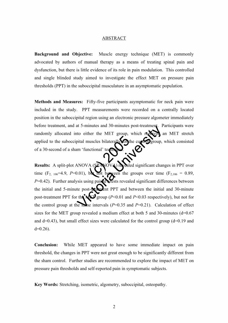
Results: A split-plot ANOVA (SPANOVA) revealed significant changes in PPT over
time (F2, 106=4.9, P=0.01), but not between the groups over time (F2,106 = 0.89,
P=0.42). Further analysis using paired t-tests revealed significant differences between
the initial and 5-minute post-treatment PPT and between the initial and 30-minute
post-treatment PPT for the MET group (P<0.01 and P<0.03 respectively), but not for
the control group at the same intervals (P=0.35 and P=0.21). Calculation of effect
sizes for the MET group revealed a medium effect at both 5 and 30-minutes (d=0.67
and d=0.43), but small effect sizes were calculated for the control group (d=0.19 and
d=0.26).
Conclusion: While MET appeared to have some immediate impact on pain
threshold, the changes in PPT were not great enough to be significantly different from
the sham control. Further studies are recommended to explore the impact of MET on
pressure pain thresholds and self-reported pain in symptomatic subjects.
Key Words: Stretching, isometric, algometry, suboccipital, osteopathy.

(c) 2
005
Victor
ia Univ
ersit
y
3
INTRODUCTION
Muscle energy technique (MET) differs from many manual techniques in that it is an
active technique requiring the patient to contribute the corrective force.1 One author of
manual therapy claims that it is one of the most valuable treatment techniques because
of the many therapeutic benefits resulting from a single procedure, including
lengthening and strengthening muscles, increasing fluid mechanics and decreasing
local oedema, and for mobilising a restricted articulation.1 MET is also claimed to be
useful for reducing pain and disability.2-4
MET involves the voluntary contraction of patient muscle in a precisely controlled
direction, at varying levels of intensity and against a distinctly executed counterforce
which is applied by the operator.1 MET has been advocated as a safer alternative
option to high velocity low amplitude (HVLA) techniques.1, 5, 6 Bourdillion et al5
warned against the use of HVLA in the upper cervical spine and suggested use of
MET as a more gentle approach to treatment in this region. McPartland et al6 stated
that non-thrusting techniques such as MET may better serve areas such as the
suboccipital region, cautioning that a poorly executed high velocity thrust may
critically damage the suboccipital tissues. Concerns for safety have also been
expressed by numerous authors in relation HVLA and the upper cervical spine, as
there is potential for such a thrust technique to damage the vertebro-basilar artery.5-7,8
As such is it imperative to determine the effectiveness of more gentle manual
techniques for treating these regions.
With origins of the technique claimed to extend back to the days of A.T Still,7 who
first proposed his philosophy and practice of osteopathy in 1874,1 and then developed
and popularised by Mitchell1, it is remarkable that research relating to MET is so
limited. Of the few studies published to date, most have examined the effect of MET
for increasing flexibility and range of motion9-13. However, since pain and discomfort
are very commonly the symptoms presenting to a manual therapist, it is important to
determine the potential of MET for pain relief.
Wilson et al2 examined the effects of MET in patients with acute low back pain.
These researchers examined whether patients with low back pain would demonstrate a
(c) 2
005
Victor
ia Univ
ersit
y
4
greater reduction in disability, as assessed by the Oswestry Disability Index (ODI),
after being treated with MET treatment coupled with supervised neuromuscular re-
education and resistance training as compared to those treated with supervised
neuromuscular re-education and resistance training alone. Wilson et al2 found that
every patient in the MET group had a higher change in ODI scores than patients in the
control group, and that the addition of MET improved the ODI scores substantially,
with mean percentage change for the MET group being 83%, compared to 65% for
the control group. The ODI is one of most frequently used tools in back pain research
and consists of 10 sections measuring a variety of activities of daily living as well as
pain intensity. 29 The present study aims to determine the effect of MET on pain
intensity through the use of an alternative research tool.
Cassidy et al14 investigated the effects of MET and HVLA manipulation of the
cervical spine in subjects with neck pain on both pain and cervical range of motion.
The researchers used a 101-point numerical rating scale to measure pain and found
that both treatments reduced pain; however, the change was larger in the manipulation
group. This study demonstrates the potential of MET for pain relief in the cervical
spine and therefore justifies the need for continuing research into non-thrusting
techniques to ensure safe and effective practice of manual therapists.
One region of particular clinical importance when assessing and treating the cervical
spine is the suboccipital region, which includes the atlanto-occipital joint and the
suboccipital muscles.15-17 Various authors have implicated the suboccipital muscles
not only as a cause of upper cervical pain, but also as a cause of chronic
headaches.5,15-17 The suboccipital muscles extend from C1 and C2 to the occiput,
including rectus capitis posterior major, rectus capitis posterior minor, obliquus
capitis superior and obliquus capitis inferior. Their actions include extension of the
head on C1, or rotation the head on C1 and C2.18
McPartland et al5 reviewed the clinical importance of the rectus capitis posterior
minor (RCPMn) muscle in balance and pain. These authors suggested that the high
density of muscle spindles found in the RCPMn may indicate that the value of these
muscles lies not only in their motor function, but also in their role as “proprioceptive
monitors” of the head and cervical spine. McPartland et al5 proposed that dysfunction
(c) 2
005
Victor
ia Univ
ersit
y
5
of this muscle may disrupt its role in proprioception and therefore may be clinically
significant. Dysfunction in the suboccipital muscles has been claimed to arise from
any trauma that causes sudden or extreme movement of the head, or simply from
chronic postural stresses, such as those occurring during slouching and the typical
“chin poking” posture.5 Hallgren et al15 examined patients with chronic headaches and
neck pain using MRI and found that some individuals exhibited replacement of
suboccipital skeletal muscle with fatty tissue. Hallgren et al15 hypothesised that this
may result in a decrease of muscle spindle and Golgi tendon organ density, with a
decrease in proprioceptive information transmitted to the central nervous system
(CNS), which may result in postural destabilisation.15 While cervical proprioception is
recognised as an essential component in maintaining balance5, adequate treatment of
the region is imperative, especially when treating the elderly community.
Both Hallgren et al15 and McPartland et al5 also recognised the possible importance of
the spinal gate theory of pain with relationship to suboccipital atrophy and chronic
pain. According to the spinal gate theory, mechanoreceptor input entering the dorsal
horn cells in the spinal cord may act as a “gate” that can modulate and inhibit
incoming nociceptive information.5,15 A substantial decrease in the proportion of
proprioceptive activity from the affected muscles may therefore result in greater
perception of pain by the patient.15 As such it would seem that the effect of MET in
the suboccipital region may be superior to many other techniques as it can strengthen
atrophied muscles resulting in greater proprioception while also modulating excessive
pain signals. Further testing into the effects of MET on pain is therefore imperative.
Accurate measurement and analysis of pain levels in individuals can often be difficult
due to the subjective nature of the sensation. The visual analogue scale and pain and
disability questionnaires, such as the McGill and Oswestry surveys, are commonly
used and validated research tools implemented as a means of monitoring patient
progress.19,20 An alternative method of evaluating pain and tenderness lies in the use
of pressure algometry. Used by many researchers,21-26 the algometer works as a
pressure gauge that quantifies the amount of pressure required to produce pain by
giving a value to the pressure-pain threshold (PPT) in an individual. The PPT refers
to the point at which the force being applied first causes a change in sensation from
pressure to pain.21-26
(c) 2
005
Victor
ia Univ
ersit
y
6
The reliability of measuring PPTs on bony landmarks and muscles has been verified
by numerous researchers.21-26 Nussbaum et al21 reported almost perfect reliability for
measurement of PPT within and across 3 days when assessing for PPTs in the biceps
brachii muscle. Nussbaum et at21 also concluded that reliability was enhanced when
all measurements were taken by one examiner. Similarly, Keating et al22 reported that
reproducibility of PPTs in the cervical and thoracic levels were excellent (ICC>0.9)
and good at the level of the lumbar spine (ICC=0.84), but standard deviations were
relatively large. Keating et al22 compared PPTs between different spinal regions and
found that the mid-thoracic segments were less tender than the cervical segments, but
more tender than lumbar segments.
Fryer et al23 adopted the use of pressure algometry as a tool for measuring the efficacy
of mobilisation and manipulation in the thoracic spine in asymptomatic subjects.
These researchers found that both procedures increased the PPT readings; however
mobilisation produced a greater immediate improvement in PPT.
Although MET is commonly advocated by authors of manual therapy for treatment of
somatic dysfunction and muscle pain, there remains little research into the effects of
MET on pain and tenderness.2-6 Previous studies have measured PPTs as a means of
investigating the efficacy of manual techniques in the lumbar and thoracic spine in
asymptomatic groups.2, 23 The present study aimed to determine the effect of MET
applied to the suboccipital muscles on PPTs in the suboccipital region.
METHOD
Subjects
Fifty-five asymptomatic volunteers were recruited from a population of student
osteopaths at the Victoria University (mean age 23 ± 5 including 16 males and 39
females). Testing was performed in the Victoria University Osteopathic Clinic.
Participants were screened and excluded if they suffered from any cervical pathology
or a current neck complaint which was determined by administration of treatment for
(c) 2
005
Victor
ia Univ
ersit
y
7
a neck complaint in the three days prior to this testing. All participants were informed
of the nature of the study, procedures to be used and any risks that may occur
throughout the study and signed a consent form. This study received approval from
the Victoria University Human Research Ethics Committee.
Measurement of Pressure Pain Thresholds
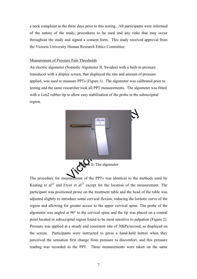
An electric algometer (Somedic Algometer II, Sweden) with a built-in pressure
transducer with a display screen, that displayed the rate and amount of pressure
applied, was used to measure PPTs (Figure 1). The algometer was calibrated prior to
testing and the same researcher took all PPT measurements. The algometer was fitted
with a 1cm2 rubber tip to allow easy stabilisation of the probe in the suboccipital
region.
Figure 1: The algometer
The procedure for measurement of the PPTs was identical to the methods used by
Keating et al22 and Fryer et al23 except for the location of the measurement. The
participant was positioned prone on the treatment table and the head of the table was
adjusted slightly to introduce some cervical flexion, reducing the lordotic curve of the
region and allowing for greater access to the upper cervical spine. The probe of the
algometer was angled at 90° to the cervical spine and the tip was placed on a central
point located in suboccipital region found to be most sensitive to palpation (Figure 2).
Pressure was applied at a steady and consistent rate of 30kPa/second, as displayed on
the screen. Participants were instructed to press a hand-held button when they
perceived the sensation first change from pressure to discomfort, and this pressure
reading was recorded as the PPT. Three measurements were taken on the same
(c) 2
005
Victor
ia Univ
ersit
y
8
location with an interval of 20 seconds between each reading. The PPT was
calculated according to PPT calculations conducted in studies by Keating et al22 and
Fryer et al23, where the PPT measurement used in the analysis was the mean of three
trials.
Figure 2: Measurement of pressure pain threshold using the algometer
Pilot Reliability Study
In order to determine the reliability of PPT measurement of the suboccipital region, a
pilot study was conducted involving 20 participants. PPT measurements were
performed as previously described. PPT measurements were recorded on 20
participants, and repeated 5 minutes and 30 minutes later. The mean differences from
the initial PPT measurement to the 5-minute and 30-minute PPT measurements were
small (7kPa, SD=43 and 11kPa, SD=52 respectively). The measurement procedure
appeared to be highly reliable, producing an average measure ICC = 0.96. The error
range of the measurement procedure (mean difference and SD) was calculated to be
50kPa (5-minutes) and 63kPa (30-minutes).
Procedure
A brief demonstration of the PPT measurement was conducted on the participant’s
forearm, and they were instructed to push the button when the pressure first became a
discomfort. The participant then lay prone on the treatment table with the cervical
region exposed. Researcher 1 identified sensitive area located centrally in the
suboccipital region and performed measurement of the PPT using the algometer.
(c) 2
005
Victor
ia Univ
ersit
y
9
Researcher 2 recorded the PPT readings and calculated the mean from the 3
measurements.
Following the initial PPT readings, participants were directed to another room where
they were allocated into the treatment or control group by lottery draw by researcher 3
(a registered osteopath), who performed all treatments. On completion of the
treatment, participants returned to the measurement room for re-testing of the PPT.
Movement of participants between testing was part of the procedure that was adopted
by each group in order to reduce the influence it may have in creating error. However
it was also considered that if walking a short distance between treatment rooms could
have such an influence on the effectiveness of a technique, then application in the
clinical setting seems redundant if the effects would have worn off by the time the
patient walked to the reception area.
Researchers 1 and 2 were blinded to the group allocation of all participants.
Participants were asked to remain in the clinic for the final measurement that was
conducted at 30-minutes post treatment.
Treatment Interventions
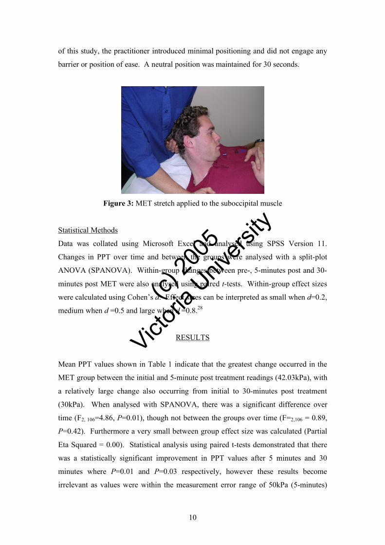
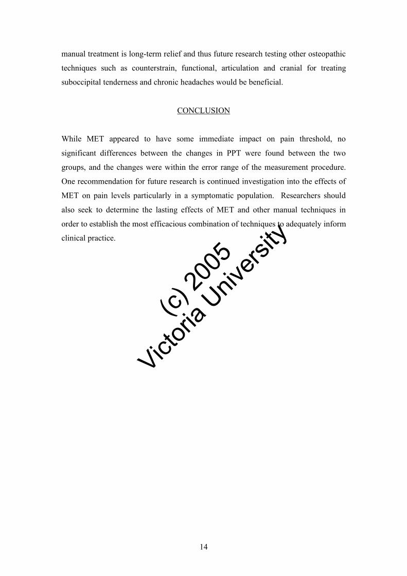
Muscle Energy Technique
Those participants allocated to the MET treatment group were requested to lie supine
on the treatment table. With the practitioner standing at the head of the table, an MET
stretch was applied to the suboccipital and trapezius muscles on both the left and right
sides. Hand contact was made with the base of the occiput with one hand, while the
other hand stabilised the shoulder (Figure 3). The head and neck were positioned in
flexion and slight side bending to the opposite side until the participant perceived a
stretch in the suboccipital region. The participant was instructed to gently push their
head back against the practitioners’ resistance as if to lay their head back on the
pillow.27 Contraction was held for around 3-5 seconds and was followed by a period
of relaxation. When the tissues relaxed, the practitioner then took up the slack to the
new point of resistance. This was performed randomly 3 times on each side.
Sham Functional Technique (Control)
The control group received a modified version of a functional technique. Correctly
applied, this indirect technique involves taking the joint away from a barrier to a
position of ease and waiting for a release in the surrounding tissues.1 For the purpose
(c) 2
005
Victor
ia Univ
ersit
y
10
of this study, the practitioner introduced minimal positioning and did not engage any
barrier or position of ease. A neutral position was maintained for 30 seconds.
Figure 3: MET stretch applied to the suboccipital muscle
Statistical Methods
Data was collated using Microsoft Excel and analysed using SPSS Version 11.
Changes in PPT over time and between the groups were analysed with a split-plot
ANOVA (SPANOVA). Within-group changes between pre-, 5-minutes post and 30-
minutes post MET were also analysed using paired t-tests. Within-group effect sizes
were calculated using Cohen’s d. Effect sizes can be interpreted as small when d=0.2,
medium when d =0.5 and large when d =0.8.28
RESULTS
Mean PPT values shown in Table 1 indicate that the greatest change occurred in the
MET group between the initial and 5-minute post treatment readings (42.03kPa), with
a relatively large change also occurring from initial to 30-minutes post treatment
(30kPa). When analysed with SPANOVA, there was a significant difference over
time (F2, 106=4.86, P=0.01), though not between the groups over time (F=2,106 = 0.89,
P=0.42). Furthermore a very small between group effect size was calculated (Partial
Eta Squared = 0.00). Statistical analysis using paired t-tests demonstrated that there
was a statistically significant improvement in PPT values after 5 minutes and 30
minutes where P=0.01 and P=0.03 respectively, however these results become
irrelevant as values were within the measurement error range of 50kPa (5-minutes)
(c) 2
005
Victor
ia Univ
ersit
y
11
and 63kPa (30-minutes). Medium pre-post effect sizes were calculated for both
intervals (d=0.67 and d=0.43).
A small increase in PPT was observed in the control group between both the initial
and 5 minute and the initial and 30 minute groups with mean changes of 15.88kPa and
16.12kPa respectively, however again changes were well within the error range of the
measurement procedure. There was not significant difference at either interval
(P=0.35 and P=0.21) and small effect sizes were calculated for both (d=0.19 and
d=0.26).
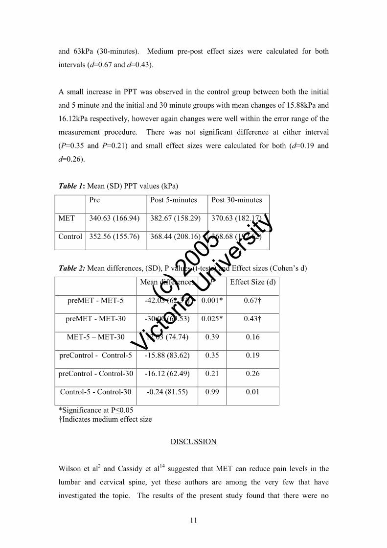
Table 1: Mean (SD) PPT values (kPa)
Pre Post 5-minutes Post 30-minutes
MET 340.63 (166.94) 382.67 (158.29) 370.63 (182.17)
Control 352.56 (155.76) 368.44 (208.16) 368.68 (192.62)
Table 2: Mean differences, (SD), P values (t-tests) and Effect sizes (Cohen’s d)
Mean differences P Effect Size (d)
preMET - MET-5 -42.03 (62.37) 0.001* 0.67†
preMET - MET-30 -30.00 (69.53) 0.025* 0.43†
MET-5 – MET-30 12.03 (74.74) 0.39 0.16
preControl - Control-5 -15.88 (83.62) 0.35 0.19
preControl - Control-30 -16.12 (62.49) 0.21 0.26
Control-5 - Control-30 -0.24 (81.55) 0.99 0.01
*Significance at P≤0.05 †Indicates medium effect size
DISCUSSION
Wilson et al2 and Cassidy et al14 suggested that MET can reduce pain levels in the
lumbar and cervical spine, yet these authors are among the very few that have
investigated the topic. The results of the present study found that there were no
(c) 2
005
Victor
ia Univ
ersit
y
12
significant differences in the change in PPT between MET and the sham control,
when analysed using a SPANOVA. However, MET produced the greatest increase in
PPT, the pre-post change was significant when analysed using the less robust t-test,
and the pre-post change produced medium effect sizes, all of which lend cautious
support to the possibility that MET may produce a change in PPT. However, it should
be noted that the mean changes in PPT after 5-minutes and after 30-minutes were
within the error range determined by the pilot reliability study (50kPa and 63kPa
respectively) and therefore it cannot be concluded that MET was effective in reducing
pain levels in this asymptomatic group tested.
Significant pre- post change in the MET group was found through the use of t-tests
and the mean PPT change at 5-minutes and 30-minutes produced a medium effect
sizes. While this gives an indication that MET has potential to produce changes in
pain thresholds, Wilson et al2 demonstrated the long term benefit of MET in a clinical
setting. These researchers found that in patients with acute low back pain, MET
combined with neuromuscular re-education and resistance training was more effective
than the re-education and training alone for reducing pain levels over a period of 8
weeks. In this study, the participants received 2 MET treatments weekly for a period
of 4 weeks. Although the present study is useful for examining and comparing the
effects of individual techniques, it does not accurately represent treatment in a clinical
setting, and researchers could improve this by using a symptomatic population and
including multiple treatments in future studies.
In the control group, small increases in PPT occurred over time (15.88kPa at 5minutes
and 16.12kPa at 30minutes) and, despite the fact that these increases were not
significant, these changes may be the result of a minor placebo effect or may be
attributed an adaptation of pain fibre signals. No treatment effect was expected from
the sham functional technique, because the researcher did not engage any perceived
‘position of ease’, but assessed this position and then moved the head back to the
neutral position. While it was expected that most participants believed the sham
functional treatment to be a legitimate technique because of the subtle leverages
involved, no follow up study was conducted to determine this. The participants in this
study were recruited from a population of student osteopaths, and despite the subtle
nature of the technique, it is possible that they may have been more aware of the
(c) 2
005
Victor
ia Univ
ersit
y
13
functional technique being a sham than if the population was from the general public.
However, over half of those assigned to the control group were junior students
untrained in the use of functional technique, and therefore would not know what to
expect of the technique. One explanation for the change maybe associated with the
error range of the measurement procedure. Calculated at 50kPa at 5-minutes and
63kPa at 30-minutes, this suggests that unless the difference in PPT is greater than
these values, the changes that do occur are most likely associated with the
measurement procedure rather than due to the effects of the technique. This may have
been more clearly indicated with the addition of a no-treatment group.
When assessing pain levels, a certain amount of variability between individuals
should be expected due to the subjective nature of the sensation. Some large
variations between the 3 PPT readings in the present study produced standard
deviations of mean values ranging from 60-75kPa. In comparison to research by
Keating et al22 and Fryer et al23, standard deviations in the present study were quite
low. Keating et al22 reported a standard deviation of 150kPa in a repeatability study
of PPT measurement in the cervical spine, and still the algometer readings were
reported to be highly reliable based on ICC and coefficient of variance (CV).
Similarly, Fryer et al23 reported standard deviations ranging from 83-97kPa when
testing PPTs in the thoracic spine. Again the measurement procedure using the
algometer was reported as reliable (ICC=0.93). A reliability study conducted prior to
the beginning of this study produced an average measure ICC=0.96, and therefore
supports previous studies which have shown that measurements of PPT can be highly
repeatable in individuals without pain, despite the large standard deviations.22, 23
The present study measured PPTs on an asymptomatic population and therefore it
must be determined whether testing the effects of MET on symptomatic subjects
would better reflect the potential of MET for pain reduction, as has been indicated in
studies conducted by Wilson et al.2 It would appear from results of the present study
that MET has little effect on pain levels after one treatment, however in the clinical
setting most treatments are applied more than once. Wilson et al2 demonstrated this
incorporating multiple applications of MET in people suffering low back pain.
Further studies must therefore investigate whether significant results would be found
when testing a longer regimen of therapy and follow-up. The therapeutic goal of
(c) 2
005
Victor
ia Univ
ersit
y
14
manual treatment is long-term relief and thus future research testing other osteopathic
techniques such as counterstrain, functional, articulation and cranial for treating
suboccipital tenderness and chronic headaches would be beneficial.
CONCLUSION
While MET appeared to have some immediate impact on pain threshold, no
significant differences between the changes in PPT were found between the two
groups, and the changes were within the error range of the measurement procedure.
One recommendation for future research is continued investigation into the effects of
MET on pain levels particularly in a symptomatic population. Researchers should
also seek to determine the lasting effects of MET and other manual techniques in
order to establish the most efficacious combination of techniques to adequately inform
clinical practice.
(c) 2
005
Victor
ia Univ
ersit
y
15
REFERENCES
1. Greenman, PE. Principles of Manual Medicine, 3rd Edition, Lippincott, Williams
& Wilkins, Sydney, 2003, pp3,93,98
2. Wilson, E. Payton, O. Donegan-Shoaf, L. Dec, K. Muscle Energy Technique in
Patients with Acute Low Back Pain: A pilot clinical trial. J Orth and Sports Phys
Ther 2003;33(9):502-12.
3. Lewit, K. Simons, DG. Myofascial Pain: Relief by Post Isometric Relaxation.
Arch Phys Med Rehab 1984;65:452-6.
4. Roberts, B. Soft Tissue Manipulation: Neuromuscular and Muscle Energy
Techniques. J of Neurosci Nurs 1997;29(2):123-7.
5. McPartland, JM. Brodeur, RR. Rectus Capitis Posterior Minor: a small but
important suboccipital muscle. J of Bodywork Movement Ther 1999;3(1):30-5.
6. Bourdillon JF, Day EA, Bookhout MR. Spinal Manipulation. 5th ed. Oxford:
Butterworth-Heinemann; 1992.
7. Ward, R.C. Foundation for Osteopathic Medicine, 2nd Edition, Williams &
Wilkins, Sydney, 2003 , pp881, 856
8. Gibbons, P. Tehan, P. Manipulation of the spine, thorax and pelvis: An
osteopathic perspective. Churchill Livingstone, 2001, p23
9. Feland, JB. Myrer, JW. Merrill, RM. Acute changes in hamstring flexibility: PNF
versus static stretch in senior athletes. Phys Ther Sport 2002;2(4): 186-93.
10. Spernoga, SG. Uhl, TL. Arnold, BL. Gansneder, BM. Duration of Maintained
Hamstring flexibility after a one-time, modified hold-relax stretching protocol. J
Athl Train 2001;36(1):44-8.
11. Schenk RJ, Adelman K, Rousselle J. The effects of muscle energy technique on
cervical range of motion. J Manual Manipulative Ther. 1994;2(4):149-55.
12. Lenehan KL, Fryer G, McLaughlin P. The effect of muscle energy technique on
gross trunk range of motion. J Osteopath Med. 2003;6(1):13-8.
13. Fryer G, Ruszkowski W. The influence of contraction duration in muscle energy
technique applied to the atlanto-axial joint. J Osteopath Med. 2004;7(2):79-84.
14. Cassidy, J.D. Lopes, A.A. Yong-Hing, K. The Immediate Effect of Manipulation
versus Mobilisation on Pain and Range of Motion in the Cervical Spine: A
Randomised Controlled Trial. J of Manipulative Physiol Ther 1992;15(9):570-5.
(c) 2
005
Victor
ia Univ
ersit
y
16
15. Hallgren, RC. Greenman, PE. Rechtien, JJ. Atrophy of suboccipital muscles in
patients with chronic pain: A pilot study. J Am Osteopath Assoc. 1994;94(12):
1032-8.
16. Such, G.W. Upper Cervical Synthesis: Integrative manual care of the Occipito-
atlantal Joint. Chiropractic Technique 1999;11(3):116-24.
17. Alix, ME. Bates, DK. A Proposed Aetiology of Cervicogenic Headache: The
Neurophysiologic Basis and Anatomic Relationship between the Dura Mater and
the Rectus Posterior Capitis Minor Muscle. J Manipulative Physiol
Ther1999;22(8):534-9.
18. Moore, KL. Dalley, AF. Clinically Oriented Anatomy. 4th Edition, Lippincott
Williams & Wilkins, Sydney, 1999, pp475-6
19. Chow, RT. Bamsley, L. Heller, GZ. Siddall, PJ. A Pilot study of Low-Power
Laser Therapy in the Management of Chronic Neck Pain. J Musculoskeletal Pain
2004;12(2):71-81.
20. Fritz, JM. Irrang, JJ. A comparison of a modified Oswestry Low Back Pain
Disability Questionnaire and the Quebec Back Pain Disability Scale. Phys Ther
2001;81(2):776-88.
21. Nussbaum, EL. Downes, L. Reliability of Clinical Pressure-Pain Algometry
Measurements Obtained on Consecutive days. Phys Ther 1998;78(2):160-9.
22. Keating, L. Lubke, C. Powell, V. Young, T. Souvlis, T. Jull, G. Mid-thoracic
tenderness: a comparison of pressure pain threshold between spinal regions, in
asymptomatic subjects. Man Ther 2001;6(1):34-39.
23. Fryer G, Carub J, McIvor S. The effect of manipulation and mobilisation on
pressure pain thresholds in the thoracic spine. J Osteopath Med 2004;7(1): 8-14.
24. Vernon, HT. Aker, P. Burns, S. Viljakaanen, S. Short, L. Pressure pain threshold
evaluation of the effect of spinal manipulation in the treatment of chronic neck
pain: a pilot study. J Manipulative Physiol Ther 1990;13(1): 13-6.
25. Kosek, E. Ekholm, J. Nordemar, R. A comparison of pressure pain thresholds in
different tissues and body regions – Long term reliability of pressure algometry in
healthy volunteers. Scand J of Rehabil Med 1993;25:117-24.
26. Vanderweeen, L. Oostendorp, RAB. Vaes, P. Duquet, W. Pressure algometry in
manual therapy. Man Ther 1996;1(5):258-65.
27. Chaitow, L. Liebenson, C. Muscle Energy Technique. 2nd Edition, Churchill
Livingstone; 2001, pp168-71
(c) 2
005
Victor
ia Univ
ersit
y
17
28. Aron, A. Aron, EN. Statistics for Psychology. 2nd Edition, Prentice Hall; 1999,
p365
29. Warfield, C.A. Bajwa, A.H. Principles and Practice of Pain Medicine. McGraw-
Hill, 2004, p76.