Embed Size (px)
Citation preview

THE EARLY TRIASSIC STEM−FROG CZATKOBATRACHUSFROM POLAND
SUSAN E. EVANS and MAGDALENA BORSUK−BIAŁYNICKA
Evans, S.E. and Borsuk−Białynicka, M. 2009. The Early Triassic stem−frog Czatkobatrachusfrom Poland. Palaeontologica Polonica 65, 79–105.
Czatkobatrachus polonicus Evans et Borsuk−Białynicka, 1998 is a stem−frog from the EarlyTriassic karst locality of Czatkowice 1 (southern Poland). It was described and named on thebasis of a small collection of vertebrae, ilia, and forelimb bones, with subsequent descriptionof the scapulocoracoid. Further skeletal elements have now been recovered. Here we presenta complete overview of the available material of Czatkobatrachus, give an extended diagno−sis of the genus, and provide an assessment of its relevance to our understanding of the earlystages of salientian evolution. Czatkobatrachus has an anuran type ilium, a urodelan−likescapulocoracoid, unfused epipodials, a moderately shortened presacral column, and a veryshort tail of separate caudal vertebrae. The strongly ossified ends of the proximal limb bonessuggest terrestrial life. The skull is unknown.
Key words: Stem−frogs, Anura, Salientia, Lissamphibia, Triassic.
Susan E. Evans [[email protected]], Research Department of Cell and Developmental Bio−logy, UCL, University College London, Gower Street, London, WC1E 6BT, UK.
Magdalena Borsuk−Białynicka [[email protected]], Instytut Paleobiologii PAN,Twarda 51/55, 00−818 Warszawa, Poland.
Received 17 November 2006, accepted 15 September 2008

INTRODUCTION
Czatkobatrachus polonicus Evans et Borsuk−Białynicka, 1998 is a stem−frog described from the EarlyTriassic microvertebrate locality of Czatkowice 1 in southern Poland (Paszkowski and Wieczorek 1982). It isthe earliest lissamphibian known from Northern Pangaea, and only the second recovered from Triassic de−posits, the roughly contemporaneous salientian Triadobatrachus massinoti (Piveteau 1936) from Madagas−car being the first. To date, only a few postcranial bones of Czatkobatrachus (ilia, distal humeri, ulnae, verte−brae, scapulocoracoid) have been described (Evans and Borsuk−Białynicka 1998; Borsuk−Białynicka et al.1999; Borsuk−Białynicka and Evans 2002). Incomplete as they are, the remains of Czatkobatrachus are gen−erally similar to those of Triadobatrachus but are significantly smaller and appear more derived (or at leastbetter ossified) in rib, vertebral structure and limb morphology.
The objective of the present paper is to describe newly discovered material of Czatkobatrachus polonicus,as well as give a more detailed description of skeletal elements only briefly discussed in the original paper,most notably parts of the vertebral column and limbs.
Institutional abbreviations. — MNHN, Museum National d'Histoire Naturelle, Paris, France; UCMP,University of California, Museum of Paleontology, Berkeley, USA; ZPAL, Institute of Paleobiology, PolishAcademy of Sciences, Warsaw, Poland.
Acknowledgments. — The authors are indebted to the team of the Institute of Geology, Jagiellonian Uni−versity, Kraków, and particularly Józef Wieczorek and Mariusz Paszkowski who discovered, and generouslytransferred the bone breccia from Czatkowice 1 to the Museum of the Earth and the Institute of Paleobiology,Polish Academy of Sciences, Warsaw. Our thanks are due also to the late Halszka Osmolska (Institute ofPaleobiology) and Teresa Maryańska (Museum of the Earth) for access to materials in their care; Jean−ClaudeRage (Museum d’Histoire Naturelle, Paris), for access to high−resolution casts of the holotype of Triado−batrachus massinoti; A.G. Jacobson (University of Texas, Austin) for information on head development in am−phibians; Borja Sanchiz (Natural History Museum, Madrid) for information on vertebral development; and CarlGans (University of Texas, Austin) and Farish Jenkins Jr (Harvard University), for discussions on the func−tional morphology of early frogs. We are grateful to the referees: F. Jenkins Jr and Zbyněk Roček (Charles Uni−versity Prague). Critical comments of the latter helped us to improve the final version of the manuscript. EwaHara (Institute of Paleobiology) carried out the acid preparation of the Czatkowice 1 breccia; Cyprian Kulicki(Institute of Paleobiology) performed the scanning electron microscope photography.
GEOLOGICAL SETTING
The Triassic deposits filling the karst fissures developed in the Early Carboniferous limestones at Czat−kowice 1 (southern Poland) were discovered by a team from the Jagiellonian University, Kraków, and werefirst described by Paszkowski and Wieczorek (1982). The material described herein comes from the largestof the karst fissures called Czatkowice 1. This material has been dated as probably Early Olenekian in age(Borsuk−Białynicka et al. 2003). A more detailed account of the geology may be found elsewhere in this vol−ume (Cook and Trueman 2009; Paszkowski 2009).
MATERIAL AND METHODS
The bone material from the karst deposits of Czatkowice 1 consists of the completely disarticulated skele−tons of several small vertebrates, most of them reptiles (Borsuk−Białynicka et al.1999). Salientian postcranialbones are easily discriminated from reptilian ones by their anatomical structure, further supported by theirsize range (all but one of the Czatkowice 1 reptiles are larger as adults); their frequency (Czatkobatrachus re−mains are comparatively scarce); and the type of bone tissue in terms of surface appearance. The skeletal ele−
80 SUSAN E. EVANS and MAGDALENA BORSUK−BIAŁYNICKA

ments were compared with those of both extant and fossil frogs, particularly the earliest known Jurassicfrogs: Notobatrachus degiustoi Reig, 1955 and Vieraella herbsti Reig, 1961 (Báez and Basso 1996), andProsalirus bitis Shubin et Jenkins, 1995 (Jenkins and Shubin 1998). Most informative was a comparison withthe Early Triassic Malagasy basal salientian Triadobatrachus massinoti (Piveteau, 1936) (Rage and Roček1989; Roček and Rage 2000).
The puzzling absence of skull bones cannot be explained as the effect of chemical preparation in aceticacid, because equally small and fine reptilian elements are perfectly preserved. It may be partly an artifact ofthe relative scarcity of Czatkobatrachus remains overall. Although all crown−group frogs have lost the lowerdentition, most basal taxa, as well as many neobatrachians, retain teeth in the maxilla, and these toothed ele−ments are both common and distinctive in microvertebrate assemblages, even when fragmented. Nonethe−less, despite a careful scrutiny of all toothed elements from Czatkowice 1, none are attributable to Czatko−batrachus. Rage and Rocek (1989) found no trace of teeth in Triadobatrachus, but only the most posteriorpart of the maxilla is preserved, and no teeth are expected in this part (Roček personal communication 2007).
The scanning electron microscope was used extensively for illustrations and studies of surface texture.Terminology used follows Sanchiz (1998).
PHYLOGENETIC BACKGROUND
No−one has ever seriously doubted the monophyly of Anura, and although the status of Triadobatrachusas a stem−frog has occasionally been challenged (e.g., Hecht 1960), most authors accept also the monophylyof Salientia, including Triadobatrachus, and now Czatkobatrachus, along with crown−group Anura (e.g.,Duellman and Trueb 1986; Milner 1988; Rage and Roček 1989; Sanchiz 1999; Roček 2000; Carroll 2007;Anderson et al. 2008). There is also a general consensus that Salientia were derived from temnospondyl, ormore precisely dissorophoid, ancestors (Bolt 1969, 1977, 1991; Bolt and Lombard 1985; Daly 1994; Milner1988, 1990, 1993; Carroll 1999; but see Laurin and Reisz 1997; Laurin et al. 2000; Yates and Warren 2000;Anderson et al. 2008).
The monophyly of the Lissamphibia as a whole (Salientia, Caudata and Gymnophiona) was proposed byParsons and Williams (1962, 1963), Szarski (1962), and Bolt (1969), and has been supported by many au−thors (e.g., Milner 1988; Rage and Janvier 1982; Gauthier et al. 1989; Trueb and Cloutier 1991; Cannatellaand Hillis 1993; Ford and Cannatella 1993; McGowan and Evans 1995; Gardner 2000). Others have rejectedor questioned lissamphibian monophyly (e.g., Shishkin 1973; Bolt and Lombard 1985; Carroll and Holmes1980; Carroll 1999; Carroll et al. 1999), and the debate is ongoing. The recovery and description of early rep−resentatives of major lissamphibian lineages are clearly critical to the discussion of their ancestry.
SYSTEMATIC PALEONTOLOGY
Class Amphibia Linné, 1758
Order Salientia Laurenti, 1768
Family uncertainGenus Czatkobatrachus Evans et Borsuk−Białynicka, 1998
Czatkobatrachus polonicus Evans et Borsuk−Białynicka, 1998
Holotype: ZPAL Ab IV/7, a right ilium.
Type horizon and locality: Olenekian karst deposits at Czatkowice Quarry (locality 1), Kraków region, Poland.
Material. — 76 catalogued specimens, including 15 presacral vertebrae, 2 sacral vertebrae, 9 scapulo−coracoids, 11 humeri, 2 ulnae, 19 ilia, 5 femora.
Emended generic and specific diagnosis. — Small stem−frog (less than 50 mm snout−vent length) thatresembles Triadobatrachus and all other salientians in having an anteriorly extended iliac shaft. It resembles
EARLY TRIASSIC STEM−FROG CZATKOBATRACHUS FROM POLAND 81
Triadobatrachus and differs from crown−group anurans in retaining relatively long neural arches, unfusedepipodials, a series of unfused caudal vertebrae instead of an urostyle (Lynch 1973; Trueb 1973; Sanchiz1998), a scapulocoracoid that is a single ossification, and an ilium with very strong dorsal prominence and aslender elongate shaft, rounded in cross−section. It differs from Triadobatrachus in having a long slenderscapular blade (short and broad in Triadobatrachus), in having a single atlas ossification with no trace of ribfacets (bipartite atlas with a rib processes reported in Triadobatrachus by Rage and Roček 1989, and Ročekand Rage 2000, but see below for different view), in having a higher level of ossification, long fused poste−rior transverse processes and sacral ribs, fully ossified components in elbow joint, ischium fused to ilium (thelast feature unique within the Salientia; Roček, personal communication 2007) despite its much smaller size,and in having longer, more slender limbs. As reconstructed, the ilio−sacral joint of Czatkobatrachus has amorphology quite distinct from that of Triadobatrachus, in that the sacral rib is short and fused to the verte−bral centrum (rather than free and posteriorly elongate), and has an expanded distal end that is bothdorsoventrally and anteroposteriorly bifurcate, with a posterior notch and groove. Czatkobatrachus also dif−fers from Triadobatrachus and resembles many crown−group frogs in having a fully ossified, capitate emi−nence (eminentia capitata) that equals or exceeds 60% of the width of the distal end of humerus, and asym−metrical epicondyles (ulnar epicondyle larger). It differs from crown−group frogs in having the anterior mar−gin of atlas pedicel notched for the exit of the first spinal nerve and spinal nerve notches or foramina in someposterior vertebrae, and in retaining an ossified remnant of the pubis, fused to the body of the pelvis and per−forated by an obturator canal.
Occurence. — Only type locality.
VERTEBRAL COLUMN
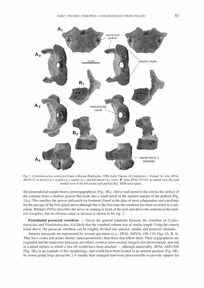
The vertebral column of Czatkobatrachus is represented by isolated elements that have been ordered (Fig.4B) partly on the basis of morphology (neural arch and centrum length, posterior zygapophysial size, trans−verse processes, and the presence or absence of free ribs) and partly by comparison with Triadobatrachus(Fig. 4A). On this basis, the atlas (Fig. 1), anterior, middle and posterior presacrals (Fig. 2), sacrals (Fig. 3),and caudals have all been identified. They are all fully ossified, and share a distinctive domed neural arch, aweak dorsal midline ridge, and a cylindrical perichordal (ectochordal) centrum perforated by a largeunconstricted notochordal canal. The anterior zygapophyses are horizontal and vary in size along the col−umn. Between them, the anterior border bears a deep V− or U−shaped notch that extends to the level of thebase of the transverse processes. Behind the notch, there are bilateral depressions, some more marked thanothers, that mark the attachment of intervertebral muscles or ligaments. More posteriorly, the neural arch be−comes strongly domed but curves down at the posterior margin. This posterior margin is usually slightly in−dented, but in the vertebrae immediately following the sacrum, it develops a small median process (Fig. 2C,G). The centrum is rounded in cross−section. Seen in anterior view, however, the lateral walls of the cylinderare usually thicker than the dorsal and ventral walls, giving the centrum a slightly depressed shape (see e.g.,Fig. 2A1, C1). In general morphology, the vertebrae closely resemble those of Triadobatrachus, except thatthe transverse processes, and in some cases the ribs, are fused to the vertebra rather than free. A few speci−mens (e.g., ZPAL AbIV/6, Fig. 2A1, A2) show a bulbous region part way along the transverse process thatmarks the point of fusion, although subsequent remodelling apparently removes this to leave a single smooth,but actually compound, process.
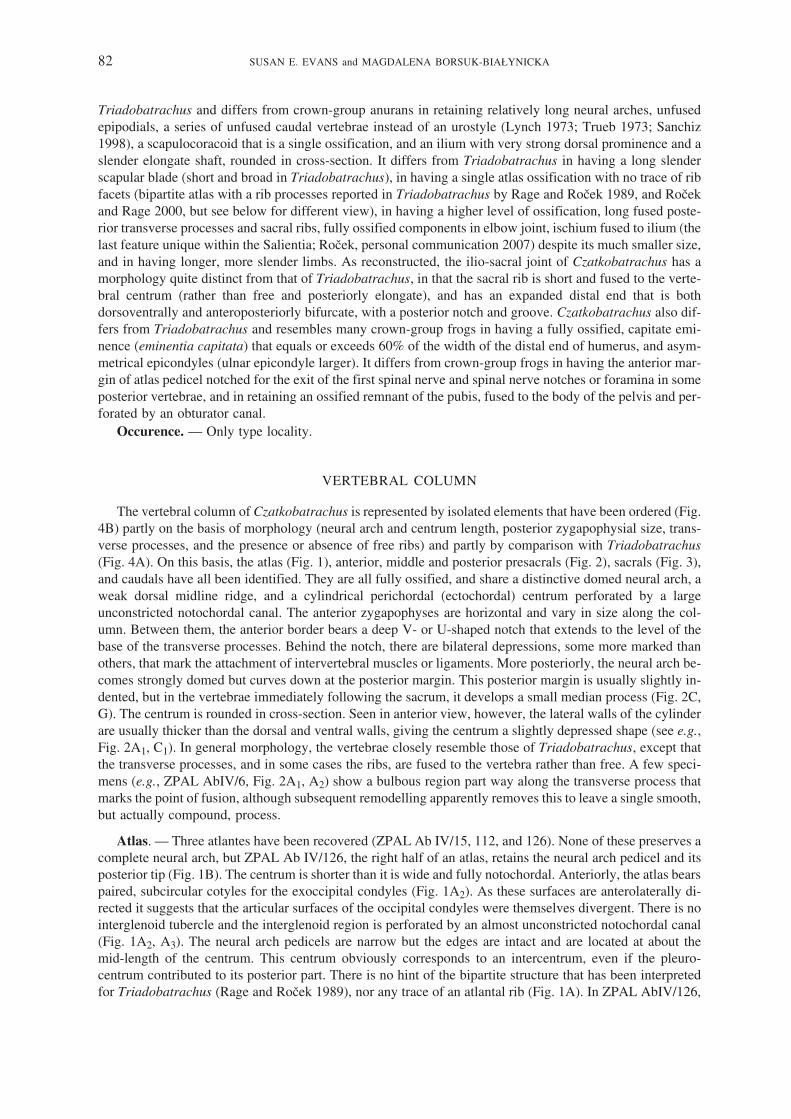
Atlas. — Three atlantes have been recovered (ZPAL Ab IV/15, 112, and 126). None of these preserves acomplete neural arch, but ZPAL Ab IV/126, the right half of an atlas, retains the neural arch pedicel and itsposterior tip (Fig. 1B). The centrum is shorter than it is wide and fully notochordal. Anteriorly, the atlas bearspaired, subcircular cotyles for the exoccipital condyles (Fig. 1A2). As these surfaces are anterolaterally di−rected it suggests that the articular surfaces of the occipital condyles were themselves divergent. There is nointerglenoid tubercle and the interglenoid region is perforated by an almost unconstricted notochordal canal(Fig. 1A2, A3). The neural arch pedicels are narrow but the edges are intact and are located at about themid−length of the centrum. This centrum obviously corresponds to an intercentrum, even if the pleuro−centrum contributed to its posterior part. There is no hint of the bipartite structure that has been interpretedfor Triadobatrachus (Rage and Roček 1989), nor any trace of an atlantal rib (Fig. 1A). In ZPAL AbIV/126,
82 SUSAN E. EVANS and MAGDALENA BORSUK−BIAŁYNICKA
the posterodorsal margin bears a postzygapophysis (Fig. 1B2). Above and lateral to the cotyles the surface ofthe centrum bears a shallow groove that leads into a small notch in the anterior margin of the pedicel (Fig.1A4). This matches the groove and notch (or foramen) found in the atlas of most salamanders and caeciliansfor the passage of the first spinal nerve although this is the first time the condition has been recorded in a sali−entian. Ritland (1955a) describes the nerve as running in front of the arch and above the centrum in the mod−ern Ascaphus, but no obvious canal or incision is shown in his fig. 2.
Postatlantal presacral vertebrae. — Given the general similarity between the vertebrae of Czatko−batrachus and Triadobatrachus, it is likely that the vertebral column was of similar length. Using the criterialisted above, the presacral vertebrae can be roughly divided into anterior, middle and posterior elements.
Anterior presacrals are represented by several specimens (e.g., ZPAL AbIV/6, 108–110; Figs 2A, B, 4).They have centra and arches shorter (anteroposteriorly) than those that follow them. Their zygapophyses areexpanded and the transverse processes are robust, ovoid in cross−section (longest axis dorsoventral), and endin a pitted surface to which a free rib would have been attached — although immovably. ZPAL AbIV/108(Fig. 2B2) is an example of this morphology, and would have been located in an anterior position (Fig. 4B).In crown−group frogs presacrals 2–4 usually bear enlarged transverse processes/ribs to provide support for
EARLY TRIASSIC STEM−FROG CZATKOBATRACHUS FROM POLAND 83
neural archpedicel
atlantal cotyles1 mm
notochordalcanal
spinal nerve 1passage
Fig. 1. Czatkobatrachus polonicus Evans et Borsuk−Białynicka, 1998, Early Triassic of Czatkowice 1, Poland. A. Atlas ZPALAb IV/15, in dorsal (A1), cranial (A2), caudal (A3), and left lateral (A4) views. B. Atlas ZPAL IV/126, in cranial view (B1) and
medial view of the left neural arch pedicel (B2). SEM stereo−pairs.

the pectoral girdle. Of these, the strongest rib/process is usually that of presacral 3 (e.g., Ritland 1955a). Thecondition in the Malagasy stem−frog Triadobatrachus is similar although whether the vertebra bearing thelargest rib is presacral 3 or 4 depends on the interpretation of the atlas as single or bipartite. According to our
84 SUSAN E. EVANS and MAGDALENA BORSUK−BIAŁYNICKA
bony spur
spinal nerveforamen
1 mm
Fig. 2. Czatkobatrachus polonicus Evans et Borsuk−Białynicka, 1998, Early Triassic of Czatkowice 1, Poland. A. Anteriordorsal ZPAL Ab IV/6, in caudal (A1) and dorsal (A2) views. B. Anterior dorsal ZPAL Ab IV/108, in caudal (B1) and ventral(B2) views. C. Anterior caudal ZPAL Ab IV/135, in posterior (C1) and dorsal (C2) views. D. Posterior presacral ZPAL AbIV/127, in dorsal view. E. Directly presacral or anterior caudal vertebra ZPAL Ab IV/115, in dorsal (E1) and left lateral (E2)views. F. Middle presacral vertebra ZPAL IV/128, in dorsal (F1) and caudal (F2) views. G. ?the second caudal vertebra ZPALAb IV/134, in caudal (G1) and dorsal (G2) views. H. Posterior caudal ZPAL AB IV/20, in left lateral view. SEM micrographs;
all but C1, C2, G1, G2 stereo−pairs.

interpretation, it is presacral 4 (Fig. 4A). Amongst the Polish material, specimen ZPAL ABIV/6 bears thelongest transverse process, probably including a fused rib. In the reconstruction (Fig. 4), this element hasbeen located posterior to ZPAL AbIV/108, which had either the third or fourth position, the exact number ofanterior vertebrae being unknown. The length of processes and their dorsoventral flattening suggest they sup−ported the pectoral girdle as they do in crown−group frogs.
Strong bifurcate ribs and/or uncinate processes (Ritland 1955a) are found on the third presacral of the Ju−rassic Vieraella and Notobatrachus (Báez and Basso 1996), and isolated ribs of similar morphology havealso been described and illustrated for the Jurassic Prosalirus (Jenkins and Shubin 1998). In the livingAscaphus, the second rib is generally the largest and possesses a sharp, posterolaterally directed uncinate pro−cess, but the fourth rib is sometimes equally large. This compares closely with the structure in Leiopelma(Ritland 1955a) and discoglossids, The bifurcations that relate to the attachment of muscles connecting thescapula to the spine (m. serratus, Ritland 1955a), have not been recorded in Czatkobatrachus.
A third set of vertebrae (e.g., ZPAL AbIV/11, 14, 26, 128, and 146; for the latter two see Figs 2F, 4B)show a fairly consistent morphology and are the most common. The neural arch is longer than it is wide (un−like the more anterior presacrals), and this is matched by a slightly longer centrum. The transverse processes
EARLY TRIASSIC STEM−FROG CZATKOBATRACHUS FROM POLAND 85
ilio-sacraljoint surface
ilio-sacral joint surface
thickenedventralridge
convexity
triangulardepression
for ilio-sacralligament
1 mm
Fig. 3. Czatkobatrachus polonicus Evans et Borsuk−Białynicka, 1998, Early Triassic of Czatkowice 1, Poland. A. Sacral ver−tebra ZPAL Ab IV/79, in dorsal (A1), caudal (A3), and cranial (A5) views; right iliac surface in lateral view (A2, A4). B. Sacralvertebra ZPAL Ab IV/76, in caudal view (B1); right iliac surface in lateral view (B2, B3). All but A2 and B2 SEM stereo−pairs.

are shorter than those on ZPAL AbIV/6, are distally subcircular in section, vary in width, and are directedposterolaterally. The anterior zygapophyses vary slightly in shape, but seem to have decreased in size to−wards the rear of the column.
A fourth group of vertebrae, e.g., ZPAL AbIV/8 and10, tentatively considered a sacral by Evans andBorsuk−Białynicka (1998, fig. 3B, E), and ZPAL AbIV/127 (Fig. 2D) is characterised by a somewhat shorterarch and centrum than those of the mid−trunk, smaller postzygapophyses that are closer to the midline (distin−guishing them from anterior presacrals) and a more robust posteriorly curved transverse processes probablywith no free ribs contacting them. We interpret these elements as posterior presacrals.
ZPAL AbIV/115 (Fig. 2E) has two unusual features. The first is a small additional bony spur (Fig. 2E1) onthe left side between the transverse process and the posterior margin of the pedicel (on the right only a slighttuberosity). The second is the presence of a distinct foramen, possibly for a spinal nerve, perforating the neu−ral arch pedicel on each side behind the transverse process (Fig. 2E2). From its morphology, this vertebra is aposterior presacral or an anterior caudal (Fig. 4B). Rage and Roček (1986) reported that such foramina werenot visible in Triadobatrachus.
Sacrum. — Two almost complete sacral vertebrae have been recovered, ZPAL AbIV/76 and 79 (Fig. 3B,A), and ZPAL AbIV/123. The main body of the bone is slightly shorter than that of the presacrals, but is oth−erwise similar in morphology. The arch has a low midline ridge and is slightly domed (Fig. 3A1). The hori−zontal anterior zygapophyses are ovoid, long axis slightly divergent, and the U−shaped notch more open thanin presacrals (Fig. 3A1). Postzygapophyses are present (as in some basal frogs, e.g., Ascaphus, Ritland1955a) but small and more closely placed than in most presacrals. The centrum is spool−shaped like those ofother vertebrae and shows a normally developed posterior joint surface (i.e., no specialised sacro−caudal ar−ticulation). The notochordal canal continued into the tail (Fig. 3A3, B1).
In spite of variation (see below), the basic, highly unusual, morphology of the sacral transverse processes(diapophyses) is the same. The processes are short and strong, proximally almost circular in cross−section, butthey expand and bifurcate distally, in both dorsal and anterior/posterior views (Fig. 3A1, A3, B1). Theposterodorsal margin of the process extends first laterally and then curves strongly posteriad (to be continuedfurther in cartilage, as shown by a pitted, unfinished surface). The anteroventral margin of the process anglesslightly posterolaterally and then curves anteriad. Between these margins, the dorsolateral surface of the pro−cess bears a triangular depression (or furrow) that opens outward into a large distal concavity facing laterally(Fig. 3A1) or dorsolaterally (Fig. 3B2, B3). The concavity creates a subhorizontal passage oriented antero−ventrally, and open posteriorly (Fig. 3A2, B3), that could have admitted the anterior tip of the ilium (the diame−ter of which is consistent). It is bordered ventrally by the thickened, shelf−like edge of the process andposterodorsally by a convexity (Fig. 3A2, A4, B2). However, its surface is completely smooth, without the pit−ting that characterises a surface bearing joint cartilages (e.g., that of the pelvic acetabulum). Rather, the mor−phology suggests that the tip of the ilium was suspended in position by ligaments, the chief of which ran fromthe dorsomedial surface of the transverse process in the triangular depression noted above (as in the type IIAiliac suspension of Emerson 1979). A small anterior tubercle may have limited forward movement (Figs 3A4,B2, 4C2). However, given the importance of sesamoid cartilages in the ilio−sacral articulation of extant frogs(Emerson 1979, 1982), there is a possibility that the distal concavity of the sacral processes included a sesamoidwhich allowed a more anterior position of the ilium (Fig. 4C1) or a shift of the ilium shaft relative to the sacrum.The position of the iliac facet varies (see above) from directly lateral facing (Fig. 3A1, the distal flange particu−larly broad dorsoventrally Fig. 3A2), to dorsolateral (Fig. 3B2, the distal flange dorsoventrally less extensiveFig. 3B3), However, according to Ritland (1955a, p. 138), “no other parts of the skeleton of Ascaphus are sub−ject to greater variation than the sacrum and the coccyx”, and the variability of the sacral processes inCzatkobatrachus is most probably intraspecific. There is nothing to suggest a ventral position of the ilium jointwith respect to the sacrum in Czatkobatrachus, but the arrangement of the iliosacral joint offers a perfect transi−tional stage towards an eventual position of the ilium ventral to the sacrum. Aquisition of this ventral positionwas an important element in the evolution of the anuran locomotor apparatus (Shubin and Jenkins 1995), al−ready present in the Early Jurassic species Prosalirus bitis. The arrangement of the ilio−sacral joint was mark−edly different from that reconstructed for Triadobatrachus (Rage and Roček 1986) in which the ilium simplyabuts the lateral surface of a sacral transverse process that is not fused to the body of the vertebra. In its largersize (relative to body size), the complete fusion of the sacral ribs, and the larger, more complex, ilio−sacral con−tact, the sacrum of Czatkobatrachus appears to be better adapted to terrestrial locomotion.
86 SUSAN E. EVANS and MAGDALENA BORSUK−BIAŁYNICKA

Caudal series. — In all crown−group frogs, the caudal vertebrae are fused into an elongated urostyle,although some (e.g., the Jurassic Notobatrachus) are recorded as retaining one free caudal vertebra behindthe sacrum (Báez and Basso 1996) and the condition occurs as a variant in the living Ascaphus (Ritland1955a). In Triadobatrachus, however, the caudal vertebrae are unfused and form a short series of at least
EARLY TRIASSIC STEM−FROG CZATKOBATRACHUS FROM POLAND 87
AB/115
Ab/79
AB/6
Ab/15
Ab/128
Ab/108 -110
Ab/146
Ab/20
Ab/135
Ab/134
acetabulum
sacral process
pubis
Ab/10
Ab/127
sacralligament
possible sacral facetor ilio-caudal muscle scar
Fig. 4. A. Triadobatrachus massinoti (Piveteau, 1936), Early Triassic of Madagascar. Vertebral column with pelvis according toRage and Roček (1989) with a hypothesised reinterpretation of two anterior cervicals based on Czatkobatrachus structure. B, C,E. Czatkobatrachus polonicus Evans et Borsuk−Białynicka, 1998, Early Triassic of Czatkowice 1, Poland. B. Vertebrae arrangedin natural sequence. C. Possible sacro−pelvic configuration with a connective tissue pad filling up the distal concavity of the sa−cral transverse process (C1), the same with the distal end of the ilium located in the concavity (C2). E. Reconstruction of rightinnominate bone, in lateral (E1) and medial (E2) views. D. Pelobates fuscus ZPAL Ab III/6, Recent, right innominate bone and
sacral process in lateral view. All but D, E in dorsal view.

6 small vertebrae showing a sharp posterior decrease in diameter and a reduction in the neural arch and trans−verse processes. Posterior caudals are thus little more than double cylinders. We have recovered two kinds ofcaudal element from Czatkowice 1 (e.g., ZPAL AbIV/134, 135, and 20; Evans and Borsuk−Białynicka 1998;Fig. 2C, G, H, respectively), and we interpret Czatkobatrachus as having had a short tail like that of Triado−batrachus, rather than a urostyle. Anterior postsacrals (e.g., ZPAL AbIV/134 and 135, Fig. 2C, G) resembleposterior presacrals in having a relatively short arch and centrum and a thickened transverse process, but dif−fer in that the postzygapophyses are even smaller and lie close to the midline on a tapering posterior spur.Further posteriorly, the transverse processes are reduced and then lost. As represented by ZPAL AbIV/20(Fig. 2H), posterior caudals also lack anterior or posterior zygapophyses, and their anteriorly and posteriorlytapering arches would have been held together by ligaments.
The robustness of the transverse processes of the vertebrae immediately in front of and behind the sacrumsuggests they were involved in some way with the support of the ilium, perhaps providing additional attach−ment points for stabilising ilio−lumbar and sacro−coccygeal musculature (Emerson and de Jongh 1980).
PECTORAL GIRDLE AND LIMB
Scapulocoracoid. — The pectoral girdle of Czatkobatrachus polonicus has been described in detail else−where (Borsuk−Białynicka and Evans 2002), but its morphology is summarised here for completeness.
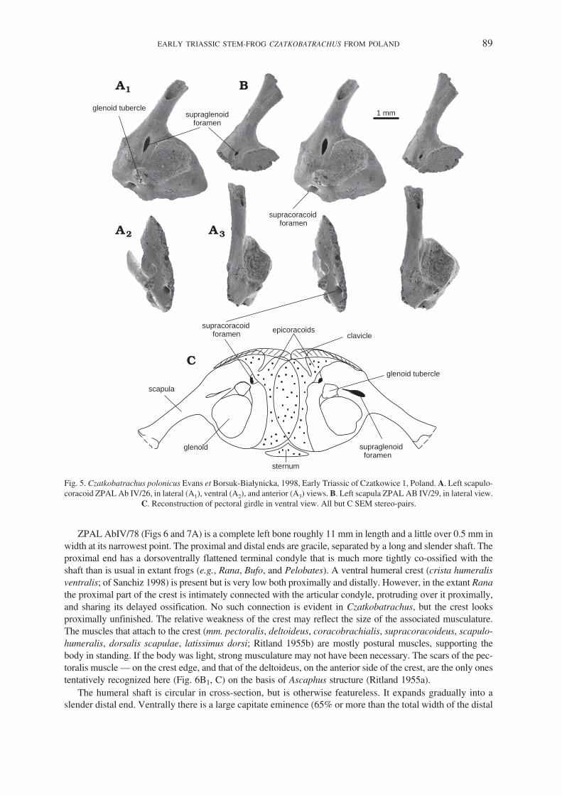
The bone is represented by many fragmentary specimens, the most complete of which is ZPAL AbIV/26(Fig. 5A). It is a single ossification and consists of a subtriangular ventral plate and a narrow blade that wid−ens distally along its vertebral border to about twice the proximal width, although no specimen has the distalend preserved. The scapular blade is unusually long and slender (ZPAL AbIV/29, Fig. 5B). In life, it was in−clined posterodorsally, its axis making an angle of about 65� with the long axis of the coracoid plate.
A large glenoid cavity occupies much of the postero−ventral region of the scapulocoracoid, extendingwell posteriorly. It is subcircular in outline, much deeper dorsoventrally than is usual in lower tetrapods, andis hemispherical rather than funnel shaped (in contrast to urodeles). It faces directly laterad, in contrast to theposterolateral orientation in non−lissamphibian temnospondyls. A distinct anteroventral glenoid tubercleseems to belong to the scapular component of the glenoid and contributes a small articular surface to itsanteroventral border.
Anterodorsal to the glenoid is a large supraglenoid foramen that forms a subvertical cleft partly separatingthe glenoid region of the scapula from its acromial part, homologous and similar in position to the scapularcleft of Anura (Borsuk−Białynicka and Evans 2003; Carroll 2007). The acromial part is a long, laterally (orslightly anterolaterally) flattened process extending ventrally from the scapula, of which it is an integral part.Ventral to the supraglenoid foramen, the acromial process fuses with the procoracoid part (anteroventrally),and with both the scapular and coracoid parts of the glenoid (posteroventrally). Separating the procoracoidregion from the coracoid, the supracoracoid foramen leads into a short canal directed toward the supra−glenoid foramen. Both open on the medial surface of the scapula in the subscapular fossa. The procoracoidregion faces almost laterally, but the coracoid itself turns ventromedially to lie at an angle of about 110� tothe plane of the scapula. Its ventromedial margin ends in a strip of unfinished bone that would have been con−tinued in cartilage in life Fig. 5C), rather than forming a transverse bar as it does in anurans. The retention ofthe supracoracoid foramen, which is included within the scaphoid fenestra in anurans, provides further evi−dence that an independent coracoid bar was not yet developed. Furthermore, it suggests that the arciferal con−tact of the Czatkobatrachus scapulocoracoids was of a caudate rather than anuran pattern. No clavicles havebeen identified for Czatkobatrachus, but they were probably present, given the high degree of ossification ofthe skeleton, and their retention in Triadobatrachus (Rage and Roček 1989). These elements have been lostin caudates, but in arciferal frogs, clavicles are necessary to retain the structural integrity of the girdle, giventhe mobility of the epicoracoid cartilages with respect to each other during locomotion (Emerson 1983).Their possible role in Triadobatrachus and Czatkobatrachus remains obscure.
Humerus. — The humerus was one of the first bones to be recognised for Czatkobatrachus because it ischaracteristically salientian. The majority of specimens preserve only the distal ends (e.g., ZPAL AbIV/2–3,12–13, and 55), but ZPAL Ab IV/78 is a complete humerus that permits a detailed description and also a dis−cussion of forelimb−hind limb ratios.
88 SUSAN E. EVANS and MAGDALENA BORSUK−BIAŁYNICKA
ZPAL AbIV/78 (Figs 6 and 7A) is a complete left bone roughly 11 mm in length and a little over 0.5 mm inwidth at its narrowest point. The proximal and distal ends are gracile, separated by a long and slender shaft. Theproximal end has a dorsoventrally flattened terminal condyle that is much more tightly co−ossified with theshaft than is usual in extant frogs (e.g., Rana, Bufo, and Pelobates). A ventral humeral crest (crista humeralisventralis; of Sanchiz 1998) is present but is very low both proximally and distally. However, in the extant Ranathe proximal part of the crest is intimately connected with the articular condyle, protruding over it proximally,and sharing its delayed ossification. No such connection is evident in Czatkobatrachus, but the crest looksproximally unfinished. The relative weakness of the crest may reflect the size of the associated musculature.The muscles that attach to the crest (mm. pectoralis, deltoideus, coracobrachialis, supracoracoideus, scapulo−humeralis, dorsalis scapulae, latissimus dorsi; Ritland 1955b) are mostly postural muscles, supporting thebody in standing. If the body was light, strong musculature may not have been necessary. The scars of the pec−toralis muscle — on the crest edge, and that of the deltoideus, on the anterior side of the crest, are the only onestentatively recognized here (Fig. 6B1, C) on the basis of Ascaphus structure (Ritland 1955a).
The humeral shaft is circular in cross−section, but is otherwise featureless. It expands gradually into aslender distal end. Ventrally there is a large capitate eminence (65% or more than the total width of the distal
EARLY TRIASSIC STEM−FROG CZATKOBATRACHUS FROM POLAND 89
sternum
glenoid
glenoid tubercle
scapula
supracoracoidforamen
supraglenoidforamen
supraglenoidforamen
supracoracoidforamen
epicoracoids
glenoid tubercle
clavicle
1 mm
Fig. 5. Czatkobatrachus polonicus Evans et Borsuk−Białynicka, 1998, Early Triassic of Czatkowice 1, Poland. A. Left scapulo−coracoid ZPAL Ab IV/26, in lateral (A1), ventral (A2), and anterior (A3) views. B. Left scapula ZPAL AB IV/29, in lateral view.
C. Reconstruction of pectoral girdle in ventral view. All but C SEM stereo−pairs.

end). The remaining some 35% of the distal end is occupied by the ulnar epicondyle, contacting the eminenceventrally through a small trochlear surface (Figs 6B, 7A3). Dorsally, there is a short triangular olecranon scar(Fig. 7B). The radial epicondyle — the site of origin of many extensor muscles (e.g., mm. extensor carpiradialis, extensor carpi ulnaris, extensor digitorum communis longus), is very small (suggesting weakextensor muscles) while the ulnar epicondyle (mostly for flexor muscles) is more prominent. These differ−ences may relate to sexual behaviour (amplexus) and may be subject to sexual dimorphism (Roček, personalcommunication 2007). The longitudinal medial and lateral distal muscle crests are quite feeble (Fig. 6A, C),and thus the forelimbs were probably not heavily muscled.
90 SUSAN E. EVANS and MAGDALENA BORSUK−BIAŁYNICKA
ventral crest
capitateeminence
ventralcubitalfossa
ulnarepicondyle
proximal condyle
deltoidmuscle scar
ventralcrest
lateralcrest
medialcrest
pectoralismuscle
scar
capitateeminence
trochlear surfaceof ulnar epicondyle
1 mm
ulnarepicondyle
Fig. 6. Czatkobatrachus polonicus Evans et Borsuk−Białynicka, 1998, Early Triassic of Czatkowice 1, Poland. Left humerusZPAL Ab IV/78, in posterior = medial (A), ventral (B), and anterior = lateral (C) views. A1, B2, C, SEM stereo−pairs.

Antebrachium. — The ulna of Czatkobatrachus is represented by three specimens of the proximal end(e.g., ZPAL AbIV/22, Fig. 7C). Like the humerus, the bone is well ossified with a strong elongated olecranonprocess, providing the lever arm for the triceps muscle (m. anconeus; Duellman and Trueb 1986) and a con−cave articular surface for the humeral articulation. The elbow joint seems to have been stable. No radius hasbeen recognized as yet.
PELVIC GIRDLE AND HIND LIMB
Pelvic girdle. — The pelvic girdle of Czatkobatrachus is represented by many distinctive ilia (some 18specimens), some specimens having the puboischiadic plate fused in place (e.g., ZPAL AbIV/111 and 129;Fig. 8D and B, respectively). At the time of the original description (Evans and Borsuk−Białynicka 1998), themost complete ilium was the holotype, ZPAL AbIV/7, but more complete specimens have been found subse−quently. ZPAL AbIV/114 (Fig. 8A) is one of the most complete, the delicate iliac shaft usually breaking at orclose to its junction with the acetabular region. The acetabular region is expanded, with a large shallow lat−eral acetabular surface, little development of a supra−acetabular buttress, and no extension of the anterior orposterior acetabular margins (into partes descendens or ascendens; Sanchiz 1998). The medial surface issmooth with no trace of an interiliac synchondrosis. The most obvious feature is a large protruding dorsalprominence (also present in Triadobatrachus; Rage and Roček 1989, fig. 3) for the gluteus magnus muscle or
EARLY TRIASSIC STEM−FROG CZATKOBATRACHUS FROM POLAND 91
capitate eminence
olecranon scar dorsal side
ulnar trochlea
olecranon
1 mm
Fig. 7. Czatkobatrachus polonicus Evans et Borsuk−Białynicka, 1998, Early Triassic of Czatkowice 1, Poland. A. Left humerusZPAL Ab IV/78, in dorsal view (A1), proximal end (A2), and distal end (A3). B. Distal part of the left humerus ZPAL Ab IV/55,
in dorsal view. C. Ulna ZPAL Ab IV/22, in anterior (C1) and lateral (C2) views. SEM stereo−pairs.
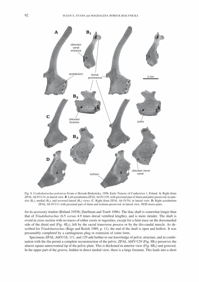
for its accessory tendon (Ritland 1955b; Duellman and Trueb 1986). The iliac shaft is somewhat longer thanthat of Triadobatrachus (6.5 versus 4.9 times dorsal vertebral lengths), and is more slender. The shaft isovoid in cross section with no traces of either crests or rugosities, except for a faint trace on the dorsomedialside of the distal end (Fig. 4E2), left by the sacral transverse process or by the ilio−caudal muscle. As de−scribed for Triadobatrachus (Rage and Roček 1989, p. 11), the end of the shaft is open and hollow. It waspresumably completed by a cartilaginous plug or extension of some form.
Specimens ZPAL AbIV/16, 111, and 129 add further to our knowledge of pelvic structure, and in combi−nation with the ilia permit a complete reconstruction of the pelvis. ZPAL AbIV/129 (Fig. 8B3) preserves thealmost square anteroventral tip of the pelvic plate. This is thickened in anterior view (Fig. 8B1) and grooved.In the upper part of the groove, hidden in direct medial view, there is a large foramen. This leads into a short
92 SUSAN E. EVANS and MAGDALENA BORSUK−BIAŁYNICKA
acetabulum
pubisobturatorforamen
ischium
obturatorcanal
entrance
obturator nervecanal
dorsalprominence 2 mm
Fig. 8. Czatkobatrachus polonicus Evans et Borsuk−Białynicka, 1998, Early Triassic of Czatkowice 1, Poland. A. Right iliumZPAL Ab IV/114, in lateral view. B. Left acetabulum ZPAL Ab IV/129, with proximal part of ilium and pubis preserved, in ante−rior (B1), medial (B2), and reversed lateral (B3) views. C. Right ilium ZPAL Ab IV/54, in lateral view. D. Right acetabulum
ZPAL Ab IV/111 with proximal part of ilium and ischium preserved, in lateral view. SEM stereo−pairs.

canal that runs through the anterior pelvic margin at roughly 45 degrees and emerges on the lateral surface(Fig. 8B3) just below the anterior rim of the acetabulum. This canal is not present in crown−group anuransand is not described for Triadobatrachus, but from its course and position must have carried the obturatornerve and blood vessels from out of the pelvic cavity. In non−lissamphibian temnospondyls and amniotes,this canal perforates the pubis, suggesting that the anterior corner of the pelvic plate in Czatkobatrachus is anossified remnant of this bone (cartilaginous in crown−group frogs), not a calcified prepubis as found in
EARLY TRIASSIC STEM−FROG CZATKOBATRACHUS FROM POLAND 93
femoralcrest
femoralcondyle
tibialarticularsurface
ventralside
femoralcrest
fibulararticularsurface
poplitealspace
ventral side
2mm
2 mm
Fig. 9. Czatkobatrachus polonicus Evans et Borsuk−Białynicka, 1998, Early Triassic of Czatkowice 1, Poland. Left femur ZPALAB IV/25, in anterior (A), posterior = lateral (B), ventral (C), proximal (D), and distal (E) views. All but B2, C2 SEM stereo−pairs.

Ascaphus and Xenopus (Ritland 1955a). Posteriorly, ZPAL AbIV/16 (not figured) and 111 (Fig. 8D) pre−serve the almost complete posteroventral, ischiadic part of the pelvic plate. They show that the acetabularsurface is not rounded in the complete pelvis but instead has a posterior triangular extension formed by theischium. This, in turn, suggests that there was an anteroposterior component to the femoral movement as wellas the dorsoventral one. The triangular ischium closely resembles the same element in Triadobatrachus, ex−cept that the ischium is free from the rest of the pelvis in the Malagasy genus.
In crown−group frogs, the two halves of the adult pelvis meet posteroventrally at a steep angle, with a deepsynchondrosis of the pubo−ischiadic plate that may or may not extend to the base of the ilium. As described byGreen (1931) for Rana, this synchondrosis deepens gradually during development, with only the edges of thepubo−ischiadic plate meeting initially but then incorporating more and more of the plate until the two halves arecompletely fused. This articulation is not discussed for Triadobatrachus (Rage and Roček 1989). The only visi−ble articular surface on the pubo−ischiadic plate of the Polish form is a recessed pitted strip along theventromedial margin (Fig. 8B2). The two pelvic plates clearly met at an angle (a horizontal orientation wouldrender the pelvis too wide), but with a relatively weak synchondrosis, like that of the larval Rana.
Femur. — The only hind limb element to be identified with any certainty is the femur (Fig. 8), but a secondelement closely matches the fibulare (Fig. 10) of modern frogs. No conjoined tibiofibulae have been recovered.Since these are among the most characteristic and common frog elements preserved in microvertebrate assem−blages, we are confident that in Czatkobatrachus, as in Triadobatrachus, these two bones were not fused.
The femur is represented by two complete specimens, ZPAL AbIV/25 (Fig. 9) and 125, and several frag−ments. By comparison with the modern Bufo punctatus, the complete specimens are both right femora. How−ever, the attribution of femora to right or left can be somewhat problematic, because of the lack of landmarks,and the variability of bone curvature in extant frogs. In Rana, the bone is S−shaped in a dorso−ventral plane,but is curved in both dorso−ventral and lateral planes in Bufo, and in only one plane in Pelobates. Perhaps thetype of locomotion, and the size have a bearing on this character, but the relationships are obscure. InCzatkobatrachus, the femur is only slightly bowed dorsoventrally (dorsally concave Fig. 9A, B), and proba−bly twisted, the axes of the ends being subperpendicular to each other. The torsion is less obvious in Bufo butis difficult to observe in extant frogs because their epiphyses are usually lost. In contrast to them, Czatko−batrachus femora have completely fused epiphyses and their articular surfaces are at least partly finished (al−though in life they were certainly coated with articular cartilage).
ZPAL Ab IV/25 (Fig. 9) is long and slender, ca. 12.5 mm in total length with small proximal and distalends of equal width (ca. 2 mm). The shaft is 0.8 mm at its narrowest point. The femur is thus only slightlylonger than the humerus and of very similar proportions. The proximal end bears a proximally convex,slightly bilaterally flattened, condyle (Fig. 9D). The femoral crest (corresponding to the trochanter) ex−tends down the ventral surface beginning proximally from a point about 1/5 of the femoral length and fad−ing out at about 3/5 femoral length. By comparison with extant frogs (Ascaphus as illustrated by Ritland
94 SUSAN E. EVANS and MAGDALENA BORSUK−BIAŁYNICKA
1 mm1 mm
Fig. 10. Czatkobatrachus polonicus Evans et Borsuk−Białynicka, 1998, Early Triassic of Czatkowice 1, Poland. Possible fibulareZPAL Ab IV/138, in proximal (A) and lateral (B) views. SEM stereo−pairs.

1955a, b), the crest is for the attachment of hip muscles (mm. pectineus, gluteus minimus, iliofemoralis,pyriformis, quadratus femoris, and obturator externus; Ritland 1955b). The distal end has its surface dif−ferentiated (Fig. 9E). By comparison with a Bufo femur, a large concavity in the anteroventral part of distalsurface was probably for the tibia, whereas a second, posterior, convexity articulated in some way with thefibula. As noted above, there is no evidence that the crural bones were fused.
Possible additional element. — ZPAL AbIV/138 (Fig. 10) is a long bone of similar overall morphologyand bone type (density and surface texture) to that of the other limb elements of Czatkobatrachus, but it ismuch shorter than either the humerus or femur (roughly 45% of the femoral length). Like the femur, the shaftis bowed, with one edge almost straight and the other strongly concave. Both heads are compressed, with apossible distal head broader than the proximal one. This bone does not show any obvious match to either ofthe epipodials in Triadobatrachus (and is relatively shorter) or modern frogs (where they are rounded but al−ways co−ossified), although it does show some resemblance to the epipodials of salamanders. The closestmatch in a modern frog is to the fibulare, one of the two ankle bones that become elongated in crown−groupfrogs. These bones are apparently only slightly elongated in Triadobatrachus so their extension in Czatko−batrachus would represent an advance, although it would be consistent with the generally longer and moregracile limbs of the Polish form.
CHARACTER ANALYSIS
Axial length. — Crown−group frogs are characterised by a very short vertebral column (typically 6–9presacrals, Griffiths 1963; Kluge and Farris 1969; Lynch 1973). The number of presacral vertebrae inCzatkobatrachus but it was probably similar to that of Triadobatrachus (14–15), because their vertebrae arevery similar both in length and morphology. If this is correct, then the body axis of basal salientians wasmuch shorter than that of many outgroup taxa (e.g., 19–24 in branchiosaurs, Boy and Sues 2000), a shorten−ing that would have involved a reduction in the number of trunk somites (Richardson et al. 1998). The re−cently described amphibamid Gerobatrachus has 17 presacrals, intermediate between the primitive conditionand that in basal salientians (Anderson et al. 2008).
The first spinal nerve. — The notch in the anterior margin of the atlas in Czatkobatrachus is reminiscentof the morphology of crown−group salamanders (but not stem taxa) and caecilians. In salamanders and cae−cilians the atlantal foramen transmits a spinal nerve, but the identity and homology of this nerve is still dis−puted. According to Duellman and Trueb (1986), the first spinal nerve (here called the transatlas nerve toavoid confusion later) emerges through the atlas in salamanders, but between the atlas and second presacralin frogs. Deuchar (1975), however, argued that the disposition of the anterior spinal nerves in frogs is similarto that in salamanders, the difference being that the first nerve (between the occiput and atlas; sometimescalled the suboccipital nerve, e.g., Mookerjee 1930, 1931; Fox 1954) usually gets lost in anuran metamor−phosis. Whether this nerve really exists in frogs, and which nerve, if any, is its equivalent in salamanders andcaecilians is a problem on which Czatkobatrachus structure sheds some light. This, however, requires a briefdiversion into cranial embryology.
In the embryonic amphibian head, there are four segments (somitomeres) (Jacobson 1993 and personalcommunication 2003), three preotic and one subotic. These are followed by two postotic somitomeres thatbecome the first two somites. A preoccipital arch forms between the first and second of these (somitomeres 5and 6), the occipital arch forms between the second and third somite (somitomeres 6 and 7), and the atlas archforms between the third and fourth somites (somitomeres 7 and 8) (Goodrich 1911; Wake and Lawson 1973).
According to Jacobson (1993) the first spinal nerve emerges from the third somite, and thus at the level ofthe anterior part of the atlas (see Burke et al. 1995 for different opinion). Francis (1934) claimed that sala−manders had a still more anterior nerve (in front of the atlas, in addition to the transatlas nerve), equivalent,according to him, to the suboccipital nerve, and thus to the spinal nerve 1. The transatlas nerve would thuscorrespond to spinal nerve 2 (although Francis chose to designate it as spinal nerve 1 as it was the first nervevisible in the adult). However, Wake and Lawson (1973) found no evidence for the existence of any nerve infront of the atlas in salamanders, and identified the suboccipital nerve with the transatlas nerve, as spinalnerve 1, with nerve 2 passing behind the atlas. This is the homology accepted herein.
EARLY TRIASSIC STEM−FROG CZATKOBATRACHUS FROM POLAND 95

This accepted homology also agrees with the pattern of innervation of the atlanto−occipital muscles, andwith morphology. The transatlas nerve of salamanders has only a ventral root, and the first spinal nerve infrogs has lost the dorsal root and dorsal root ganglion during metamorphosis (Mookerjee 1930, 1931; Fox1954). Wake and Lawson (1973) also suggested that the first spinal nerve originally lay in front of the atlasarch in salamanders (within the range of the third somite), moving back into the atlas pedicel through time(possibly to protect it from the cranio−cervical joint surfaces). A similar process has been assumed by Ritland(1955a) in crown−group frogs (e.g., Rana and Xenopus) in which the fibers of the first spinal nerve fuse withthe spinal nerve 2 (Ritland 1955, pp. 160–163) completely losing their individuality during metamorphosis,except in Ascaphus and Leiopelma in which they are very much reduced. According to Ritland (1955a, p.163) “the way in which change may occur at any level of the cord is by a gradual shift of fibers to the nextposterior exit from the spinal canal”.
The alternative scenario by Francis (1934) would require that the second spinal nerve moved forwardsthrough the atlas from back to front (to become transatlas nerve i.e., his spinal nerve 1 in adults). This is not thepattern seen in the embryonic development of salamanders, where the notch in the front edge of the atlas gradu−ally becomes enclosed (e.g., Mookerjee 1930, 1931). The same transition is seen in salamander evolutionwhere an anteriorly open notch (SEE) may present an intermediate stage between the unnotched condition insome karaurids ( Evans et al. 1988, SEE personal observations) and the perforating foramen of crown−groupurodeles (SEE). The presence of a notch in the anterior margin of the atlas in Czatkobatrachus provides evi−dence that the condition in stem−frogs was not dissimilar to the condition in most salamanders, with the first spi−nal nerve (= suboccipital nerve, i.e., transatlas nerve) notching the anterior margin of the atlas. A fully enclosedatlantal foramen is also found in basal caecilians (Jenkins and Walsh 1993; Evans and Sigogneau−Russell2001), and could be a basal lissamphibian feature. However, the absence of either a notch or foramen in thestem−caudate Marmorerpeton (Evans et al. 1988, and SEE personal observations) renders this character prob−lematic, as does the two−state condition (anterior incision or closed foramen) in salamanders, and its onto−genetic variation.
Subdivision of the atlas. — Rage and Roček (1989) and Roček and Rage (2000) interpreted the atlas ofTriadobatrachus as being bipartite, with a single arch but with the centrum divided into anterior and posteriorparts, and with a dichocephalous atlas rib (see also Estes and Reig 1973) meeting both components. Thiswould be an unusual morphology, and it is certainly not the condition in Czatkobatrachus where the atlantalcentrum is a single entity with a short neural arch bearing the postzygapophysis but no rib. According toShishkin (2000, pp. 543–544) one of the few apomorphies that can be attributed with confidence totemnospondyls (including lissamphibians, Duellman and Trueb 1986) is the absence of transverse processeson the atlantal neural arch. Fusion of the first two vertebrae can occur in frogs (e.g., palaeobatrachids,rhinodermatids, brachycephalids, and some bufonids, myobatrachids, and pipids, Duellman and Trueb 1986,p. 470; rarely in Ascaphus, Ritland 1955a), and is probably associated with a strong shortening of both thecentra and neural arches (Ritland 1955a, fig. 4). Such fusion does lead to the presence of a transverse process,but it is associated with the second vertebral segment. Contrary to the opinion of Rage and Roček (1989), theneural arches were certainly present on the first two vertebrae in Triadobatrachus, as shown by what is prob−ably the pedicel of atlantal arch, and by the postzygapophyses on the second vertebra (Fig. 4A). The state ofpreservation of Triadobatrachus does not permit a full understanding of its neck structure. The second verte−bra of Triadobatrachus is damaged (Roček and Rage 2000, fig. 3), but what remains appears to be quite con−sistent with the morphology of the same element in Czatkobatrachus (Fig. 4A).
Tuberculum interglenoideum. — As most frogs, Czatkobatrachus lacks an interglenoid tubercle betweenthe atlantal cotyles, having instead a flat surface perforated by the notochord. However, an anterior median pro−cess reported by Báez and Basso (1996, p. 143) in Notobatrachus and a nubbin−like process in the same posi−tion of Prosalirus (Jenkins and Shubin 1998, p. 500 and fig. 3A) may represent remnants of the interglenoid tu−bercle. The tubercle is present in most crown group salamanders (secondarily lost in some paedomorphic taxa,e.g., batrachosauroids, Estes 1981), in albanerpetontid amphibians (SEE personal observations), in stem−cae−cilians (Jenkins and Walsh 1993; Evans and Sigogneau−Russell 2001), and in the derived amphibamidGerobatrachus (Anderson et al. 2008). It is absent in the Middle Jurassic karaurid Marmorerpeton (Evans et al.1988), but has been described as present in the Upper Jurassic Karaurus from Kazachstan. Its presence mightbe a synapomorphy linking caecilians, crown−group salamanders and albanerpetontids (e.g., Feller and Hedges
96 SUSAN E. EVANS and MAGDALENA BORSUK−BIAŁYNICKA

1998; but see Hay et al. 1995), or, given its presence in Gerobatrachus (Anderson et al. 2008), a character ofGerobatrachus and Batrachia (sensu Anderson et al. 2008). This would require a subsequent reversal incrown−group caecilians, in salientians, and in Marmorerpeton. However, the tubercle can also occur in otheramphibian lineages (e.g., some microsaurs, Carroll and Gaskill 1978), and it may be of functional significance.
Caudals. — On present evidence, the fusion of the caudal vertebrae to form the urostyle is a derived char−acter of crown−group Anura, although some early taxa retained a single postsacral vertebra in front of theurostyle (e.g., some Notobatrachus, Báez and Basso 1996). According to Ritland (1955a), the extantAscaphus also retains a strong tendency for the possession of discrete post−sacral vertebrae. In Czatko−batrachus the presence of discrete caudals is considered plesiomorphic, as is their presence in Triado−batrachus, but the reduced caudal number they probably share is derived. The widely open notochordal canalin Czatkobatrachus caudals implies the persistence of the notochord into the tail.
Transverse processes/ribs. — Developmental studies on living frogs (Blanco and Sanchiz 2000) haveshown that the transverse process and small free rib, where present, are part of the same anlagen. Differencesbetween clades reflect different degrees and patterns of ossification. In neobatrachian frogs, the transverse pro−cess ossifies as a single unit and fuses to the vertebral body. Three extant families (ascaphids, discoglossids andpipids: Trueb 1973), and virtually all Mesozoic anurans (Rocek 2000, including Vieraella, Notobatrachus,Prosalirus, Eodiscoglossus), retain small ribs on the anterior presacrals. In Triadobatrachus, all vertebrae, ex−cept the atlas (but see Rage and Roček 1989 for different opinion) bear free ribs, and no vertebra has more thana short transverse process to which the rib attaches. This is presumably the primitive salientian condition since itis also found in stem caudates (Evans, unpublished data). Czatkobatrachus resembles primitive modern frogs inhaving short transverse processes that probably contacted free ribs (not yet recognized, but interpreted from thepitted terminal surfaces of the processes) on a few anterior presacrals (ZPAL Ab IV/108–110), but fused oneson posterior vertebrae (ZPAL Ab IV/6, 10, 127, 128, 146) as shown by areas of incomplete fusion in ZPALAB/IV/6 (Fig. 2A). These processes are directed laterally in some anterior vertebrae and posteriorly in most ofthe others; they are slender mid−trunk, but become thickened immediately in front of and behind the sacrum.The state of transverse processes/ribs in Czatkobatrachus is here considered derived.
Pectoral girdle. — The pectoral girdle of Czatkobatrachus has been reconstructed (Borsuk−Białynickaand Evans 2002) as a pair of undivided scapulocoracoid plates ventrally connected in an arciferal manner(Fig. 5C), no clavicle, cleithrum or sternum fragments having been identified. Crown−group frogs, with botharciferal and firmisternal girdles, have a separate scapula and coracoid. This is in contrast to non−liss−amphibian temnospondyls (Borsuk−Bialynicka and Evans 2002 and references herein) and to caudates thathave a single scapulocoracoid, this state being considered plesiomorphic. Within Anura, more basal cladeslike Ascaphidae, Discoglossidae and Pipidae display short scapulae, in contrast to neobatrachian frogs wherethe scapula may be longer and more slender (Trueb 1973), but never to the extent seen in Czatkobatrachus.In ascaphids and pipids the scapulae are proximally uncleft while being cleft or bicapitate in almost all otherfrogs (Trueb 1973). The widely held opinion (e.g., Trueb 1973) that short uncleft scapulae are primitive forfrogs has been challenged by the structure of the scapulocoracoid in Czatkobatrachus. Its elongate scapularblade is proximally perforated by the supraglenoid foramen. This separates the anterior acromial part fromthe posterior, glenoid part, and is most probably homologous to the scapular cleft (Borsuk−Białynicka andEvans 2002) or a scapular cleft in statu nascendi.
Trueb (1973) stated explicitly that the arciferal type of pectoral girdle was plesiomorphic for salientians,and that firmisterny is derived. Emerson (1983) was more cautious. However, the consistently arciferal struc−ture of the caudate pectoral girdle suggests that this is the plesiomorphic state, and we accept this view.Firmisterny is derived, and is probably better as a shock−absorbing device Although earlier reconstructions ofTriadobatrachus (Rage and Roček 1989) suggested it had a tripartite pectoral girdle like that of modernfrogs, our studies have shown that the pectoral girdle of the Malagasy specimen might be reinterpreted as asingle structure (Borsuk−Białynicka and Evans 2002) like that of Czatkobatrachus.
Ilium and ilio−sacral joint. — An elongate anteriorly directed ilium is synapomorphic for the Salientia(Triadobatrachus included) and is shared by Czatkobatrachus, but its contact with the sacrum is probablystill plesiomorphic or intermediate. Emerson (1979) was probably the first to realize that the ilia articulateventral to the sacrum in extant frogs. They do also in Notobatrachus (Báez and Basso 1996, fig. 13). As re−
EARLY TRIASSIC STEM−FROG CZATKOBATRACHUS FROM POLAND 97

constructed herein, the contact in Czatkobatrachus occurs between the medial surface of the ilium and thelateral concavity of the sacral process whereas the anteroventral extension of the transverse process props theilium from the ventral side. However, a posterior extension of the transverse process does overlap the iliumshaft dorsally and may represent a rudiment of the modern configuration.
Puboischiadic plate. — In salamanders, the pubis is unossified and the same is generally true of frogs(Trueb 1973), although Ritland (1955a) reported it as ossified or calcified in Ascaphus (calcified according toTrueb 1973). It is also ossified in all pipids (Roček, personal communication 2007). In albanerpetontids(McGowan and Evans 1995) an ossified pubis remains, providing evidence that it was still present in thelissamphibian ancestor (assuming monophyly), although Anderson et al. (2008) report it as unossified in theamphibamid Gerobatrachus (but this could be due to immaturity). In living frogs, the pubis is restricted to asmall unossified region of the ventral pelvic plate between the ilium and ischium. The pelvis of Czatko−batrachus thus represents an intermediate stage in which the pubis has been greatly reduced (as in modernfrogs) but remains ossified. It is also intermediate in the presence of the perforating canal.
In amniotes and in most non−lissamphibian temnospondyls, the ossified pubis contains a conspicuous fo−ramen for the obturator nerve and its accompanying blood vessels. In salamanders, this nerve supplies apuboischiofemoralis internus muscle on the inner face of the pelvis (Francis 1934) and then sends branchesto puboischiofemoralis externus on the outside. The nerve is said to be reduced in salamanders compared toother tetrapods (Noble 1922) and is absent in living frogs (Green 1931; Ritland 1955a), with its role takenover by a branch of the femoral nerve. This change has been linked to metamorphosis, when there is a loss ofsome spinal nerves (Green 1931). The presence of a homologue of the obturator canal in Czatkobatrachusimplies retention of the nerve. This would be consistent with the fact that there has been less reduction of thepresacral region in Czatkobatrachus than in crown group frogs.
Sacro−caudal joint. — According to Trueb (1973) the anuran sacro−urostylar joint is subject to consider−able variation. Most crown−group frogs have a bicondylar sacro−urostylar joint, and this was regarded asprimitive by Trueb (1973, see also Duellman and Trueb 1986). However, the Jurassic Prosalirus, and the ex−tant Ascaphus and Leiopelma, retain a simple fibrocartilaginous intercentral connection that is identical tothose within the presacral column. This condition is considered to be more primitive by Jenkins and Shubin(1998), and is that found in Czatkobatrachus. The Czatkobatrachus sacrum retains postzygapophyses, as aplesiomorphic character, as does Notobatrachus (Báez and Basso 1996). These are absent in Prosalirus(Jenkins and Shubin 1998) and in most extant frogs (Trueb 1973; exceptions include some extant pelobatidsand discoglossids).
Limb bones. — Both the humerus and femur display a strikingly anuran morphology that may be at leastpartly size−dependent. Which, if any, of their characters are synapomorphic for Czatkobatrachus andcrown−group anurans is a question that requires more extensive studies of the out−groups. The large capitateeminence of the anuran humerus is shared not only by caudates and albanerpetontids (SEE personal observa−tions) but also by the Dissorophoidea and by a more−inclusive group of temnospondyls (the Euskelia of Yatesand Warren 2000), and is thus plesiomorphic at the level of Lissamphibia. The size and shape of the ventralcrest of the humerus is also similar in both anurans and caudates, but salientians lack the dorsal crest of thehumerus, that bears an attachment for the humeral retractor (subscapularis muscle) in caudates. The slenderelongate salientian−type femur bears a low ventral crest for the attachment of hip muscles (mm. pectineus,gluteus, iliofemoralis, pyriformis, quadratus femoris, and obturator externus; Ritland 1955b) instead of theprotruding finger−like trochanter serving the same function in caudates. The development of prominenthumeral and femoral crests is probably derived for salamanders, with salientians showing the primitive state.
FUNCTIONAL MORPHOLOGY
General axial characters. — Shortening the axial skeleton limits its capacity for lateral undulation dur−ing locomotion and implies that both Czatkobatrachus and Triadobatrachus had already modified their loco−motor strategy towards a gait in which there was a greater dependency on the limbs.
98 SUSAN E. EVANS and MAGDALENA BORSUK−BIAŁYNICKA

Transverse processes. — Reduction of lateral mobility of the vertebral column in Czatkobatrachus isalso supported by the fusion and, sometimes, by elongation of transverse processes, which suggest the spinewas more stiffened in the horizontal plane than that of Triadobatrachus. Enlargement of the peri−sacral trans−verse processes may be associated with strengthening of the ilio−lumbaris (anteriorly) and of caudal muscula−ture homologous to coccygeo−iliacus and coccygeo−sacralis (posteriorly) (Emerson and De Jongh 1980).These may have provided additional support and stabilisation to the ilio−sacral articulation during locomotion(see also for lumbo−dorsal fascia below).
Caudal vertebrae. — The reduction of the tail in Triadobatrachus, and presumably also in Czatko−batrachus (see above), shows that these animals had moved away from a primarily undulatory mode of loco−motion, for which the tail is of great importance. Despite the absence of the urostyle in both these animals,the juxtaposition of a shortened tail between elongate, anteriorly directed ilia may, according to Jenkins andShubin (1998), represent a primitive stage in the evolution of the anuran caudopelvic musculature linkage.We concur with this view. The absence of any ridges or tuberosities on the cylindrical terminal caudal ele−ments suggests they were not tightly held together and probably retained some flexibility (but see also the in−formation on transverse processes above).
Elements of the caudopelvic mechanism. — Saltation is, without doubt, the most distinctive feature ofcrown−group frogs and was present in the earliest known and most primitive anuran taxa (Prosalirus,Vieraella, Notobatrachus). The key finding of Emerson and De Jongh (1980) was that the typical anuran sal−tation mechanism requires a body divided into two components — anterior (head, forelimbs, presacral col−umn and sacrum) and posterior (pelvis, urostyle and hind limbs) separated by a joint (sacro−urostylar) thatpermitted dorsoventral flexion and extension. In jumping, the sacro−urostylar joint extends to bring the sa−crum, presacral series and head into line with the urostyle and the long axis of the ilia.
Sacro−caudal joint. — In extant frogs a loss of sacral postzygapophyses and the development of abicondylar, uniaxial, joint between the sacral centrum and the urostyle (Jenkins and Shubin 1998) allow forextension−flexion movements in the sacro−urostylic joint. In Czatkobatrachus, the retention of both a simplesacro−caudal joint and sacral postzygapophyses do not exclude, but do limit, the potential for dorsoventralexcursion of the spine at this joint (Jenkins and Shubin 1998). Modern anurans that lack this capacity alsolack true saltation, and have locomotor patterns involving walking, swimming, climbing, or burrowing. Thisis likely to have been the case for Czatkobatrachus.
Ilium. — The presence of an elongate ilium, the shaft of which extends well anterior to the level of theacetabulum, obviously contributed to the stiffening of the posterior body segment. This important element ofthe frog−type locomotory mechanism had already developed in Czatkobatrachus and, to a lesser degree (Fig.4), in Triadobatrachus. The rotation and elongation of the iliac shaft, that occurs prior to metamorphosis inextant frogs (Green 1931), was also one of the first postcranial characters developed in salientian phylogeny.As this iliac elongation evolved well before true saltation, its selective advantage remains obscure, particu−larly as it now functions in many different locomotory types including jumping, walking, and swimming.
The ilia of Czatkobatrachus and Triadobatrachus are distinctive in having a hypertrophied dorsal tuber−cle — dorsal prominence (according to Sanchíz’s 1998 terminology). In extant frogs, the dorsal prominenceis associated with the origin of the gluteus magnus muscle, a powerful extensor of the hip. The function ofhip (and knee) extension in leaping is obvious, but it is less clear why this tubercle should be so enlarged intwo taxa that were clearly not saltatory. In most frogs that possess a dorsal prominence, this structure isaligned with the iliac blade and is not protruding. It is, however, hypertrophied in living pipids and somebufonids, apparently in relation to the presence of an accessory tendon of the gluteus magnus muscle that at−taches to it (Dunlap 1960; Emerson and Jongh 1980). This tendon is said to be a part of the dorsolumbar fas−cia and has a role in limiting movement of the urostyle in relation to the pelvic girdle. Both pipids andbufonids are locomotor specialists, swimming or walking respectively. Since they are not related, this is aconvergent adaptation. The accessory tendon is not present in either Ascaphus or Leiopelma, and the samewas presumably true of early crown group frogs as the dorsal prominence is not hypertrophied in Prosalirus(Jenkins and Shubin 1998), Vieraella, or Notobatrachus (Báez and Basso 1996). Whether the enlargement ofthe dorsal prominence of stem−frogs is comparable to that of pipids and bufonids is, of course, impossible todetermine but it does suggest the gluteus magnus, or an accessory tendon, had an important role in stem−frog
EARLY TRIASSIC STEM−FROG CZATKOBATRACHUS FROM POLAND 99

locomotion, perhaps (as in the living taxa) in stabilisation of the pelvis against the sacrum and tail. This couldhave been important to taxa like Czatkobatrachus and Triadobatrachus in which the ilio−sacral joint was stillvery weak (see below).
Ilio−sacral joint. — Emerson (1979, 1982), Emerson and De Jongh (1980), and Jenkins and Shubin(1998) have dealt with this topic in some depth. The ilio−sacral joint of crown−group frogs is not asynchondrosis or an abutting joint as it is in most tetrapods. Instead, the sacral diapophysis lies above theiliac blade, suspending it by means of a specialised ligament system that frequently contains a sesamoid.Emerson (1982) described three principle types of ilio−sacral joint in frogs, ranging from the specialisedjoint of pipids (her type I) that permits fore−aft sliding between an expanded sacral diapophysis and a longilium, to the mechanically simplest kind (her type IIB) in which the sacral diapophysis is only slightly di−lated and the iliac blade is suspended from it by means of a ligament that runs from the distal end of the sa−cral rib to the ventral surface of the ilium. This arrangement, found in basal ascaphids and derived ranids(as well as the Jurassic Prosalirus, Jenkins and Shubin 1998), is said to maximise dorsoventral rotation ofthe pelvis on the sacrum and tends to limit movement to this plane. There is a variant of this arrangement(Type IIA) in which distally expanded sacral processes with arcuate distal edges are connected to the iliumby means of a ligament inserting dorsally near the base of the sacrum rib. The difference between types IIA and IIB in the position of the ligament origin reflect a difference in mobility, type IIA allowing for a lat−eral swing of the pelvis on the sacrum. This type, found mostly in walking frogs, tends to increase the rangeof protraction (and thus stride length), but at the expense of jumping ability (since the body would be lessstable in the leap). It could be predicted that early frogs and stem−frogs might have an arrangement inter−mediate between type IIA and IIB, so that there was both dorso−ventral and lateral movement, permittingwalking and imperfect jumping, in the absence of the specialised sacro−urostylar system. This is clearly notthe condition in Triadobatrachus where the sacral ribs remain separate from the vertebral body and the dis−tal ends are elongated, posteriorly directed processes that have a long abutting contact with the medial sideof the ilium (Rage and Roček 1989), much like the arrangement in more basal amphibians. Czatko−batrachus has sacral diapophyses that are fused to the vertebral body. Each of them has a widely extendeddistal end containing a dorsolaterally to laterally facing concavity that probably received the ilium in a lat−eral (rather than ventral) position. A dorsal ligament furrow, extending medially, well towards the shaft ofthe sacral process, clearly recalls Emerson’s type IIA and suggests lateral mobility rather than jumping.According to Jenkins and Shubin (1998), the ventral position of the ilia with respect to the sacral transverseprocesses in crown−group frogs positions the pelvis advantageously to transmit vertical thrust to the axialskeleton during jumping. A lateral, rather than ventral, position of the ilia with respect to sacral transverseprocess, as reconstructed for Czatkobatrachus, contributes to the hypothesis that this stem salientian was awalker rather than a jumper.
Musculoskeletal information. — Among the muscles that fire during the initial phase of take−off, and arepositively correlated with the height of jump in extant frogs (Emerson and Jongh 1980), only m. longissimusdorsi could have functioned in frog style in Czatkobatrachus, i.e., to straighten the back, but this is its normalfunction. The short, rather strong transverse processes show that it was well developed. Two other muscles thatcontribute to the caudopelvic mechanism in extant frogs, the mm. coccygeo−sacralis and coccygeo−iliacus, wereprobably no different in morphology and function from their homologues in other tetrapods, as shown by thelack of a urostyle in Czatkobatrachus. The same is probably true of the pyriformis muscle that, in frogs, contrib−utes to posteroventral rotation of the urostyle at take−off. In Czatkobatrachus it probably still acted like thecaudifemoralis muscle of caudates, to retract the femur and flex what was left of the tail.
Pectoral girdle. — The nature of the arciferal pectoral girdle organization ascribed to Czatkobatrachus(Fig. 5C) is difficult to interpret in functional terms. According to Emerson (1983), the ventral cartilages ofarciferal frogs rotate in a horizontal plane, but the exact biomechanical significance of this mobility remainsobscure. There is no obvious correlation between locomotion type and girdle type, jumping frogs being botharciferal and firmisternal as are the hopping/walking types (Emerson 1983). In Czatkobatrachus, the slenderproportions of the scapula are somewhat similar to those of some neobatrachians (Bufo, Rana, and Rhino−derma; Trueb 1973, fig. 2−9c, d, e), but also to the early Jurassic frog Vieraella herbsti (Báez and Basso1996,figs 6, 7), and contrast with short scapulae of ascaphids, discoglossids and pipids (Trueb 1973, fig. 2−9i, j, g).The shape is also very different from that of Triadobatrachus (Rage and Roček 1989). This difference must
100 SUSAN E. EVANS and MAGDALENA BORSUK−BIAŁYNICKA

be interpreted in functional, rather than phylogenetic terms, but the function remains obscure. More conclu−sive is the structure of the glenoid discussed in detail elsewhere (Borsuk−Białynicka and Evans 2002). In es−sence, the structure of the glenoid, which is relatively large in vertical diameter, suggests a greater range ofdorsoventral mobility for the forelimb than that of outgroup temnospondyl clades where forelimb movementwas primarily horizontal. The directly lateral orientation of the glenoid results in a humeral resting positionthat was perpendicular to the body axis rather than oblique. This lateral orientation of the glenoid differs fromthe more posterolateral position of outgroup taxa and the more posterior position of crown−group frogs, but issimilar to that of caudates. It has been tentatively interpreted as an early adaptation towards maintaining bal−ance, preventing an animal burdened with a heavy head from falling forwards (Borsuk−Białynicka and Evans2002). Overall, therefore, the scapulocoracoid of Czatkobatrachus suggests terrestrial locomotion in whichthe forelimbs had a role in raising the body off the ground.
Limb bones. — The humerus of Czatkobatrachus is slender and elongate, and apparently more stronglyossified than is usual in extant frogs. It has rather elaborate ends that are completely co−ossified with theshaft. The proximal end of the Czatkobatrachus humerus may have been more heavily loaded and subjectedto a greater degree of stress, perhaps because the body was less balanced (although it was a very light ani−mal). The distal end is also strongly ossified (as is the corresponding ulna joint) but is more closely similar inits morphology to that of extant frogs, except that the antebrachial bones were not fused. The asymmetry ofthe epicondyles in Czatkobatrachus is interesting, given that symmetrical radial and ulnar epicondyles, asfound in Ascaphus and Notobatrachus (Báez and Basso 1996), were thought to be primitive. However, in liv−ing frogs the development of the epicondyles, and of the associated medial and lateral crests, can be size andsex dependent (Ritland 1955a). Nonetheless, since the ulnar epicondyle provides the common origin for theflexor muscles of the hand, this movement may have been more important in Czatkobatrachus. The signifi−cant length and ossification of the olecranon reflects the size of the triceps muscles extending the elbow joint,an important action for anterior body elevation. This process is also well−developed in the amphibamidGerobatrachus (Anderson et al. 2008).
Gans and Parsons (1965) suggested that stem−frogs may have waited for prey with the body propped upon the forelimbs, thrusting forward to grab food using the forelimbs as fixed points. The morphology of theCzatkobatrachus forelimbs would be consistent with this hypothesis.
According to Jenkins and Shubin (1998), the length of the femur in the earliest recorded crown−groupfrog, Prosalirus, is about 9.9 of that of a dorsal vertebra, with that in Notobatrachus at 8.5, Bombina at 8.1,Bufo at 7, and Triadobatrachus at 4.9 times the dorsal length. In Czatkobatrachus , as preserved, this value isaround 6.5. Czatkobatrachus femur is longer and more slender than that of the Malagasy stem frog, butshorter than in crown−group leapers, although given the significantly longer vertebral centra of Czatko−batrachus, the difference is not significant. The humerus of Czatkobatrachus is unusually long (83%) in rela−tion to femoral length, by comparison with Triadobatrachus (77%), Bufo marinus (75%), Prosalirus(70.6%), Notobatrachus (65%), and Rana temporaria (62.5%). Frogs moving primarily by saltation have rel−atively shorter fore−limbs than those, like Bufo, which do not.
CONCLUSIONS
The detailed study of the postcranial remains of the early Triassic genus Czatkobatrachus have not alteredthe original hypotheses (Evans and Borsuk−Białynicka 1998) with respect to the position of this genus in sali−entian phylogeny (Fig. 11). Czatkobatrachus shows a combination of primitive and derived character statesthat together place it between purported stem−lissamphibians (e.g., Gerobatrachus) and crown−group anu−rans. However, its position with respect to Triadobatrachus remains unclear. Many of its characters (humeralmorphology, ilio−sacral morphology, scapula shape, the fusion of the transverse processes) appear to positionCzatkobatrachus above Triadobatrachus on the anuran stem, but these differences could relate to functionand lifestyle, if Triadobatrachus were less terrestrial.
The most obvious derived characters shared by Czatkobatrachus, Triadobatrachus and the Anura relateto the pelvis (i.e., the anteriorly elongate ilium and, consequently, posterior position of the acetabulum with
EARLY TRIASSIC STEM−FROG CZATKOBATRACHUS FROM POLAND 101

respect to the ilio−sacral joint, and the reduced pubis). The ilium is less elongate than it is in the crown−groupfrogs but more so than in Triadobatrachus.
A shortened presacral column and the reduction in length of the tail, would be further synapomorphies ofthe Salientia, if our reconstruction, based on vertebra size and structure in Czatkobatrachus, is confirmed bythe recovery of further articulated material. Similarly, long slender propodials and the loss of the interglenoidtubercle are also putative salientian synapomorphies. The posterior shift of the first spinal nerve, to a positionthat notches the anterior margin of the neural pedicel of the atlas, is an expression of a lissamphibian evolu−tionary trend.
Although the material is still limited, we have data on four key regions in the evolution of salientian loco−motion: the axial skeleton, the pectoral girdle, the pelvis, and the limb proportions. These suggest Czatko−batrachus had taken the following steps:
(1) A shift in locomotor pattern away from lateral undulation toward greater dependence on the limbs (fu−sion of the ribs to the vertebrae, strong transverse processes, the tail probably reduced in length).
(2) The development of a slender scapula, and a large circular glenoid facing laterad within a basicallyarciferal pectoral girdle. These characters might be Czatkobatrachus autapomorphies related to the impor−tance of the forelimbs in propping up the body, in a stage transitional between caudate−like locomotion andthat of anurans. The strongly and fully ossified elbow joint suggests that this was an important part of thesame functional complex for this small amphibian.
(3) The development of an anteriorly directed cylindrical iliac shaft, with a large dorsal prominence (asso−ciated with gluteus magnus or its stabilising tendon). The orientation of the iliac blade implies that thrustforces from the legs were already being directed anteriorly parallel to the body axis, even if the animal wasnot yet leaping. The ilio−sacral joint had been modified from an abutting cartilage covered joint into a primar−ily suspensory one, the two components being fastened by a ligament. The ilium was still positioned lateral tothe sacral transverse process at the ilio−sacral joint, but was stabilised both dorsally and ventrally by parts ofthe sacral diapophysis. However, the posterodorsal convexity might represent a rudiment of the sacral flangethat overlaps the ilium dorsally in crown−group frogs. The combination of a pelvis modified to transmit thrustanteriorly, with a weak ilio−sacral joint and a short but probably flexible tail, would have placed considerablereliance on the soft tissues holding these elements together. This may explain the increased size of theperi−sacral transverse processes and the enlargement of the dorsal prominence of the ilium (for a stabilisingtendon of gluteus magnus).
102 SUSAN E. EVANS and MAGDALENA BORSUK−BIAŁYNICKA
Ca
ud
ata
Tria
do
ba
tra
ch
us
Cza
tko
ba
tra
ch
us
Via
ere
lla
No
tob
atr
ach
us
Pro
sa
liru
s
anuran-type pelvis obturator foramenstill preserved ilium elongate
separate coracoid zeugopodia fusion
ilium ventral to sacrum
pubis cartilaginous
urostyle
column further shortened
column shortened reduction in tail size
ilium further lengthened
Fig. 11. A possible sequence of character states in salientian phylogeny. Phylogenetic diagram after Evans and Borsuk−Białynicka (1998).

(4) The development of longer, more gracile limbs without strong muscle attachment surfaces. The reten−tion of fore− and hind limbs of similar length is a primitive character, compared to the disparity found incrown−group anurans. However, the elongation of the humeri, by comparison with both Triadobatrachus andcrown−group frogs, may represent another autapomorphy of Czatkobatrachus, reflecting an unusual, transi−tional, locomotor pattern.
In combination, these features suggest Czatkobatrachus was a small agile terrestrial animal that walked,and perhaps hopped, with a stiffened back, and a reduced tail. It thus shows many of the morphological fea−tures predicted by Gans and Parsons (1965) for the ancestral stem−frog stage. Czatkobatrachus was substan−tially smaller than Triadobatrachus, was longer limbed, and would have been more agile. It was probablyalso more fully terrestrial, judging by the higher degree of ossification throughout the skeleton (even by com−parison with basal crown−group taxa).
The reconstructed palaeoenvironment for Czatkobatrachus (Borsuk−Białynicka et al. 1999; Cook andTrueman 2009) was a small non−permanent water body (or group of such water bodies), surrounded locallyby vegetation (oasis), but in a rather arid environment. This is precisely the environment envisaged by Hecht(1962) and Gans and Parsons (1965) for the stem−group frog stage. Food, in the form of insects, is likely tohave been concentrated in the vegetation around the water, providing a focus for small vertebrates.
Together Triadobatrachus and Czatkobatrachus demonstrate that the evolution of the pelvis (elongate ante−riorly directed blades, reduced pubis) and some aspects of the ilio−sacral joint (loosening of the ilio−sacral con−tact, development of a suspensory system), preceded the evolution of those features uniquely associated withsaltation (the urostyle, fusion and elongation of the antebrachial and tibiofibular bones, a separate coracoid ele−ment and more parasagittal position of the fore−limb for jumping). The saltatory functional complex hadevolved by the Early Jurassic (Prosalirus Shubin et Jenkins, 1995), but further Triassic material is needed in or−der to determine the sequence of intermediate steps. These may, in turn, shed light on the peculiar functionalcomplex presented by Czatkobatrachus, as would the recovery of more of its Permian antecedents (Gero−batrachus being an important recent addition). Since very few localities of appropriate age yield articulatedspecimens, microvertebrate localities like Czatkowice probably offer the best chance of filling the void.
REFERENCES
Anderson, J.S., Reisz, R.R., Scott, D., Fröbisch, N.B., and Sumida, S.S. 2008. A stem batrachian from the Early Permian ofTexas and the origin of frogs and salamanders. Nature 453, 515–518.
Báez, A.M. and Basso, N.G. 1996. The earliest known frogs of the Jurassic of South America: Review and cladistic appraisal oftheir relationships. Münchener Geowissenschaftliche Abhandlungen A 30,131–158.
Blanco, M.J. and Sanchiz, B. 2000. Evolutionary mechanisms of rib loss in anurans: a comparative developmental approach.Journal of Morphology 244, 57–67.
Bolt, J.R. 1969. Lissamphibian origins: possible protolissamphibians from the Lower Permian of Oklahoma. Science 166,888–891.
Bolt, J.R. 1977. Dissorophoid relationships and ontogeny, and the origin of the Lissamphibia. Journal of Paleontology 51,235–249.
Bolt, J.R. 1991. Lissamphibian origin. In: H.−P. Schultze and L. Trueb (eds) Origin of Higher Groups of Tetrapods: Contro−versy and Consensus, 194–222. Comstock Publishing Associates, Ithaca and London.
Bolt, J.R. and Lombard, R.E. 1985. Evolution of the tympanic ear and the origin of frogs. Biological Journal of the Linnean So−ciety 24, 83–99.
Borsuk−Białynicka, M. and Evans, S.E. 2002. The scapulocoracoid of an Early Triassic stem−frog from Poland. Acta Palaeonto−logica Polonica 47, 79–96.
Borsuk−Białynicka, M., Cook, E., Evans, S.E., and Maryańska, T. 1999. A microvertebrate assemblage from the Early Triassicof Poland. Acta Palaeontologica Polonica 44, 167–188.
Borsuk−Białynicka, M., Maryańska, T., and Shishkin, M.A. 2003. New data on the age of the bone breccia from the localityCzatkowice 1 (Cracow Upland, Poland). Acta Palaeontologica Polonica 48, 153–155.
Boy, J.A. and Sues, H.−D. 2000. Branchiosaurs: larvae, metamorphosis and heterochrony in temnospondyls and seymouria−morphs. In: H. Heatwole and R. L. Carroll (eds) Amphibian Biology, Volume 4, Palaeontology, 1150–1197. Surrey Beattyand Sons, Chipping Norton.
Burke, A.C., Nelson, C.E., Morgan, B.A., and Tabin, C. 1995. Hox genes and the evolution of vertebrate axial morphology. De−velopment 121, 333–346.
EARLY TRIASSIC STEM−FROG CZATKOBATRACHUS FROM POLAND 103

Cannatella, D.C. and Hillis, D.M. 1993. Amphibian relationships: phylogenetic analysis of morphology and molecules.Herpetological Monographs 7, 1–7.
Carroll, R.L. 1999. The ancestry and interrelationships of modern amphibians. Canadian Association of Herpetologists Bulletin13, 2–7.
Carroll, R.L. and Gaskill, P. 1978. The order Microsauria. Memoirs of the American Philosophical Society 126, 1–211.Carroll, R.L. and Holmes, R. 1980. The skull and jaw musculature as guides to the ancestry of salamanders. Zoological Journal
of the Linnean Society 68, 1–40.Carroll, R.L., Kuntz, A., and Albright, K. 1999. Vertebral development and amphibian evolution. Evolution and Development
1, 136–48.Cook, E. and Trueman, C. 2009. Taphonomy and geochemistry of a vertebrate microremains assemblage from the Early Trias−
sic karst deposits at Czatkowice 1, southern Poland. Palaeontologia Polonica 65, 17–30.Daly, E. 1994. The Amphibamidae (Amphibia: Temnospondyli), with a description of a new genus from the Upper Pennsylva−
nian of Kansas. The University of Kansas Museum of Natural History Miscellaneous Publications 85, 1–55.Deuchar, E. 1975. Xenopus: The South African Clawed Toad. 246 pp. John Wiley and Sons, London.Duellman, W.E. and Trueb, L. 1986. Biology of the Amphibians. 669 pp. McGraw−Hill Book Company, New York.Dunlap, D.G. 1960. The comparative myology of the pelvic appendage in the Salientia. Journal of Morphology 106, 1–76.Emerson, S.B. 1979. The ilio−sacral articulation in frogs: form and function. Biological Journal of the Linnean Society 11,
153–168.Emerson, S.B. 1982. Frog postcranial morphology: identification of a functional complex. Copeia 1982, 603–613.Emerson, S.B. 1983. Functional analysis of frog pectoral girdles. The epicoracoid cartilages. Journal of Zoology, London 201,
293–308.Emerson, S.B. and De Jongh, H.J. 1980. Muscle activity at the ilio−sacral articulation of frogs. Journal of Morphology 166,
129–144.Estes, R. 1981. Gymnophiona, Caudata. In: P. Wellnhofer (ed.) Handbuch der Paläoherpetologie, 2, 1–113. Gustav Fischer
Verlag, Stuttgart.Estes, R.and Reig, O. 1973. The early fossil record of frogs; a review of the evidence. In: J.L. Vial (ed.) Evolutionary Biology of
the Anurans, 11–63. University of Missouri Press, Columbia.Evans S.E. and Borsuk−Białynicka, M. 1998. A stem−group frog from the Early Triassic of Poland. Acta Palaeontologica
Polonica 43, 573–580.Evans, S.E. and Sigogneau−Russell, D. 2001. A stem−group caecilian (Lissamphibia: Gymnophiona) from the Lower Creta−
ceous of North Africa. Palaeontology 44, 259–273.Evans, S.E., Milner, A.R, and Mussett, F. 1988. The earliest known salamanders (Amphibia: Caudata): a record from the Mid−
dle Jurassic of England. Geobios 21: 539–552.Feller, A.E. and Hedges, S.B. 1998. Molecular evidence for the early history of living amphibians. Molecular Phylogenetics
and Evolution 9, 509–516.Ford, L.S. and Cannatella, D.C. 1993. The major clades of frogs. Herpetological Monographs 7, 94–117.Fox, H. 1954. Development of the skull and associated structures in the Amphibia with special reference to the urodeles. Trans−
actions of the Zoological Society of London 28, 241–304.Francis, E.T.B. 1934. The Anatomy of the Salamander. 381 pp. Clarendon Press, Oxford.Gans, C. and Parsons, T.S. 1965. On the origin of the jumping mechanism in frogs. Evolution 20, 92–99.Gardner, J. 2000. Revised taxonomy of albanerpetontid amphibians. Acta Palaeontologica Polonica 45, 55–70.Gauthier, J., Cannatella, D., de Queiroz, K., Kluge, A.G., and Rowe, T. 1989. Tetrapod phylogeny. In: B. Fernholm, K. Bremer,
and H. Jornvall (eds) The Hierarchy of Life, 337–353. Elsevier Science Publishers B.V., New York.Goodrich, E.S. 1911. On the segmentation of the occipital region of the head in the Batrachia Urodela. Proceedings of the Zoo−
logical Society of London 1911, 101–120.Green, T.L 1931. On the pelvis of the Anura: a study in adaptation and recapitulation. Proceedings of the Zoological Society of
London 4, 1259–1290.Griffiths, I. 1963. The phylogeny of the Salientia. Biological Reviews of the Cambridge Philosophical Society 38, 241–292.Hay, J.M., Ruvinsky, I., Hedges, S.B., and Maxson, R.L. 1995. Phylogenetic relationships of the amphibian families inferred
from DNA sequences of mitochondrial 12S and 16S ribosomal RNA genes. Molecular Biology and Evolution 12,928–937.
Hecht, M.K. 1960. The history of the frogs. Anatomical Record 138, 365.Hecht, M.K. 1962. A reevaluation of the early history of the frogs. Part I. Systematic Zoology 11, 39–44.Jacobson, A.G. 1993. Somitomeres: mesodermal segments of the head and trunk. In: J. Hanken and B.K. Hall (eds) The Skull,
Volume 1, Development, 42–76. The University of Chicago Press, Chicago.Jenkins, F. Jr and Shubin, N. 1998. Prosalirus bitis and the anuran caudopelvic mechanism. Journal of Vertebrate Paleontology
18, 495–510.Jenkins, F. Jr and Walsh, D.M. 1993. An early Jurassic caecilian with limbs. Nature 363, 501–502.Kluge, A.G. and Farris, J.S. 1969. Quantitative phyletics and the evolution of anurans. Systematic Zoology 18, 1–32.Laurin, M. and Reisz, R.R. 1997. A new perspective in tetrapod phylogeny. In: S. Sumida and K.L.M. Martin (eds) Amniote Or−
igins, 9–59. Academic Press, San Diego.Laurin, M., Girondot, M. and De Ricqlès, A. 2000. Early tetrapod evolution. Trends in Ecology and Evolution 15, 118–123.Lynch, J.D. 1973. The transition from archaic to advanced frogs. In: J.L. Vial (ed.) Evolutionary Biology of the Anurans, 133–182.
University of Missouri Press, Columbia.
104 SUSAN E. EVANS and MAGDALENA BORSUK−BIAŁYNICKA

McGowan, G.J. and Evans, S.E. 1995. Albanerpetontid amphibians from the early Cretaceous of Spain. Nature 373, 143–145.Milner, A.R. 1988. The relationships and origin of living amphibians In: M.J. Benton (ed.) The Phylogeny and Classification of
the Tetrapods, Vol. 1. Systematics Association Special Volume 35A, 59–102. Clarendon Press, Oxford.Milner, A.R. 1990. The radiation of temnospondyl amphibians. In: P.D. Taylor and G.P. Larwood (eds) Major Evolutionary
Radiations, 321–349. Clarendon Press, Oxford.Milner, A.R. 1993. The Paleozoic relatives of lissamphibians. Herpetological Monographs 7, 8–27.Mookerjee, H.K. 1930. On the development of the vertebral column in Urodela. Philosophical Transactions of the Royal Soci−
ety of London B 218, 415–446.Mookerjee, H.K. 1931. On the development of the vertebral column of Anura. Philosophical Transactions of the Royal Society
of London B 219, 165–196.Noble, G.K. 1922. The phylogeny of the Salientia. I. The osteology and the thigh musculatur; their bearing on the classification
and phylogeny. Bulletin of the American Museum of Natural History 46, 1–87.Parsons, T.S. and Williams, E.E. 1962. The teeth of Amphibia and their relationship to amphibian phylogeny. Journal of Mor−
phology 110, 375–389.Parsons, T.S. and Williams, E.E. 1963. The relationships of the modern Amphibia: a reexamination. Quarternary Review of Bi−
ology 38, 26–53.Paszkowski, M. 2009. The Early Triassic karst of Czatkowice 1, southern Poland. Palaeontologia Polonica 65, 7–16.Paszkowski, M. and Wieczorek, J. 1982. Fossil karst with Mesosoic bone breccia in Czatkowice 1 (Cracow Upland, Poland).
Kras i speleologia 3, 32–38.Piveteau, J. 1937. Paléontologie de Madagascar. XXIII. Un Amphibien du Trias inferieur. Annales de Paléontologie 26,
135–177.Rage, J−C. and Janvier, Ph. 1982. Le probleme de la monophylie des Amphibiens actuels, à la lumiere des nouvelles données sur
les affinites des Tetrapodes. Geobios, Mémoire speciale 6, 65–83.Rage, J.−C. and Roček, Z. 1989. Redescription of Triadobatrachus massinoti (Piveteau, 1936) an anuran amphibian from the
Early Triasic. Palaeontographica, Abteilung Palăozoologie−Stratigraphie 206, 1–16.Richardson, M.K., Allen, S.P., Wright, G.M., Raynaud, A., and Hanken, J. 1998. Somite number and vertebrate evolution. De−
velopment 125, 151–160.Ritland, R.M. 1955a. Studies of the post−cranial morphology of Ascaphus truei. — Part I Skeleton and spinal nerves. Journal of
Morphology 97, 119–177.Ritland, R.M. 1955b. Studies of the post−cranial morphology of Ascaphus truei. — Part II Myology. Journal of Morphology 97,
215–282.Roček, Z. and Rage, J.−C. 2000. The anatomical transformations from temnospondyl to proanuran stages. In: H. Heatwole and
R.L. Carroll (eds) Palaeontology: The Evolutionary History of Amphibians, 1274–1294. Surrey Beatty and Sons, ChippingNorton, Australia.
Sanchíz, B. 1998. Salientia. In: P. Wellnhofer (ed.) Handbuch der Paläontologie 4, 1–275. Verlag Dr. Friedrich Pfeil, München.Shishkin, M.A. [�iœkin, M.A.] 1973. Morfologi¹ drevnih zemnovodnyh i nekatorye problemy êvolócii nizwih
tetrapod [The morphology of the early amphibians and some problems of the lower tetrapod evolution]. Trudy Pale−
ontologiæeskogo Instituta AN SSSR 137, 1–260.Shishkin, M.A. 2000. Evolution of the cervical vertebrae in temnospondyl amphibians and differentiationm of the early tetra−
pods. Palaeontological Journal 34, 534–546.Shubin, N.H. and Jenkins, F.A. Jr. 1995. An Early Jurassic jumping frog. Nature 377, 49–51.Szarski, H. 1962. The origin of Amphibia. Quarterly Review of Biology 37, 189–241.Trueb, L. 1973. Bones, frogs and evolution. In: J.L. Vial (ed.) Evolutionary Biology of the Anurans, 65–132. University of Mis−
souri Press, Columbia.Trueb, L. and Cloutier, R. 1991. A phylogenetic investigation of the inter− and intrarelationships of the Lissamphibia (Amphibia,
Temnospondyli). In: H.−P. Schultze and L. Trueb (eds) Origin of the Higher Groups of Tetrapods — Controversy and Con−sensus, 223–313. Cornell University Press, Ithaca.
Yates, A.M. and Warren, A.A. 2000. The phylogeny of the “higher” temnospondyls (Vertebrata: Choanata) and its implicationsfor the monophyly and origins of the Stereospondyli. Zoological Journal of the Linnean Society 128, 77–121.
Wake, D.B. and Lawson, R. 1973. Developmental and adult morphology of the vertebral column in the plethodontid sala−mander Eurycea bislineata, with comments on vertebral evolution in the Amphibia. Journal of Morphology 139,251–299.
EARLY TRIASSIC STEM−FROG CZATKOBATRACHUS FROM POLAND 105